Retrospective evaluation of canine conjunctival mast cell tumors
Retrospective evaluation of canine conjunctival mast cell tumors
Retrospective evaluation of canine conjunctival mast cell tumors

You also want an ePaper? Increase the reach of your titles
YUMPU automatically turns print PDFs into web optimized ePapers that Google loves.
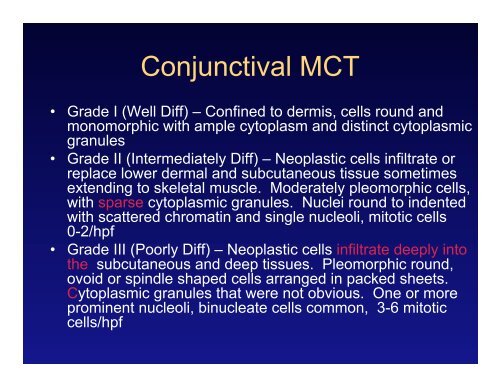
Conjunctival MCT<br />
• Grade I (Well Diff) – Confined to dermis, <strong>cell</strong>s round and<br />
monomorphic with ample cytoplasm and distinct cytoplasmic<br />
granules<br />
• Grade II (Intermediately Diff) – Neoplastic <strong>cell</strong>s infiltrate or<br />
replace lower dermal and subcutaneous tissue sometimes<br />
extending to skeletal muscle. Moderately pleomorphic <strong>cell</strong>s,<br />
with sparse cytoplasmic granules. Nuclei round to indented<br />
with scattered chromatin and single nucleoli, mitotic <strong>cell</strong>s<br />
0-2/hpf<br />
• Grade III (Poorly Diff) – Neoplastic <strong>cell</strong>s infiltrate deeply into<br />
the subcutaneous and deep tissues. Pleomorphic round,<br />
ovoid or spindle shaped <strong>cell</strong>s arranged in packed sheets.<br />
Cytoplasmic granules that were not obvious. One or more<br />
prominent nucleoli, binucleate <strong>cell</strong>s common, 3-6 mitotic<br />
<strong>cell</strong>s/hpf