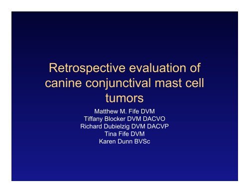
Retrospective evaluation of canine conjunctival mast cell tumors
Retrospective evaluation of canine conjunctival mast cell tumors
Retrospective evaluation of canine conjunctival mast cell tumors

Create successful ePaper yourself
Turn your PDF publications into a flip-book with our unique Google optimized e-Paper software.
<strong>Retrospective</strong> <strong>evaluation</strong> <strong>of</strong><br />
<strong>canine</strong> <strong>conjunctival</strong> <strong>mast</strong> <strong>cell</strong><br />
<strong>tumors</strong><br />
Matthew M. Fife DVM<br />
Tiffany Blocker DVM DACVO<br />
Richard Dubielzig DVM DACVP<br />
Tina Fife DVM<br />
Karen Dunn BVSc
Conjunctival MCT<br />
• MCT represents the most common<br />
cutaneous neoplasm <strong>of</strong> the dog (16-21%)<br />
• Behavior <strong>of</strong> cutaneous <strong>tumors</strong> is well<br />
studied<br />
• Conjunctival <strong>tumors</strong> poorly described,<br />
behavior not documented
Conjunctival MCT<br />
• 1988 Johnson et al. - Reported two<br />
<strong>conjunctival</strong> <strong>mast</strong> <strong>cell</strong> <strong>tumors</strong> originating<br />
from the dorsal bulbar conj. Local<br />
resection was curative at 4 years and 6<br />
months<br />
• 2007 Barsotti et al. – Single case report <strong>of</strong><br />
palpebral MCT in a Labrador Retriever.<br />
Local excision curative at 29 months
Conjunctival MCT<br />
• Behavior and prognosis <strong>of</strong> MCT best predicted<br />
by histologic grade<br />
• Additionally, size <strong>of</strong> tumor, location <strong>of</strong> tumor,<br />
clinical stage and growth rate <strong>of</strong> tumor as well as<br />
AgNOR’s, and Ki67 can be prognostic indicators<br />
• Therapy is directed primarily by the histologic<br />
grade <strong>of</strong> the tumor
Conjunctival MCT<br />
• Tumor location<br />
– Historically <strong>tumors</strong> originating at mucosal,<br />
inguinal and subungual locations were<br />
thought to carry a poorer prognosis in spite <strong>of</strong><br />
a lack <strong>of</strong> support in the literature. Two recent<br />
case series, however, found a comparable<br />
survival time for dogs with inguinal and<br />
perineal <strong>mast</strong> <strong>cell</strong>s <strong>tumors</strong> to those at other<br />
cutaneous locations.
Conjunctival MCT<br />
• Grade I (Well Diff) – Confined to dermis, <strong>cell</strong>s round and<br />
monomorphic with ample cytoplasm and distinct cytoplasmic<br />
granules<br />
• Grade II (Intermediately Diff) – Neoplastic <strong>cell</strong>s infiltrate or<br />
replace lower dermal and subcutaneous tissue sometimes<br />
extending to skeletal muscle. Moderately pleomorphic <strong>cell</strong>s,<br />
with sparse cytoplasmic granules. Nuclei round to indented<br />
with scattered chromatin and single nucleoli, mitotic <strong>cell</strong>s<br />
0-2/hpf<br />
• Grade III (Poorly Diff) – Neoplastic <strong>cell</strong>s infiltrate deeply into<br />
the subcutaneous and deep tissues. Pleomorphic round,<br />
ovoid or spindle shaped <strong>cell</strong>s arranged in packed sheets.<br />
Cytoplasmic granules that were not obvious. One or more<br />
prominent nucleoli, binucleate <strong>cell</strong>s common, 3-6 mitotic<br />
<strong>cell</strong>s/hpf
Conjunctival MCT<br />
• Treatment – Low and Intermediate Grade<br />
– Surgical excision with 2-3cm lateral margins plus 1<br />
fascial plane deep<br />
• If complete excision is not possible primary treatment with<br />
external beam radiation-therapy or a combination <strong>of</strong> surgical<br />
excision and radiation therapy is recommended<br />
• Treatment – High Grade<br />
– Surgical excision combined with chemotherapy +/-<br />
regional node radiation-therapy<br />
– Incomplete surgical margins warrant radiation-therapy<br />
<strong>of</strong> primary site
Conjunctival MCT<br />
• Evaluation <strong>of</strong> the importance <strong>of</strong> clean<br />
surgical margins have demonstrated a<br />
similar long term outcome for completely<br />
and incompletely excised <strong>tumors</strong> <strong>of</strong> low to<br />
intermediate grade<br />
– Michels et. al. Prognosis following surgical excision <strong>of</strong> <strong>canine</strong> cutaneous <strong>mast</strong><br />
<strong>cell</strong> <strong>tumors</strong> with histopathologically tumor-free versus nontumor-free margins: a<br />
retrospective study <strong>of</strong> 31 cases. JAAHAv38n5p458
Conjunctival MCT<br />
Grade<br />
1 year survival<br />
(%)<br />
2 year survival<br />
(%)<br />
Low (#46) 100 100<br />
Intermed (#121) 92 89<br />
High (#28) 46 36<br />
Murphy S. et al. Relationship between the histological grade <strong>of</strong> cutaneous <strong>mast</strong> <strong>cell</strong> tumours in dogs, their<br />
survival and the efficacy <strong>of</strong> surgical resection. Vet Rec v154p 743-746 ‘04
Conjunctival MCT<br />
• 33 dogs with 35 MCT identified<br />
– Labrador (7) Labrador cross (5) most common<br />
– 20 FS, 9CM, 2F, 1M<br />
– 27 cases had duration reported – Range 1 week-2<br />
years (mean 4.6 months)<br />
• Site<br />
– bulbar (12), dorsal palpebral (9), third eyelid (8),<br />
ventral palpebral (3), medial canthal (1), unknown (1)<br />
• Grade<br />
– 11 low grade, 20 intermediate grade, 3 high grade
Conjunctival MCT<br />
• <strong>Retrospective</strong> <strong>evaluation</strong> <strong>of</strong> <strong>canine</strong> <strong>conjunctival</strong><br />
MCT<br />
– COPLOW - 24 cases<br />
– Eye Path Lab - 7 cases<br />
– Eye Care for Animals - 3 cases (Histopathology by<br />
ANTECH)<br />
• Questionnaires sent to submitting veterinarian<br />
for follow up information. Referring veterinarian<br />
and/or owners contacted for additional<br />
information where needed
Conjunctival MCT<br />
• Surgery performed<br />
– Local excision in 25 cases<br />
– Excision with H-plasty in 2 cases<br />
– TEL excision in 3 cases<br />
– Enucleation in 2 cases (1 following recurrence)<br />
• Margins<br />
– Dirty margins in 25 cases, narrow in 5 cases and clean in 1 case<br />
(enucleated)<br />
• Adjunctive Therapy<br />
– External beam radiation performed in one case<br />
– Vincristine/Cyclophosphamide administered in one case<br />
– Cryotherapy performed <strong>of</strong> surgical site at time <strong>of</strong> surgery in 5 cases
Conjunctival MCT<br />
• Follow up information was received for 25<br />
animals<br />
– 15 currently disease free (21.4 months)<br />
– 4 disease free but lost to follow up (13<br />
months)<br />
– 4 died <strong>of</strong> unrelated causes (20.25 months)<br />
– 2 local recurrences
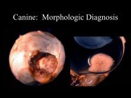
Palpebral
Third Eyelid
Low Grade
Intermediate Grade
High Grade
Conjunctival MCT
Conjunctival MCT<br />
• Conclusion<br />
– MCT <strong>of</strong> the conjunctiva are typically <strong>of</strong> low to<br />
intermediate grade and appear to follow a<br />
benign clinical course<br />
– Local surgical excision seems to yield good<br />
clinical results even with incomplete surgical<br />
margins
Acknowledments