Supplement I to the Japanese Pharmacopoeia Fourteenth Edition
Supplement I to the Japanese Pharmacopoeia Fourteenth Edition
Supplement I to the Japanese Pharmacopoeia Fourteenth Edition

You also want an ePaper? Increase the reach of your titles
YUMPU automatically turns print PDFs into web optimized ePapers that Google loves.
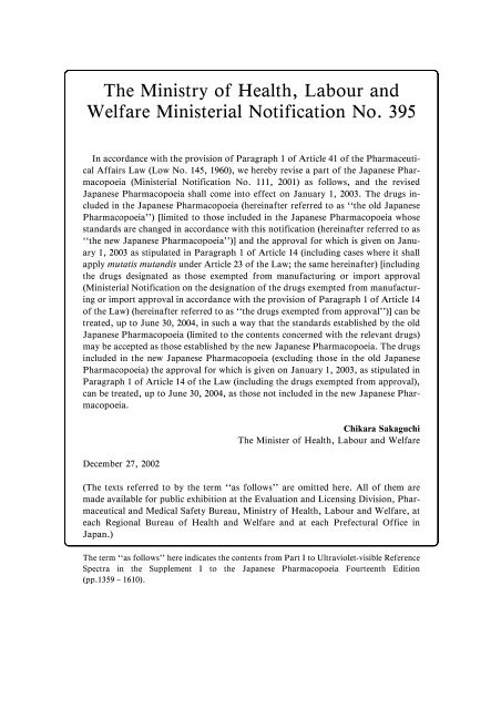
The Ministry of Health, Labour and<br />
Welfare Ministerial Notification No. 395<br />
In accordance with <strong>the</strong> provision of Paragraph 1 of Article 41 of <strong>the</strong> Pharmaceutical<br />
Affairs Law (Low No. 145, 1960), we hereby revise a part of <strong>the</strong> <strong>Japanese</strong> <strong>Pharmacopoeia</strong><br />
(Ministerial Notification No. 111, 2001) as follows, and <strong>the</strong> revised<br />
<strong>Japanese</strong> <strong>Pharmacopoeia</strong> shall come in<strong>to</strong> effect on January 1, 2003. The drugs included<br />
in <strong>the</strong> <strong>Japanese</strong> <strong>Pharmacopoeia</strong> (hereinafter referred <strong>to</strong> as ``<strong>the</strong> old <strong>Japanese</strong><br />
<strong>Pharmacopoeia</strong>'') [limited <strong>to</strong> those included in <strong>the</strong> <strong>Japanese</strong> <strong>Pharmacopoeia</strong> whose<br />
standards are changed in accordance with this notification (hereinafter referred <strong>to</strong> as<br />
``<strong>the</strong> new <strong>Japanese</strong> <strong>Pharmacopoeia</strong>'')] and <strong>the</strong> approval for which is given on January<br />
1, 2003 as stipulated in Paragraph 1 of Article 14 (including cases where it shall<br />
apply mutatis mutandis under Article 23 of <strong>the</strong> Law; <strong>the</strong> same hereinafter) [including<br />
<strong>the</strong> drugs designated as those exempted from manufacturing or import approval<br />
(Ministerial Notification on <strong>the</strong> designation of <strong>the</strong> drugs exempted from manufacturing<br />
or import approval in accordance with <strong>the</strong> provision of Paragraph 1 of Article 14<br />
of <strong>the</strong> Law) (hereinafter referred <strong>to</strong> as ``<strong>the</strong> drugs exempted from approval'')] can be<br />
treated, up <strong>to</strong> June 30, 2004, in such a way that <strong>the</strong> standards established by <strong>the</strong> old<br />
<strong>Japanese</strong> <strong>Pharmacopoeia</strong> (limited <strong>to</strong> <strong>the</strong> contents concerned with <strong>the</strong> relevant drugs)<br />
may be accepted as those established by <strong>the</strong> new <strong>Japanese</strong> <strong>Pharmacopoeia</strong>. The drugs<br />
included in <strong>the</strong> new <strong>Japanese</strong> <strong>Pharmacopoeia</strong> (excluding those in <strong>the</strong> old <strong>Japanese</strong><br />
<strong>Pharmacopoeia</strong>) <strong>the</strong> approval for which is given on January 1, 2003, as stipulated in<br />
Paragraph 1 of Article 14 of <strong>the</strong> Law (including <strong>the</strong> drugs exempted from approval),<br />
can be treated, up <strong>to</strong> June 30, 2004, as those not included in <strong>the</strong> new <strong>Japanese</strong> <strong>Pharmacopoeia</strong>.<br />
December 27, 2002<br />
Chikara Sakaguchi<br />
The Minister of Health, Labour and Welfare<br />
(The texts referred <strong>to</strong> by <strong>the</strong> term ``as follows'' are omitted here. All of <strong>the</strong>m are<br />
made available for public exhibition at <strong>the</strong> Evaluation and Licensing Division, Pharmaceutical<br />
and Medical Safety Bureau, Ministry of Health, Labour and Welfare, at<br />
each Regional Bureau of Health and Welfare and at each Prefectural Office in<br />
Japan.)<br />
The term ``as follows'' here indicates <strong>the</strong> contents from Part I <strong>to</strong> Ultraviolet-visible Reference<br />
Spectra in <strong>the</strong> <strong>Supplement</strong> I <strong>to</strong> <strong>the</strong> <strong>Japanese</strong> <strong>Pharmacopoeia</strong> <strong>Fourteenth</strong> <strong>Edition</strong><br />
(pp.1359 – 1610).
CONTENTS<br />
Preface ...................................................... i<br />
<strong>Supplement</strong> I <strong>to</strong> The <strong>Japanese</strong> <strong>Pharmacopoeia</strong>,<br />
<strong>Fourteenth</strong> <strong>Edition</strong>, Part I................. 1359–1540<br />
General Notices ................................... 1359<br />
General Rules for Preparations ............... 1361<br />
General Tests, Processes and Apparatus ... 1363<br />
23. Infrared Spectropho<strong>to</strong>metry.............. 1363<br />
52. Residue on Ignition Test .................. 1363<br />
66. Vitamin A Assay ............................ 1364<br />
70. Reference Standards; Reagents, Test Solutions;<br />
Standard Solutions for Volumetric Analysis;<br />
Standard Solutions; Matching Fluids for Color;<br />
Optical Filters for Wavelength and Transmission<br />
Rate Calibration; and Measuring Instruments,<br />
Appliances ............................ 1365<br />
(1) Reference Standards................. 1365<br />
(2) Reagents, Test Solutions ........... 1365<br />
(3) Standard Solutions for Volumetric<br />
Analysis................................. 1373<br />
72. Conductivity Measurement................ 1373<br />
73. Determination of Bulk and Tapped<br />
Densities ....................................... 1375<br />
Official Monographs............................. 1377<br />
<strong>Supplement</strong> I <strong>to</strong> The <strong>Japanese</strong> <strong>Pharmacopoeia</strong>,<br />
<strong>Fourteenth</strong> <strong>Edition</strong>, Part II................ 1541–1566<br />
General Rules for Crude Drugs ............... 1541<br />
Official Monographs............................. 1543<br />
Infrared Reference Spectra ................ 1567–1586<br />
Part I ................................................ 1567<br />
Ultraviolet-visible Reference Spectra .... 1587–1610<br />
Part I ................................................ 1587<br />
General Information<br />
1. Aris<strong>to</strong>lochic Acid............................ 1611<br />
5. International Harmonization Implemented<br />
in <strong>the</strong> <strong>Japanese</strong> <strong>Pharmacopoeia</strong> <strong>Fourteenth</strong><br />
<strong>Edition</strong> ......................................... 1611<br />
12. Preservatives-Effectiveness Tests ........ 1616<br />
17. Basic Requirements for Viral Safety of<br />
Biotechnological/Biological Products<br />
listed in <strong>Japanese</strong> <strong>Pharmacopoeia</strong> ...... 1618<br />
18. Qualification of Animals as Origin of<br />
Animal-derived Medicinal Products<br />
provided in <strong>the</strong> General Notices 39 of<br />
<strong>Japanese</strong> <strong>Pharmacopoeia</strong> and O<strong>the</strong>r<br />
Standards...................................... 1631<br />
19. SDS-Polyacrylamide Gel<br />
Electrophoresis............................... 1634<br />
Appendix .............................................. 1641<br />
Index.................................................... 1643<br />
Index in <strong>Japanese</strong>.................................... 1659
Preface<br />
The <strong>Fourteenth</strong> <strong>Edition</strong> of <strong>the</strong> <strong>Japanese</strong> <strong>Pharmacopoeia</strong><br />
was promulgated on March 30, 2001 by<br />
Ministerial Notiˆcation No. 111 of <strong>the</strong> Ministry of<br />
Health, Labour and Welfare. To keep pace with<br />
progress in medical and pharmaceutical sciences, in<br />
November 2001, <strong>the</strong> Council, at a meeting of <strong>the</strong><br />
Committee on <strong>Japanese</strong> <strong>Pharmacopoeia</strong> (JP), established<br />
<strong>the</strong> basic principles for <strong>the</strong> preparation of <strong>the</strong><br />
JP Fifteenth <strong>Edition</strong>, setting out <strong>the</strong> characteristics<br />
and roles of <strong>the</strong> JP, <strong>the</strong> deˆnite measures for <strong>the</strong> revision,<br />
<strong>the</strong> date of <strong>the</strong> revision, and <strong>the</strong> organization of<br />
<strong>the</strong> Subcommittee on JP.<br />
At <strong>the</strong> above meeting, <strong>the</strong> following ``ˆve pillars''<br />
were established as <strong>the</strong> basic principles of <strong>the</strong> JP:<br />
Making it more substantial by including all drugs<br />
which are important from <strong>the</strong> viewpoint of health care<br />
and medical treatment; Making prompt partial revision<br />
as necessary and facilitating smooth administrative<br />
operation; Promoting international harmonization;<br />
Ensuring transparency regarding <strong>the</strong> revision and<br />
dissemination <strong>to</strong> <strong>the</strong> public of <strong>the</strong> JP; and Positively<br />
introducing contemporary analytical tests and developing<br />
reference standards. It was decided at <strong>the</strong><br />
meeting that each panel set up under <strong>the</strong> Subcommittee<br />
on JP should make eŠorts, on <strong>the</strong> basis of <strong>the</strong>se<br />
principles, <strong>to</strong> ensure that <strong>the</strong> JP is used more eŠectively<br />
in <strong>the</strong> ˆelds of health care and medical treatment by<br />
taking appropriate measures, including getting <strong>the</strong> understanding<br />
and cooperation of o<strong>the</strong>r parties concerned.<br />
The JP should comprise an o‹cial standard being<br />
required <strong>to</strong> assure <strong>the</strong> quality of drugs in this country<br />
in response <strong>to</strong> <strong>the</strong> progress in science and technology<br />
and clinical demands at <strong>the</strong> time, it should deˆne <strong>the</strong><br />
standards for speciˆcations as well as <strong>the</strong> methods of<br />
tests <strong>to</strong> assure <strong>the</strong> overall quality of all drugs in principle,<br />
and it should have a role in clarifying <strong>the</strong> criteria<br />
for quality of drugs which are recognized <strong>to</strong> be important<br />
from <strong>the</strong> viewpoint of medical treatment.<br />
At <strong>the</strong> same time, it was agreed that <strong>the</strong> JP should<br />
be prepared with <strong>the</strong> aid of <strong>the</strong> knowledge and experience<br />
of many persons involved in <strong>the</strong> pharmaceuticals,<br />
that it should have <strong>the</strong> characteristics of an o‹-<br />
cial standard, which might be widely used by all parties<br />
concerned, that it should provide information and<br />
understanding about <strong>the</strong> quality of drugs <strong>to</strong> <strong>the</strong> public,<br />
and that it should be conducive <strong>to</strong> smooth and<br />
eŠective government control of <strong>the</strong> quality of drugs,<br />
and <strong>to</strong> securing and maintaining international consistency.<br />
It was also agreed that JP articles should cover<br />
drugs which are important from <strong>the</strong> viewpoint of<br />
health care and medical treatment, clinical results and<br />
frequency of use, as soon as possible after <strong>the</strong>y reach<br />
<strong>the</strong> market.<br />
It was also decided <strong>to</strong> make a deˆnite rule for selection<br />
of articles, by clarifying <strong>the</strong> signiˆcance of, and<br />
standards for selection. The JP Fifteenth <strong>Edition</strong> was<br />
decided <strong>to</strong> be slated for completion in April 2006.<br />
Under <strong>the</strong> Subcommittee on JP, <strong>the</strong> following<br />
twelve panels and two provisional panels were established<br />
at ˆrst: Panel on Planning and Revisions; Panel<br />
on <strong>the</strong> Selection of Articles; Panel on Nomenclature;<br />
Panel on Excipients; First Panel on Medicinal Chemicals;<br />
Second Panel on Medicinal Chemicals; Panel on<br />
Biologically Derived Drugs; Panel on Biological Tests;<br />
Panel on Physico-chemical Tests; Panel on Material<br />
Sciences; Panel on Preparations; Panel on Crude<br />
Drugs; Provisional First Panel on Antibiotics;<br />
Provisional First Panel on Crude Drugs. Some of <strong>the</strong><br />
names of <strong>the</strong> above panels were changed as follows,<br />
due <strong>to</strong> <strong>the</strong> reorganization of <strong>the</strong> Pharmaceutical<br />
AŠairs and Food Sanitation Council (PAFSC) in<br />
November 2001: Panel on Nomenclature <strong>to</strong> Panel on<br />
Nomenclature for Pharmaceutical Chemicals; First<br />
Panel on Medicinal Chemicals and Second Panel on<br />
Medicinal Chemicals were integrated in<strong>to</strong> Panel on<br />
Medicinal Chemicals; Panel on Physico-chemical<br />
Tests, Panel on Material Sciences and Panel on Preparations<br />
were integrated in<strong>to</strong> Panel on Physico-chemical<br />
Tests; Panel on Crude Drugs and Provisional First<br />
Panel on Crude Drugs were integrated in<strong>to</strong> Panel on<br />
Crude Drugs; Provisional First Panel on Antibiotics<br />
<strong>to</strong> Panel on Antibiotics. Under <strong>the</strong> Panel on Planning<br />
and Revisions, <strong>the</strong> following two panels were newly established:<br />
Panel on General Revisions and Panel on<br />
<strong>Pharmacopoeia</strong>l Harmonization (PDG).<br />
In <strong>the</strong> Committee on <strong>Japanese</strong> <strong>Pharmacopoeia</strong>,<br />
Tadao Terao <strong>to</strong>ok <strong>the</strong> role of chairman from November<br />
1997 <strong>to</strong> December 2000 and Mitsuru Uchiyama<br />
from January 2001 <strong>to</strong> December 2002.<br />
With <strong>the</strong> reform of central government ministries<br />
and agencies in January 2001, <strong>the</strong> Ministry of Health<br />
and Welfare became <strong>the</strong> Ministry of Health, Labour<br />
and Welfare, and <strong>the</strong> Committee on <strong>Japanese</strong> <strong>Pharmacopoeia</strong><br />
(CJP) came under <strong>the</strong> authority of <strong>the</strong><br />
i
ii<br />
Preface<br />
<strong>Supplement</strong> I, JP XIV<br />
Minister of Health, Labour and Welfare. At <strong>the</strong> same<br />
time, <strong>the</strong> Central Pharmaceutical AŠairs Council<br />
became <strong>the</strong> Pharmaceutical AŠairs and Food Sanitation<br />
Council (PAFSC) and Mitsuru Uchiyama was<br />
nominated as chairman of <strong>the</strong> CJP.<br />
It was decided that <strong>the</strong> JP will be revised not only<br />
every ˆve years, in line with <strong>the</strong> basic principles for <strong>the</strong><br />
preparation of <strong>the</strong> JP Fifteenth <strong>Edition</strong>, but also as<br />
necessary <strong>to</strong> take account of recent progress of science<br />
and in <strong>the</strong> interests of international harmonization. In<br />
accordance with <strong>the</strong> revision principles, <strong>the</strong> panels<br />
continued discussions on selection of articles, and revisions<br />
for General Notices, General Rules for Preparations,<br />
General Tests, and monographs on drugs.<br />
Among <strong>the</strong> items discussed, <strong>the</strong> following two revision<br />
drafts were separately examined by <strong>the</strong> CommitteeonJPinNovember2001,followedbyPAFSCin<br />
December 2001, and <strong>the</strong>n submitted <strong>to</strong> <strong>the</strong> Minister of<br />
Health, Labour and Welfare, and Ministerial Notiˆcation<br />
No.151 promulgated <strong>the</strong>se revisions on March 29,<br />
2002: Addition of a paragraph ``In principle, animals<br />
used as a source of materials for preparing pharmaceutical<br />
drugs must be healthy.'' <strong>to</strong> General Notices;<br />
and Deletion of a monograph ``Phenacetin''.<br />
Draft revisions covering subjects in General Notices,<br />
General Rules for Preparations, General Rules<br />
for Crude Drugs, General Tests, and monographs on<br />
drugs, for which discussions were ˆnished between<br />
June 2000 and February 2001, were prepared for a<br />
supplement <strong>to</strong> <strong>the</strong> book. They were examined by <strong>the</strong><br />
Committee on JP in September 2002, followed by<br />
PAFSC in December 2002, and <strong>the</strong>n submitted <strong>to</strong> <strong>the</strong><br />
Minister of Health, Labour and Welfare.<br />
Numbers of discussions in <strong>the</strong> panels <strong>to</strong> prepare<br />
supplement drafts were as follows: Panel on <strong>the</strong> Principles<br />
of Revisions, 6 times; Panel on <strong>the</strong> Selection of<br />
Articles, 2 times; Panel on Nomenclature, 11 times;<br />
Panel on Excipients, 10 times; First Panel on Medicinal<br />
Chemicals, 10 times; Second Panel on Medicinal<br />
Chemicals, 16 times; Panel on Biologically Derived<br />
Drugs, 8 times; Panel on Biological Tests, 9 times;<br />
Panel on Physico-chemical Tests, 8 times; Panel on<br />
Material Sciences, 8 times; Panel on Preparations, 5<br />
times; Panel on Crude Drugs, 6 times; Provisional<br />
First Panel on Antibiotics, 27 times; Provisional First<br />
Panel on Crude Drugs, 7 times. Numbers of discussion<br />
in <strong>the</strong> panels, which were renamed due <strong>to</strong> <strong>the</strong> reorganization<br />
of PAFSC, were as follows: Panel on<br />
Nomenclature for Pharmaceutical Chemicals, 2 times;<br />
Panel on Physico-chemical Tests, 1 time; Panel on<br />
Crude Drags, 3 times; Panel on General Revisions, 1<br />
time.<br />
It should be noted that in <strong>the</strong> preparation of <strong>the</strong><br />
drafts for <strong>the</strong> <strong>Supplement</strong> I, generous cooperation was<br />
given by <strong>the</strong> Technical Committee of <strong>the</strong> Pharmaceutical<br />
Manufacturer's Association of Tokyo and of<br />
Osaka, <strong>the</strong> Crude Drugs Association of Tokyo, <strong>the</strong><br />
Japan Pharmaceutical Excipients Council, <strong>the</strong> Federation<br />
of Crude Drugs Associations of Japan, <strong>the</strong> Japan<br />
Antibiotics Research Association, <strong>the</strong> Japan Flavor<br />
and Fragrance Manufacturer's Association, <strong>the</strong> Japan<br />
Medical Plants Federation, <strong>the</strong> Japan Pharmaceutical<br />
Manufacturer's Association, <strong>the</strong> <strong>Japanese</strong> Society of<br />
Hospital Pharmacists, <strong>the</strong> Japan Pharmaceutical Association,<br />
and <strong>the</strong> Japan Oilseed Processors Association.<br />
In consequence of this revision, <strong>the</strong> JP <strong>Fourteenth</strong><br />
<strong>Edition</strong> carries 881 articles in Part I owing <strong>to</strong> <strong>the</strong> addition<br />
of 31 articles and <strong>the</strong> deletion of 8 articles; and<br />
481 articles in Part II owing <strong>to</strong> <strong>the</strong> addition of 15 articles<br />
and <strong>the</strong> deletion of 3 articles.<br />
The principles of description and <strong>the</strong> salient points<br />
of <strong>the</strong> revision in this volume are as follows:<br />
1. The <strong>Supplement</strong> I <strong>to</strong> JP <strong>Fourteenth</strong> <strong>Edition</strong><br />
comprises <strong>the</strong> following items, in order: Notiˆcation<br />
of <strong>the</strong> Ministry of Health, Labour and Welfare; Contents;<br />
Preface; followed by General Notices; General<br />
Rules for Preparations; General Tests, Processes and<br />
Apparatus; Monographs on Drugs in Part I, and<br />
General Notices; General Rules for Crude Drugs;<br />
General Rules for Preparations; General Tests,<br />
Processes and Apparatus; Monographs on Drugs in<br />
Part II, <strong>the</strong>n followed by Infrared Reference Spectra<br />
in Part I; Ultraviolet-visible Reference Spectra in Part<br />
I; General Information, and as appendix, <strong>the</strong> Ministry<br />
of Health, Labour and Welfare Ministerial Notiˆcation<br />
No. 151 (March 2002); and Cumulative Index<br />
containing references <strong>to</strong> <strong>the</strong> main volume and <strong>the</strong> supplement<br />
I.<br />
2. The articles in General Rules for Preparations,<br />
in General Tests, Processes and Apparatus, Monographs<br />
on Drugs, Infrared Reference Spectra and<br />
Ultraviolet-visible Reference Spectra are respectively<br />
placed in alphabetical order.<br />
3. The following items in each monograph are put<br />
in <strong>the</strong> order shown below, except that unnecessary i-<br />
tems are omitted depending on <strong>the</strong> nature of <strong>the</strong> drug:<br />
(1) English title<br />
(2) Commonly used name(s)<br />
(3) Latin title (only for Crude Drugs)<br />
(4) Title in <strong>Japanese</strong><br />
(5) Structural formula or empirical formula<br />
(6) Molecular formula and molecular mass<br />
(7) Chemical name
<strong>Supplement</strong> I, JP XIV<br />
Preface<br />
iii<br />
(8) Origin<br />
(9) Limits of <strong>the</strong> content of <strong>the</strong> ingredient(s)<br />
and/or <strong>the</strong> unit of potency<br />
(10) Labeling requirements<br />
(11) Method of preparation<br />
(12) Description<br />
(13) Identiˆcation tests<br />
(14) Speciˆc physical and/or chemical values<br />
(15) Purity tests<br />
(16) Loss on drying, loss on ignition, and/or water<br />
(17) Residue on ignition, <strong>to</strong>tal ash, and/or acid-insoluble<br />
ash<br />
(18) Special tests<br />
(19) Isomer ratio<br />
(20) Assay or <strong>the</strong> content of <strong>the</strong> ingredient(s)<br />
(21) Containers and s<strong>to</strong>rage<br />
(22) Expiration date<br />
(23) O<strong>the</strong>rs<br />
4. In each monograph on a drug, <strong>the</strong> following<br />
physical and chemical values representing <strong>the</strong> properties<br />
and quality of <strong>the</strong> drug are given in <strong>the</strong> order indicated<br />
below, except that unnecessary items are omitted<br />
depending on <strong>the</strong> nature of <strong>the</strong> drug:<br />
(1) Alcohol number<br />
(2) Absorbance<br />
(3) Congealing point<br />
(4) Refractive index<br />
(5) Osmolarity<br />
(6) Optical rotation<br />
(7) Viscosity<br />
(8) pH<br />
(9) Speciˆc gravity<br />
(10) Boiling point<br />
(11) Melting point<br />
(12) Acid value<br />
(13) Saponiˆcation value<br />
(14) Ester value<br />
(15) Hydroxyl value<br />
(16) Iodine value<br />
5. Identiˆcation tests comprise <strong>the</strong> following<br />
items, which are generally put in <strong>the</strong> order given below:<br />
(1) Coloration reactions<br />
(2) Precipitation reactions<br />
(3) Decomposition reactions<br />
(4) Derivatives<br />
(5) Visible, ultraviolet or infrared spectra<br />
(6) Special reactions<br />
(7) Cations<br />
(8) Anions<br />
6. Purity tests comprise <strong>the</strong> following items, which<br />
are generally put in <strong>the</strong> order given below, except that<br />
unnecessary items are omitted depending on <strong>the</strong> nature<br />
of <strong>the</strong> drug:<br />
(1) Color<br />
(2) Odor<br />
(3) Clarity and/or color of solution<br />
(4) Acidity or alkalinity<br />
(5) Acid<br />
(6) Alkali<br />
(7) Chloride<br />
(8) Sulfate<br />
(9) Sulˆte<br />
(10) Nitrate<br />
(11) Nitrite<br />
(12) Carbonate<br />
(13) Bromide<br />
(14) Iodide<br />
(15) Soluble halide<br />
(16) Thiocyanide<br />
(17) Selenium<br />
(18) Cationic salts<br />
(19) Ammonium<br />
(20) Heavy metals<br />
(21) Iron<br />
(22) Manganese<br />
(23) Chromium<br />
(24) Bismuth<br />
(25) Tin<br />
(26) Aluminum<br />
(27) Zinc<br />
(28) Cadmium<br />
(29) Mercury<br />
(30) Copper<br />
(31) Lead<br />
(32) Silver<br />
(33) Alkaline earth metals<br />
(34) Arsenic<br />
(35) Foreign matter<br />
(36) Related substances<br />
(37) O<strong>the</strong>r mixtures<br />
(38) Readily carbonizable substances<br />
7. Revisions in <strong>the</strong> General Notices are as follows:<br />
(1) In paragraph 6, an abbreviation ``lx'' for lux<br />
was added as <strong>the</strong> main unit.<br />
(2) In paragraph 8, <strong>the</strong> deˆnition for ``a cold<br />
place'' was revised <strong>to</strong> as ``a place having a temperature<br />
between 1 and 159C''.<br />
8. Revision in <strong>the</strong> General Rules for Preparations<br />
is as follows:<br />
(1) Injections: The description with respect <strong>to</strong> <strong>the</strong><br />
injection having 50 mL or more was deleted<br />
from section (9).<br />
9. The following items of <strong>the</strong> General Tests,
iv<br />
Preface<br />
<strong>Supplement</strong> I, JP XIV<br />
Processes and Apparatus were partially revised:<br />
(1) Infrared Spectropho<strong>to</strong>metry<br />
(2) Residue on Ignition Test<br />
(3) Vitamin A Assay<br />
10. The following tests were added <strong>to</strong> <strong>the</strong> General<br />
Tests, Processes and Apparatus:<br />
(1) Conductivity Measurement<br />
(2) Determination of Bulk and Tapped Densities<br />
11. The following Reference Standard was deleted:<br />
Drostanolone Propionate<br />
12. The following Reference Standard was renamed:<br />
Kitasamycin <strong>to</strong> Leucomycine A 5<br />
13. The following Reference Standards were added:<br />
Aceglutamide<br />
Acetylspiramycin II<br />
Aclarubicin<br />
Actinomycin D<br />
Arbekacin Sulfate<br />
Astromicin Sulfate<br />
Bacitracin<br />
Bekanamycin Sulfate<br />
Benzylpenicillin Potassium<br />
Benzylpenicillin Sodium<br />
Bleomycin A 2 Hydrochloride<br />
Carumonam Sodium<br />
Cefaclor<br />
Cefaloridin<br />
Cefalotin Sodium<br />
Cefamandole Lithium<br />
Cefbuperazone<br />
Cefmenoxime Hydrochloride<br />
Cefodizime Sodium<br />
Cefotaxime<br />
Cefotetan<br />
Cefotiam Hexetil Hydrochloride<br />
Cefoxitin<br />
Cefpiramide<br />
Cefpodoxime Proxetil<br />
Cefroxadine<br />
Cefteram Pivoxil Mesitylenesulfonate<br />
Cefuroxime Axetil<br />
Chloramphenicol<br />
Chloramphenicol Palmitate<br />
Chloramphenicol Succinate<br />
Ciclacillin<br />
Clindamycin Hydrochloride<br />
Clindamycin Phosphate<br />
Colistin Sulfate<br />
Daunorubicin Hydrochloride<br />
Demethylchlortetracycline Hydrochloride<br />
Dibekacin Sulfate<br />
Diethanolamine Fusidate<br />
Doxorubicin Hydrochloride<br />
Doxycycline Hydrochloride<br />
Enviomycin Sulfate<br />
Epirubicin Hydrochloride<br />
Flomoxef Triethylammonium<br />
Fradiomycin Sulfate<br />
Gentamicin Sulfate<br />
Gramicidin<br />
Griseofulvin<br />
Imipenem<br />
Josamycin Propionate<br />
Kanamycin Monosulfate<br />
Latamoxef Ammonium<br />
Lenampicillin Hydrochloride<br />
Lincomycin Hydrochloride<br />
Lysozyme<br />
Micronomicin Sulfate<br />
Mi<strong>to</strong>mycin C<br />
Oxytetracycline Hydrochloride<br />
Peplomycin Sulfate<br />
L-Phenethicillin Potassium<br />
Pimaricin<br />
Pirarubicin<br />
Pivmecillinam Hydrochloride<br />
Polymixin B Sulfate<br />
Puerarin<br />
Pyrrolnitrin<br />
Ranitidine Hydrochloride<br />
Retinol Acetate<br />
Retinol Palmitate<br />
Ribostamycin Sulfate<br />
Rifampicin<br />
Sennoside A<br />
Sennoside B<br />
Siccanin<br />
Spectinomycin Hydrochloride<br />
Strep<strong>to</strong>mycin Sulfate<br />
Sulbenicillin Sodium<br />
Talampicillin Hydrochloride<br />
Tobramycin<br />
Trichomycin<br />
Vancomycin Hydrochloride<br />
14. English and Latin titles of drugs are derived,<br />
in principle, from International Nonproprietary<br />
Names (INN) for Pharmaceutical Substances recommended<br />
by <strong>the</strong> World Health Organization. <strong>Japanese</strong><br />
titles are derived from <strong>the</strong> <strong>Japanese</strong> version of this<br />
book. The chemical names are based on <strong>the</strong> rules of<br />
<strong>the</strong> International Union of Pure and Applied Chemis-
<strong>Supplement</strong> I, JP XIV<br />
Preface<br />
v<br />
try (IUPAC).<br />
15. Molecular formulas of organic compounds beginwithCand<strong>the</strong>nH,followedbyo<strong>the</strong>rinvolvedelements<br />
in <strong>the</strong> alphabetical order of <strong>the</strong> symbols of <strong>the</strong><br />
elements.<br />
16. Structural formulas of drugs represent, as far<br />
as possible, steric conˆgurations.<br />
17. Test procedures in monographs in Part I are,<br />
in principle, written in full even in corresponding<br />
monographs in Part II, and vice versa. The test procedures<br />
in monographs for preparations are also written<br />
in full even within <strong>the</strong> same part, except in <strong>the</strong> monographs<br />
for preparations having a corresponding<br />
monograph of <strong>the</strong>ir principal material substances.<br />
18. In O‹cial Monographs, names of some of <strong>the</strong><br />
reagents and <strong>the</strong> test solutions were changed <strong>to</strong> <strong>the</strong><br />
latest names based on <strong>the</strong> JIS.<br />
19. The following articles were deleted from O‹-<br />
cial Monographs<br />
Part I<br />
Drostanolone Propionate<br />
Drostanolone Propionate Injection<br />
Floctafenine<br />
Iopanoic Acid<br />
Iopanoic Acid Tablets<br />
Simˆbrate<br />
Sodium Iopodate<br />
Sodium Iopodate Capsules<br />
Part II<br />
Phenovalin and Magnesium Oxide Powder<br />
Compound Scopolia Extract and Tannic Acid Ointment<br />
Compound Scopolia Extract and Tannic Acid Supposi<strong>to</strong>ries<br />
20. The following articles were newly added <strong>to</strong><br />
O‹cial Monographs:<br />
Part I<br />
Aceglutamide Aluminum<br />
Bacitracin<br />
Benzylpenicillin Benzathine<br />
Cefodizime Sodium<br />
Cefpodoxime Proxetil<br />
Chloramphenicol Palmitate<br />
Chloramphenicol Sodium Succinate<br />
Clindamycin Hydrochloride<br />
Colistin Sulfate<br />
Daunorubicin Hydrochloride<br />
Demethylchlortetracycline Hydrochloride<br />
Enviomycin Sulfate<br />
Epirubicin Hydrochloride<br />
Erythromycin Lac<strong>to</strong>bionate<br />
Etizolam<br />
Gramicidin<br />
Kanamycin Monosulfate<br />
Ke<strong>to</strong>tifen Fumarate<br />
Kitasamycin Tartrate<br />
Lenampicillin Hydrochloride<br />
Lysozyme Hydrochloride<br />
O‰oxacin<br />
Phenethicillin Potassium<br />
Pimaricin<br />
Pirarubicin<br />
Pyrrolnitrin<br />
Ranitidine Hydrochloride<br />
Siccanin<br />
Sodium Fusidate<br />
Spectinomycin Hydrochloride<br />
Trimebutine Maleate<br />
Part II<br />
Alpinia O‹cinarum Rhizome<br />
Asparagus Tuber<br />
Burdock Fruit<br />
Chrysan<strong>the</strong>mum Flower<br />
Clematis Root<br />
Eucommia Bark<br />
Fritillaria Bulb<br />
Gastrodia Tuber<br />
Hemp Fruit<br />
Jujube Seed<br />
Loquat Leaf<br />
Magnolia Flower<br />
No<strong>to</strong>pterygium Rhizome<br />
Polygonum Root<br />
Uncaria Thorn<br />
21. The following monographs were revised by an<br />
addition or a change in <strong>the</strong> Description or o<strong>the</strong>r items:<br />
Part I<br />
Acetylkitasamycin<br />
Acetylspiramycin<br />
Aclarubicin Hydrochloride<br />
Actinomycin D<br />
Amoxicillin<br />
Ampicillin<br />
Anhydrous Ampicillin<br />
Ampicillin Sodium<br />
Arbekacin Sulfate<br />
Astromicin Sulfate<br />
Bacampicillin Hydrochloride<br />
Bekanamycin Sulfate<br />
Benzbromarone<br />
Benzylpenicillin Potassium<br />
Betamethasone Sodium Phosphate
vi<br />
Preface<br />
<strong>Supplement</strong> I, JP XIV<br />
Betamethasone Valerate<br />
Bisacodyl Supposi<strong>to</strong>ries<br />
Bleomycin Hydrochloride<br />
Bleomycin Sulfate<br />
Calcium Chloride Injection<br />
Calcium Polystyrene Sulfonate<br />
d-Camphor<br />
dl-Camphor<br />
Carbazochrome Sodium Sulfonate<br />
Carbidopa<br />
Carumonam Sodium<br />
Cefaclor<br />
Cefaloridin<br />
Cefalotin Sodium<br />
Cefamandole Sodium<br />
Cefbuperazone Sodium<br />
Cefmenoxime Hydrochloride<br />
Cefotaxime Sodium<br />
Cefotetan<br />
Cefotiam Hexetil Hydrochloride<br />
Cefoxitin Sodium<br />
Cefpiramide Sodium<br />
Cefroxadine<br />
Cefteram Pivoxil<br />
Ceftibuten<br />
Ceftriaxone Sodium<br />
Cefuroxime Axetil<br />
Cetraxate Hydrochloride<br />
Chloramphenicol<br />
Chlordiazepoxide Powder<br />
Chlordiazepoxide Tablets<br />
Chlormadinone Acetate<br />
Chlorphenesin Carbamate<br />
Ciclacillin<br />
Citric Acid<br />
Anhydrous Citric Acid<br />
Clindamycin Phosphate<br />
Clomifene Citrate<br />
Cloxacillin Sodium<br />
1z Codeine Phosphate Powder<br />
10z Codeine Phosphate Powder<br />
Codeine Phosphate Tablets<br />
Dibekacin Sulfate<br />
Diclofenamide<br />
Diethylcarbamazine Citrate Tablets<br />
1z Dihydrocodeine Phosphate Powder<br />
10z Dihydrocodeine Phosphate Powder<br />
Dihydroergo<strong>to</strong>xine Mesilate<br />
Diltiazem Hydrochloride<br />
Dipyridamole<br />
Dobutamine Hydrochloride<br />
Dopamine Hydrochloride Injection<br />
Doxorubicin Hydrochloride<br />
Doxycycline Hydrochloride<br />
Droperidol<br />
En‰urane<br />
Ergocalciferol<br />
Erythromycin<br />
Estradiol Benzoate<br />
Estriol<br />
Famotidine for Injection<br />
Flomoxef Sodium<br />
Flurbiprofen<br />
Folic Acid<br />
Fradiomycin Sulfate<br />
Gentamicin Sulfate<br />
Glycerin<br />
Concentrated Glycerin<br />
Griseofulvin<br />
Haloxazolam<br />
Homochlorcyclizine Hydrochloride<br />
Hydrochlorothiazide<br />
Hydrocortisone Acetate<br />
Hydrocortisone Sodium Phosphate<br />
Idarubicin Hydrochloride<br />
Idoxuridine Ophthalmic Solution<br />
Imipenem<br />
Indometacin Supposi<strong>to</strong>ries<br />
Isoniazid Injection<br />
Isoniazid Tablets<br />
Josamycin<br />
Josamycin Propionate<br />
Kanamycin Sulfate<br />
Kitasamycin<br />
Latamoxef Sodium<br />
Lincomycin Hydrochloride<br />
Liothyronine Sodium Tablets<br />
Magnesium Sulfate Injection<br />
D-Manni<strong>to</strong>l Injection<br />
Menatetrenone<br />
Mepitiostane<br />
Methotrexate<br />
Meticrane<br />
Micronomicin Sulfate<br />
Migrenin<br />
Minocycline Hydrochloride<br />
Mi<strong>to</strong>mycin C<br />
Morphine Hydrochloride<br />
Morphine Hydrochloride Injection<br />
Morphine Hydrochloride Tablets<br />
Norepinephrine<br />
Norepinephrine Injection<br />
Norgestrel and Ethinylestradiol Tablets<br />
Oxytetracycline Hydrochloride<br />
Peplomycin Sulfate<br />
Pethidine Hydrochloride
<strong>Supplement</strong> I, JP XIV<br />
Preface<br />
vii<br />
Phy<strong>to</strong>nadione<br />
Pipemidic Acid Trihydrate<br />
Piperacillin Sodium<br />
Pivmecillinam Hydrochloride<br />
Polymixin B Sulfate<br />
Potassium Clavulanate<br />
Povidone-Iodine<br />
Prednisolone Acetate<br />
Prednisolone Sodium Succinate for Injection<br />
Primidone<br />
Procaine Hydrochloride Injection<br />
Procarbazine Hydrochloride<br />
Pyrazinamide<br />
Reserpine<br />
Retinol Acetate<br />
Retinol Palmitate<br />
Ribostamycin Sulfate<br />
Rifampicin<br />
Roxithromycin<br />
Scopolamine Butylbromide<br />
Sisomicin Sulfate<br />
Sodium Bicarbonate Injection<br />
Sodium Chloride<br />
10z Sodium Chloride Injection<br />
Strep<strong>to</strong>mycin Sulfate<br />
Sulbenicillin Sodium<br />
Talampicillin Hydrochloride<br />
Tegafur<br />
Teicoplanin<br />
Theophylline<br />
Thiamine Hydrochloride<br />
Thiamine Nitrate<br />
Thiopental Sodium<br />
Tipepidine Hibenzate<br />
Tobramycin<br />
Tocopherol<br />
Tocopherol Acetate<br />
Triamcinolone<br />
Trichomycin<br />
Trime<strong>to</strong>quinol Hydrochloride<br />
Vancomycin Hydrochloride<br />
Xyli<strong>to</strong>l Injection<br />
Part II<br />
Acacia<br />
Powdered Acacia<br />
Aloe<br />
Powdered Aloe<br />
Areca<br />
Asiasarum Root<br />
Capsicum<br />
Powdered Capsicum<br />
Cinnamon Bark<br />
Powdered Cinnamon Bark<br />
Cod Liver Oil<br />
Coptis Rhizome<br />
Powdered Coptis Rhizome<br />
Gardenia Fruit<br />
Glycyrrhiza<br />
Powdered Glycyrrhiza<br />
Imperata Rhizome<br />
Powdered <strong>Japanese</strong> Gentian<br />
Magnolia Bark<br />
Powdered Magnolia Bark<br />
Morphine and Atropine Injection<br />
Mulberry Bark<br />
Opium Alkaloids Hydrochlorides<br />
Powdered Panax Rhizome<br />
Peony Root<br />
Powdered Peony Root<br />
Phellodendron Bark<br />
Powdered Phellodendron Bark<br />
Compound Phellodendron Powder for Cataplasm<br />
Phellodendron, Albumin Tannate and Bismuth<br />
Subnitrate Powder<br />
Pueraria Root<br />
Rhubarb<br />
Powdered Rhubarb<br />
Scopolia Extract and Ethyl Aminobenzoate Powder<br />
Scutellaria Root<br />
Powdered Scutellaria Root<br />
Senna Leaf<br />
Powdered Senna Leaf<br />
Sinomenium Stem<br />
Swertia Herb<br />
Powdered Swertia Herb<br />
Swertia and Sodium Bicarbonate Powder<br />
Toad Venom<br />
Vitamin A Oil<br />
Vitamin A Oil Capsules<br />
22. The monographs, which use in Identiˆcation<br />
<strong>the</strong> newly introduced Infrared Reference Spectra, are<br />
as follows:<br />
Part I<br />
Acetylspiramycin<br />
Aclarubicin Hydrochloride<br />
Ampicillin<br />
Anhydrous Ampicillin<br />
Ampicillin Sodium<br />
Benzylpenicillin Benzathine<br />
Benzylpenicillin Potassium<br />
Bleomycin Hydrochloride<br />
Bleomycin Sulfate<br />
Carumonam Sodium<br />
Cefaclor<br />
Cefaloridin<br />
Cefalotin Sodium
viii<br />
Preface<br />
<strong>Supplement</strong> I, JP XIV<br />
Cefamandole Sodium<br />
Cefmenoxime Hydrochloride<br />
Cefodizime Sodium<br />
Cefotaxime Sodium<br />
Cefotetan<br />
Cefpodoxime Proxetil<br />
Cefuroxime Axetil<br />
Chloramphenicol<br />
Chloramphenicol Sodium Succinate<br />
Ciclacillin<br />
Citric Acid<br />
Anhydrous Citric Acid<br />
Clindamycin Phosphate<br />
Demethylchlortetracycline Hydrochloride<br />
Doxorubicin Hydrochloride<br />
Doxycycline Hydrochloride<br />
Erythromycin<br />
Etizolam<br />
Flomoxef Sodium<br />
Glycerin<br />
Concentrated Glycerin<br />
Griseofulvin<br />
Homochlorcyclizine Hydrochloride<br />
Imipenem<br />
Ke<strong>to</strong>tifen Fumarate<br />
Kitasamycin Tartrate<br />
Latamoxef Sodium<br />
Lenampicillin Hydrochloride<br />
Lincomycin Hydrochloride<br />
Mi<strong>to</strong>mycin C<br />
Norepinephrine<br />
O‰oxacin<br />
Phenethicillin Potassium<br />
Phy<strong>to</strong>nadione<br />
Pivmecillinam Hydrochloride<br />
Pyrazinamide<br />
Pyrrolnitrin<br />
Ranitidine Hydrochloride<br />
Rifampicin<br />
Siccanin<br />
Sodium Fusidate<br />
Sulbenicillin Sodium<br />
Talampicillin Hydrochloride<br />
Theophylline<br />
Trimebutine Maleate<br />
Vancomycin Hydrochloride<br />
23. The monographs, which use in Identiˆcation<br />
<strong>the</strong> newly introduced Ultraviolet-visible Reference<br />
Spectra, are as follows:<br />
Part I<br />
Acetylspiramycin<br />
Aclarubicin Hydrochloride<br />
Actinomycin D<br />
Benzbromarone<br />
Benzylpenicillin Benzathine<br />
Benzylpenicillin Potassium<br />
Bleomycin Hydrochloride<br />
Bleomycin Sulfate<br />
Carumonam Sodium<br />
Cefaclor<br />
Cefaloridin<br />
Cefalotin Sodium<br />
Cefamandole Sodium<br />
Cefbuperazone Sodium<br />
Cefmenoxime Hydrochloride<br />
Cefodizime Sodium<br />
Cefotaxime Sodium<br />
Cefotetan<br />
Cefotiam Hexetil Hydrochloride<br />
Cefoxitin Sodium<br />
Cefpiramide Sodium<br />
Cefpodoxime Proxetil<br />
Cefroxadine<br />
Cefteram Pivoxil<br />
Ceftriaxone Sodium<br />
Cefuroxime Axetil<br />
Chloramphenicol<br />
Chloramphenicol Palmitate<br />
Chloramphenicol Sodium Succinate<br />
Cloxacillin Sodium<br />
Daunorubicin Hydrochloride<br />
Demethylchlortetracycline Hydrochloride<br />
Doxorubicin Hydrochloride<br />
Enviomycin Sulfate<br />
Epirubicin Hydrochloride<br />
Etizolam<br />
Flomoxef Sodium<br />
Gramicidin<br />
Griseofulvin<br />
Homochlorcyclizine Hydrochloride<br />
Imipenem<br />
Josamycin<br />
Josamycin Propionate<br />
Ke<strong>to</strong>tifen Fumarate<br />
Kitasamycin Tartrate<br />
Latamoxef Sodium<br />
Lysozyme Hydrochloride<br />
Minocycline Hydrochloride<br />
Mi<strong>to</strong>mycin C<br />
Norepinephrine<br />
O‰oxacin<br />
Oxytetracycline Hydrochloride<br />
Peplomycin Sulfate<br />
Phenethicillin Potassium<br />
Pimaricin<br />
Pirarubicin
<strong>Supplement</strong> I, JP XIV<br />
Preface<br />
ix<br />
Potassium Clavulanate<br />
Pyrrolnitrin<br />
Ranitidine Hydrochloride<br />
Rifampicin<br />
Siccanin<br />
Theophylline<br />
Trimebutine Maleate<br />
Vancomycin Hydrochloride<br />
24. The following monographs were revised in origin:<br />
Part I<br />
Acetylspiramycin<br />
Aclarubicin Hydrochloride<br />
Actinomycin D<br />
Amoxicillin<br />
Ampicillin<br />
Anhydrous Ampicillin<br />
Ampicillin Sodium<br />
Arbekacin Sulfate<br />
Astromicin Sulfate<br />
Bacampicillin Hydrochloride<br />
Bekanamycin Sulfate<br />
Benzbromarone<br />
Benzylpenicillin Potassium<br />
Bleomycin Hydrochloride<br />
Bleomycin Sulfate<br />
Calcium Chloride Injection<br />
Carumonam Sodium<br />
Cefaclor<br />
Cefaloridin<br />
Cefalotin Sodium<br />
Cefamandole Sodium<br />
Cefbuperazone Sodium<br />
Cefmenoxime Hydrochloride<br />
Cefotaxime Sodium<br />
Cefotetan<br />
Cefotiam Hexetil Hydrochloride<br />
Cefoxitin Sodium<br />
Cefpiramide Sodium<br />
Cefroxadine<br />
Cefteram Pivoxil<br />
Cefuroxime Axetil<br />
Chloramphenicol<br />
Ciclacillin<br />
Citric Acid<br />
Anhydrous Citric Acid<br />
Clindamycin Phosphate<br />
Dibekacin Sulfate<br />
Doxorubicin Hydrochloride<br />
Doxycycline Hydrochloride<br />
Erythromycin<br />
Flomoxef Sodium<br />
Fradiomycin Sulfate<br />
Gentamicin Sulfate<br />
Glycerin<br />
Concentrated Glycerin<br />
Griseofulvin<br />
Imipenem<br />
Josamycin<br />
Josamycin Propionate<br />
Kanamycin Sulfate<br />
Latamoxef Sodium<br />
Lincomycin Hydrochloride<br />
Micronomicin Sulfate<br />
Mi<strong>to</strong>mycin C<br />
Oxytetracycline Hydrochloride<br />
Peplomycin Sulfate<br />
Pipemidic Acid Trihydrate<br />
Pivmecillinam Hydrochloride<br />
Polymixin B Sulfate<br />
Potassium Clavulanate<br />
Procarbazine Hydrochloride<br />
Pyrazinamide<br />
Retinol Acetate<br />
Retinol Palmitate<br />
Ribostamycin Sulfate<br />
Rifampicin<br />
Sodium Chloride<br />
Strep<strong>to</strong>mycin Sulfate<br />
Sulbenicillin Sodium<br />
Talampicillin Hydrochloride<br />
Teicoplanin<br />
Tobramycin<br />
Trichomycin<br />
Trime<strong>to</strong>quinol Hydrochloride<br />
Vancomycin Hydrochloride<br />
Part II<br />
Aloe<br />
Cod Liver Oil<br />
Evodia Fruit<br />
Gardenia Fruit<br />
Pueraria Root<br />
Senna Leaf<br />
Powdered Senna Leaf<br />
Vitamin A Oil<br />
Vitamin A Oil Capsules
x<br />
Preface<br />
<strong>Supplement</strong> I, JP XIV<br />
Those who were engaged in <strong>the</strong> preparation of <strong>the</strong><br />
<strong>Supplement</strong> I <strong>to</strong> JP <strong>Fourteenth</strong> <strong>Edition</strong> are as follows:<br />
Norio Aimi<br />
Mitsuo Aoki<br />
Kiichi Aonuki<br />
Nobuo Aoyagi<br />
Yoshichika Arakawa<br />
Keiko Arimo<strong>to</strong><br />
Kazuhide Ashizawa<br />
Shinichiro Aso<br />
Kunihiro Fujita<br />
Hiroshi Fujiwara<br />
Goro Funamo<strong>to</strong><br />
Yukihiro Goda<br />
Morio Hamashima<br />
Ruri Hanajiri<br />
Kouji Hasegawa<br />
Ryuichi Hasegawa<br />
Takao Hayakawa<br />
Masahiro Hayashi<br />
Fusayoshi Hirayama<br />
Yukio Hiyama<br />
Kunimo<strong>to</strong> Hotta<br />
Takanori Ichikawa<br />
Nobukazu Igoshi<br />
Toshio Imanari<br />
Kenichi Inui<br />
Mumio Ishibashi<br />
Shigeru Itai<br />
Mitsuo I<strong>to</strong><br />
Yuji I<strong>to</strong><br />
Takashi I<strong>to</strong>h<br />
Shozo Iwagami<br />
Akemi Kai<br />
Kazuaki Kakehi<br />
Shozo Kamiya<br />
Takemine Kanai<br />
Nahoko Kaniwa<br />
Mo<strong>to</strong>ko Kanke<br />
Yoshiaki Ka<strong>to</strong><br />
Mitsunori Ka<strong>to</strong>h<br />
Noriko Ka<strong>to</strong>ri<br />
Nobuo Kawahara<br />
Toru Kawanishi<br />
Toshiaki Kawanishi<br />
Nana Kawasaki<br />
Toshisuke Kawasaki<br />
Yoshiaki Kawashima<br />
Keiji Kijima<br />
Takao Kiyohara<br />
Toyohiko Kobayashi<br />
Masayoshi Kohase<br />
Shigeo Kojima<br />
Hiroyasu Kokubo<br />
Seizo Kondo<br />
Hideki Kumakura<br />
Takao Kunisada<br />
Mitsuo Kurashige<br />
Takeshi Kurata<br />
Masaaki Kurihara<br />
Haruo Kuriyama<br />
Fumiyo Kusu<br />
Masako Maeda<br />
Tamio Maitani<br />
Toshio Masaoka<br />
Toshihiko Matsubara<br />
Yoshihisa Matsuda<br />
Norio Matsuki<br />
Shigeru Matsuki<br />
Hayashi Matsukura<br />
Katsu<strong>to</strong>shi Mise<br />
Hiro<strong>to</strong> Miyamo<strong>to</strong><br />
Naoki Miyata<br />
Michinao Mizugaki<br />
Kaoru Morikawa<br />
Osamu Morita<br />
Takashi Morita<br />
Toshimi Murai<br />
Shigeru Muraki<br />
Emi Nakajima<br />
Hiroshi Nakamura<br />
Tatsuya Nakano<br />
Hiroyuki Nakazawa<br />
Masaaki Naotsuka<br />
Masao Nasu<br />
Koji Nishijima<br />
Mo<strong>to</strong>hiro Nishijima<br />
Tatsumi Nishiyama<br />
Takashi Nomo<strong>to</strong><br />
Hiroyasu Ogata<br />
Yoshiyuki Ogawa<br />
Masaru Ohno<br />
Yasuo Ohno<br />
Yoshiro Ohtani<br />
Masakazu Ootani<br />
Minoru Okada<br />
Sa<strong>to</strong>shi Okada<br />
Tsuneo Okubo<br />
Haruhiro Okuda<br />
Masami Otsuka<br />
Tadashi Ouchi<br />
Kazuhiko Sagara<br />
Eiji Sakai<br />
Hideki Sasaki<br />
Tsuguo Sasaki<br />
Mo<strong>to</strong>yoshi Satake<br />
Akihiro Sa<strong>to</strong><br />
Setsuko Sekita<br />
Yasuo Shimada<br />
Kesamitsu Shimizu<br />
Kyoko Shimura<br />
Fumi<strong>to</strong>shi Shincho<br />
Osamu Shirota<br />
Kouichi Shudo<br />
*Chairman, Committee on JP<br />
**Acting Chairman, Committee on JP<br />
Hisashi Sonobe<br />
Shoko Sueyoshi<br />
Hisakazu Sunada<br />
Hideyo Suzuki<br />
Senji Suzuki<br />
Yukio Tabuchi<br />
Yoshikazu Takahashi<br />
Tadahiro Takeda<br />
Yasushi Takeda**<br />
Hiroshi Tokunaga<br />
Toyoshige Tanabe<br />
Haruo Tanaka<br />
Toshihiro Tanaka<br />
Kenichi Tanamo<strong>to</strong><br />
Tsuyoshi Tanimo<strong>to</strong><br />
Susumu Terabayashi<br />
Tadao Terao*<br />
Hiroshi Terashima<br />
Kunikazu Teshima<br />
Kiyoshi Tomioka<br />
Mo<strong>to</strong>wo Tomita<br />
Tatsuru Tomizawa<br />
Nobuchika Tsumagari<br />
Mitsuru Uchiyama*<br />
Yoshimasa Uehara<br />
Takashi Unno<br />
Morimasa Yagisawa<br />
Takehiko Yajima<br />
Teruhide Yamaguchi<br />
Keiichi Yamamo<strong>to</strong><br />
Keiji Yamamo<strong>to</strong><br />
Kenichi Yamazaki<br />
Takeshi Yamazaki<br />
Hikaru Yoden<br />
Chikako Yomota<br />
Hi<strong>to</strong>o Yoshida<br />
Kazumasa Yoshikawa<br />
Sumie Yoshioka
General Notices<br />
Change <strong>the</strong> paragraph 6 <strong>to</strong> read:<br />
6. The following abbreviations are used for <strong>the</strong><br />
main units.<br />
meter<br />
m<br />
centimeter<br />
cm<br />
millimeter<br />
mm<br />
micrometer<br />
mm<br />
nanometer<br />
nm<br />
kilogram<br />
kg<br />
gram<br />
g<br />
milligram<br />
mg<br />
microgram<br />
mg<br />
nanogram<br />
ng<br />
picogram<br />
pg<br />
Celsius degree<br />
9C<br />
square centimeter cm 2<br />
liter<br />
L<br />
milliliter<br />
mL<br />
microliter<br />
mL<br />
megahertz<br />
MHz<br />
per centimeter cm -1<br />
new<strong>to</strong>n<br />
N<br />
kilopascal<br />
kPa<br />
mole per liter<br />
mol/L<br />
millipascal second<br />
mPa・s<br />
square millimeter second mm 2 /s<br />
lux<br />
lx<br />
mass per cent<br />
z<br />
mass parts per million<br />
ppm<br />
mass parts per billion<br />
ppb<br />
volume per cent<br />
volz<br />
volume parts per million vol ppm<br />
mass per volume per cent w/vz<br />
hydrogen ion concentration pH<br />
Endo<strong>to</strong>xin unit<br />
EU<br />
Note: ``ppm'' used in <strong>the</strong> Nuclear Magnetic<br />
Resonance Spectroscopy ( 1 H) indicates <strong>the</strong> chemical<br />
shift, and ``w/vz'' is used in <strong>the</strong> formula or composition<br />
of preparations.<br />
Change <strong>the</strong> paragraph 8 <strong>to</strong> read:<br />
8. Standard temperature, ordinary temperature,<br />
room temperature, and lukewarm are deˆned as 209C,<br />
15 – 259C, 1–309C, and 30–409C, respectively. A<br />
cold place, unless o<strong>the</strong>rwise speciˆed, shall be a place<br />
having a temperature of 1 – 159C.<br />
The temperatures of cold water, lukewarm water,<br />
warm water, and hot water are deˆned as not exceeding<br />
109C, 30 – 409C, 60–709C, and about 1009C,<br />
respectively.<br />
The term ``heated solvent'' or ``hot solvent'' means<br />
a solvent heated almost <strong>to</strong> <strong>the</strong> boiling point of <strong>the</strong> solvent,<br />
and <strong>the</strong> term ``warmed solvent'' or ``warm solvent''<br />
usually means a solvent heated <strong>to</strong> a temperature<br />
between 609C and709C. The term ``heat on or in a<br />
water bath'' indicates, unless o<strong>the</strong>rwise speciˆed,<br />
heating with a boiling water bath or a steam bath at<br />
about 1009C.<br />
Cold extraction and warm extraction are usually<br />
performed at temperatures of 15 – 259C and35–45<br />
9C, respectively.<br />
1359
11. Injections<br />
Change <strong>the</strong> paragraph (9) <strong>to</strong> read:<br />
(9) Unless o<strong>the</strong>rwise speciˆed, Injections meet <strong>the</strong><br />
requirements of <strong>the</strong> Sterility Test. In <strong>the</strong> case of drugs<br />
<strong>to</strong> be dissolved before use, carry out <strong>the</strong> test with <strong>the</strong><br />
solution obtained by dissolving <strong>the</strong> contents in <strong>the</strong> attached<br />
solvent.<br />
General Rules<br />
for Preparations<br />
1361
General Tests, Processes<br />
and Apparatus<br />
Change <strong>to</strong> read:<br />
General Tests, Processes and Apparatus includes common<br />
methods for tests and o<strong>the</strong>r articles related <strong>to</strong> <strong>the</strong>m. Unless<br />
o<strong>the</strong>rwise speciˆed, <strong>the</strong> procedures for absorbance determination,<br />
absorbance ratio determination, acid-neutralizing<br />
capacity determination of gastrointestinal medicines, alcohol<br />
number determination, ammonium determination, arsenic<br />
determination, a<strong>to</strong>mic absorption spectropho<strong>to</strong>metry,<br />
test for bacterial endo<strong>to</strong>xins, boiling point determination,<br />
distilling range determination, chloride determination, conductivity<br />
measurement, congealing point determination, test<br />
for content uniformity, determination of bulk and tapped<br />
densities, digestion test, disintegration test, dissolution test,<br />
endpoint detection in titrimetry, ‰ame coloration, ‰uorometry,<br />
foreign insoluble matter test for injections, gas chroma<strong>to</strong>graphy,<br />
heavy metals determination, infrared spectropho<strong>to</strong>metry,<br />
insoluble particulate matter test for injections,<br />
insoluble particulate matter test for ophthalmic solutions,<br />
iron determination, liquid chroma<strong>to</strong>graphy, loss on<br />
drying determination, loss on ignition determination, mass<br />
variation test, melting point determination, methanol determination,<br />
methoxyl assay, test for microbial limit, test for<br />
microbial limit for crude drugs, microbiological potency determination<br />
for antibiotics, mineral oil determination, nitrogen<br />
determination, nuclear magnetic resonance spectroscopy,<br />
optical rotation determination, osmolarity determination,<br />
oxygen ‰ask combustion method, paper chroma<strong>to</strong>graphy,<br />
particle size distribution test for preparations,<br />
pH determination, powder particle size determination, test<br />
for pyrogen, qualitative test, test for readily carbonizable<br />
substances, refractive index determination, residual solvents<br />
test, residue on ignition determination, speciˆc gravity and<br />
density determination, speciˆc surface area determination,<br />
test for sterility, sulfate determination, test for glass containers<br />
for injections, test for metal particles in ophthalmic ointments,<br />
test for plastic containers, test for rubber closure for<br />
aqueous infusions, test for <strong>to</strong>tal organic carbon, <strong>the</strong>rmal<br />
analysis, thin-layer chroma<strong>to</strong>graphy, viscosity determination,<br />
vitamin A assay, test for volatile contaminants in<br />
ethanol, water determination, and X-ray powder diŠraction<br />
are performed as directed in <strong>the</strong> corresponding articles under<br />
<strong>the</strong> General Tests, Processes and Apparatus. The tests for<br />
melting point of fats, congealing point of fatty acids, speciˆc<br />
gravity, acid value, saponiˆcation value, ester value,<br />
hydroxyl value, unsaponiˆable matter and iodine value of<br />
fats and fatty oils are performed as directed in <strong>the</strong> corresponding<br />
items under <strong>the</strong> Fats and Fatty oils Test, and <strong>the</strong><br />
tests for foreign matter and loss on drying, <strong>to</strong>tal ash, acid-insoluble<br />
ash, extract content, essential oil content of crude<br />
drugs are performed as directed in <strong>the</strong> corresponding items<br />
under <strong>the</strong> Crude Drugs Test.<br />
23. Infrared Spectropho<strong>to</strong>metry<br />
Change <strong>the</strong> Instrument and adjustment <strong>to</strong> read:<br />
Instrument and adjustment<br />
Several models of dispersive infrared spectropho<strong>to</strong>meters<br />
or Fourier-transform infrared spectropho<strong>to</strong>meters are available.<br />
The instruments, adjusted according <strong>to</strong> <strong>the</strong> instruction<br />
manual of each individual instrument, should comply with<br />
<strong>the</strong> following test for resolving power, transmittance reproducibility<br />
and wave number reproducibility. When <strong>the</strong> spectrum<br />
of a polystyrene ˆlm about 0.04 mm thick is recorded,<br />
<strong>the</strong> depth of <strong>the</strong> trough from <strong>the</strong> maximum absorption at<br />
about 2850 cm -1 <strong>to</strong> <strong>the</strong> minimum at about 2870 cm -1 should<br />
be not less than 18z transmittance and that from <strong>the</strong> maximum<br />
at about 1583 cm -1 <strong>to</strong> <strong>the</strong> minimum at about 1589<br />
cm -1 should be not less than 12z transmittance.<br />
The wave number (cm -1 ) scale is usually calibrated by <strong>the</strong><br />
use of several characteristic absorption wave numbers<br />
(cm -1 ) of a polystyrene ˆlm shown below. The number in<br />
paren<strong>the</strong>ses indicates <strong>the</strong> permissible range.<br />
3060.0 (±1.5) 2849.5 (±1.5) 1942.9 (±1.5)<br />
1601.2 (±1.0) 1583.0 (±1.0) 1154.5 (±1.0)<br />
1028.3 (±1.0)<br />
When <strong>the</strong> dispersive infrared spectropho<strong>to</strong>meter is used,<br />
<strong>the</strong> permissible range of <strong>the</strong> absorption wave unmbers at<br />
1601.2 cm -1 and at 1028.3 cm -1 should be both within ±2.0<br />
cm -1 .<br />
As <strong>the</strong> repeatability of transmittance and wave number,<br />
<strong>the</strong> diŠerence of transmittance should be within 0.5z when<br />
<strong>the</strong> spectrum of a polystyrene ˆlm is measured twice at several<br />
wave numbers from 3000 <strong>to</strong> 1000 cm -1 , and <strong>the</strong> diŠerence<br />
of wave number should be withun 5 cm -1 at about 3000<br />
cm -1 andwithin1cm -1 at about 1000 cm -1 .<br />
Change <strong>to</strong> read:<br />
52. Residue on Ignition Test<br />
The Residue on Ignition Test is a method <strong>to</strong> measure <strong>the</strong><br />
mass of <strong>the</strong> residual substance not volatilized when <strong>the</strong> sample<br />
is ignited with sulfuric acid by <strong>the</strong> method described be-<br />
1363
1364 General Tests, Processes and Apparatus<br />
<strong>Supplement</strong> I, JP XIV<br />
low. Generally, this test is intended for determining <strong>the</strong> content<br />
of inorganic substances contained as impurities in an organic<br />
substance.<br />
The description, for example, ``not more than 0.10z (1<br />
g),'' in a monograph, indicates that <strong>the</strong> mass of <strong>the</strong> residue is<br />
not more than 1.0 mg per 1 g of <strong>the</strong> substance in <strong>the</strong> test in<br />
which about 1 g of <strong>the</strong> substance is weighed accurately and<br />
ignited by <strong>the</strong> procedure described below, and ``after<br />
drying'' indicates that <strong>the</strong> sample is tested after being dried<br />
under <strong>the</strong> conditions speciˆed in <strong>the</strong> test for Loss on drying.<br />
Procedure<br />
Previously ignite a crucible of platinum, quartz or porcelain<br />
at 600 ± 509C for 30 minutes, and weigh accurately<br />
after cooling in a desicca<strong>to</strong>r (silica gel).<br />
Take <strong>the</strong> sample of <strong>the</strong> amount directed in <strong>the</strong> monograph,<br />
transfer in<strong>to</strong> <strong>the</strong> ignited crucible, and weigh accurately.<br />
When <strong>the</strong> quantity of <strong>the</strong> sample <strong>to</strong> be taken is indicated<br />
in a volume, pipet exactly <strong>the</strong> amount directed in <strong>the</strong> monograph<br />
and transfer in<strong>to</strong> <strong>the</strong> above crucible. When directed as<br />
``after evaporating,'' heat properly <strong>to</strong> evaporate <strong>the</strong> solution.<br />
Moisten <strong>the</strong> sample with a small amount of sulfuric acid,<br />
usually 1 mL, <strong>the</strong>n heat slowly at a temperature as low as<br />
practicable until <strong>the</strong> sample is completely carbonized, and<br />
cool. Moisten again with a small amount of sulfuric acid,<br />
heat gently until white fumes are no longer evolved, and ignite<br />
at 600 ± 509C until <strong>the</strong> residue is completely incinerated.<br />
Proceed with care <strong>to</strong> not burn with a ‰ame. Cool <strong>the</strong><br />
crucible in a desicca<strong>to</strong>r (silica gel), and reweigh accurately <strong>to</strong><br />
calculate <strong>the</strong> amount of <strong>the</strong> residue.<br />
When <strong>the</strong> amount of <strong>the</strong> residue obtained above exceeds<br />
<strong>the</strong> limit speciˆed in <strong>the</strong> monograph, unless o<strong>the</strong>rwise speci-<br />
ˆed, ignite repeatedly <strong>to</strong> constant mass.<br />
Change <strong>to</strong> read:<br />
66. Vitamin A Assay<br />
The Vitamin A Assay is a method <strong>to</strong> determine vitamin A<br />
in Retinol Acetate, Retinol Palmitate, Vitamin A Oil, Cod<br />
Liver Oil and o<strong>the</strong>r preparations. Method 1 is for <strong>the</strong> assay<br />
of syn<strong>the</strong>tic vitamin A esters, using <strong>the</strong> ultraviolet-visible<br />
spectropho<strong>to</strong>metry (Method 1-1) or <strong>the</strong> liquid chroma<strong>to</strong>graphy<br />
(Method 1-2). Method 2 is for <strong>the</strong> assay of vitamin<br />
A of natural origin, containing many geometrical<br />
isomers, using <strong>the</strong> ultraviolet-visible spectropho<strong>to</strong>metry <strong>to</strong><br />
determine vitamin A as vitamin A alcohol obtained by<br />
saponiˆcation in an alkaline solution and extraction.<br />
One Vitamin A Unit (equal <strong>to</strong> 1 vitamin A I.U.) is equivalent<br />
<strong>to</strong> 0.300 mg of vitamin A (all-trans vitamin A alcohol).<br />
Procedure<br />
All procedures should be carried out quickly and care<br />
should be taken as far as possible <strong>to</strong> avoid exposure <strong>to</strong> light,<br />
air, oxidants, oxidizing catalysts (e.g. copper, iron), acids<br />
and heat. If necessary, light-resistant vessels may be used.<br />
Generally, for syn<strong>the</strong>tic vitamin A esters apply Method<br />
1-1 or Method 1-2, but if <strong>the</strong> assay conditions required for<br />
Method 1-1 are not suitable, apply Method 2.<br />
Method 1-1<br />
Weigh accurately about 0.1 g of <strong>the</strong> sample, and dissolve<br />
in 2-propanol for vitamin A assay <strong>to</strong> make exactly 50 mL.<br />
Dilute this solution with 2-propanol for vitamin A assay <strong>to</strong><br />
make a solution so that each mL contains 10 <strong>to</strong> 15 vitamin A<br />
Units, and use this solution as <strong>the</strong> sample solution. Determine<br />
<strong>the</strong> absorption spectrum of <strong>the</strong> sample solution between<br />
220 nm and 400 nm as directed under <strong>the</strong> Ultravioletvisible<br />
Spectropho<strong>to</strong>metry <strong>to</strong> obtain <strong>the</strong> wavelength of <strong>the</strong><br />
maximum absorption and <strong>the</strong> absorbances at 300 nm, 310<br />
nm, 320 nm, 326 nm, 330 nm, 340 nm and 350 nm. When<br />
<strong>the</strong> maximum absorption lies between 325 nm and 328 nm,<br />
and <strong>the</strong> ratios, A li /A 326 , of each absorbance, A li ,at300nm,<br />
310 nm, 320 nm, 330 nm, 340 nm and 350 nm <strong>to</strong> <strong>the</strong> absorbance,<br />
A 326 , at 326 nm are within <strong>the</strong> range of ±0.030 of <strong>the</strong><br />
values in <strong>the</strong> Table, <strong>the</strong> potency of vitamin A in Units per g<br />
of <strong>the</strong> sample is calculated from <strong>the</strong> following equation.<br />
UnitsofvitaminAin1g= A 326<br />
W × V<br />
100 × 1900<br />
A 326 : Absorbance at 326 nm<br />
V: Total volume (mL) of <strong>the</strong> sample solution<br />
W: Amount (g) of sample in V mL of <strong>the</strong> sample solution<br />
1900: Conversion fac<strong>to</strong>r from speciˆc absorbance of<br />
retinol ester <strong>to</strong> IU (Unit/g)<br />
This method is applied <strong>to</strong> drugs or preparations containing<br />
vitamin A esters (retinol acetate or retinol palmitate) as<br />
<strong>the</strong> main component. However, when <strong>the</strong> wavelength of<br />
maximum absorption does not lie between 325 nm and 328<br />
nm, or when <strong>the</strong> absorbance ratio A li /A 326 is not within <strong>the</strong><br />
range of ±0.030 of <strong>the</strong> values in <strong>the</strong> Table, apply Method 2.<br />
Table Absorbance Ratio, A li /A 326 , of retinol acetate and<br />
retinol palmitate<br />
l i (nm)<br />
300<br />
310<br />
320<br />
330<br />
340<br />
350<br />
A li /A 326<br />
Retinol acetate Retinol palmitate<br />
0.578<br />
0.815<br />
0.948<br />
0.972<br />
0.786<br />
0.523<br />
0.590<br />
0.825<br />
0.950<br />
0.981<br />
0.795<br />
0.527<br />
Method 1-2<br />
Proceed with an appropriate amount of sample as directed<br />
under <strong>the</strong> Liquid Chroma<strong>to</strong>graphy.<br />
For <strong>the</strong> assay of retinol acetate and retinol palmitate use<br />
Retinol Acetate Reference Standard and Retinol Palmitate<br />
Reference Standard, respectively, and ˆx appropriately <strong>the</strong><br />
operating procedure, <strong>the</strong> operating conditions and <strong>the</strong> system<br />
suitability based on <strong>the</strong> characteristics of <strong>the</strong> substance<br />
<strong>to</strong> be tested and <strong>the</strong> species and amount of coexisting substances.
<strong>Supplement</strong> I, JP XIV<br />
General Tests, Processes and Apparatus<br />
1365<br />
Method 2<br />
Unless o<strong>the</strong>rwise speciˆed, weigh accurately a sample containing<br />
not less than 500 Units of vitamin A, and not more<br />
than 1 g of fat, transfer <strong>to</strong> a ‰ask, and add 30 mL of aldehyde-free<br />
ethanol and 1 mL of a solution of pyrogallol in<br />
ethanol (95) (1 in 10). Then add 3 mL of a solution of potassium<br />
hydroxide (9 in 10), attach a re‰ux condenser, and heat<br />
on a water bath for 30 minutes <strong>to</strong> saponify. Cool quickly <strong>to</strong><br />
ordinary temperature, add 30 mL of water, transfer <strong>to</strong> a<br />
separa<strong>to</strong>r A, wash <strong>the</strong> ‰ask with 10 mL of water and <strong>the</strong>n 40<br />
mL of diethyl e<strong>the</strong>r, transfer <strong>the</strong> washings <strong>to</strong> <strong>the</strong> separa<strong>to</strong>r<br />
A, shake well, and allow <strong>to</strong> stand. Transfer <strong>the</strong> water layer<br />
so obtained <strong>to</strong> a separa<strong>to</strong>r B, wash <strong>the</strong> ‰ask with 30 mL of<br />
diethyl e<strong>the</strong>r, add <strong>the</strong> washing <strong>to</strong> <strong>the</strong> separa<strong>to</strong>r B, and extract<br />
by shaking. Transfer <strong>the</strong> water layer <strong>to</strong> a ‰ask, add <strong>the</strong><br />
diethyl e<strong>the</strong>r layer <strong>to</strong> <strong>the</strong> separa<strong>to</strong>r A, transfer <strong>the</strong> water layer<br />
in <strong>the</strong> ‰ask <strong>to</strong> <strong>the</strong> separa<strong>to</strong>r B, add 30 mL of diethyl e<strong>the</strong>r,<br />
and extract by shaking. Transfer <strong>the</strong> diethyl e<strong>the</strong>r layer so<br />
obtained <strong>to</strong> <strong>the</strong> separa<strong>to</strong>r A, add 10 mL of water, allow <strong>the</strong><br />
separa<strong>to</strong>r A <strong>to</strong> stand after gentle turning upside-down 2 or 3<br />
times, and remove <strong>the</strong> water layer. Wash <strong>the</strong> content of <strong>the</strong><br />
separa<strong>to</strong>r A with three 50-mL portions of water with increasingly<br />
vigorous shaking as <strong>the</strong> washing proceeds. Fur<strong>the</strong>r<br />
wash with 50-mL portions of water until <strong>the</strong> washing no longer<br />
shows a pink color with phenolphthalein TS, and allow<br />
<strong>to</strong> stand for 10 minutes. Remove remaining water as far as<br />
possible, transfer <strong>the</strong> diethyl e<strong>the</strong>r <strong>to</strong> an Erlenmeyer ‰ask,<br />
wash <strong>the</strong> separa<strong>to</strong>r with two 10-mL portions of diethyl e<strong>the</strong>r,<br />
add <strong>the</strong> washings <strong>to</strong> <strong>the</strong> ‰ask, add 5 g of anhydrous sodium<br />
sulfate <strong>to</strong> <strong>the</strong> ‰ask, mix by shaking, and transfer <strong>the</strong> diethyl<br />
e<strong>the</strong>r <strong>to</strong> a round-bot<strong>to</strong>med ‰ask by decantation. Wash <strong>the</strong><br />
remaining sodium sulfate in <strong>the</strong> ‰ask with two or more<br />
10-mL portions of diethyl e<strong>the</strong>r, and transfer <strong>the</strong> washings<br />
<strong>to</strong> <strong>the</strong> ‰ask. Evaporate <strong>the</strong> diethyl e<strong>the</strong>r in a water bath at 45<br />
9C while swirling <strong>the</strong> ‰ask, using an aspira<strong>to</strong>r, <strong>to</strong> about 1<br />
mL, immediately add an exactly measured amount of 2-<br />
propanol for vitamin A assay <strong>to</strong> make a solution containing<br />
6 <strong>to</strong> 10 vitamin A Units per mL, and designate <strong>the</strong> solution<br />
as <strong>the</strong> sample solution. Determine <strong>the</strong> absorbances, A 310 at<br />
310 nm, A 325 at 325 nm, and A 334 at 334 nm, of <strong>the</strong> sample<br />
solution as directed under <strong>the</strong> Ultraviolet-visible Spectropho<strong>to</strong>metry.<br />
Units of vitamin A in 1 g of <strong>the</strong> sample<br />
= A 325<br />
W × V<br />
100 × f × 1830<br />
f = 6.815 - 2.555 × A 310<br />
A 325<br />
- 4.260 × A 334<br />
A 325<br />
A 325 : Absorbance at 325 nm<br />
V: Total volume (mL) of <strong>the</strong> sample solution<br />
W: Amount (g) of sample in V mL of <strong>the</strong> sample solution<br />
f: Correction fac<strong>to</strong>r<br />
1830: Conversion fac<strong>to</strong>r from speciˆc absorbance of<br />
retinol alcohol <strong>to</strong> IU (Unit/g)<br />
70. Reference Standards;<br />
Reagents, Test Solutions; Standard<br />
Solutions for Volumetric Analysis;<br />
Standard Solutions; Matching<br />
Fluids for Color; Optical Filters for<br />
Wavelength and Transmission Rate<br />
Calibration; and Measuring<br />
Instruments, Appliances<br />
(1) Reference Standards<br />
Delete <strong>the</strong> following Reference Standards:<br />
Drostanolone Propionate, Kitasamycin<br />
Add <strong>the</strong> following:<br />
Aceglutamide, Acetylspiramycin II, Aclarubicin, Actinomycin<br />
D, Arbekacin Sulfate, Astromicin Sulfate, Bacitracin,<br />
Bekanamycin Sulfate, Benzylpenicillin Potassium, Benzylpenicillin<br />
Sodium, Bleomycin A 2 Hydrochloride, Carumonam<br />
Sodium, Cefaclor, Cefaloridin, Cefalotin Sodium, Cefamandole<br />
Lithium, Cefbuperazone, Cefmenoxime Hydrochloride,<br />
Cefodizime Sodium, Cefotaxime, Cefotetan,<br />
Cefotiam Hexetil Hydrochloride, Cefoxitin, Cefpiramide,<br />
Cefpodoxime Proxetil, Cefroxadine, Cefteram Pivoxil Mesitylenesulfonate,<br />
Cefuroxime Axetil, Chloramphenicol, Chloramphenicol<br />
Palmitate,ChloramphenicolSuccinate,Ciclacillin,<br />
Clindamycin Hydrochloride, Clindamycin Phosphate,<br />
Colistin Sulfate, Daunorubicin Hydrochloride, Demethylchlortetracycline<br />
Hydrochloride, Dibekacin Sulfate,Diethanolamine<br />
Fusidate, Doxorubicin Hydrochloride, Doxycycline<br />
Hydrochloride, Enviomycin Sulfate, Epirubicin Hydrochloride,<br />
Flomoxef Triethylammonium, Fradiomycin Sulfate,<br />
Gentamicin Sulfate, Gramicidin, Griseofulvin, Imipenem,<br />
Josamycin Propionate, Kanamycin Monosulfate, Latamoxef<br />
Ammonium, Lenampicillin Hydrochloride, Leucomycin A 5 ,<br />
Lincomycin Hydrochloride, Lysozyme, Micronomicin Sulfate,<br />
Mi<strong>to</strong>mycin C, Oxytetracycline Hydrochloride, Peplomycin<br />
Sulfate, L-Phenethicillin Potassium, Pimaricin,<br />
Pirarubicin, Pivmecillinam Hydrochloride, Polymixin B Sulfate,<br />
Puerarin, Pyrrolnitrin, Ranitidine Hydrochloride,<br />
Retinol Acetate, Retinol Palmitate, Ribostamycin Sulfate,<br />
Rifampicin, Sennoside A, Sennoside B, Siccanin, Spectinomycin<br />
Hydrochloride, Strep<strong>to</strong>mycin Sulfate, Sulbenicillin<br />
Sodium, Talampicillin Hydrochloride, Tobramycin,<br />
Trichomycin, Vancomycin Hydrochloride<br />
(2) Reagents, Test Solutions<br />
Delete <strong>the</strong> following Solution:<br />
0.1 mol/L Phosphate buŠer solution for ceftibuten, pH<br />
8.0
1366 General Tests, Processes and Apparatus<br />
<strong>Supplement</strong> I, JP XIV<br />
Change <strong>the</strong> following:<br />
Arecoline hydrobromide for thin-layer chroma<strong>to</strong>graphy<br />
C 8 H 13 NO 2 .HBr White crystals. Freely soluble in water,<br />
soluble in methanol, and practically insoluble in diethyl<br />
e<strong>the</strong>r.<br />
Melting point: 169–1719C<br />
Purity Related substances—Dissolve 50 mg of arecoline<br />
hydrobromide for thin-layer chroma<strong>to</strong>graphy in exactly 10<br />
mL of methanol. Perform <strong>the</strong> test with 10 mL of this solution<br />
as directed in <strong>the</strong> Identiˆcation under Areca: any spot o<strong>the</strong>r<br />
than <strong>the</strong> principal spot at <strong>the</strong> Rf value of about 0.4 does not<br />
appear.<br />
N-Demethylroxithromycin C 40 H 74 N 2 O 15 White powder.<br />
Identiˆcation—Determine <strong>the</strong> infrared absorption spectrum<br />
of a solution of <strong>the</strong> substance <strong>to</strong> be tested in chloroform<br />
(1 in 20) as directed in <strong>the</strong> solution method under <strong>the</strong><br />
Infrared Spectropho<strong>to</strong>metry using a 0.1-mm cell made of<br />
potassium bromide: it exhibits absorption at <strong>the</strong> wave numbers<br />
of about 3600 cm -1 , 3520 cm -1 ,3450cm -1 , 3340 cm -1 ,<br />
1730 cm -1 and 1627 cm -1 .<br />
Geniposide for thin-layer chroma<strong>to</strong>graphy C 17 H 24 O 10<br />
White crystals or crystalline powder. Melting point: 159 –<br />
1639C.<br />
Purity Related substances—Dissolve 1.0 mg of geniposide<br />
for thin-layer chroma<strong>to</strong>graphy in exactly 1 mL of<br />
methanol, and perform <strong>the</strong> test with 20 mLofthissolutionas<br />
directed in <strong>the</strong> Identiˆcation (2) under Gardenia Fruit: any<br />
spot o<strong>the</strong>r than <strong>the</strong> principal spot at <strong>the</strong> Rf value of about<br />
0.3 does not appear.<br />
Imidazole TS Dissolve 8.25 g of imidazole in 65 mL of<br />
water, adjust <strong>the</strong> pH <strong>to</strong> 6.8 with 5 mol/L hydrochloric acid<br />
TS, and add water <strong>to</strong> make 100 mL.<br />
Add <strong>the</strong> following:<br />
2-Acetamidoglutarimide C 7 H 10 N 2 O 3 : 170.17<br />
Identiˆcation—Determine <strong>the</strong> infrared absorption spectrum<br />
of 2-acetamidoglutarimide as directed in <strong>the</strong> potassium<br />
bromide disk method under <strong>the</strong> Infrared Spectropho<strong>to</strong>metry:<br />
it exhibits absorption at <strong>the</strong> wave numbers of about 3350<br />
cm -1 , 1707 cm -1 , 1639 cm -1 and 1545 cm -1 .<br />
Purity Related substances—Dissolve 10 mg of 2-<br />
acetamidoglutarimide in 100 mL of <strong>the</strong> mobile phase, and<br />
use this solution as <strong>the</strong> sample solution. Pipet 1 mL of <strong>the</strong><br />
sample solution, add <strong>the</strong> mobile phase <strong>to</strong> make exactly 100<br />
mL, and use this solution as <strong>the</strong> standard solution. Proceed<br />
with 20 mL each of <strong>the</strong> sample solution and <strong>the</strong> standard solution<br />
as directed in <strong>the</strong> Purity (3) under Aceglutamide Aluminum:<br />
<strong>the</strong> <strong>to</strong>tal of <strong>the</strong> peak areas o<strong>the</strong>r than 2-<br />
acetamidoglutarimide from <strong>the</strong> sample solution is not more<br />
than <strong>the</strong> peak area from <strong>the</strong> standard solution.<br />
Content: not less than 98.0z. Assay—Weigh accurately<br />
about 20 mg of 2-acetamidoglutarimide, and perform <strong>the</strong><br />
test as directed under <strong>the</strong> Nitrogen Determination.<br />
Each mL of 0.01 mol/L sulfuric acid VS<br />
= 0.8509 mg of C 7 H 10 N 2 O 3<br />
Acetaminophen C 8 H 9 NO 2 [Same as <strong>the</strong> namesake<br />
monograph]<br />
Acetate buŠer solution, pH 5.4 To 5.78 mL of acetic<br />
acid (100) add water <strong>to</strong> make 1000 mL (solution A). Dissolve<br />
8.2 g of anhydrous sodium acetate in water <strong>to</strong> make 1000<br />
mL (solution B). Mix 176 mL of <strong>the</strong> solution A and 824 mL<br />
of <strong>the</strong> solution B, and adjust, if necessary, <strong>the</strong> pH <strong>to</strong> 5.4<br />
with <strong>the</strong> solution A or <strong>the</strong> solution B.<br />
0.02 mol/L Acetic acid-sodium acetate TS Dissolve 2.74<br />
g of sodium acetate trihydrate in a suitable amount of water,<br />
and add 2 mL of acetic acid (100) and water <strong>to</strong> make 1000<br />
mL.<br />
Albi‰orin C 23 H 28 O 11 .x H 2 O Colorless powder having<br />
no odor. Freely soluble in water and in methanol, and practically<br />
insoluble in diethyl e<strong>the</strong>r.<br />
Purity—Dissolve 1 mg in diluted methanol (1 in 2) <strong>to</strong><br />
make 10 mL, and use this solution as <strong>the</strong> sample solution.<br />
Perform <strong>the</strong> test with 10 mL of <strong>the</strong> sample solution as directed<br />
in <strong>the</strong> Assay under Peony Root; <strong>the</strong> <strong>to</strong>tal area of <strong>the</strong><br />
peaks o<strong>the</strong>r than albi‰orin and o<strong>the</strong>r than <strong>the</strong> solvent is not<br />
larger than 1/10 of <strong>the</strong> <strong>to</strong>tal area of <strong>the</strong> peaks o<strong>the</strong>r than <strong>the</strong><br />
solvent peak.<br />
0.05 mol/L Ammonium formate buŠer solution, pH 4.0<br />
Dissolve 3.5 g of ammonium formate in about 750 mL of<br />
water, adjust <strong>the</strong> pH <strong>to</strong> 4.0 with formic acid, and add water<br />
<strong>to</strong> make 1000 mL.<br />
Amoxicillin C 16 H 19 N 3 O 5 S.3H 2 O [Same as <strong>the</strong> namesake<br />
monograph]<br />
a-Apooxytetracycline C 22 H 22 N 2 O 8 Yellow-brown <strong>to</strong><br />
green powder.<br />
Melting point: 200–2059C<br />
b-Apooxytetracycline C 22 H 22 N 2 O 8 Yellow-brown <strong>to</strong><br />
brown powder.<br />
Purity Related substances—Dissolve 8 mg of b-apooxytetracycline<br />
in 5 mL of 0.01 mol/L sodium hydroxide TS,<br />
add 0.01 mol/L hydrochloric acid TS <strong>to</strong> make 100 mL, and<br />
use this solution as <strong>the</strong> sample solution. Proceed <strong>the</strong> test<br />
with 20 mL of <strong>the</strong> sample solution as directed in <strong>the</strong> Purity<br />
(2) under Oxytetracycline Hydrochloride, determine each<br />
peak area by <strong>the</strong> au<strong>to</strong>matic integration method, and calculate<br />
<strong>the</strong> amounts of <strong>the</strong>m by <strong>the</strong> area percentage method: <strong>the</strong><br />
<strong>to</strong>tal amount of <strong>the</strong> peaks o<strong>the</strong>r than β-apooxytetracycline<br />
is not more than 10z.<br />
L-Arabinose C 5 H 10 O 5 [K 8054: 1991, L(+)-Arabinose,<br />
Special class]<br />
Aris<strong>to</strong>lochic acid I for crude drugs purity test<br />
C 17 H 11 NO 7 Yellow crystalline powder. Melting point:<br />
about 2809C (with decomposition).<br />
Absorbance E 1z<br />
1cm (318 nm): 384 – 424 (1 mg, methanol,<br />
100 mL).<br />
Purity Related substances—Dissolve 1.0 mg of aris<strong>to</strong>-
<strong>Supplement</strong> I, JP XIV<br />
General Tests, Processes and Apparatus<br />
1367<br />
lochic acid I for crude drugs purity test in 100 mL of diluted<br />
methanol (3 in 4), and use this solution as <strong>the</strong> sample solution.<br />
Pipet 1 mL of this solution, add diluted methanol (3 in<br />
4) <strong>to</strong> make exactly 100 mL, and use this solution as <strong>the</strong> standard<br />
solution. Perform <strong>the</strong> test with exactly 10 mL eachof<br />
<strong>the</strong> sample solution and <strong>the</strong> standard solution as directed under<br />
<strong>the</strong> Liquid Chroma<strong>to</strong>graphy according <strong>to</strong> <strong>the</strong> following<br />
conditions, and determine each peak area by <strong>the</strong> au<strong>to</strong>matic<br />
integration method: <strong>the</strong> <strong>to</strong>tal area of <strong>the</strong> peaks o<strong>the</strong>r than<br />
aris<strong>to</strong>lochic acid I obtained from <strong>the</strong> sample solution is not<br />
more than <strong>the</strong> peak area of aris<strong>to</strong>lochic acid I from <strong>the</strong> standard<br />
solution.<br />
Operating conditions<br />
Detec<strong>to</strong>r, column, column temperature, mobile phase,<br />
and ‰ow rate: Proceed as directed in <strong>the</strong> operating conditions<br />
in <strong>the</strong> Purity (3) under Asiasarum Root.<br />
Time span of measurement: About 3 times as long as <strong>the</strong><br />
retention time of aris<strong>to</strong>lochic acid I after <strong>the</strong> solvent peak.<br />
System suitability<br />
Proceed as directed in <strong>the</strong> system suitability in <strong>the</strong> Purity<br />
(3) under Asiasarum Root.<br />
Barbaloin for component determination Use barbaloin<br />
for thin-layer chroma<strong>to</strong>graphy meeting <strong>the</strong> following additional<br />
speciˆcations.<br />
Absorbance E 1z<br />
1cm (360nm):260–290[10mgdriedina<br />
desicca<strong>to</strong>r (in vacuum, phosphorus (V) oxide) for not less<br />
than 24 hours, methanol, 500 mL].<br />
Purity Related substances—Dissolve 10 mg of <strong>the</strong> substance<br />
<strong>to</strong> be tested in 10 mL of methanol, and use this solution<br />
as <strong>the</strong> sample solution. Pipet 1 mL of <strong>the</strong> sample solution,<br />
add methanol <strong>to</strong> make exactly 100 mL, and use this solution<br />
as <strong>the</strong> standard solution (1). Perform <strong>the</strong> test with<br />
exactly 20 mL each of <strong>the</strong> sample solution and <strong>the</strong> standard<br />
solution (1) as directed under <strong>the</strong> Liquid Chroma<strong>to</strong>graphy<br />
according <strong>to</strong> <strong>the</strong> following conditions, and measure each<br />
peak area of <strong>the</strong> both solutions by <strong>the</strong> au<strong>to</strong>matic integration<br />
method: <strong>the</strong> <strong>to</strong>tal area of <strong>the</strong> peaks o<strong>the</strong>r than barbaloin<br />
from <strong>the</strong> sample solution is not larger than <strong>the</strong> peak area of<br />
barbaloin from <strong>the</strong> standard solution (1).<br />
Operating conditions<br />
Proceed <strong>the</strong> operating conditions in <strong>the</strong> Component determination<br />
under Aloe except wavelength, detection sensitivity<br />
andtimespanofmeasurement.<br />
Wavelength: 300 nm<br />
Detection sensitivity: Pipet 1 mL of <strong>the</strong> standard solution<br />
(1), add methanol <strong>to</strong> make exactly 20 mL, and use this solution<br />
as <strong>the</strong> standard solution (2). Adjust <strong>the</strong> detection sensitivity<br />
so that <strong>the</strong> peak area of barbaloin obtained from 20<br />
mL of <strong>the</strong> standard solution (2) can be measured by <strong>the</strong> au<strong>to</strong>matic<br />
integration method and <strong>the</strong> peak height of barbaloin<br />
obtained from 20 mL of <strong>the</strong> standard solution (1) shows<br />
about 20z of <strong>the</strong> full scale.<br />
Time span of measurement: About 3 times as long as <strong>the</strong><br />
retention time of barbaloin after <strong>the</strong> solvent peak.<br />
[Same as <strong>the</strong> namesake mono-<br />
Becanamycin sulfate<br />
graph]<br />
Benzalphthalide C 15 H 10 O 2 Yellow crystalline powder.<br />
Melting point: 99 – 1029C.<br />
4-Chlorobenzenediazonium TS Dissolve 0.5 g of 4-chloroaniline<br />
in 1.5 mL of hydrochloric acid, and add water <strong>to</strong><br />
make 100 mL. To 10 mL of this solution add 10 mL of sodium<br />
nitrite TS and 5 mL of ace<strong>to</strong>ne. Prepare before use.<br />
Cinnamic acid C 9 H 8 O 2 White crystalline powder, having<br />
a characteristic odor.<br />
Melting point: 132–1359C<br />
Citric acid-phosphate-ace<strong>to</strong>nitrile TS Dissolve 2.1 g of<br />
citric acid monohydrate, 13.4 g of dipotassium hydrogen<br />
phosphate and 3.1 g of potassium dihydrogen phosphate in<br />
1000 mL of a mixture of water and ace<strong>to</strong>nitrile (3:1).<br />
N-Demethylerythromycin C 36 H 65 NO 13 White <strong>to</strong> light<br />
yellowish white powder.<br />
Deuterated formic acid for nuclear magnetic resonance<br />
spectroscopy DCOOD Prepared for nuclear magnetic<br />
resonance spectroscopy.<br />
Deuterated pyridine for nuclear magnetic resonance spectroscopy<br />
C 5 D 5 N Prepared for nuclear magnetic<br />
resonance spectroscopy.<br />
Dibekacin sulfate<br />
[Same as <strong>the</strong> namesake monograph]<br />
1,3-Dihydroxynaphthalene C 10 H 8 O 2 Purple-brown,<br />
crystals or powder. Freely soluble in water and in ethanol<br />
(95).<br />
Melting point: about 1259C<br />
N,N-Dimethylacetamide CH 3 CON(CH 3 ) 2 Clear and<br />
colorless liquid.<br />
Boiling point: 163 – 1659C<br />
Speciˆc gravity: 0.938 – 0.945 (Method 3).<br />
Water: not more than 0.2z (0.1 g, Coulometric titration).<br />
Purity—Perform <strong>the</strong> test with 3 mL of N,Ndimethylacetamide<br />
as directed under <strong>the</strong> Gas Chroma<strong>to</strong>graphy<br />
according <strong>to</strong> <strong>the</strong> following conditions, determine<br />
each peak area by <strong>the</strong> au<strong>to</strong>matic integration method,<br />
and calculate <strong>the</strong> amount of N,N-dimethylacetamide by <strong>the</strong><br />
area percentage method: not less than 98.0z.<br />
Operating conditions<br />
Detec<strong>to</strong>r: A hydrogen ‰ame-ionization detec<strong>to</strong>r.<br />
Column: A fused silica column 0.25 mm in inside diameter<br />
and 30 m in length, coated <strong>the</strong> inside surface 0.5 mm<br />
in thickness with polyethylene glycol 20 M for gas chroma<strong>to</strong>graphy.<br />
Column temperature: The sample is injected at a constant<br />
temperature of about 709C, keep this temperature for 1<br />
minute, <strong>the</strong>n raise <strong>to</strong> 2009C inarateof109C perminute,<br />
and keep 2009C for 3 minutes.<br />
Carrier gas: Helium<br />
Flow rate (linear velocity): About 30 cm/sec.<br />
Time span of measurement: About 2 times as long as <strong>the</strong><br />
retention time of N,N-dimethylacetamide.<br />
System suitability
1368 General Tests, Processes and Apparatus<br />
<strong>Supplement</strong> I, JP XIV<br />
Test for required detection: To exactly 1.0 g of N,Ndimethylacetamide<br />
add ace<strong>to</strong>ne <strong>to</strong> make exactly 100 mL.<br />
Pipet 5 mL of this solution, and add ace<strong>to</strong>ne <strong>to</strong> make exactly<br />
50 mL. Conˆrm that <strong>the</strong> peak area of N,Ndimethylacetamide<br />
obtained from 3 mL ofthissolutionisequivalent<strong>to</strong>40<strong>to</strong>60z<br />
of <strong>the</strong> full-scale.<br />
System repeatability: When <strong>the</strong> test is repeated with 3 mL<br />
of N,N-dimethylacetamide under <strong>the</strong> above operating conditions,<br />
<strong>the</strong> relative standard deviation of <strong>the</strong> peak area of<br />
N,N-dimethylacetamide is not more than 2.0z.<br />
4-Dimethylaminoantipyrine C 13 H 17 N 3 O Colorless or<br />
white crystals, or a white crystalline powder.<br />
Purity—Proceed <strong>the</strong> test with 5 mL ofasolutionof4-<br />
dimethylaminoantipyrine (1 in 2000) as directed in <strong>the</strong> Assay<br />
under Cefpiramide Sodium, determine each peak area in a<br />
range of about 2 times as long as <strong>the</strong> retention time of 4-<br />
dimethylaminoantipyrine after <strong>the</strong> solvent peak by <strong>the</strong> au<strong>to</strong>matic<br />
integration method, and calculate <strong>the</strong> <strong>to</strong>tal amount of<br />
<strong>the</strong> peaks o<strong>the</strong>r than 4-dimethylaminoantipyrine by <strong>the</strong> area<br />
percentage method: not more than 1.0z.<br />
9,10-Diphenylanthracene C 26 H 18 Yellow crystalline<br />
powder. Soluble in diethyl e<strong>the</strong>r, and practically insoluble in<br />
water.<br />
Melting point: about 2489C<br />
1,4-Diphenylbenzene C 18 H 14 White scaly crystals, having<br />
a slight aromatic odor. It is freely soluble in ethanol<br />
(99.5), and slightly soluble in water.<br />
Identiˆcation—Determine <strong>the</strong> infrared absorption spectrum<br />
of 1,4-diphenylbenzene as directed in <strong>the</strong> potassium<br />
bromide disk method under <strong>the</strong> Infrared Spectropho<strong>to</strong>metry:<br />
it exhibits absorption at <strong>the</strong> wave numbers of about 3050<br />
cm -1 ,3020cm -1 , 1585 cm -1 , 1565 cm -1 , 1476 cm -1 , 1450<br />
cm -1 ,995cm -1 ,834cm -1 ,740cm -1 and 680 cm -1 .<br />
Divinylbenzene-methacrylate co-polymer for liquid chroma<strong>to</strong>graphy<br />
Prepared for liquid chroma<strong>to</strong>graphy.<br />
6-Epidoxycycline hydrochloride C 22 H 24 N 2 O 8 .HCl<br />
Yellow <strong>to</strong> dark yellow, crystals or crystalline powder.<br />
Purity Related substances—Dissolve 20 mg of 6-epioxycycline<br />
hydrochloride in 25 mL of 0.01 mol/L hydrochloric<br />
acid TS, and use this solution as <strong>the</strong> sample solution. Proceed<br />
<strong>the</strong> test with 20 mL of <strong>the</strong> sample solution as directed in<br />
<strong>the</strong> Purity (2) under Doxycycline Hydrochloride, determine<br />
each peak area by <strong>the</strong> au<strong>to</strong>matic integration method, and<br />
calculate <strong>the</strong> amounts of <strong>the</strong>m by <strong>the</strong> area percentage<br />
method: <strong>the</strong> <strong>to</strong>tal area of <strong>the</strong> peaks o<strong>the</strong>r than 6-epidoxycycline<br />
is not more than 10z.<br />
4-Epioxytetracycline C 22 H 24 N 2 O 9 Green-brown <strong>to</strong> brown<br />
powder.<br />
Purity Related substances—Dissolve 20 mg of 4-epioxytetracycline<br />
in 25 mL of 0.01 mol/L hydrochloric acid TS,<br />
and use this solution as <strong>the</strong> sample solution. Proceed <strong>the</strong> test<br />
with 20 mL of <strong>the</strong> sample solution as directed in <strong>the</strong> Purity<br />
(2) under Oxytetracycline Hydrochloride, determine each<br />
peak area by <strong>the</strong> au<strong>to</strong>matic integration method, and calculate<br />
<strong>the</strong> amounts of <strong>the</strong>m by <strong>the</strong> area percentage method: <strong>the</strong><br />
<strong>to</strong>tal amount of <strong>the</strong> peaks o<strong>the</strong>r than 4-epioxytetracycline is<br />
not more than 10z.<br />
Erythromycin B C 37 H 67 NO 12 White <strong>to</strong> light yellowish<br />
white powder.<br />
Purity Related substances—Dissolve 10 mg of erythromycin<br />
B in 1 mL of methanol, add a mixture of phosphate<br />
buŠer solution, pH 7.0 and methanol (15:1) <strong>to</strong> make 5 mL,<br />
and use this solution as <strong>the</strong> sample solution. Pipet 1 mL of<br />
<strong>the</strong> sample solution, add a mixture of phosphate buŠer solution,<br />
pH 7.0 and methanol (15:1) <strong>to</strong> make exactly 20 mL,<br />
and use this solution as <strong>the</strong> standard solution. Proceed with<br />
exactly 100 mL each of <strong>the</strong> sample solution and <strong>the</strong> standard<br />
solution as directed in <strong>the</strong> Purity (3) Related substances under<br />
Erythromycin, and determine each peak area from <strong>the</strong><br />
solutions by <strong>the</strong> au<strong>to</strong>matic integration method: <strong>the</strong> <strong>to</strong>tal of<br />
areas of <strong>the</strong> peaks o<strong>the</strong>r than erythromycin B from <strong>the</strong> sample<br />
solution is not more than <strong>the</strong> peak area of erythromycin<br />
B from <strong>the</strong> standard solution.<br />
Erythromycin C C 36 H 65 NO 13 White <strong>to</strong> light yellowish<br />
white powder.<br />
Purity Related substances—Dissolve 10 mg of erythromycin<br />
C in 1 mL of methanol, add a mixture of phosphate<br />
buŠer solution, pH 7.0 and methanol (15:1) <strong>to</strong> make 5 mL,<br />
and use this solution as <strong>the</strong> sample solution. Pipet 1 mL of<br />
<strong>the</strong> sample solution, add a mixture of phosphate buŠer solution,<br />
pH 7.0 and methanol (15:1) <strong>to</strong> make exactly 20 mL,<br />
and use this solution as <strong>the</strong> standard solution. Proceed with<br />
exactly 100 mL each of <strong>the</strong> sample solution and <strong>the</strong> standard<br />
solution as directed in <strong>the</strong> Purity (3) Related substances under<br />
Erythromycin, and determine each peak area from <strong>the</strong><br />
solutions by <strong>the</strong> au<strong>to</strong>matic integration method: <strong>the</strong> <strong>to</strong>tal of<br />
areas of <strong>the</strong> peaks o<strong>the</strong>r than erythromycin C from <strong>the</strong> sample<br />
solution is not more than <strong>the</strong> peak area of erythromycin<br />
C from <strong>the</strong> standard solution.<br />
3-Ethoxy-4-hydroxybenzaldehyde C 9 H 10 O 3 White <strong>to</strong><br />
pale yellowish white crystalline. Freely soluble in ethanol<br />
(95), and slightly soluble in water.<br />
Melting point: 76–789C<br />
Content: notlessthan98.0z. Assay—Weigh accurately<br />
about 0.3 g of 3-ethoxy-4-hydroxybenzaldehyde, previously<br />
dried in a desicca<strong>to</strong>r (phosphorous (V) oxide) for 4 hours,<br />
dissolve in 50 mL of N,N-dimethylacetamide, and titrate<br />
with 0.1 mol/L sodium methoxide VS (indica<strong>to</strong>r: thymol<br />
blue TS).<br />
Each mL of 0.1 mol/L sodium methoxide VS<br />
= 16.62 mg of C 9 H 10 O 3<br />
2-Formylbenzoic acid CHOC 6 H 4 COOH White crystals.<br />
Melting point: 97 – 999C<br />
Content: not less than 99.0z. Assay—Weigh accurately<br />
about 0.3 g of 2-formylbenzoic acid, previously dried (in<br />
vacuum, phosphorus (V) oxide, 3 hours), dissolve in 50 mL<br />
of freshly boiled and cooled water, and titrate with 0.1<br />
mol/L sodium hydroxide VS (indica<strong>to</strong>r: 3 drops of phenol<br />
red TS).
<strong>Supplement</strong> I, JP XIV<br />
General Tests, Processes and Apparatus<br />
1369<br />
Each mL of 0.1 mol/L sodium hydroxide VS<br />
= 15.01 mg of C 8 H 6 O 3<br />
Geniposide for component determination Use geniposide<br />
for thin-layer chroma<strong>to</strong>graphy meeting <strong>the</strong> following<br />
additional speciˆcations.<br />
Absorbance E 1z<br />
1cm (240nm):249–269[10mgdriedina<br />
desicca<strong>to</strong>r (reduced pressure of not exceeding 0.67 kPa,<br />
phosphorus (V) oxide) for 24 hours, diluted methanol (1 in<br />
2), 500 mL].<br />
Purity Related substances—Dissolve 5 mg of geniposide<br />
for component determination in 50 mL of diluted methanol<br />
(1 in 2), and use this solution as <strong>the</strong> sample solution. Pipet 1<br />
mL of <strong>the</strong> sample solution, add diluted methanol (1 in 2) <strong>to</strong><br />
make exactly 100 mL, and use this solution as <strong>the</strong> standard<br />
solution (1). Perform <strong>the</strong> test with exactly 10 mL eachof<strong>the</strong><br />
sample solution and <strong>the</strong> standard solution (1) as directed under<br />
<strong>the</strong> Liquid Chroma<strong>to</strong>graphy according <strong>to</strong> <strong>the</strong> following<br />
conditions, and measure each peak area of <strong>the</strong> both solutions<br />
by <strong>the</strong> au<strong>to</strong>matic integration method: <strong>the</strong> <strong>to</strong>tal area of<br />
<strong>the</strong> peaks o<strong>the</strong>r than geniposide from <strong>the</strong> sample solution is<br />
not larger than <strong>the</strong> peak area of geniposide from <strong>the</strong> standard<br />
solution (1).<br />
Operating conditions<br />
Proceed as directed in <strong>the</strong> Component determination under<br />
Gardenia Fruit except detection sensitivity and time span<br />
of measurement.<br />
Detection sensitivity: Pipet 1 mL of <strong>the</strong> standard solution<br />
(1), add diluted methanol (1 in 2) <strong>to</strong> make exactly 20 mL,<br />
and use this solution as <strong>the</strong> standard solution (2). Adjust <strong>the</strong><br />
detection sensitivity so that <strong>the</strong> peak area of geniposide obtained<br />
from 10 mL of <strong>the</strong> standard solution (2) can be measured<br />
by <strong>the</strong> au<strong>to</strong>matic integration method and <strong>the</strong> peak<br />
height of geniposide obtained from 10 mL of <strong>the</strong> standard<br />
solution (1) shows about 20z of <strong>the</strong> full scale.<br />
Time span of measurement: About 3 times as long as <strong>the</strong><br />
retention time of geniposide after <strong>the</strong> solvent peak.<br />
Guaifenesin C 10 H 14 O 4 [Same as <strong>the</strong> namesake monograph]<br />
Hirsutine C 22 H 28 N 2 O 3 White <strong>to</strong> light yellow, crystals<br />
or crystalline powder. Melting point: about 1009C.<br />
Optical rotation [a] 20<br />
D : about +579(10 mg, methanol, 1<br />
mL).<br />
Absorbance E 1z<br />
1cm (245 nm): 354 – 379 (5 mg calculated<br />
on <strong>the</strong> anhydrous basis, a mixture of methanol and dilute<br />
acetic acid (7:3), 500 mL).<br />
Purity Related substances-Dissolve 5 mg of hirsutine<br />
in 100 mL of a mixture of methanol and dilute acetic acid<br />
(7:3), proceed with 20 mL ofthissolutionasdirectedin<strong>the</strong><br />
Component determination under Uncaria Thorn, and perform<br />
<strong>the</strong> Liquid Chroma<strong>to</strong>graphy: <strong>the</strong> sum of <strong>the</strong> peak areas<br />
except <strong>the</strong> areas of hirsutine and <strong>the</strong> solvent is not more than<br />
1/10 of <strong>the</strong> sum of <strong>the</strong> peak areas except <strong>the</strong> solvent.<br />
0.01 mol/L Hydrochloric acid-methanol TS To 20 mL<br />
of 0.5 mol/L hydrochloric acid TS add methanol <strong>to</strong> make<br />
1000 mL.<br />
0.05 mol/L Hydrochloric acid-methanol TS To 100 mL<br />
of 0.5 mol/L hydrochloric acid add methanol <strong>to</strong> make 1000<br />
mL.<br />
1-(2-Hydroxyethyl)-1H-tetrazol-5-thiol C 3 H 6 N 4 OS<br />
White, crystals or powder.<br />
Melting point: 136–1419C<br />
Purity Related substances—Dissolve 0.10 g of 1-(2-<br />
hydroxyethyl)-1H-tetrazol-5-thiol in 1 mL of water, and use<br />
this solution as <strong>the</strong> sample solution. Pipet 0.5 mL of <strong>the</strong><br />
sample solution, add water <strong>to</strong> make exactly 25 mL, and use<br />
this solution as <strong>the</strong> standard solution. Perform <strong>the</strong> test with<br />
<strong>the</strong>se solutions as directed under <strong>the</strong> Thin-layer Chroma<strong>to</strong>graphy.<br />
Spot 1 mL each of <strong>the</strong> sample solution and <strong>the</strong><br />
standard solution on a plate of silica gel with ‰uorescent indica<strong>to</strong>r<br />
for thin-layer chroma<strong>to</strong>graphy, develop with a mixture<br />
of ethyl acetate, water, methanol and formic acid<br />
(60:10:7:6) <strong>to</strong> a distance of about 10 cm, and air-dry <strong>the</strong><br />
plate. Examine under ultraviolet light (main wavelength: 254<br />
nm): <strong>the</strong> spot o<strong>the</strong>r than <strong>the</strong> principal spot obtained from<br />
<strong>the</strong> sample solution is not more intense than <strong>the</strong> spot from<br />
<strong>the</strong> standard solution.<br />
Isoniazid C 6 H 7 N 3 O [Same as <strong>the</strong> namesake monograph]<br />
Josamycin C 42 H 69 NO 15 [Same as <strong>the</strong> namesake monograph]<br />
Josamycin propionate C 45 H 73 NO 16 [Same as <strong>the</strong><br />
namesake monograph]<br />
Lac<strong>to</strong>bionic acid C 12 H 22 O 12 Colorless crystals or white<br />
crystalline powder, having no odor.<br />
Melting point: 113–1189C<br />
Purity—Dissolve 0.10 g of lac<strong>to</strong>bionic acid in 10 mL of a<br />
mixture of methanol and water (3:2), and perform <strong>the</strong> test<br />
with 10 mL of this solution as directed in <strong>the</strong> Identiˆcation<br />
(2) under Erythromycin Lac<strong>to</strong>bionate: <strong>the</strong> spot o<strong>the</strong>r than<br />
<strong>the</strong> principal spot is not found.<br />
Luteolin for thin-layer chroma<strong>to</strong>graphy C 15 H 10 O 6<br />
Light yellow <strong>to</strong> yellow-brown crystalline powder. Slightly<br />
soluble in methanol and in ethanol (99.5), and practically insoluble<br />
in water. Melting point: about 3109C (with decomposition).<br />
Purity Related substances—Dissolve 1.0 mg of luteolin<br />
for thin-layer chroma<strong>to</strong>graphy in 1 mL of methanol. Proceed<br />
<strong>the</strong> test with 10 mL of this solution as directed in <strong>the</strong><br />
Identiˆcation under Chrysan<strong>the</strong>mum Flower: any spot o<strong>the</strong>r<br />
than <strong>the</strong> principal spot of Rf about 0.7 does not appear.<br />
D-Mannose C 6 H 12 O 6 White crystal or crystalline powder.<br />
It is very soluble in water.<br />
Melting point: about 1329C (with decomposition).<br />
Optical rotation [a] 20<br />
D : +13.7 – +14.79(4 g, diluted ammonia<br />
TS (1 in 200), 20 mL, 100 mm).<br />
Metacycline hydrochloride C 22 H 22 N 2 O 8 .HCl Yellow<br />
<strong>to</strong> dark yellow, crystals or crystalline powder.<br />
Purity Related substances—Dissolve 20 mg of metacycline<br />
hydrochloride in 25 mL of 0.01 mol/L hydrochloric
1370 General Tests, Processes and Apparatus<br />
<strong>Supplement</strong> I, JP XIV<br />
acid TS, and use this solution as <strong>the</strong> sample solution. Proceed<br />
<strong>the</strong> test with 20 mL of <strong>the</strong> sample solution as directed in<br />
<strong>the</strong> Purity (2) under Doxycycline Hydrochloride, determine<br />
each peak area by <strong>the</strong> au<strong>to</strong>matic integration method, and<br />
calculate <strong>the</strong> amounts of <strong>the</strong>m by <strong>the</strong> area percentage<br />
method: <strong>the</strong> <strong>to</strong>tal area of peaks o<strong>the</strong>r than metacycline is not<br />
more than 10z.<br />
1-Methyl-1H-tetrazole-5-thiol for liquid chroma<strong>to</strong>graphy<br />
C 2 H 4 N 4 S White, crystals or crystalline powder. Very soluble<br />
in methanol, and freely soluble in water.<br />
Melting point: 123–1279C<br />
Loss on drying: not more than 1.0z (1 g, in vacuum,<br />
phosphorous (V) oxide, 2 hours).<br />
Content: not less than 99.0z. Assay—Weigh accurately<br />
about 0.2 g of 1-methyl-1H-tetrazole-5-thiol, previously d-<br />
ried, dissolve in 80 mL of N,N-dimethylformamide, and titrate<br />
with 0.1 mol/L sodium methoxide VS (indica<strong>to</strong>r: 3<br />
drops of thymol blue-N,N-dimethylformamide TS). Perform<br />
a blank determination, and make any necessary correction.<br />
Each mL of 0.1 mol/L sodium methoxide VS<br />
= 11.61 mg of C 2 H 4 N 4 S<br />
[Same as <strong>the</strong> namesake mono-<br />
Morphine Hydrochloride<br />
graph]<br />
2-Naphthalenesulfonic acid C 10 H 8 O 3 S.H 2 O White <strong>to</strong><br />
pale yellowish white powder. Very soluble in water, in<br />
methanol and in ethanol (95), and sparingly soluble in<br />
diethyl e<strong>the</strong>r and in chloroform.<br />
Water: 7.0–11.5z (0.5 g, volumetric titration, direct<br />
titration).<br />
Content: not less than 95.0z, calculatedon<strong>the</strong>anhydrous<br />
basis. Assay—Weigh accurately about 0.5 g of 2-<br />
naphthalenesulfonic acid, dissolve in 30 mL of water, and titrate<br />
with 0.1 mol/L sodium hydroxide VS (indica<strong>to</strong>r: 3<br />
drops of bromothymol blue TS). Perform a blank determination,<br />
and make any necessary correction.<br />
Each mL of 0.1 mol/L soduim hydroxide VS<br />
= 20.82 mg of C 10 H 8 O 3 S<br />
1-Naphthol-sulfuric acid TS Dissolve 1.5 g of 1-<br />
naphthol in 50 mL of ethanol (95), add 3 mL of water and 7<br />
mL of sulfuric acid, and mix well. Prepare before use.<br />
Ninhydrin-acetic acid TS Dissolve 1.0 g of ninhydrin in<br />
50 mL of ethanol (95), and add 10 mL of acetic acid (100).<br />
Octadecylsilanized silica gel for thin-layer chroma<strong>to</strong>graphy<br />
Octadecylsilanized silica gel prepared for<br />
thin-layer chroma<strong>to</strong>graphy.<br />
Octadecylsilanized silica gel with ‰uorescent indica<strong>to</strong>r for<br />
thin-layer chroma<strong>to</strong>graphy Octadecylsilanized silica gel<br />
for thin-layer chroma<strong>to</strong>graphy containing ‰uorescent indica<strong>to</strong>r.<br />
O‰oxacin demethyl substance (±)-9-Fluoro-2,3-dihydro-<br />
3-methyl-7-oxo-7H-10-(1-piperazinyl)-pirido[1, 2, 3-de] [1,<br />
4]benzoxazine-6-carboxylic acid C 17 H 18 FN 3 O 4 White<br />
<strong>to</strong> light green-yellowish white, crystals or crystalline powder.<br />
Identiˆcation—Determine <strong>the</strong> infrared absorption spectrum<br />
of o‰oxacin demethyl substance as directed in <strong>the</strong><br />
potassium bromide disk method under <strong>the</strong> Infrared Spectropho<strong>to</strong>metry:<br />
it exhibits absorption at <strong>the</strong> wave numbers<br />
of about 3050 cm -1 , 2840 cm -1 , 1619 cm -1 , 1581 cm -1 ,<br />
1466 cm -1 , 1267 cm -1 , 1090 cm -1 , 1051 cm -1 and 816<br />
cm -1 .<br />
Phenol red TS, dilute To 235 mL of a solution of ammonium<br />
nitrate (1 in 9400) add 105 mL of 2 mol/L sodium<br />
hydroxide TS and 135 mL of a solution prepared by dissolving<br />
24 g of acetic acid (100) in water <strong>to</strong> make 200 mL. To this<br />
solution add 25 mL of a solution prepared by dissolving 33<br />
mg of phenol red in 1.5 mL of 2 mol/L sodium hydroxide<br />
TS and adding water <strong>to</strong> make 100 mL. If necessary, adjust<br />
<strong>the</strong> pH <strong>to</strong> 4.7.<br />
50z Phenyl-50z methylpolysiloxane for gas chroma<strong>to</strong>graphy<br />
Prepared for gas chroma<strong>to</strong>graphy.<br />
0.01 mol/L Phosphate buŠer solution, pH 6.8 Dissolve<br />
1.36 g of potassium dihydrogen phosphate in 900 mL of<br />
water, adjust <strong>the</strong> pH <strong>to</strong> 6.8 with 0.2 mol/L sodium<br />
hydroxide TS, and add water <strong>to</strong> make 1000 mL.<br />
0.03 mol/L Phosphate buŠer solution, pH 7.5 Dissolve<br />
4.083 g of potassium dihydrogen phosphate in 800 mL of<br />
water, adjust <strong>the</strong> pH <strong>to</strong> 7.5 with 0.2 mol/L sodium<br />
hydroxide TS, and add water <strong>to</strong> make 1000 mL.<br />
1/15 mol/L Phosphate buŠer solution, pH 5.6 Dissolve<br />
9.07 g of potassium dihydrogen phosphate in about 750 mL<br />
of water, adjust <strong>the</strong> pH <strong>to</strong> 5.6 with potassium hydroxide TS,<br />
and add water <strong>to</strong> make 1000 mL.<br />
0.1 mol/L Phosphate buŠer solution, pH 5.3 Dissolve<br />
0.44 g of disodium hydrogen phosphate 12-water and 13.32<br />
g of potassium dihydrogen phosphate in 750 mL of water,<br />
adjust <strong>the</strong> pH <strong>to</strong> 5.3 with sodium hydroxide TS or phosphoric<br />
acid, and add water <strong>to</strong> make 1000 mL.<br />
0.1 mol/L Phosphate buŠer solution, pH 6.8 Dissolve<br />
6.4 g of potassium dihydrogen phosphate and 18.9 g of disodium<br />
hydrogen phosphate 12-water in about 750 mL of<br />
water, adjust <strong>the</strong> pH <strong>to</strong> 6.8 with sodium hydroxide TS if<br />
necessary, and add water <strong>to</strong> make 1000 mL.<br />
0.1 mol/L Phosphate buŠer solution for antibiotics, pH<br />
8.0 Dissolve 16.73 g of dipotassium hydrogen phosphate<br />
and 0.523 g of potassium dihydrogen phosphate in about<br />
750 mL of water, adjust <strong>the</strong> pH <strong>to</strong> 8.0 with phosphoric acid,<br />
and add water <strong>to</strong> make 1000 mL.<br />
Phosphate buŠer solution, pH 3.1 Dissolve 136.1 g of<br />
potassium dihydrogen phosphate in 500 mL of water, and<br />
add 6.3 mL of phosphoric acid and water <strong>to</strong> make 1000 mL.<br />
Phosphate buŠer solution, pH 5.9 Dissolve 6.8 g of<br />
potassium dihydrogen phosphate in 800 mL of water, adjust<br />
<strong>the</strong> pH <strong>to</strong> 5.9 with diluted potassium hydroxide TS (1 in 10),<br />
and add water <strong>to</strong> make 1000 mL.
<strong>Supplement</strong> I, JP XIV<br />
General Tests, Processes and Apparatus<br />
1371<br />
Phosphate buŠer solution, pH 6.2 Dissolve 9.08 g of<br />
potassium dihydrogen phosphate in 1000 mL of water (solution<br />
A). Dissolve 9.46 g of disodium hydrogen phosphate in<br />
1000 mL of water (solution B). Mix 800 mL of <strong>the</strong> solution<br />
A and 200 mL of <strong>the</strong> solution B, and adjust <strong>the</strong> pH <strong>to</strong> 6.2<br />
with <strong>the</strong> solution A or <strong>the</strong> solution B if necessary.<br />
Phosphate buŠer solution for antibiotics, pH 6.5<br />
Dissolve 10.5 g of disodium hydrogen phosphate 12-water<br />
and 5.8 g of potassium dihydrogen phosphate in 750 mL of<br />
water, adjust <strong>the</strong> pH <strong>to</strong> 6.5 with sodium hydroxide TS, and<br />
add water <strong>to</strong> make 1000 mL.<br />
Phosphate TS Dissolve 2.0 g of dipotassium hydrogen<br />
phosphate and 8.0 g of potassium dihydrogen phosphate in<br />
water <strong>to</strong> make 1000 mL.<br />
Phthalein purple C 32 H 32 N 2 O 12 .x H 2 O Yellowish white<br />
<strong>to</strong> brown power. Soluble in ethanol (95), and practically insoluble<br />
in water.<br />
Sensitivity test—Dissolve 10 mg of phthalein purple in 1<br />
mL of ammonia solution (28), and add water <strong>to</strong> make 100<br />
mL. To 5 mL of this solution add 95 mL of water, 4 mL of<br />
ammonia solution (28), 50 mL of ethanol (95) and 0.1 mL of<br />
diluted barium chloride TS (1 in 5): <strong>the</strong> solution shows a<br />
blue-purple color which disappears on <strong>the</strong> addition of 0.15<br />
mL of 0.1 mol/L disodium dihydrogen ethylenediamine<br />
tetraacetate TS.<br />
Phthalimide C 8 H 5 NO 2 White<strong>to</strong>palebrowncrystalsor<br />
powder.<br />
Melting point: 232–2379C<br />
Clarity—1.0 g of phthalimide dissolves in 20 mL of sodium<br />
hydroxide TS as a slight turbid solution.<br />
Content: notlessthan98.0z. Assay—Weigh accurately<br />
about 0.3 g of <strong>the</strong> substance <strong>to</strong> be tested, dissolve in 40 mL<br />
of N,N-dimethylformamide, and titrate with 0.1 mol/L sodium<br />
methoxide VS (potentiometric titration). Perform a<br />
blank determination, and make any necessary correction.<br />
Each mL of 0.1 mol/L sodium methoxide VS<br />
= 14.71 mg of C 8 H 5 NO 2<br />
Pre-<br />
Polyethylene glycol 600 for gas chroma<strong>to</strong>graphy<br />
pared for gas chroma<strong>to</strong>graphy.<br />
Pre-<br />
Polyethylene glycol 1500 for gas chroma<strong>to</strong>graphy<br />
pared for gas chroma<strong>to</strong>graphy.<br />
Polyoxyethylene (23) lauryl e<strong>the</strong>r<br />
C 12 H 25 (OCH 2 CH 2 )nOH White masses. Melting point:<br />
about 409C<br />
Potassium chloride for conductivity measurement<br />
[K 8121, Potassium chloride for conductivity measurement]<br />
0.1 mol/L Potassium dihydrogen phosphate TS, pH 2.0<br />
Dissolve 13.6 g of potassium dihydrogen phosphate in water<br />
<strong>to</strong> make 1000 mL. Adjust <strong>the</strong> pH <strong>to</strong> 2.0 with phosphoric<br />
acid.<br />
0.25 mol/L Potassium dihydrogen phosphate TS, pH 3.5<br />
Dissolve 34 g of potassium dihydrogen phosphate in 900 mL<br />
of water, adjust <strong>the</strong> pH <strong>to</strong> 3.5 with phosphoric acid, and add<br />
water <strong>to</strong> make 1000 mL.<br />
0.33 mol/L Potassium dihydrogen phosphate TS Dissolve<br />
4.491 g of potassium dihydrogen phosphate in water <strong>to</strong><br />
make 100 mL.<br />
Potassium iodide TS, saturated Saturate 20 g of potassium<br />
iodide in 10 mL of ‰eshly boiled and cooled water. Prepare<br />
before use.<br />
2-Propanol for vitamin A assay (CH 3 ) 2 CHOH [K<br />
8839, Special class] When <strong>the</strong> absorbances at 300 nm and<br />
between 320 nm and 350 nm are determined as directed under<br />
<strong>the</strong> Ultraviolet-visible Spectropho<strong>to</strong>metry, using water<br />
as <strong>the</strong> control, <strong>the</strong>y are not more than 0.05 and not more<br />
than 0.01, respectively. If necessary, purify by distillation.<br />
Ranitidinediamine (C 10 H 18 N 2 OS) 2 .C 4 H 4 O 4 White <strong>to</strong><br />
pale yellow crystalline powder.<br />
Identiˆcation—Determine <strong>the</strong> infrared absorption spectrum<br />
of ranitidinediamine as directed in <strong>the</strong> paste method<br />
under <strong>the</strong> Infrared Spectropho<strong>to</strong>metry: it exhibits absorption<br />
at <strong>the</strong> wave numbers of about 2780 cm -1 , 1637 cm -1 ,<br />
1015 cm -1 and 788 cm -1 .<br />
Content: notlessthan95z. Assay—Weigh accurately<br />
about 0.1 g of ranitidinediamine, dissolve in 50 mL of acetic<br />
acid (100), and titrate with 0.1 mol/L perchloric acid VS until<br />
<strong>the</strong> color of <strong>the</strong> solution changes from purple <strong>to</strong> green<br />
through blue (indica<strong>to</strong>r: crystal violet TS). Perform <strong>the</strong><br />
blank determination in <strong>the</strong> same manner, and make any<br />
necessary correction.<br />
Each mL of 0.1 mol/L perchloric acid VS<br />
= 13.62 mg of (C 10 H 18 N 2 OS) 2 .C 4 H 4 O 4<br />
Resibufogenin for thin-layer chroma<strong>to</strong>graphy<br />
C 24 H 32 O 4 .nH 2 O White crystalline powder having no odor.<br />
It is freely soluble in ace<strong>to</strong>ne and in methanol.<br />
Purity Related substances—Dissolve 5.0 mg of <strong>the</strong> substance<br />
<strong>to</strong> be tested in exactly 5 mL of ace<strong>to</strong>ne. Perform <strong>the</strong><br />
test with 5 mL of this solution as directed in <strong>the</strong> Identiˆcation<br />
under Toad Venom: no o<strong>the</strong>r spots than <strong>the</strong> principal spot<br />
of around Rf 0.4appear.<br />
L-Rhamnose monohydrate C 6 H 12 O.H 2 O White crystalline<br />
powder having sweet taste. Freely soluble in water,<br />
and sparingly soluble in ethanol (95).<br />
Optical rotation [a] 20<br />
D : +7.8 – +8.39 (1 g, 20 mL of<br />
water, 2 drops of ammonia TS, 100 mm).<br />
Melting point: 87–919C<br />
Purity Related substances—Dissolve 1.0 mg of L-rhamnose<br />
monohydrate in 1 mL of water, and add methanol <strong>to</strong><br />
make exactly 10 mL. Proceed with 20 mL of this solution as<br />
directed in <strong>the</strong> Identiˆcation (2) under Acacia: any spot<br />
o<strong>the</strong>r than <strong>the</strong> principal spot at <strong>the</strong> Rf value of about 0.5<br />
does not appear.<br />
Rhynchophylline for component determination<br />
C 22 H 28 N 2 O 4 White, crystals or crystalline powder. Sparingly<br />
soluble in ethanol (99.5) and in ace<strong>to</strong>ne, and practically<br />
insoluble in water. Melting point: 205 – 2099C
1372 General Tests, Processes and Apparatus<br />
<strong>Supplement</strong> I, JP XIV<br />
Absorbance E 1z<br />
1cm (245 nm): 473 – 502 (5 mg dried in a<br />
desicca<strong>to</strong>r (silica gel) for 24 hours, a mixture of methanol<br />
and dilute acetic acid (7:3), 500 mL).<br />
Purity Related substances—<br />
(1) Dissolve 1.0 mg of rhynchophylline for component<br />
determination in 1 mL of ace<strong>to</strong>ne, and perform <strong>the</strong> test with<br />
this solution as directed under <strong>the</strong> Thin-layer Chroma<strong>to</strong>graphy.<br />
Spot 10 mL of <strong>the</strong> solution on a plate of silica<br />
gel with ‰uorescent indica<strong>to</strong>r for thin-layer chroma<strong>to</strong>graphy,<br />
develop with a mixture of 1-butanol, water and<br />
acetic acid (100) (7:2:1) <strong>to</strong> a distance of about 10 cm, and<br />
air-dry <strong>the</strong> plate. Examine under ultraviolet light (main<br />
wavelength: 254 nm): any spot o<strong>the</strong>r than <strong>the</strong> principal spot<br />
of Rf about 0.5 does not appear.<br />
(2) Dissolve 5 mg of rhynchophylline for component determination<br />
in 100 mL of a mixture of methanol and dilute<br />
acetic acid (7:3), and use this solution as <strong>the</strong> sample solution.<br />
Pipet 1 mL of <strong>the</strong> sample solution, add a mixture of<br />
methanol and dilute acetic acid (7:3) <strong>to</strong> make exactly 100<br />
mL, and use this solution as <strong>the</strong> standard solution. Perform<br />
<strong>the</strong> test with exactly 20 mL each of <strong>the</strong> sample solution and<br />
<strong>the</strong> standard solution as directed under <strong>the</strong> Liquid Chroma<strong>to</strong>graphy<br />
according <strong>to</strong> <strong>the</strong> following conditions. Determine<br />
each peak area obtained from <strong>the</strong>se solutions by <strong>the</strong><br />
au<strong>to</strong>matic integration method: <strong>the</strong> sum of <strong>the</strong> peak areas except<br />
<strong>the</strong> areas of rhynchophylline and <strong>the</strong> solvent obtained<br />
from <strong>the</strong> sample solution is not more than <strong>the</strong> peak area of<br />
rhynchophylline from <strong>the</strong> standard solution.<br />
Operating conditions<br />
Detec<strong>to</strong>r, column, column temperature, mobile phase,<br />
and ‰ow rate: Proceed as directed in <strong>the</strong> operating conditions<br />
in <strong>the</strong> Component determination under Uncaria<br />
Thorn.<br />
Time span of measurement: About 4 times as long as <strong>the</strong><br />
retention time of rhynchophylline after <strong>the</strong> solvent peak.<br />
System suitability<br />
Test for required detectability: Measure exactly 1 mL of<br />
<strong>the</strong> standard solution, add a mixture of methanol and dilute<br />
acetic acid (7:3) <strong>to</strong> make exactly 20 mL. Conˆrm that <strong>the</strong><br />
peak area of rhynchophylline obtained from 20 mL ofthis<br />
solution is equivalent <strong>to</strong> 3.5 <strong>to</strong> 6.5z of that from 20 mL of<br />
<strong>the</strong> standard solution.<br />
System performance, and system repeatability: Proceed as<br />
directed in <strong>the</strong> operating conditions in <strong>the</strong> Component determination<br />
under Uncaria Thorn.<br />
Sodium dihydrogen phosphate TS, pH 2.5 Dissolve 2.7<br />
g of sodium dihydrogen phosphate dihydrate in 1000 mL of<br />
water, and adjust <strong>the</strong> pH <strong>to</strong> 2.5 with phosphoric acid.<br />
6 mol/L Sodium hydroxide TS Dissolve 252 g of sodium<br />
hydroxide in water <strong>to</strong> make 1000 mL. Preserve in a polyethylene<br />
bottle.<br />
Sodium 1-methyl-1H-tetrazole-5-thiolate<br />
C 2 H 3 N 4 NaS.2H 2 O White, crystals or crystalline powder.<br />
Melting point: 90–949C<br />
Purity Related substances—Dissolve 10 mg of sodium 1-<br />
methyl-1H-tetrazole-5-thiolate in 10 mL of water, and use<br />
this solution as <strong>the</strong> sample solution. Perform <strong>the</strong> test with<br />
this solution as directed under <strong>the</strong> Thin-layer Chroma<strong>to</strong>graphy.<br />
Spot 5 mL of <strong>the</strong> sample solution on a plate of<br />
silica gel with ‰uorescent indica<strong>to</strong>r for thin-layer chroma<strong>to</strong>graphy.<br />
Develop <strong>the</strong> plate with a mixture of ethyl<br />
acetate, ace<strong>to</strong>ne, water and acetic acid (100) (10:2:1:1) <strong>to</strong> a<br />
distance of about 10 cm, and air-dry <strong>the</strong> plate. Examine under<br />
ultraviolet light (main wavelength: 254 nm): any spot<br />
o<strong>the</strong>r than <strong>the</strong> principal spot does not appear.<br />
Sodium 2-naphthalenesulfonate C 10 H 7 NaO 3 S Pale<br />
brown, crystals or powder.<br />
Content: not less than 98.0z.<br />
Sodium periodate TS Dissolve 60.0 g of sodium periodate<br />
in 120 mL of 0.05 mol/L sulfuric acid TS, and add<br />
water <strong>to</strong> make 1000 mL. If <strong>the</strong> solution is not clear, ˆlter this<br />
through a glass-ˆlter. Keep in a light-resistant vessel.<br />
Strongly acidic ion-exchange silica gel for liquid chroma<strong>to</strong>graphy<br />
Prepared for liquid chroma<strong>to</strong>graphy.<br />
Substrate solution for lysozyme hydrochloride To a suitable<br />
amount of dried cells of Micrococcus luteus add a suitable<br />
amount of phosphate buŠer solution, pH 6.2, gently<br />
shake <strong>to</strong> make a suspension, and add <strong>the</strong> substrate cells or<br />
<strong>the</strong> same buŠer solution so that <strong>the</strong> absorbance of <strong>the</strong> suspension<br />
at 640 nm is about 0.65. Prepare before use.<br />
Tetracycline Hydrochloride C 22 H 24 N 2 O 8 .HCl Yellow,<br />
crystals or crystalline powder.<br />
Purity Related substances—Dissolve 20 mg of tetracycline<br />
hydrochloride in 25 mL of 0.01 mol/L hydrochloric<br />
acid TS, and use this solution as <strong>the</strong> sample solution. Proceed<br />
<strong>the</strong> test with 20 mL of <strong>the</strong> sample solution as directed in<br />
<strong>the</strong> Purity (2) under Oxytetracycline Hydrochloride, determine<br />
each peak area by <strong>the</strong> au<strong>to</strong>matic integration method,<br />
and calculate <strong>the</strong> amounts of <strong>the</strong>m by <strong>the</strong> area percentage<br />
method: <strong>the</strong> <strong>to</strong>tal amount of <strong>the</strong> peaks o<strong>the</strong>r than tetracycline<br />
is not more than 10z.<br />
Tetra‰uoroethylene polymer for gas chroma<strong>to</strong>graphy<br />
Prepared for gas chroma<strong>to</strong>graphy.<br />
Tetrahydrofuran for liquid chroma<strong>to</strong>graphy C 4 H 8 O<br />
Clear and colorless liquid.<br />
Refractive index n 20<br />
D : 1.406 – 1.409<br />
Density (209C): 0.884 – 0.889 g/mL<br />
Purity Ultraviolet absorbing substances—Determine <strong>the</strong><br />
absorption spectrum of tetrahydrofuran for liquid chroma<strong>to</strong>graphy<br />
as directed under <strong>the</strong> Ultraviolet-visible Spectropho<strong>to</strong>metry,<br />
using water as <strong>the</strong> blank: <strong>the</strong> absorbences at<br />
240 nm, 254 nm, 280 nm, 290 nm, and between 300 nm and<br />
400 nm are not more than 0.35, 0.20, 0.05, 0.02 and 0.01, respectively.<br />
Peroxide—Perform <strong>the</strong> test according <strong>to</strong> <strong>the</strong> method described<br />
in JIS K 9705: not more than 0.01z.<br />
Tetrakishydroxypropylethylenediamine for gas chroma<strong>to</strong>graphy<br />
Prepared for gas chroma<strong>to</strong>graphy.
<strong>Supplement</strong> I, JP XIV<br />
General Tests, Processes and Apparatus<br />
1373<br />
Tetra-n-propylammonium bromide<br />
[CH 3 CH 2 CH 2 ] 4 NBr White, crystals or crystalline powder.<br />
Purity Clarity and color of solution—Dissolve 1.0 g of<br />
tetra-n-propylammonium bromide in 20 mL of water: <strong>the</strong><br />
solution is clear and colorless.<br />
Content: not less than 98.0z. Assay—Weigh accurately<br />
about 0.4 g of tetra-n-propylammonium bromide, dissolve<br />
in 50 mL of water, add 5 mL of dilute nitric acid, and titrate<br />
with 0.1 mol/L silver nitrate VS while shaking strongly<br />
(potentiometric titration).<br />
Each mL of 0.1 mol/L silver nitrate VS<br />
= 26.63 mg of C 12 H 28 NBr<br />
Thymine C 5 H 6 N 2 O 2 : 126.11<br />
Identiˆcation—Determine <strong>the</strong> infrared absorption spectrum<br />
of thymine, previously dried at 1059C for 3 hours, as<br />
directed in <strong>the</strong> potassium bromide disk method under <strong>the</strong> Infrared<br />
Spectropho<strong>to</strong>metry: it exhibits absorption at <strong>the</strong> wave<br />
numbers of about 3030 cm -1 , 1734 cm -1 , 1676 cm -1 , 1446<br />
cm -1 and 814 cm -1 .<br />
Purity Related substances—Dissolve 50 mg of thymine<br />
in 100 mL of methanol. To 10 mL of this solution add <strong>the</strong><br />
mobile phase <strong>to</strong> make 100 mL, and use this solution as <strong>the</strong><br />
sample solution. Proceed with 10 mL of <strong>the</strong> sample solution<br />
as directed in <strong>the</strong> Purity (3) under Aceglutamide Aluminum:<br />
any peak does not appear at <strong>the</strong> retention time of<br />
aceglutamide.<br />
Tin (II) chloride-hydrochloric acid TS To20goftinadd<br />
85 mL of hydrochloric acid, heat until hydrogen gas no longer<br />
are evolved, and allow <strong>to</strong> cool. Mix 1 volume of this solution<br />
and 10 volume of dilute hydrochloric acid. Prepare<br />
before use.<br />
Triethylamine buŠer solution, pH 3.2 To 4 mL of<br />
triethylamine add 2000 mL of water, and adjust <strong>the</strong> pH <strong>to</strong><br />
3.2 with phosphoric acid.<br />
Trimethylsilanized silica gel for liquid chroma<strong>to</strong>graphy<br />
Prepared for liquid chroma<strong>to</strong>graphy.<br />
[Same as <strong>the</strong> namesake mono-<br />
Water, sterile puriˆed<br />
graph in Part II]<br />
Add <strong>the</strong> following:<br />
(3) Standard Solutions<br />
for Volumetric Analysis<br />
Barium chloride, 0.1 mol/L<br />
1000 mL of this solution contains 24.426 g of barium<br />
chloride dihydrate (BaCl 2 .2H 2 O: 244.26).<br />
Preparation—Dissolve 24.5 g of barium chloride dihydrate<br />
in water <strong>to</strong> make 1000 mL, and standardize <strong>the</strong> solution<br />
as follows:<br />
Standardization—Measure exactly 20 mL of <strong>the</strong> prepared<br />
solution, add 3 mL of hydrochloric acid, and warm <strong>the</strong> mixture.<br />
Add 40 mL of diluted sulfuric acid (1 in 130), previously<br />
warmed, heat <strong>the</strong> mixture on a water bath for 30 minutes,<br />
and allow it <strong>to</strong> stand overnight. Filter <strong>the</strong> mixture, wash <strong>the</strong><br />
precipitate on <strong>the</strong> ˆlter paper with water until <strong>the</strong> last washing<br />
shows no turbidity with silver nitrate TS, transfer <strong>the</strong><br />
precipitate <strong>to</strong>ge<strong>the</strong>r with <strong>the</strong> ˆlter paper <strong>to</strong> a tared crucible,<br />
and <strong>the</strong>n heat strongly <strong>to</strong> ashes. After cooling, add 2 drops<br />
of sulfuric acid, and heat again at about 7009C for 2 hours.<br />
After cooling, weigh accurately <strong>the</strong> mass of <strong>the</strong> residue, and<br />
calculate <strong>the</strong> molarity fac<strong>to</strong>r as barium sulfate (BaSO 4 ).<br />
Each mL of 0.1 mol/L barium chloride VS<br />
= 23.34 mg of BaSO 4<br />
Add <strong>the</strong> following:<br />
72. Conductivity Measurement<br />
Conductivity measurement is a method for <strong>the</strong> measuring<br />
<strong>the</strong> ‰owability of electric current in an aqueous solution.<br />
The measurement is made with a conductivity meter or a resistivity<br />
meter, and is used, for example, in <strong>the</strong> purity tests in<br />
monographs. The method is applied <strong>to</strong> evaluate <strong>the</strong> test item<br />
``Conductivity (Electrical Conductivity)'' speciˆed in <strong>the</strong><br />
monographs. Fur<strong>the</strong>r it is also used for moni<strong>to</strong>ring <strong>the</strong> quality<br />
of water in <strong>the</strong> preparation of highly puriˆed water.<br />
However, when applying this method for moni<strong>to</strong>ring <strong>the</strong><br />
quality of water, <strong>the</strong> details of measurement should be speci-<br />
ˆed by <strong>the</strong> user, based on <strong>the</strong> principles described here.<br />
Conductivity of a solution k(S・m -1 ) is deˆned as <strong>the</strong><br />
reciprocal of resistivity r(Q・m), which is an indica<strong>to</strong>r of <strong>the</strong><br />
strength of ionic conductivity for a ‰uid conduc<strong>to</strong>r. Resistivity<br />
is deˆned as <strong>the</strong> product of electrical resistance per unit<br />
length and cross-sectional area of a conduc<strong>to</strong>r. When resistivity<br />
is r, cross-section area A (m 2 ), and length l (m),<br />
resistance R (Q) can be expressed by <strong>the</strong> following equation.<br />
R = r(l/A)<br />
Thus, conductivity k is expressed as follows,<br />
k = 1/r = (1/R )(l/A)<br />
If l/A is known, <strong>the</strong> conductivity k can be obtained by<br />
measuring resistance R or conductance G (= R -1 ).<br />
In <strong>the</strong> International System (SI), <strong>the</strong> unit of conductivity<br />
is <strong>the</strong> Siemens per meter (S・m -1 ). In practice, conductivity<br />
of a solution is generally expressed by mS・cm -1 , and resistivity<br />
by Q・cm.<br />
Unless o<strong>the</strong>rwise speciˆed, <strong>the</strong> reference temperature for<br />
<strong>the</strong> expression of conductivity or resistivity is 209C.<br />
Apparatus<br />
A conductivity meter or a resistivity meter is composed of<br />
an indica<strong>to</strong>r part (operating panel, display, recording unit)<br />
and a detec<strong>to</strong>r part, <strong>the</strong> latter of which includes a conductivity<br />
cell. In <strong>the</strong> conductivity cell a pair of platinum electrodes<br />
is embedded. The cell is immersed in a solution, and <strong>the</strong><br />
resistance or <strong>the</strong> resistivity of <strong>the</strong> liquid column between <strong>the</strong><br />
electrodes is measured. Alternating current is supplied <strong>to</strong><br />
this apparatus <strong>to</strong> avoid <strong>the</strong> eŠects of electrode polarization.<br />
Fur<strong>the</strong>r, a temperature compensation system is generally
1374 General Tests, Processes and Apparatus<br />
<strong>Supplement</strong> I, JP XIV<br />
contained in <strong>the</strong> apparatus.<br />
Conductivity measurement is generally performed by using<br />
an immersion-type cell. A pair of platinum electrodes,<br />
<strong>the</strong> surfaces of which are coated with platinum black, is ˆxed<br />
in parallel. Both electrodes are generally protected by a glass<br />
tube <strong>to</strong> prevent physical shocks.<br />
When <strong>the</strong> surface area of <strong>the</strong> electrode is A (cm 2 ), and <strong>the</strong><br />
separation distance of <strong>the</strong> two electrodes is l (cm), <strong>the</strong> cell<br />
constant C (cm -1 ) is given by <strong>the</strong> following equation.<br />
C = a・(l/A )<br />
a is a dimensionless numerical coe‹cient, and it is characteristic<br />
of <strong>the</strong> cell design.<br />
In addition <strong>to</strong> <strong>the</strong> immersion-type cell, <strong>the</strong>re are ‰owthrough-type<br />
and insert-in-pipe-type cells. These cells are set<br />
or inserted in an appropriate position in <strong>the</strong> ‰ow system for<br />
moni<strong>to</strong>ring <strong>the</strong> quality of water continuously or intermittently,<br />
during <strong>the</strong> preparation of highly puriˆed water.<br />
Standard Solution of Potassium Chloride<br />
After pulverizing an appropriate amount of potassium<br />
chloride for conductivity measurement, dry it at 500 – 6009C<br />
for 4 hours. Take an indicated amount of <strong>the</strong> dried potassium<br />
chloride, as shown in Table 1, dissolve it in distilled or<br />
puriˆed water (conductivity less than 2 mS・cm -1 ), previously<br />
boiled and cooled, and adjust <strong>to</strong> make 1000.0 g, for<br />
preparation of <strong>the</strong> standard solutions. The conductivity and<br />
<strong>the</strong> resistivity of <strong>the</strong> respective standard solutions at 209C<br />
are indicated in Table 1. These standard solutions should be<br />
kept in tightly closed polyethylene or hard glass bottles.<br />
Table 1. Conductivity and Resistivity of <strong>the</strong> Standard<br />
Solutions of Potassium Chloride at 209C<br />
Concentration<br />
(g/1000.0 g)<br />
Conductivity k<br />
(mS・cm -1 )<br />
Resistivity r<br />
(Q・cm)<br />
0.7455 1330 752<br />
0.0746 133.0 7519<br />
0.0149 26.6 37594<br />
When measurement at 209C cannotbedone,<strong>the</strong>indicated<br />
value of conductivity for <strong>the</strong> respective standard solution<br />
(Table 1), can be corrected by using <strong>the</strong> equation below.<br />
However, <strong>the</strong> equation is valid only within <strong>the</strong> range of 20 ±<br />
59C.<br />
k T = k 20 [1 + 0.021(T - 20)]<br />
T: Measuring temperature speciˆed in <strong>the</strong> monograph<br />
k T : Calculated conductivity of <strong>the</strong> KCl standard solution<br />
at T9C<br />
k 20 : Conductivity of <strong>the</strong> KCl standard solution at 209C<br />
Operating Procedure<br />
(1) Cell Constant<br />
An appropriate conductivity cell should be chosen according<br />
<strong>to</strong> <strong>the</strong> expected conductivity of <strong>the</strong> sample solution. The<br />
higher <strong>the</strong> expected conductivity, <strong>the</strong> larger <strong>the</strong> cell constant<br />
required for <strong>the</strong> conductivity cell, so that <strong>the</strong> electrical<br />
resistance is within <strong>the</strong> measuring range of <strong>the</strong> apparatus<br />
being used. Conductivity cells with a cell constant of <strong>the</strong> order<br />
of 0.1 cm -1 ,1cm -1 ,or10cm -1 , are generally used.<br />
For determination or conˆrmation of <strong>the</strong> cell constant, an<br />
appropriate KCl standard solution should be chosen and<br />
prepared, taking account of <strong>the</strong> expected conductivity of <strong>the</strong><br />
sample solution <strong>to</strong> be measured. Rinse <strong>the</strong> cell several times<br />
with distilled water. Next, after rinsing <strong>the</strong> cell 2 – 3 times<br />
with<strong>the</strong>standardsolutionusedfor<strong>the</strong>cellconstantdetermination,<br />
immerse <strong>the</strong> cell in <strong>the</strong> standard solution contained<br />
in a measuring vessel. After conˆrming that <strong>the</strong> temperature<br />
of <strong>the</strong> standard solution is maintained at 20 ± 0.19C orat<br />
<strong>the</strong> temperature speciˆed in <strong>the</strong> monograph, measure <strong>the</strong><br />
resistance R KCl or <strong>the</strong> conductance G KCl of <strong>the</strong> standard solution,<br />
and calculate <strong>the</strong> cell constant C (cm -1 )byuseof<strong>the</strong><br />
following equation.<br />
C = R KCl ・k KCl or C = k KCl /G KCl<br />
R KCl : Measured resistance (MQ)<br />
G KCl : Measured conductance (mS)<br />
k KCl : Conductivity of <strong>the</strong> standard solution being used<br />
(mS・cm -1 )<br />
The measured cell constant should be consistent with <strong>the</strong><br />
given value within 5z. Ifitisnotconsistent,coat<strong>the</strong>electrodes<br />
with platinum black again, or replace <strong>the</strong> cell with a<br />
new one.<br />
(2) Suitability Test for <strong>the</strong> Apparatus<br />
Using an appropriate KCl standard solution according <strong>to</strong><br />
<strong>the</strong> expected conductivity of <strong>the</strong> sample solution, perform<br />
<strong>the</strong> suitability test for <strong>the</strong> apparatus. Rinse <strong>the</strong> conductivity<br />
cell several times with distilled water, and rinse again 2 – 3<br />
times with <strong>the</strong> selected standard solution. Fill <strong>the</strong> standard<br />
solution in <strong>the</strong> measuring vessel. After conˆrming that <strong>the</strong><br />
temperature of <strong>the</strong> measuring system is maintained at 20 ±<br />
0.19C, measure <strong>the</strong> conductivity of <strong>the</strong> standard solution.<br />
When this measuring procedure is repeated several times, <strong>the</strong><br />
average conductivity should be consistent with an indicated<br />
value in Table 1 within 5z. Fur<strong>the</strong>r, <strong>the</strong> relative standard<br />
deviation should be less than 2z.<br />
(3) Measurement<br />
After conˆrmation of <strong>the</strong> suitability of <strong>the</strong> apparatus, perform<br />
<strong>the</strong> conductivity measurement for <strong>the</strong> sample solution.<br />
Unless o<strong>the</strong>rwise speciˆed, <strong>the</strong> preparation method for sample<br />
solution should be as speciˆed in <strong>the</strong> respective monograph.<br />
Rinse <strong>the</strong> conductivity cell several times with distilled<br />
water, and rinse again 2 – 3 times with sample solution. Immerse<br />
<strong>the</strong> cell in <strong>the</strong> sample solution placed in a measuring<br />
vessel. If necessary, agitate gently <strong>the</strong> sample solution. After<br />
conˆrming that <strong>the</strong> temperature of <strong>the</strong> sample solution is<br />
maintained at 20 ± 0.19C or at <strong>the</strong> temperature speciˆed in<br />
<strong>the</strong> monograph, measure <strong>the</strong> resistance R T (MQ) or conductance<br />
G T (mS) of <strong>the</strong> sample solution, and calculate <strong>the</strong> conductivity<br />
k T by using <strong>the</strong> following equation.<br />
k T = CG T or k T = C/R T<br />
Items such as <strong>the</strong> sample preparation method, <strong>the</strong> necessity<br />
of blank correction, <strong>the</strong> calculation method, <strong>the</strong> speciˆcation<br />
value, and <strong>the</strong> measuring temperature should be described<br />
in <strong>the</strong> monograph, if necessary.
<strong>Supplement</strong> I, JP XIV<br />
General Tests, Processes and Apparatus<br />
1375<br />
Add <strong>the</strong> following:<br />
73. Determination of Bulk and<br />
Tapped Densities<br />
Determination of bulk and tapped densities is a method <strong>to</strong><br />
determine <strong>the</strong> bulk densities of powdered drugs under loose<br />
and tapped packing conditions respectively. Loose packing<br />
is deˆned as <strong>the</strong> state obtained by pouring a powder sample<br />
in<strong>to</strong> a vessel without any consolidation, and tapped packing<br />
is deˆned as <strong>the</strong> state obtained when <strong>the</strong> vessel containing<br />
<strong>the</strong> powder sample is <strong>to</strong> be repeatedly dropped a speciˆed<br />
distance at a constant drop rate until <strong>the</strong> apparent volume of<br />
sample in <strong>the</strong> vessel becomes almost constant. The bulk density<br />
is expressed in mass per unit apparent volume of powder<br />
(g/mL). Because <strong>the</strong> bulk density is one of <strong>the</strong> measures of<br />
packing properties, compressibility and ‰ow properties, and<br />
is dependent on <strong>the</strong> ``his<strong>to</strong>ry'' of <strong>the</strong> powder, it is essential<br />
<strong>to</strong> report <strong>the</strong> bulk density <strong>to</strong> specify how <strong>the</strong> determination<br />
was made.<br />
Bulk density<br />
The bulk density is an apparent density obtained by pouring<br />
a powder sample in<strong>to</strong> a vessel without any consolidation.<br />
The determination of bulk density is achieved by measuring<br />
<strong>the</strong> apparent volume of a powder sample having a known<br />
mass in a graduated cylinder (Method 1) or by measuring <strong>the</strong><br />
massofpowderinavesselhavingaknownvolume(Method<br />
2).<br />
Method 1 (Constant mass method)<br />
Unless o<strong>the</strong>rwise speciˆed, pass a quantity of sample<br />
su‹cient <strong>to</strong> complete <strong>the</strong> test through a 1000-mm (No.16)<br />
screen <strong>to</strong> break up agglomerates that may have formed during<br />
s<strong>to</strong>rage. Weigh accurately about 30 g of test sample, and<br />
pour it in<strong>to</strong> a dry 100-mL graduated glass cylinder (readable<br />
<strong>to</strong> 1 mL). Carefully level <strong>the</strong> powder without consolidation,<br />
if necessary, and read <strong>the</strong> unsettled apparent volume, V 0 ,<strong>to</strong><br />
<strong>the</strong> nearest graduated unit. Calculate <strong>the</strong> bulk density r B by<br />
<strong>the</strong> formula:<br />
r B = M V 0<br />
r B : Bulk density by constant mass method (g/mL)<br />
M :Massofpowdersample(g)<br />
V 0 : Apparent volume of powder sample (mL)<br />
Record <strong>the</strong> average of 3 determinations using 3 diŠerent<br />
powder samples. If a 30-g sample is <strong>to</strong>o large <strong>to</strong> determine,<br />
adjust <strong>the</strong> mass of sample so as <strong>to</strong> provide an apparent<br />
volume of 60 – 100 mL.<br />
Method 2 (Constant volume method)<br />
Unless o<strong>the</strong>rwise speciˆed, pass a quantity of sample<br />
su‹cient <strong>to</strong> complete <strong>the</strong> test through a 1000-mm (No.16)<br />
screen <strong>to</strong> break up agglomerates that may have formed during<br />
s<strong>to</strong>rage. Allow an excess of sample powder <strong>to</strong> pour in<strong>to</strong><br />
<strong>the</strong> measuring vessel having <strong>the</strong> volume of V and mass of<br />
M 0 . Carefully scrape excess powder from <strong>the</strong> <strong>to</strong>p of <strong>the</strong> vessel<br />
by smoothly moving across it <strong>the</strong> edge of a slide glass or<br />
o<strong>the</strong>r <strong>to</strong>ol. Remove any material from <strong>the</strong> sides of <strong>the</strong> vessel,<br />
and determine <strong>the</strong> <strong>to</strong>tal mass M t . Calculate <strong>the</strong> bulk density<br />
r B by <strong>the</strong> formula:<br />
r B = M t - M 0<br />
V<br />
r B : Bulk density by constant volume method (g/mL)<br />
M t : Total mass of powder and measuring vessel (g)<br />
M 0 : Mass of measuring vessel (g)<br />
V : Volume of measuring vessel (mL)<br />
Record <strong>the</strong> average of 3 determinations using 3 diŠerent<br />
powder samples.<br />
Tapped density<br />
Tapped density is an apparent density obtained by<br />
mechanically tapping a measuring vessel containing a powder<br />
sample. The determination of tapped density is achieved<br />
by measuring <strong>the</strong> apparent volume of a powder sample having<br />
a known mass in a vessel after tapping (Method 1) or by<br />
measuring <strong>the</strong> mass of powder in a vessel having a known<br />
volume after tapping (Method 2).<br />
Method 1 (Constant mass method)<br />
Unless o<strong>the</strong>rwise speciˆed, pass a quantity of sample<br />
su‹cient <strong>to</strong> complete <strong>the</strong> test through a 1000-mm (No.16) or<br />
a710-mm (No.22) screen <strong>to</strong> break up agglomerates that may<br />
have formed during s<strong>to</strong>rage. Weigh accurately about 100 g<br />
of test sample, and pour it in<strong>to</strong> a 250-mL graduated glass<br />
cylinder (readable <strong>to</strong> 2 mL) without consolidation. If it is not<br />
possible <strong>to</strong> use 100 g, proceed according <strong>to</strong> <strong>the</strong> same procedure<br />
as that described above by using a 100-mL graduated g-<br />
lass cylinder (readable <strong>to</strong> 1 mL). It is essential <strong>to</strong> select appropriate<br />
masses of <strong>the</strong> cylinder support, holder and cylinder<br />
so as <strong>to</strong> ensure <strong>the</strong> dynamic stability of <strong>the</strong> apparatus<br />
during tapping. After attaching <strong>the</strong> glass cylinder containing<br />
<strong>the</strong> powder sample <strong>to</strong> <strong>the</strong> tapping apparatus, carry out tapping<br />
under <strong>the</strong> measuring conditions (tapping rate and drop<br />
height) speciˆed for each apparatus.<br />
Unless o<strong>the</strong>rwise speciˆed, repeat increments of 50 taps or<br />
1 minute until <strong>the</strong> diŠerence between succeeding measurements<br />
is less than 2z, and determine <strong>the</strong> ˆnal apparent<br />
volume, V f . Calculate <strong>the</strong> tapped density r T by <strong>the</strong> formula:<br />
r T = M V f<br />
r T : Tapped density by constant mass method (g/mL)<br />
M :Massofpowdersample(g)<br />
V f : Final apparent volume of sample after tapping (mL)<br />
Record <strong>the</strong> average of 3 determinations using 3 diŠerent<br />
powder samples.<br />
Method 2 (Constant volume method)<br />
Unless o<strong>the</strong>rwise speciˆed, pass a quantity of sample<br />
su‹cient <strong>to</strong> complete <strong>the</strong> test through a 1000-mm (No.16)<br />
screen <strong>to</strong> break up agglomerates that may have formed during<br />
s<strong>to</strong>rage. Attach a supplementary cylinder <strong>to</strong> <strong>the</strong> stainless<br />
steel measuring vessel having a known mass of M 0 and a<br />
volume of V (Fig.1),and<strong>the</strong>npouranexcessof<strong>the</strong>sample<br />
in<strong>to</strong> <strong>the</strong> vessel. After setting up <strong>the</strong> vessel in an adequate tap-
1376 General Tests, Processes and Apparatus<br />
<strong>Supplement</strong> I, JP XIV<br />
ping apparatus with a ˆxed drop height, carry out tapping at<br />
<strong>the</strong> rate and cumulative tap number speciˆed for each apparatus.<br />
Then remove <strong>the</strong> supplementary cylinder from <strong>the</strong><br />
vessel and carefully scrape excess powder from <strong>the</strong> <strong>to</strong>p of <strong>the</strong><br />
vessel by smoothly moving across it <strong>the</strong> edge of a slide glass<br />
or o<strong>the</strong>r <strong>to</strong>ol. Remove any material from <strong>the</strong> sides of <strong>the</strong> vessel,<br />
and determine <strong>the</strong> <strong>to</strong>tal mass M t . Calculate <strong>the</strong> tapped<br />
density r T by <strong>the</strong> formula:<br />
r T = M t - M 0<br />
V<br />
r T : Tapped density by constant volume method (g/mL)<br />
M t : Total mass of powder and measuring vessel (g)<br />
M 0 : Mass of measuring vessel (g)<br />
V : Volume of measuring vessel (mL)<br />
Record <strong>the</strong> average of 3 determinations and <strong>the</strong> relative<br />
standard deviation using 3 diŠerent powder samples. If <strong>the</strong><br />
relative standard deviation is not less than 2z, repeat <strong>the</strong><br />
test with fur<strong>the</strong>r tapping.<br />
Balances: Use balances readable <strong>to</strong> <strong>the</strong> nearest 0.1 g.<br />
Fig. 1<br />
Measuring vessel (left) and supplementary cylinder (right)
O‹cial Monographs<br />
for Part I<br />
Add <strong>the</strong> following:<br />
Aceglutamide Aluminum<br />
アセグルタミドアルミニウム<br />
C 35 H 59 Al 3 N 10 O 24 : 1084.84<br />
Pentakis[(2S)-2-acetylamino-4-<br />
carbamoylbutanoa<strong>to</strong>]tetrahydroxotrialuminium<br />
[12607-92-0]<br />
Aceglutamide Aluminum contains not less than<br />
85.4z and not more than 87.6z of aceglutamide<br />
(C 7 H 12 N 2 O 4 : 188.18), and not less than 7.0z and not<br />
more than 8.0z of aluminum (Al: 26.98), calculated<br />
on <strong>the</strong> dried basis.<br />
Description Aceglutamide Aluminum occurs as a white<br />
powder, having astringent bitter taste.<br />
It is freely soluble in water, and practically insoluble in<br />
ethanol (99.5).<br />
It dissolves in dilute hydrochloric acid.<br />
It is hygroscopic.<br />
Identiˆcation (1) Dissolve 0.03 g each of Aceglutamide<br />
Aluminum and Aceglutamide Reference Standard in 5 mL<br />
of water, and use <strong>the</strong>se solutions as <strong>the</strong> sample solution and<br />
<strong>the</strong> standard solution. Perform <strong>the</strong> test with <strong>the</strong>se solutions<br />
as directed under <strong>the</strong> Thin-layer Chroma<strong>to</strong>graphy. Spot<br />
5 mL each of <strong>the</strong> sample solution and <strong>the</strong> standard solution<br />
on a plate of cellulose for thin-layer chroma<strong>to</strong>graphy.<br />
Develop <strong>the</strong> plate with a mixture of 1-propanol, water and<br />
acetic acid (100) (16:8:1) <strong>to</strong> a distance of about 10 cm, and<br />
air-dry <strong>the</strong> plate. Spray evenly a solution of bromocresol<br />
green in ethanol (95) (1 in 1000), <strong>the</strong>n spray evenly diluted<br />
ammonia solution (28) (1 in 100): <strong>the</strong> spots from <strong>the</strong> sample<br />
solution and <strong>the</strong> standard solution show a light yellow and<br />
have <strong>the</strong> same Rf value.<br />
(2) A solution of Aceglutamide Aluminum in dilute<br />
hydrochloric acid (1 in 20) responds <strong>to</strong> <strong>the</strong> Qualitative Tests<br />
for aluminum salt.<br />
Optical rotation [a] 20<br />
D : -5.5 – -7.59(2gcalculatedon<strong>the</strong><br />
dried basis, water, 50 mL, 100 mm).<br />
Purity (1) Heavy metals—Put 1.0 g of Aceglutamide<br />
Aluminum in a porcelain crucible, cover <strong>the</strong> crucible loosely,<br />
and heat gently <strong>to</strong> carbonize. After cooling, add 2 mL of<br />
nitric acid and 1 mL of sulfuric acid, heat gently until <strong>the</strong><br />
white fumes no more evolve, and heat <strong>to</strong> incinerate at 500 <strong>to</strong><br />
6009C. If <strong>the</strong> incineration is not accomplished, add 2 mL of<br />
nitric acid and 1 mL of sulfuric acid, heat in <strong>the</strong> same manner<br />
as above, <strong>the</strong>n ignite at 500 <strong>to</strong> 6009C <strong>to</strong> incinerate. After<br />
cooling, add 2 mL of hydrochloric acid, proceed with this<br />
solution according <strong>to</strong> Method 2, and perform <strong>the</strong> test. Prepare<br />
<strong>the</strong> control solution as follows: proceed in <strong>the</strong> same<br />
manner as <strong>the</strong> preparation of <strong>the</strong> test solution with <strong>the</strong> same<br />
amount of <strong>the</strong> reagents, and add 2.0 mL of Standard Lead<br />
Solution and water <strong>to</strong> make 50 mL (not more than 20 ppm).<br />
(2) Arsenic—Prepare <strong>the</strong> test solution with 1.0 g of<br />
Aceglutamide Aluminum according <strong>to</strong> Method 1, and perform<br />
<strong>the</strong> test (not more than 2 ppm).<br />
(3) Related substances—Dissolve 0.10 g of Aceglutamide<br />
Aluminum in <strong>the</strong> mobile phase <strong>to</strong> make exactly<br />
100 mL, and use this solution as <strong>the</strong> sample solution. Pipet<br />
1 mL of <strong>the</strong> sample solution, add <strong>the</strong> mobile phase <strong>to</strong> make<br />
exactly 100 mL, and use this solution as <strong>the</strong> standard<br />
solution (1). Separately, dissolve 10 mg of 2-acetamidoglutarimide<br />
in <strong>the</strong> mobile phase <strong>to</strong> make exactly 100 mL.<br />
Pipet 3 mL of this solution, add <strong>the</strong> mobile phase <strong>to</strong> make<br />
exactly 100 mL, and use this solution as <strong>the</strong> standard solution<br />
(2). Perform <strong>the</strong> test with exactly 20 mL eachof<strong>the</strong><br />
sample solution, <strong>the</strong> standard solution (1) and <strong>the</strong> standard<br />
solution (2) as directed under <strong>the</strong> Liquid Chroma<strong>to</strong>graphy<br />
according <strong>to</strong> <strong>the</strong> following conditions, and determine each<br />
peak area by <strong>the</strong> au<strong>to</strong>matic integration method: <strong>the</strong> peak<br />
area of 2-acetamidoglutarimide from <strong>the</strong> sample solution is<br />
not more than that from <strong>the</strong> standard solution (2), <strong>the</strong> peak<br />
areas o<strong>the</strong>r than aceglutamide and 2-acetamidoglutarimide<br />
from <strong>the</strong> sample solution are not more than 3/10 times <strong>the</strong><br />
peak area of aceglutamide from <strong>the</strong> standard solution (1),<br />
and <strong>the</strong> <strong>to</strong>tal of <strong>the</strong> peak areas o<strong>the</strong>r than aceglutamide and<br />
2-acetamidoglutarimide from <strong>the</strong> sample solution is not<br />
more than <strong>the</strong> peak area of aceglutamide from <strong>the</strong> standard<br />
solution (1).<br />
Operating conditions—<br />
Detec<strong>to</strong>r, column, column temperature, mobile phase,<br />
and ‰ow rate: Proceed as directed in <strong>the</strong> operating conditions<br />
in <strong>the</strong> Assay.<br />
Time span of measurement: About 3 times as long as <strong>the</strong><br />
retention time of aceglutamide.<br />
System suitability—<br />
Test for required detection: To exactly 5 mL of <strong>the</strong><br />
standard solution (1) add <strong>the</strong> mobile phase <strong>to</strong> make exactly<br />
1377
1378 O‹cial Monographs for Part I<br />
<strong>Supplement</strong> I, JP XIV<br />
50 mL. Conˆrm that <strong>the</strong> peak area of aceglutamide obtained<br />
from 20 mL ofthissolutionisequivalent<strong>to</strong>7<strong>to</strong>13z of that<br />
of aceglutamide obtained from 20 mL of <strong>the</strong> standard solution.<br />
System performance: Proceed as directed in <strong>the</strong> system<br />
suitability in <strong>the</strong> Assay.<br />
System repeatability: When <strong>the</strong> test is repeated 6 times<br />
with 20 mL of <strong>the</strong> standard solution (1) under <strong>the</strong> above<br />
operating conditions, <strong>the</strong> relative standard deviation of <strong>the</strong><br />
peak area of aceglutamide is not more than 2.0z.<br />
Loss on drying<br />
Not more than 5.0z (1 g, 1309C, 5 hours).<br />
Assay (1) Aceglutamide—Weigh accurately about 50 mg<br />
of Aceglutamide Aluminum, dissolve in a suitable amount<br />
of <strong>the</strong> mobile phase, add exactly 10 mL of <strong>the</strong> internal standard<br />
solution and <strong>the</strong> mobile phase <strong>to</strong> make 50 mL, and use<br />
this solution as <strong>the</strong> sample solution. Separately, weigh accurately<br />
about 45 mg of Aceglutamide Reference Standard,<br />
dissolve in a suitable amount of <strong>the</strong> mobile phase, add exactly<br />
10 mL of <strong>the</strong> internal standard solution and <strong>the</strong> mobile<br />
phase <strong>to</strong> make 50 mL, and use this solution as <strong>the</strong> standard<br />
solution. Perform <strong>the</strong> test with 10 mL eachof<strong>the</strong>sample<br />
solution and <strong>the</strong> standard solution as directed under <strong>the</strong> Liquid<br />
Chroma<strong>to</strong>graphy according <strong>to</strong> <strong>the</strong> following conditions,<br />
and determine <strong>the</strong> ratios, Q T and Q S , of <strong>the</strong> peak area of<br />
aceglutamide <strong>to</strong> that of <strong>the</strong> internal standard.<br />
Amount (mg) of aceglutamide (C 7 H 12 N 2 O 4 ) = W S × Q T<br />
Q S<br />
W S : Amount (mg) of Aceglutamide Reference Standard<br />
Internal standard solution—A solution of thymine in<br />
methanol (1 in 4000).<br />
Operating conditions—<br />
Detec<strong>to</strong>r: An ultraviolet absorption pho<strong>to</strong>meter (wavelength:<br />
210 nm).<br />
Column: A stainless steel column 4.6 mm in inside diameter<br />
and 25 cm in length, packed with octadecylsilanized<br />
silica gel for liquid chroma<strong>to</strong>graphy (5 mm in particle diameter).<br />
Column temperature: A constant temperature of about<br />
259C.<br />
Mobile phase: A mixture of diluted perchloric acid (1 in<br />
1000) and methanol (99:1).<br />
Flow rate: Adjust <strong>the</strong> ‰ow rate so that <strong>the</strong> retention time<br />
of aceglutamide is about 5 minutes.<br />
System suitability—<br />
System performance: When <strong>the</strong> procedure is run with<br />
10 mL of <strong>the</strong> standard solution under <strong>the</strong> above operating<br />
conditions, aceglutamide and <strong>the</strong> internal standard are eluted<br />
in this order with <strong>the</strong> resolution between <strong>the</strong>se peaks<br />
being not less than 11.<br />
System repeatability: When <strong>the</strong> test is repeated 6 times<br />
with 10 mL of <strong>the</strong> standard solution under <strong>the</strong> above operating<br />
conditions, <strong>the</strong> relative standard deviation of <strong>the</strong> ratios<br />
of <strong>the</strong> peak area of aceglutamide <strong>to</strong> that of <strong>the</strong> internal standard<br />
is not more than 1.0z.<br />
(2) Aluminum—Weigh accurately about 3.0 g of<br />
Aceglutamide Aluminum, add 20 mL of dilute hydrochloric<br />
acid, and heat on a water bath for 60 minutes. After cooling,<br />
add water <strong>to</strong> make exactly 200 mL. Pipet 20 mL of this solution,<br />
add exactly 25 mL of 0.05 mol/L disodium dihydrogen<br />
ethylenediamine tetraacetate VS and 20 mL of acetic acidammonium<br />
acetate buŠer solution, pH 4.8, and boil for<br />
5 minutes. After cooling, add 50 mL of ethanol (95), and<br />
titrate with 0.05 mol/L zinc acetate VS until <strong>the</strong> color of <strong>the</strong><br />
solution changes from light dark green <strong>to</strong> light red (indica<strong>to</strong>r:<br />
2 mL of dithizone TS). Perform a blank determination<br />
in<strong>the</strong>samemanner.<br />
Each mL of 0.05 mol/L disodium dihydrogen<br />
ethylenediamine tetraacetate VS = 1.349 mg of Al<br />
Containers and s<strong>to</strong>rage<br />
Acetylkitasamycin<br />
アセチルキタサマイシン<br />
Change <strong>the</strong> Assay (3) <strong>to</strong> read:<br />
Containers—Tight containers.<br />
Assay<br />
(3) Standard solution-Weigh accurately an amount of<br />
Leucomycin A 5 Reference Standard equivalent <strong>to</strong> about<br />
30 mg (potency), dissolve in 10 mL of methanol, add water<br />
<strong>to</strong> make exactly 100 mL, and use this solution as <strong>the</strong> standard<br />
s<strong>to</strong>ck solution. Keep <strong>the</strong> standard s<strong>to</strong>ck solution at 59C<br />
or below and use within 3 days. Take exactly a suitable<br />
amount of <strong>the</strong> standard s<strong>to</strong>ck solution before use, add<br />
0.1 mol/L phosphate buŠer solution, pH 8.0 <strong>to</strong> make solutions<br />
so that each mL contains 30 mg (potency) and 7.5 mg<br />
(potency), and use <strong>the</strong>se solutions as <strong>the</strong> high concentration<br />
standard solution and <strong>the</strong> low concentration standard solution,<br />
respectively.<br />
Acetylspiramycin<br />
アセチルスピラマイシン<br />
Change <strong>to</strong> read except <strong>the</strong> structural formula<br />
and chemical name:<br />
Acetylspiramycin contains not less than 900 mg<br />
(potency) per mg, calculated on <strong>the</strong> dried basis. The<br />
potency of Acetylspiramycin is expressed as mass<br />
(potency) of acetylspiramycin II (C 47 H 78 N 2 O 16 :<br />
927.13). 1 mg (potency) of Acetylspiramycin is<br />
equivalent <strong>to</strong> 0.7225 mg of acetylspiramycin II<br />
(C 47 H 78 N 2 O 16 ).<br />
Description Acetylspiramycin occurs as a white <strong>to</strong> light<br />
yellowish white powder.<br />
It is very soluble in ace<strong>to</strong>nitrile and in methanol, freely<br />
soluble in ethanol (99.5), and practically insoluble in water.<br />
Identiˆcation (1) Determine <strong>the</strong> absorption spectrum of<br />
a solution of Acetylspiramycin in methanol (1 in 50,000) as
<strong>Supplement</strong> I, JP XIV<br />
O‹cial Monographs for Part I<br />
1379<br />
directed under <strong>the</strong> Ultraviolet-visible Spectropho<strong>to</strong>metry,<br />
and compare <strong>the</strong> spectrum with <strong>the</strong> Reference Spectrum:<br />
both spectra exhibit similar intensities of absorption at <strong>the</strong><br />
same wavelengths.<br />
(2) Determine <strong>the</strong> infrared absorption spectrum of<br />
Acetylspiramycin as directed in <strong>the</strong> potassium bromide disk<br />
method under <strong>the</strong> Infrared Spectropho<strong>to</strong>metry, and compare<br />
<strong>the</strong> spectrum with <strong>the</strong> Reference Spectrum: both spectra<br />
exhibit similar intensities of absorption at <strong>the</strong> same wave<br />
numbers.<br />
Content ratio of <strong>the</strong> active principle Dissolve 25 mg of<br />
Acetylspiramycin in 25 mL of <strong>the</strong> mobile phase, and use this<br />
solution as <strong>the</strong> sample solution. Perform <strong>the</strong> test with 5 mL<br />
of <strong>the</strong> sample solution as directed under <strong>the</strong> Liquid Chroma<strong>to</strong>graphy<br />
according <strong>to</strong> <strong>the</strong> following conditions, and<br />
determine <strong>the</strong> areas, A II , A III , A IV , A V , A VI and A VII ,of<strong>the</strong><br />
peaks of acetylspiramycin II, acetylspiramycin III, acetylspiramycin<br />
IV, acetylspiramycin V, acetylspiramycin VI and<br />
acetylspiramycin VII, respectively, by <strong>the</strong> au<strong>to</strong>matic integration<br />
method, and calculate <strong>the</strong> ratios of <strong>the</strong> amounts of A II ,<br />
A IV and <strong>the</strong> <strong>to</strong>tal of A III and A V <strong>to</strong> <strong>the</strong> <strong>to</strong>tal amount of all<br />
<strong>the</strong>se peaks: <strong>the</strong> amount of A II is30–45z, A IV is30–45z,<br />
and <strong>the</strong> <strong>to</strong>tal of A III and A V is not more than 25z. Therelative<br />
retention times of acetylspiramycin III, acetylspiramycin<br />
IV, acetylspiramycin V, acetylspiramycin VI and acetylspiramycin<br />
VII with respect <strong>to</strong> acetylspiramycin II are 1.3,<br />
1.7, 2.3, 0.85 and 1.4, respectively.<br />
Operating conditions—<br />
Detec<strong>to</strong>r: An ultraviolet absorption pho<strong>to</strong>meter (wavelength:<br />
231 nm).<br />
Column: A stainless steel column 6 mm in inside diameter<br />
and 15 cm in length, packed with octadecylsilanized silica gel<br />
for liquid chroma<strong>to</strong>graphy (3 mm inparticlediameter).<br />
Column temperature: A constant temperature of about<br />
359C.<br />
Mobile phase: A mixture of ace<strong>to</strong>nitrile, 0.02 mol/L<br />
potassium dihydrogen phosphate TS and a solution of<br />
dipotassium hydrogen phosphate (87 in 25,000) (26:7:7).<br />
Flow rate: Adjust <strong>the</strong> ‰ow rate so that <strong>the</strong> retention time<br />
of acetylspiramycin II is about 10 minutes.<br />
System suitability—<br />
System performance: Dissolve 25 mg of Acetylspiramycin<br />
II Reference Standard in <strong>the</strong> mobile phase <strong>to</strong> make 100 mL.<br />
When <strong>the</strong> procedure is run with 5 mL of this solution under<br />
<strong>the</strong> above operating conditions, <strong>the</strong> number of <strong>the</strong>oretical<br />
plates and <strong>the</strong> symmetry coe‹cient of <strong>the</strong> peak of acetylspiramycin<br />
II are not less than 14,500 and not more than 2.0,<br />
respectively.<br />
System repeatability: When <strong>the</strong> test is repeated 6 times<br />
with 5 mL of <strong>the</strong> sample solution under <strong>the</strong> above operating<br />
conditions, <strong>the</strong> relative standard deviation of <strong>the</strong> peak area<br />
of acetylspiramycin II is not more than 2.0z.<br />
Purity (1) Heavy metals—Proceed with 1.0 g of Acetylspiramycin<br />
according <strong>to</strong> Method 2, and perform <strong>the</strong> test.<br />
Prepare <strong>the</strong> control solution with 1.0 mL of Standard Lead<br />
Solution (not more than 10 ppm).<br />
(2) Arsenic—Prepare <strong>the</strong> test solution with 2.0 g of<br />
Acetylspiramycin according <strong>to</strong> Method 3, and perform <strong>the</strong><br />
test (not more than 1 ppm).<br />
Loss on drying Not more than 3.0z (1 g, in vacuum,<br />
phosphorus (V) oxide, 609C, 3 hours).<br />
Residue on ignition<br />
Not more than 0.50z (1.0 g).<br />
Assay Perform <strong>the</strong> test according <strong>to</strong> <strong>the</strong> Cylinder-plate<br />
method as directed under <strong>the</strong> Microbial Assay for Antibiotics<br />
according <strong>to</strong> <strong>the</strong> following conditions.<br />
(1) Test organism—Bacillus subtilis ATCC 6633<br />
(2) Culture medium—Use <strong>the</strong> medium i in 1) Medium<br />
for test organism [5] under (1) Agar media for seed and base<br />
layer.<br />
(3) Standard solutions—Weigh accurately an amount<br />
of Acetylspiramycin II Reference Standard, equivalent <strong>to</strong><br />
about 50 mg (potency), dissolve in 20 mL of methanol, add<br />
0.1 mol/L phosphate buŠer solution for antibiotics, pH 8.0<br />
<strong>to</strong> make exactly 50 mL, and use this solution as <strong>the</strong> standard<br />
s<strong>to</strong>ck solution. Keep <strong>the</strong> standard s<strong>to</strong>ck solution at not exceeding<br />
59C, and use within 3 days. Take exactly a suitable<br />
amount of <strong>the</strong> standard s<strong>to</strong>ck solution before use, add<br />
0.1 mol/L phosphate buŠer solution for antibiotics, pH 8.0<br />
<strong>to</strong> make solutions so that each mL contains 80 mg (potency)<br />
and 20 mg (potency), and use <strong>the</strong>se solutions as <strong>the</strong> high concentration<br />
standard solution and <strong>the</strong> low concentration standard<br />
solution, respectively.<br />
(4) Sample solutions—Weigh accurately an amount of<br />
Acetylspiramycin, equivalent <strong>to</strong> about 50 mg (potency), dissolve<br />
in 20 mL of methanol, and add 0.1 mol/L phosphate<br />
buŠer solution for antibiotics, pH 8.0 <strong>to</strong> make exactly<br />
50 mL. Take exactly a suitable amount of this solution, add<br />
0.1 mol/L phosphate buŠer solution for antibiotics, pH 8.0<br />
<strong>to</strong> make solutions so that each mL contains 80 mg (potency)<br />
and 20 mg (potency), and use <strong>the</strong>se solutions as <strong>the</strong> high concentration<br />
sample solution and <strong>the</strong> low concentration sample<br />
solution, respectively.<br />
Containers and s<strong>to</strong>rage<br />
Containers—Tight containers.<br />
Aclarubicin Hydrochloride<br />
塩 酸 アクラルビシン<br />
Change <strong>to</strong> read except <strong>the</strong> structural formula<br />
and chemical name:<br />
Aclarubicin Hydrochloride contains not less than<br />
860 mg (potency) per mg, calculated on <strong>the</strong> anhydrous<br />
basis. The potency of Aclarubicin Hydrochloride<br />
is expressed as mass (potency) of aclarubicin<br />
(C 42 H 53 NO 15 :811.87).<br />
Description Aclarubicin Hydrochloride occurs as a yellow<br />
<strong>to</strong> pale orange-yellow powder.<br />
It is very soluble in chloroform and in methanol, freely<br />
soluble in water, and slightly soluble in ethanol (95).
1380 O‹cial Monographs for Part I<br />
<strong>Supplement</strong> I, JP XIV<br />
Identiˆcation (1) Determine <strong>the</strong> absorption spectrum of<br />
a solution of Aclarubicin Hydrochloride in diluted methanol<br />
(4 in 5) (3 in 100,000) as directed under <strong>the</strong> Ultravioletvisible<br />
Spectropho<strong>to</strong>metry, and compare <strong>the</strong> spectrum with<br />
<strong>the</strong> Reference Spectrum: both spectra exhibit similar intensities<br />
of absorption at <strong>the</strong> same wavelengths.<br />
(2) Determine <strong>the</strong> infrared absorption spectrum of<br />
Aclarubicin Hydrochloride as directed in <strong>the</strong> potassium<br />
bromide disk method under <strong>the</strong> Infrared Spectropho<strong>to</strong>metry,<br />
and compare <strong>the</strong> spectrum with <strong>the</strong> Reference Spectrum:<br />
both spectra exhibit similar intensities of absorption at <strong>the</strong><br />
same wave numbers.<br />
(3) A solution of Aclarubicin Hydrochloride in<br />
methanol (1 in 200) responds <strong>to</strong> <strong>the</strong> Qualitative Test (2) for<br />
chloride.<br />
Optical rotation [a] 20<br />
D : -146 – -1629(0.05 g calculated on<br />
<strong>the</strong> anhydrous basis, water, 10 mL, 100 mm).<br />
pH The pH of a solution obtained by dissolving 0.05 g of<br />
Aclarubicin Hydrochloride in 10 mL of water is between 5.5<br />
and 6.5.<br />
Purity (1) Clarity and color of solution-Dissolve 0.10 g<br />
of Aclarubicin Hydrochloride in 10 mL of water: <strong>the</strong> solution<br />
is clear and yellow <strong>to</strong> pale orange-yellow.<br />
(2) Heavy metals—Proceed with 1.0 g of Aclarubicin<br />
Hydrochloride according <strong>to</strong> Method 2, and perform <strong>the</strong> test.<br />
Prepare <strong>the</strong> control solution with 2.0 mL of Standard Lead<br />
Solution (not more than 20 ppm).<br />
(3) Related substances—Dissolve 10 mg of Aclarubicin<br />
Hydrochloride in 10 mL of <strong>the</strong> mobile phase, and use this<br />
solution as <strong>the</strong> sample solution. Perform <strong>the</strong> test with 20 mL<br />
of <strong>the</strong> sample solution as directed under <strong>the</strong> Liquid Chroma<strong>to</strong>graphy<br />
according <strong>to</strong> <strong>the</strong> following conditions, and<br />
determine each peak area by <strong>the</strong> au<strong>to</strong>matic integration<br />
method. Calculate <strong>the</strong> amount of <strong>the</strong> related substances by<br />
<strong>the</strong> area percentage method: <strong>the</strong> amount of aklavinone having<br />
<strong>the</strong> relative retention time of about 0.6 <strong>to</strong> aclarubicin is<br />
not more than 0.2z, aclacinomycin L1 having <strong>the</strong> relative<br />
retention time of about 0.75 <strong>to</strong> aclarubicin is not more than<br />
0.5z, 1-deoxypyrromycin having <strong>the</strong> relative retention time<br />
of about 1.7 <strong>to</strong> aclarubicin is not more than 1.5z and<br />
aclacinomycin S1 having <strong>the</strong> relative retention time of about<br />
2.3 <strong>to</strong> aclarubicin is not more than 0.5z, and <strong>the</strong> <strong>to</strong>tal<br />
amount of <strong>the</strong> peaks o<strong>the</strong>r than aclarubicin and <strong>the</strong> peaks<br />
mentioned above is not more than 1.0z of <strong>the</strong> peak area of<br />
aclarubicin.<br />
Operating conditions—<br />
Detec<strong>to</strong>r: A visible absorption pho<strong>to</strong>meter (wavelength:<br />
436 nm).<br />
Column: A stainless steel column 3.9 mm in inside diameter<br />
and 30 cm in length, packed with silica gel for liquid<br />
chroma<strong>to</strong>graphy (10 mm in particle diameter).<br />
Column temperature: A constant temperature of about<br />
259C.<br />
Mobile phase: A mixture of chloroform, methanol, acetic<br />
acid (100), water and triethylamine (6800:2000:1000:200:1).<br />
Flow rate: Adjust <strong>the</strong> ‰ow rate so that <strong>the</strong> retention time<br />
of aclarubicin is about 5 minutes.<br />
Time span of measurement: As long as about 4 times of<br />
<strong>the</strong> retention time of aclarubicin after <strong>the</strong> solvent peak.<br />
System suitability—<br />
Test for required detectability: Pipet 1 mL of <strong>the</strong> sample<br />
solution, add <strong>the</strong> mobile phase <strong>to</strong> make exactly 100 mL, and<br />
use this solution as <strong>the</strong> solution for system suitability test.<br />
Pipet 1 mL of <strong>the</strong> solution for system suitability test, and<br />
add <strong>the</strong> mobile phase <strong>to</strong> make exactly 10 mL. Conˆrm that<br />
<strong>the</strong> peak area of aclarubicin obtained from 20 mL of this solution<br />
is equivalent <strong>to</strong> 7 <strong>to</strong> 13z of that from 20 mL of<strong>the</strong>solution<br />
for system suitability test.<br />
System performance: Dissolve 5 mg of Aclarubicin<br />
Hydrochloride in 10 mL of 0.1 mol/L hydrochloric acid TS,<br />
and allow <strong>to</strong> stand for 60 minutes. To 1.0 mL of this solution<br />
add 1.0 mL of 0.2 mol/L sodium hydroxide TS, 1.0 mL<br />
of phosphate buŠer solution, pH 8.0 and 1.0 mL of chloroform,<br />
shake vigorously, and take <strong>the</strong> chloroform layer.<br />
When <strong>the</strong> procedure is run with 20 mL of <strong>the</strong> chloroform<br />
under <strong>the</strong> above operating conditions, aclarubicin and 1-<br />
deoxypyrromycin are eluted in this order with <strong>the</strong> resolution<br />
between <strong>the</strong>se peaks being not less than 3.0.<br />
System repeatability: When <strong>the</strong> test is repeated 5 times<br />
with 20 mL of <strong>the</strong> sample solution under <strong>the</strong> above operating<br />
conditions, <strong>the</strong> relative standard deviation of <strong>the</strong> peak area<br />
of aclarubicin is not more than 2.0z.<br />
Water Not more than 3.5z (0.1 g, volumetric titration,<br />
direct titration).<br />
Residue on ignition<br />
Not more than 0.1z (1 g).<br />
Assay Weigh accurately an amount of Aclarubicin<br />
Hydrochloride, equivalent <strong>to</strong> about 20 mg (potency), and<br />
dissolve in diluted methanol (4 in 5) <strong>to</strong> make exactly 100 mL.<br />
Pipet 15 mL of this solution, add diluted methanol (4 in 5) <strong>to</strong><br />
make exactly 100 mL, and use this solution as <strong>the</strong> sample<br />
solution. Separately, weigh accurately an amount of<br />
Aclarubicin Reference Standard, equivalent <strong>to</strong> about 20 mg<br />
(potency), add 0.6 mL of diluted hydrochloric acid (1 in 250)<br />
and diluted methanol (4 in 5) <strong>to</strong> make exactly 100 mL. Pipet<br />
15 mL of this solution, add diluted methanol (4 in 5) <strong>to</strong> make<br />
exactly 100 mL, and use this solution as <strong>the</strong> standard solution.<br />
Perform <strong>the</strong> test with <strong>the</strong> sample solution and <strong>the</strong> standard<br />
solution as directed under <strong>the</strong> Ultraviolet-visible Spectropho<strong>to</strong>metry,<br />
and determine <strong>the</strong> absorbencies, A T and A S ,<br />
at 433 nm.<br />
Amount [mg (potency)] of aclarubicin (C 42 H 53 NO 15 )<br />
= W S × A T<br />
A S<br />
× 1000<br />
W S : Amount [mg (potency)] of Aclarubicin Reference<br />
Standard<br />
Containers and s<strong>to</strong>rage Containers—Tight containers.<br />
S<strong>to</strong>rage—Light-resistant and at 59C orbelow.
<strong>Supplement</strong> I, JP XIV<br />
O‹cial Monographs for Part I<br />
1381<br />
Change <strong>to</strong> read:<br />
Actinomycin D<br />
アクチノマイシンD<br />
<strong>the</strong> test with exactly 25 mL each of <strong>the</strong> sample solution and<br />
<strong>the</strong> standard solution as directed under <strong>the</strong> Liquid Chroma<strong>to</strong>graphy<br />
according <strong>to</strong> <strong>the</strong> following conditions, and<br />
determine <strong>the</strong> peak area of actinomycin D, A T and A S ,of<br />
both solutions.<br />
Amount [mg (potency)] of C 62 H 86 N 12 O 16<br />
= W S × A T<br />
× 1000<br />
A S<br />
W S : Amount [mg (potency)] of Actinomycin D Reference<br />
Standard<br />
C 62 H 86 N 12 O 16 : 1255.42<br />
[50-76-0]<br />
Actinomycin D, when dried, contains not less than<br />
950 mg (potency) per mg. The potency of Actinomycin<br />
D is expressed as mass (potency) of actinomycin D<br />
(C 62 H 86 N 12 O 16 ).<br />
Description Actinomycin D occurs as an orange-red <strong>to</strong> red<br />
crystalline powder.<br />
It is freely soluble in ace<strong>to</strong>ne, sparingly soluble in ace<strong>to</strong>nitrile<br />
and in methanol, slightly soluble in ethanol (99.5), and<br />
very slightly soluble in water.<br />
Identiˆcation (1) Determine <strong>the</strong> absorption spectrum of<br />
a solution of Actinomycin D in methanol (3 in 100,000) as<br />
directed under <strong>the</strong> Ultraviolet-visible Spectropho<strong>to</strong>metry,<br />
and compare <strong>the</strong> spectrum with <strong>the</strong> Reference Spectrum or<br />
<strong>the</strong> spectrum of a solution of Actinomycin D Reference<br />
Standard prepared in <strong>the</strong> same manner as <strong>the</strong> sample solution:<br />
both spectra exhibit similar intensities of absorption at<br />
<strong>the</strong>samewavelengths.<br />
(2) Dissolve 0.1 g each of Actinomycin D and Actinomycin<br />
D Reference Standard in 10 mL of ace<strong>to</strong>ne, and use <strong>the</strong>se<br />
solutions as <strong>the</strong> sample solution and <strong>the</strong> standard solution.<br />
Perform <strong>the</strong> test with <strong>the</strong>se solutions as directed under <strong>the</strong><br />
Thin-layer Chroma<strong>to</strong>graphy. Spot 10 mL eachof<strong>the</strong>sample<br />
solution and <strong>the</strong> standard solution on a plate of silica gel<br />
with ‰uorescent indica<strong>to</strong>r for thin-layer chroma<strong>to</strong>graphy.<br />
Develop <strong>the</strong> plate with a mixture of 1-butanol, water and<br />
methanol (4:2:1) <strong>to</strong> a distance of about 10 cm, and air-dry<br />
<strong>the</strong> plate. Examine under ultraviolet light (main wavelength:<br />
254 nm): <strong>the</strong> Rf value of <strong>the</strong> principal spot from <strong>the</strong> sample<br />
solution is <strong>the</strong> same as that from <strong>the</strong> standard solution.<br />
Optical rotation [a] 20<br />
D : -292 – -3179(after drying, 10 mg,<br />
methanol, 10 mL, 100 mm).<br />
Loss on drying<br />
609C, 3 hours).<br />
Not more than 5.0z (1 g, in vacuum,<br />
Assay Weigh accurately an amount of Actinomycin D<br />
and Actinomycin D Reference Standard, previously dried,<br />
equivalent <strong>to</strong> about 60 mg (potency), dissolve each in <strong>the</strong><br />
mobile phase <strong>to</strong> make exactly 50 mL, and use <strong>the</strong>se solutions<br />
as <strong>the</strong> sample solution and <strong>the</strong> standard solution. Perform<br />
Operating conditions—<br />
Detec<strong>to</strong>r: An ultraviolet absorption pho<strong>to</strong>meter (wavelength:<br />
254 nm).<br />
Column: A stainless steel column 3.9 mm in inside diameter<br />
and 30 cm in length, packed with octadecylsilanized<br />
silica gel for liquid chroma<strong>to</strong>graphy (10 mm in particle<br />
diameter).<br />
Column temperature: A constant temperature of about<br />
259C.<br />
Mobile phase: A mixture of 0.02 mol/L acetic acidsodium<br />
acetate TS and ace<strong>to</strong>nitrile (25:23).<br />
Flow rate: Adjust <strong>the</strong> ‰ow rate so that <strong>the</strong> retention time<br />
of actinomycin D is about 23 minutes.<br />
System suitability—<br />
System performance: When <strong>the</strong> procedure is run with<br />
25 mL of <strong>the</strong> standard solution under <strong>the</strong> above operating<br />
conditions, <strong>the</strong> number of <strong>the</strong>oretical plates and <strong>the</strong> symmetrical<br />
coe‹cient of <strong>the</strong> peak of actinomycin D are not less<br />
than 2000 steps and not more than 1.5, respectively.<br />
System repeatability: When <strong>the</strong> test is repeated 5 times<br />
with 25 mL of <strong>the</strong> standard solution under <strong>the</strong> above operating<br />
conditions, <strong>the</strong> relative standard deviation of <strong>the</strong> peak<br />
area of actinomycin D is not more than 2.0z.<br />
Containers and s<strong>to</strong>rage Containers—Tight containers.<br />
S<strong>to</strong>rage—Light-resistant.<br />
Amoxicillin<br />
アモキシシリン<br />
Change <strong>the</strong> origin/limits of content <strong>to</strong> read:<br />
Amoxicillin contains not less than 950 mg (potency)<br />
per mg, calculated on <strong>the</strong> anhydrous basis. The potency<br />
of Amoxicillin is expressed as mass (potency) of<br />
amoxicillin (C 16 H 19 N 3 O 5 S: 365.40).<br />
Add <strong>the</strong> following next <strong>to</strong> Identiˆcation:<br />
Optical rotation [a] 20<br />
D : +290 – +3159(0.1 g calculated on<br />
<strong>the</strong> anhydrous basis, water, 100 mL, 100 mm).<br />
Add<strong>the</strong>followingnext<strong>to</strong>Purity(2):<br />
Purity<br />
(3) Related substances—Dissolve 0.10 g of Amoxicillin
1382 O‹cial Monographs for Part I<br />
<strong>Supplement</strong> I, JP XIV<br />
in 50 mL of a solution of sodium tetraborate decahydrate<br />
(1 in 200), and use this solution as <strong>the</strong> sample solution. Pipet<br />
1 mL of <strong>the</strong> sample solution, add a solution of sodium<br />
tetraborate decahydrate (1 in 200) <strong>to</strong> make exactly 100 mL,<br />
and use this solution as <strong>the</strong> standard solution. Perform <strong>the</strong><br />
test with exactly 10 mL eachof<strong>the</strong>samplesolutionand<strong>the</strong><br />
standard solution as directed under <strong>the</strong> Liquid Chroma<strong>to</strong>graphy<br />
according <strong>to</strong> <strong>the</strong> following conditions, and<br />
determine each peak area by <strong>the</strong> au<strong>to</strong>matic integration<br />
method: <strong>the</strong> area of <strong>the</strong> peak o<strong>the</strong>r than amoxicillin obtained<br />
from <strong>the</strong> sample solution is not more than <strong>the</strong> peak area of<br />
amoxicillin from <strong>the</strong> standard solution.<br />
Operating conditions—<br />
Detec<strong>to</strong>r: An ultraviolet absorption pho<strong>to</strong>meter (wavelength:<br />
254 nm).<br />
Column: A stainless steel column 4 mm in inside diameter<br />
and 30 cm in length, packed with octadecylsilanized silica gel<br />
for liquid chroma<strong>to</strong>graphy (10 mm in particle diameter).<br />
Column temperature: A constant temperature of about<br />
259C.<br />
Mobile phase: Dissolve 1.361 g of sodium acetate trihydrate<br />
in 750 mL of water, adjust <strong>the</strong> pH <strong>to</strong> 4.5 with acetic<br />
acid (31), and add water <strong>to</strong> make 1000 mL. To 950 mL of<br />
this solution add 50 mL of methanol.<br />
Flow rate: Adjust <strong>the</strong> ‰ow rate so that <strong>the</strong> retention time<br />
of amoxicillin is about 8 minutes.<br />
Time span of measurement: About 4 times as long as <strong>the</strong><br />
retention time of amoxicillin.<br />
System suitability—<br />
Test for required detection: To exactly 1 mL of <strong>the</strong> standard<br />
solution add a solution of sodium tetraborate decahydrate<br />
(1 in 200) <strong>to</strong> make exactly 10 mL. Conˆrm that <strong>the</strong><br />
peak area of amoxicillin obtained from 10 mL ofthissolution<br />
is equivalent <strong>to</strong> 7 <strong>to</strong> 13z of that of amoxicillin obtained<br />
from 10 mL of <strong>the</strong> standard solution.<br />
System performance: When <strong>the</strong> procedure is run with<br />
10 mL of <strong>the</strong> standard solution under <strong>the</strong> above operating<br />
conditions, <strong>the</strong> number of <strong>the</strong>oretical plates of <strong>the</strong> peak of<br />
amoxicillin is not less than 2500.<br />
System repeatability: When <strong>the</strong> test is repeated 6 times<br />
with 10 mL of <strong>the</strong> standard solution under <strong>the</strong> above operating<br />
conditions, <strong>the</strong> relative standard deviation of <strong>the</strong> peak<br />
area of amoxicillin is not more than 1.0z.<br />
Change <strong>the</strong> Assay <strong>to</strong> read:<br />
Assay Weigh accurately an amount of Amoxicillin and<br />
Amoxicillin Reference Standard, equivalent <strong>to</strong> about 30 mg<br />
(potency), dissolve each in a solution of boric acid (1 in 200)<br />
<strong>to</strong> make exactly 100 mL, and use <strong>the</strong>se solutions as <strong>the</strong> sample<br />
solution and <strong>the</strong> standard solution. Perform <strong>the</strong> test with<br />
exactly 10 mL each of <strong>the</strong> sample solution and <strong>the</strong> standard<br />
solution as directed under <strong>the</strong> Liquid Chroma<strong>to</strong>graphy according<br />
<strong>to</strong> <strong>the</strong> following conditions, and calculate <strong>the</strong> peak<br />
areas, A T and A S , of amoxicillin of each solution.<br />
Amount [mg (potency)] of C 16 H 19 N 3 O 5 S<br />
= W S × A T<br />
A S<br />
× 1000<br />
W S : Amount [mg (potency)] of Amoxicillin Reference<br />
Standard<br />
Operating conditions—<br />
Detec<strong>to</strong>r: An ultraviolet absorption pho<strong>to</strong>meter (wavelength:<br />
230 nm).<br />
Column: A stainless steel column 4.6 mm in inside diameter<br />
and 15 cm in length, packed with octadecylsilanized<br />
silica gel for liquid chroma<strong>to</strong>graphy (5 mm in particle<br />
diameter).<br />
Column temperature: A constant temperature of about<br />
259C.<br />
Mobile phase: Dissolve 1.361 g of sodium acetate trihydrate<br />
in 750 mL of water, adjust <strong>the</strong> pH <strong>to</strong> 4.5 with acetic<br />
acid (31), and add water <strong>to</strong> make 1000 mL. To 950 mL of<br />
this solution add 50 mL of methanol.<br />
Flow rate: Adjust <strong>the</strong> ‰ow rate so that <strong>the</strong> retention time<br />
of amoxicillin is about 8 minutes.<br />
System suitability—<br />
System performance: When <strong>the</strong> procedure is run with<br />
10 mL of <strong>the</strong> standard solution under <strong>the</strong> above operating<br />
conditions, <strong>the</strong> number of <strong>the</strong>oretical plates of <strong>the</strong> peak of<br />
amoxicillin is not less than 2500.<br />
System repeatability: When <strong>the</strong> test is repeated 6 times<br />
with 10 mL of <strong>the</strong> standard solution under <strong>the</strong> above operating<br />
conditions, <strong>the</strong> relative standard deviation of <strong>the</strong> peak<br />
area of amoxicillin is not more than 1.0z.<br />
Ampicillin<br />
アンピシリン<br />
Change <strong>to</strong> read except <strong>the</strong> structural formula<br />
and chemical name:<br />
Ampicillin contains not less than 960 mg (potency)<br />
per mg, calculated on <strong>the</strong> anhydrous basis. The potency<br />
of Ampicillin is expressed as mass (potency) of<br />
ampicillin (C 16 H 19 N 3 O 4 S: 349.40).<br />
Description Ampicillin occurs as a white <strong>to</strong> light yellowish<br />
white, crystals or crystalline powder.<br />
It is sparingly soluble in water, slightly soluble in<br />
methanol, very slightly soluble in ethanol (95), and practically<br />
insoluble in ace<strong>to</strong>nitrile.<br />
Identiˆcation Determine <strong>the</strong> infrared absorption spectrum<br />
of Ampicillin as directed in <strong>the</strong> potassium bromide disk<br />
method under <strong>the</strong> Infrared Spectropho<strong>to</strong>metry, and compare<br />
<strong>the</strong> spectrum with <strong>the</strong> Reference Spectrum or <strong>the</strong> spectrum<br />
of Ampicillin Reference Standard: both spectra exhibit<br />
similar intensities of absorption at <strong>the</strong> same wave numbers.<br />
Optical rotation [a] 20<br />
D : +280 – +3059(0.5 g calculated on<br />
<strong>the</strong> anhydrous basis, water, 100 mL, 100 mm).<br />
pH The pH of a solution obtained by dissolving 1.0 g of<br />
Ampicillin in 400 mL of water is between 3.5 and 5.5.<br />
Purity (1) Heavy metals—Proceed with 1.0 g of Ampicil-
<strong>Supplement</strong> I, JP XIV<br />
O‹cial Monographs for Part I<br />
1383<br />
lin according <strong>to</strong> Method 2, and perform <strong>the</strong> test. Prepare <strong>the</strong><br />
control solution with 2.0 mL of Standard Lead Solution (not<br />
more than 20 ppm).<br />
(2) Arsenic—Prepare <strong>the</strong> test solution with 1.0 g of<br />
Ampicillin according <strong>to</strong> Method 3, and perform <strong>the</strong> test (not<br />
more than 2 ppm).<br />
(3) Related substances—Dissolve 50 mg of Ampicillin in<br />
<strong>the</strong> mobile phase <strong>to</strong> make 50 mL, and use this solution as <strong>the</strong><br />
sample solution. Pipet 1 mL of <strong>the</strong> sample solution, add <strong>the</strong><br />
mobile phase <strong>to</strong> make exactly 100 mL, and use this solution<br />
as <strong>the</strong> standard solution. Perform <strong>the</strong> test with exactly 10 mL<br />
each of <strong>the</strong> sample solution and <strong>the</strong> standard solution as<br />
directed under <strong>the</strong> Liquid Chroma<strong>to</strong>graphy according <strong>to</strong> <strong>the</strong><br />
following conditions, and determine each peak area by <strong>the</strong><br />
au<strong>to</strong>matic integration method: each peak area o<strong>the</strong>r than<br />
that of ampicillin obtained from <strong>the</strong> sample solution is not<br />
more than <strong>the</strong> peak area of ampicillin from <strong>the</strong> standard<br />
solution.<br />
Operating conditions—<br />
Detec<strong>to</strong>r, column, column temperature, mobile phase,<br />
and ‰ow rate: Proceed as directed in <strong>the</strong> operating conditions<br />
in <strong>the</strong> Assay.<br />
Time span of measurement: About 10 times as long as <strong>the</strong><br />
retention time of ampicillin.<br />
System suitability—<br />
System performance, and system repeatability: Proceed as<br />
directed in <strong>the</strong> system suitability in <strong>the</strong> Assay.<br />
Test for required detectability: Measure exactly 1 mL of<br />
<strong>the</strong> standard solution, and add <strong>the</strong> mobile phase <strong>to</strong> make<br />
exactly 10 mL. Conˆrm that <strong>the</strong> peak area of ampicillin<br />
obtained from 10 mL ofthissolutionisequivalent<strong>to</strong>7<strong>to</strong><br />
13z of that from 10 mL of <strong>the</strong> standard solution.<br />
(4) N,N-Dimethylaniline—Weigh accurately about 1 g<br />
of Ampicillin, dissolve in 5 mL of sodium hydroxide TS,<br />
add exactly 1 mL of <strong>the</strong> internal standard solution, shake<br />
vigorously for 1 minute, and use <strong>the</strong> upper layer liquid<br />
obtained after allowing it <strong>to</strong> stand as <strong>the</strong> sample solution.<br />
Separately, weigh accurately about 50 mg of N,Ndimethylaniline,<br />
dissolve in 2 mL of hydrochloric acid and<br />
20 mL of water, add water <strong>to</strong> make exactly 50 mL, and use<br />
this solution as <strong>the</strong> standard s<strong>to</strong>ck solution. Pipet 5 mL of<br />
<strong>the</strong> standard s<strong>to</strong>ck solution, and add water <strong>to</strong> make exactly<br />
250 mL. Pipet 1 mL of this solution, add 5 mL of sodium<br />
hydroxide TS and exactly 1 mL of <strong>the</strong> internal standard<br />
solution, shake vigorously for 1 minute, and use <strong>the</strong> upper<br />
layer liquid obtained after allowing it <strong>to</strong> stand as <strong>the</strong> standard<br />
solution. Perform <strong>the</strong> test with 1 mL eachof<strong>the</strong>sample<br />
solution and <strong>the</strong> standard solution as directed under <strong>the</strong> Gas<br />
Chroma<strong>to</strong>graphy according <strong>to</strong> <strong>the</strong> following conditions,<br />
determine <strong>the</strong> ratios, Q T and Q S , of <strong>the</strong> peak area of N,Ndimethylaniline<br />
<strong>to</strong> that of <strong>the</strong> internal standard, and calculate<br />
<strong>the</strong> amount of N,N-dimethylaniline by <strong>the</strong> following<br />
equation: not more than 20 ppm.<br />
Amount (ppm) of N,N-dimethylaniline = W S<br />
W T<br />
× Q T<br />
Q S<br />
× 400<br />
W S :Amount(g)ofN,N-dimethylaniline<br />
W T : Amount (g) of <strong>the</strong> sample<br />
Internal standard solution—A solution of naphthalene in<br />
cyclohexane (1 in 20,000).<br />
Operating conditions—<br />
Detec<strong>to</strong>r: A hydrogen ‰ame-ionization detec<strong>to</strong>r.<br />
Column: A glass column 2.6 mm in inside diameter and<br />
2 m in length, packed with siliceous earth for gas chroma<strong>to</strong>graphy<br />
(180 – 250 mm in particle diameter) coated with<br />
50z phenyl-50z methyl polysiloxane for gas chroma<strong>to</strong>graphy<br />
at <strong>the</strong> ratio of 3z.<br />
Column temperature: A constant temperature of about<br />
1209C.<br />
Carrier gas: Helium<br />
Flow rate: Adjust <strong>the</strong> ‰ow rate so that <strong>the</strong> retention time<br />
of N,N-dimethylaniline is about 5 minutes.<br />
System suitability—<br />
Test for required detectability: Measure exactly 1 mL of<br />
<strong>the</strong> standard s<strong>to</strong>ck solution, and add water <strong>to</strong> make exactly<br />
250 mL. Pipet 1 mL of this solution, add 5 mL of sodium<br />
hydroxide TS and exactly 1 mL of <strong>the</strong> internal standard<br />
solution, shake vigorously for 1 minute, and use <strong>the</strong> upper<br />
layer liquid obtained after allowing it <strong>to</strong> stand for <strong>the</strong> test.<br />
Conˆrm that when <strong>the</strong> procedure is run with 1 mL of<strong>the</strong><br />
upper layer liquid under <strong>the</strong> above operating conditions, <strong>the</strong><br />
ratio of <strong>the</strong> peak area of N,N-dimethylaniline <strong>to</strong> that of <strong>the</strong><br />
internal standard is equivalent <strong>to</strong> 15 – 25z of <strong>the</strong> ratio of<br />
<strong>the</strong> peak area of N,N-dimethylaniline <strong>to</strong> that of <strong>the</strong> internal<br />
standard obtained from <strong>the</strong> standard solution.<br />
System performance: Dissolve 50 mg of N,N-dimethylaniline<br />
in cyclohexane <strong>to</strong> make 50 mL. To 1 mL of this solution<br />
add <strong>the</strong> internal standard solution <strong>to</strong> make 50 mL, and use<br />
this solution as <strong>the</strong> solution for system performance test.<br />
When <strong>the</strong> procedure is run with 1 mL of <strong>the</strong> solution for system<br />
performance test under <strong>the</strong> above operating conditions,<br />
N,N-dimethylaniline and <strong>the</strong> internal standard are eluted in<br />
this order with <strong>the</strong> resolution between <strong>the</strong>se peaks being not<br />
less than 3.<br />
System repeatability: When <strong>the</strong> test is repeated 6 times<br />
with 1 mL of <strong>the</strong> solution for system performance test under<br />
<strong>the</strong> above operating conditions, <strong>the</strong> relative standard deviation<br />
of <strong>the</strong> ratios of <strong>the</strong> peak area of N,N-dimethylaniline <strong>to</strong><br />
that of <strong>the</strong> internal standard is not more than 2.0z.<br />
Water 12.0 – 15.0z (0.1 g, volumetric titration, direct<br />
titration).<br />
Assay Weigh accurately an amount of Ampicillin and<br />
Ampicillin Reference Standard, equivalent <strong>to</strong> about 50 mg<br />
(potency), dissolve in a suitable volume of <strong>the</strong> mobile phase,<br />
add exactly 5 mL each of <strong>the</strong> internal standard solution and<br />
<strong>the</strong>mobilephase<strong>to</strong>make50mL,anduse<strong>the</strong>sesolutionsas<br />
<strong>the</strong> sample solution and <strong>the</strong> standard solution. Perform <strong>the</strong><br />
test with 10 mL each of <strong>the</strong> sample solution and <strong>the</strong> standard<br />
solution as directed under <strong>the</strong> Liquid Chroma<strong>to</strong>graphy<br />
according <strong>to</strong> <strong>the</strong> following conditions, and determine <strong>the</strong><br />
ratios, Q T and Q S , of <strong>the</strong> peak area of ampicillin <strong>to</strong> that of<br />
<strong>the</strong> internal standard.<br />
Amount [mg (potency)] of C 16 H 19 N 3 O 4 S = W S × Q T<br />
Q S<br />
× 1000<br />
W S : Amount [mg (potency)] of Ampicillin Reference
1384 O‹cial Monographs for Part I<br />
<strong>Supplement</strong> I, JP XIV<br />
Standard<br />
Internal standard solution—A solution of guaifenesin in <strong>the</strong><br />
mobile phase (1 in 200).<br />
Operating conditions—<br />
Detec<strong>to</strong>r: An ultraviolet absorption pho<strong>to</strong>meter (wavelength:<br />
230 nm).<br />
Column: A stainless steel column 4.6 mm in inside diameter<br />
and 15 cm in length, packed with octadecylsilanized<br />
silica gel for liquid chroma<strong>to</strong>graphy (5 mm in particle<br />
diameter).<br />
Column temperature: A constant temperature of about<br />
259C.<br />
Mobile phase: Dissolve 5.94 g of diammonium hydrogen<br />
phosphate in 850 mL of water, add 100 mL of ace<strong>to</strong>nitrile,<br />
adjust <strong>the</strong> pH <strong>to</strong> 5.0 with phosphoric acid, and add water <strong>to</strong><br />
make exactly 1000 mL.<br />
Flow rate: Adjust <strong>the</strong> ‰ow rate so that <strong>the</strong> retention time<br />
of ampicillin is about 6 minutes.<br />
System suitability—<br />
System performance: When <strong>the</strong> procedure is run with<br />
10 mL of <strong>the</strong> standard solution under <strong>the</strong> above operating<br />
conditions, ampicillin and <strong>the</strong> internal standard are eluted in<br />
this order with <strong>the</strong> resolution between <strong>the</strong>se peaks being not<br />
less than 40.<br />
System repeatability: When <strong>the</strong> test is repeated 6 times<br />
with 10 mL of <strong>the</strong> standard solution under <strong>the</strong> above operating<br />
conditions, <strong>the</strong> relative standard deviation of <strong>the</strong> ratios<br />
of <strong>the</strong> peak area of ampicillin <strong>to</strong> that of <strong>the</strong> internal standard<br />
is not more than 1.0z.<br />
Containers and s<strong>to</strong>rage<br />
Anhydrous Ampicillin<br />
無 水 アンピシリン<br />
Containers—Tight containers.<br />
Change <strong>to</strong> read except <strong>the</strong> structural formula<br />
and chemical name:<br />
Anhydrous Ampicillin contains not less than 960 mg<br />
(potency) per mg, calculated on <strong>the</strong> anhydrous basis.<br />
The potency of Anhydrous Ampicillin is expressed as<br />
mass (potency) of ampicillin (C 16 H 19 N 3 O 4 S).<br />
Description Anhydrous Ampicillin occurs as white <strong>to</strong> light<br />
yellowish white, crystals or crystalline powder. It is sparingly<br />
soluble in water, slightly soluble in methanol, very slightly<br />
soluble in ethanol (95), and practically insoluble in ace<strong>to</strong>nitrile.<br />
Identiˆcation Determine <strong>the</strong> infrared absorption spectrum<br />
of Anhydrous Ampicillin as directed in <strong>the</strong> potassium<br />
bromide disk method under <strong>the</strong> Infrared Spectropho<strong>to</strong>metry,<br />
and compare <strong>the</strong> spectrum with <strong>the</strong> Reference Spectrum:<br />
both spectra exhibit similar intensities of absorption at <strong>the</strong><br />
same wave numbers.<br />
<strong>the</strong><br />
Optical rotation [a] 20<br />
D : +280 – +3059(0.5 g calculated on<br />
anhydrous basis, water, 100 mL, 100 mm).<br />
pH The pH of a solution obtained by dissolving 1.0 g of<br />
Anhydrous Ampicillin in 100 mL of water is between 4.0 and<br />
5.5.<br />
Purity (1) Heavy metals—Proceed with 1.0 g of Anhydrous<br />
Ampicillin according <strong>to</strong> Method 2, and perform <strong>the</strong><br />
test. Prepare <strong>the</strong> control solution with 2.0 mL of Standard<br />
Lead Solution (not more than 20 ppm).<br />
(2) Arsenic—Prepare <strong>the</strong> test solution with 1.0 g of Anhydrous<br />
Ampicillin according <strong>to</strong> Method 3, and perform <strong>the</strong><br />
test (not more than 2 ppm).<br />
(3) Related substances—Dissolve 0.05 g of Anhydrous<br />
Ampicillin in <strong>the</strong> mobile phase <strong>to</strong> make 50 mL, and use this<br />
solution as <strong>the</strong> sample solution. Pipet 1 mL of <strong>the</strong> sample<br />
solution, add <strong>the</strong> mobile phase <strong>to</strong> make exactly 100 mL, and<br />
use this solution as <strong>the</strong> standard solution. Perform <strong>the</strong> test<br />
with exactly 10 mL each of <strong>the</strong> sample solution and <strong>the</strong> standard<br />
solution as directed under <strong>the</strong> Liquid Chroma<strong>to</strong>graphy<br />
according <strong>to</strong> <strong>the</strong> following conditions, and determine each<br />
peak area by <strong>the</strong> au<strong>to</strong>matic integration method: <strong>the</strong> area of<br />
each peak o<strong>the</strong>r than ampicillin from <strong>the</strong> sample solution is<br />
not more than <strong>the</strong> peak area of ampicillin from <strong>the</strong> standard<br />
solution.<br />
Operating conditions—<br />
Detec<strong>to</strong>r, column, column temperature, mobile phase,<br />
and ‰ow rate: Proceed as directed in <strong>the</strong> operating conditions<br />
in <strong>the</strong> Assay.<br />
Time span of measurement: As long as about 10 times of<br />
<strong>the</strong> retention time of ampicillin.<br />
System suitability—<br />
Test for required detectability: To exactly 1 mL of <strong>the</strong><br />
standard solution add <strong>the</strong> mobile phase <strong>to</strong> make exactly<br />
10 mL. Conˆrm that <strong>the</strong> peak area of ampicillin obtained<br />
from 10 mL of this solution is equivalent <strong>to</strong> 7 <strong>to</strong> 13z of that<br />
from 10 mL of <strong>the</strong> standard solution.<br />
System performance, and system repeatability: Proceed as<br />
directed in <strong>the</strong> system suitability in <strong>the</strong> Assay.<br />
Water Not more than 2.0z (2.5 g, volumetric titration,<br />
direct titration).<br />
Assay Weigh accurately an amount of Anhydrous Ampicillin<br />
and Ampicillin Reference Standard, equivalent <strong>to</strong><br />
about 50 mg (potency), add exactly 5 mL each of <strong>the</strong> internal<br />
standard solution and <strong>the</strong> mobile phase <strong>to</strong> make 50 mL, and<br />
use <strong>the</strong>se solutions as <strong>the</strong> sample solution and <strong>the</strong> standard<br />
solution. Perform <strong>the</strong> test with 10 mL eachof<strong>the</strong>sample<br />
solution and <strong>the</strong> standard solution as directed under <strong>the</strong><br />
Liquid Chroma<strong>to</strong>graphy according <strong>to</strong> <strong>the</strong> following conditions,<br />
and determine <strong>the</strong> ratios, Q T and Q S , of <strong>the</strong> peak area<br />
of ampicillin <strong>to</strong> that of <strong>the</strong> internal standard.<br />
Amount [mg (potency)] of ampicillin (C 16 H 19 N 3 O 4 S)<br />
= W S × Q T<br />
Q S<br />
× 1000<br />
W S : Amount [mg (potency)] of Ampicillin Reference<br />
Standard<br />
Internal standard solution—A solution of guaifenesin in <strong>the</strong>
<strong>Supplement</strong> I, JP XIV<br />
O‹cial Monographs for Part I<br />
1385<br />
mobile phase (1 in 200).<br />
Operating conditions—<br />
Detec<strong>to</strong>r: An ultraviolet absorption pho<strong>to</strong>meter (wavelength:<br />
230 nm).<br />
Column: A stainless steel column 4.6 mm in inside diameter<br />
and 15 cm in length, packed with octadecylsilanized<br />
silica gel for liquid chroma<strong>to</strong>graphy (5 mm in particle<br />
diameter).<br />
Column temperature: A constant temperature of about<br />
259C.<br />
Mobile phase: Dissolve 5.94 g of diammonium hydrogen<br />
phosphate in 850 mL of water, add 100 mL of ace<strong>to</strong>nitrile,<br />
adjust <strong>the</strong> pH <strong>to</strong> 5.0 with phosphoric acid, and add water <strong>to</strong><br />
make exactly 1000 mL.<br />
Flow rate: Adjust <strong>the</strong> ‰ow rate so that <strong>the</strong> retention time<br />
of ampicillin is about 6 minutes.<br />
System suitability—<br />
System performance: When <strong>the</strong> procedure is run with<br />
10 mL of <strong>the</strong> standard solution under <strong>the</strong> above operating<br />
conditions, ampicillin and <strong>the</strong> internal standard are eluted in<br />
this order with <strong>the</strong> resolution between <strong>the</strong>se peaks being not<br />
less than 40.<br />
System repeatability: When <strong>the</strong> test is repeated 6 times<br />
with 10 mL of <strong>the</strong> standard solution under <strong>the</strong> above operating<br />
conditions, <strong>the</strong> relative standard deviation of <strong>the</strong> ratios<br />
of <strong>the</strong> peak area of ampicillin <strong>to</strong> that of <strong>the</strong> internal standard<br />
is not more than 1z.<br />
Containers and s<strong>to</strong>rage<br />
Ampicillin Sodium<br />
アンピシリンナトリウム<br />
Containers—Tight containers.<br />
Change <strong>to</strong> read except <strong>the</strong> structural formula<br />
and chemical name:<br />
Ampicillin Sodium contains not less than 850 mg<br />
(potency) per mg, calculated on <strong>the</strong> anhydrous basis.<br />
The potency of Ampicillin Sodium is expressed as<br />
mass (potency) of ampicillin (C 16 H 19 N 3 O 4 S: 349.40).<br />
Description Ampicillin Sodium occurs as white <strong>to</strong> light yellowish<br />
white, crystals or crystalline powder.<br />
It is very soluble in water, and sparingly soluble in ethanol<br />
(99.5).<br />
Identiˆcation (1) Determine <strong>the</strong> infrared absorption<br />
spectrum of Ampicillin Sodium, previously dried in a desicca<strong>to</strong>r<br />
(reduced pressure not exceeding 0.67 kPa, 609C) for<br />
3 hours, as directed in <strong>the</strong> potassium bromide disk method<br />
under <strong>the</strong> Infrared Spectropho<strong>to</strong>metry, and compare <strong>the</strong><br />
spectrum with <strong>the</strong> Reference Spectrum: both spectra exhibit<br />
similar intensities of absorption at <strong>the</strong> same wave numbers.<br />
(2) Ampicillin Sodium responds <strong>to</strong> <strong>the</strong> Qualitative Test<br />
(1) for sodium salt.<br />
Optical rotation [a] 20<br />
D : +246 – +2729(1 g calculated on <strong>the</strong><br />
anhydrous basis, water, 100 mL, 100 mm).<br />
pH The pH of a solution obtained by dissolving 1.0 g of<br />
Ampicillin Sodium in 10 mL of water is between 8.0 and<br />
10.0.<br />
Purity (1) Clarity and color of solution—Dissolve 1.0 g<br />
of Ampicillin Sodium in 10 mL of water: <strong>the</strong> solution is clear<br />
and colorless or pale yellow.<br />
(2) Heavy metals—Proceed with 1.0 g of Ampicillin<br />
Sodium according <strong>to</strong> Method 1, and perform <strong>the</strong> test.<br />
Prepare <strong>the</strong> control solution with 2.0 mL of Standard Lead<br />
Solution (not more than 20 ppm).<br />
(3) Arsenic—Prepare <strong>the</strong> test solution with 1.0 g of<br />
Ampicillin Sodium according <strong>to</strong> Method 1, and perform <strong>the</strong><br />
test (not more than 2 ppm).<br />
(4) Related substances—Dissolve 50 mg of Ampicillin<br />
Sodium in 50 mL of <strong>the</strong> mobile phase, and use this solution<br />
as <strong>the</strong> sample solution. Pipet 1 mL of <strong>the</strong> sample solution,<br />
add <strong>the</strong> mobile phase <strong>to</strong> make exactly 100 mL, and use this<br />
solution as <strong>the</strong> standard solution. Perform <strong>the</strong> test with<br />
exactly 10 mL each of <strong>the</strong> sample solution and <strong>the</strong> standard<br />
solution as directed under <strong>the</strong> Liquid Chroma<strong>to</strong>graphy<br />
according <strong>to</strong> <strong>the</strong> following conditions, and determine each<br />
peak area by <strong>the</strong> au<strong>to</strong>matic integration method: <strong>the</strong> peak<br />
area o<strong>the</strong>r than ampicillin obtained from <strong>the</strong> sample solution<br />
is not more than <strong>the</strong> peak area of ampicillin from <strong>the</strong> standard<br />
solution.<br />
Operating conditions—<br />
Detec<strong>to</strong>r: An ultraviolet absorption pho<strong>to</strong>meter (wavelength:<br />
230 nm).<br />
Column: A stainless steel column 4.6 mm in inside diameter<br />
and 15 cm in length, packed with octadecylsilanized<br />
silica gel for liquid chroma<strong>to</strong>graphy (5 mm in particle<br />
diameter).<br />
Column temperature: A constant temperature of about<br />
259C.<br />
Mobile phase: Dissolve 5.94 g of diammonium hydrogen<br />
phosphate in 850 mL of water, add 100 mL of ace<strong>to</strong>nitrile,<br />
adjust <strong>the</strong> pH <strong>to</strong> 5.0 with phosphoric acid, and add water <strong>to</strong><br />
make exactly 1000 mL.<br />
Flow rate: Adjust <strong>the</strong> ‰ow rate so that <strong>the</strong> retention time<br />
of ampicillin is about 6 minutes.<br />
Time span of measurement: About 10 times as long as <strong>the</strong><br />
retention time of ampicillin.<br />
System suitability—<br />
Test for required detectability: Measure exactly 1 mL of<br />
<strong>the</strong> standard solution, and add <strong>the</strong> mobile phase <strong>to</strong> make<br />
exactly 10 mL. Conˆrm that <strong>the</strong> peak area of ampicillin<br />
obtained from 10 mL ofthissolutionisequivalent<strong>to</strong>7<strong>to</strong><br />
13z of that of ampicillin obtained from 10 mL of<strong>the</strong>standard<br />
solution.<br />
System performance: Dissolve 50 mg of Ampicillin<br />
Reference Standard in a suitable amount of <strong>the</strong> mobile<br />
phase, add 5 mL of a solution of guaifenesin in <strong>the</strong> mobile<br />
phase (1 in 200) and <strong>the</strong> mobile phase <strong>to</strong> make 50 mL, and<br />
use this solution as <strong>the</strong> solution for system suitability test.<br />
When <strong>the</strong> procedure is run with 10 mL of <strong>the</strong> solution for system<br />
suitability test under <strong>the</strong> above operating conditions,<br />
ampicillin and guaifenesin are eluted in this order with <strong>the</strong>
1386 O‹cial Monographs for Part I<br />
<strong>Supplement</strong> I, JP XIV<br />
resolution between <strong>the</strong>se peaks being not less than 35.<br />
System repeatability: When <strong>the</strong> test is repeated 6 times<br />
with 10 mL of <strong>the</strong> solution for system suitability test under<br />
<strong>the</strong> above operating conditions, <strong>the</strong> relative standard deviation<br />
of <strong>the</strong> ratios of <strong>the</strong> peak area of ampicillin <strong>to</strong> that of<br />
guaifenesin is not more than 1.0z.<br />
Water Not more than 2.0z (0.2 g, volumetric titration,<br />
direct titration).<br />
Assay Perform <strong>the</strong> test according <strong>to</strong> <strong>the</strong> Cylinder-plate<br />
method as directed under <strong>the</strong> Microbial Assay for Antibiotics<br />
according <strong>to</strong> <strong>the</strong> following conditions.<br />
(1) Test organism—Bacillus subtilis ATCC 6633<br />
(2) Culture medium—Use <strong>the</strong> medium i in 1) Medium<br />
for test organism [5] under (1) Agar media for seed and base<br />
layer, having pH 6.5 <strong>to</strong> 6.6 after sterilization.<br />
(3) Standard solutions—Weigh accurately an amount of<br />
Ampicillin Reference Standard, equivalent <strong>to</strong> about 25 mg<br />
(potency), and dissolve in phosphate buŠer solution, pH 6.0<br />
<strong>to</strong> make exactly 50 mL. Take exactly a suitable amount of<br />
this solution, add phosphate buŠer solution, pH 6.0 <strong>to</strong> make<br />
solutions so that each mL contains 5 mg (potency) and<br />
1.25 mg (potency), and use <strong>the</strong>se solutions as <strong>the</strong> high concentration<br />
standard solution and <strong>the</strong> low concentration standard<br />
solution, respectively.<br />
(4) Sample solutions—Weigh accurately an amount of<br />
Ampicillin Sodium, equivalent <strong>to</strong> about 25 mg (potency),<br />
and dissolve in phosphate buŠer solution, pH 6.0 <strong>to</strong> make<br />
exactly 50 mL. Take exactly a suitable amount of this solution,<br />
add phosphate buŠer solution, pH 6.0 <strong>to</strong> make solutions<br />
so that each mL contains 5 mg (potency) and 1.25 mg<br />
(potency), and use <strong>the</strong>se solutions as <strong>the</strong> high concentration<br />
sample solution and <strong>the</strong> low concentration sample solution,<br />
respectively.<br />
Containers and s<strong>to</strong>rage<br />
Arbekacin Sulfate<br />
硫 酸 アルベカシン<br />
Containers—Tight containers.<br />
Change <strong>to</strong> read except <strong>the</strong> structural formula<br />
and chemical name:<br />
Arbekacin Sulfate contains not less than 670 mg<br />
(potency) per mg, calculated on <strong>the</strong> dried basis. The<br />
potency of Arbekacin Sulfate is expressed as mass<br />
(potency) of arbekacin (C 22 H 44 N 6 O 10 :552.62).<br />
Description Arbekacin Sulfate occurs as a white powder.<br />
It is very soluble in water, and practically insoluble in<br />
ethanol (99.5).<br />
Identiˆcation (1) Dissolve 10 mg each of Arbekacin Sulfate<br />
and Arbekacin Sulfate Reference Standard in 1 mL of<br />
water, and use <strong>the</strong>se solutions as <strong>the</strong> sample solution and <strong>the</strong><br />
standard solution. Perform <strong>the</strong> test with <strong>the</strong>se solutions as<br />
directed under <strong>the</strong> Thin-layer Chroma<strong>to</strong>graphy. Spot 2 mL<br />
each of <strong>the</strong> sample solution and <strong>the</strong> standard solution on a<br />
plate of silica gel for thin-layer chroma<strong>to</strong>graphy. Develop<br />
<strong>the</strong> plate with a mixture of ammonia solution (28),<br />
methanol, chloroform and ethanol (95) (7:6:4:1) <strong>to</strong> a distance<br />
of about 10 cm, and air-dry <strong>the</strong> plate. Spray evenly<br />
0.2z ninhydrin-water saturated 1-butanol TS on <strong>the</strong> plate,<br />
and heat at 1009C for 10 minutes: <strong>the</strong> principal spot obtained<br />
from <strong>the</strong> sample solution and <strong>the</strong> spot from <strong>the</strong> standard<br />
solution are purple-brown in color and <strong>the</strong>ir Rf values<br />
are <strong>the</strong> same.<br />
(2) A solution of Arbekacin Sulfate (1 in 50) responds <strong>to</strong><br />
<strong>the</strong> Qualitative Test (1) for sulfate.<br />
Optical rotation [a] 20<br />
D : +69 – +799(0.25 g after drying,<br />
water, 25 mL, 100 mm).<br />
pH The pH of a solution obtained by dissolving 0.75 g of<br />
Arbekacin Sulfate in 10 mL of water is between 6.0 and 8.0.<br />
Purity (1) Clarity and color of solution—A solution<br />
obtained by dissolving 1.0 g of Arbekacin Sulfate in 5 mL of<br />
waterisclearandcolorless.<br />
(2) Heavy metals—Proceed with 2.0 g of Arbekacin Sulfate<br />
according <strong>to</strong> Method 1, and perform <strong>the</strong> test. Prepare<br />
<strong>the</strong> control solution with 2.0 mL of Standard Lead Solution<br />
(not more than 10 ppm).<br />
(3) Dibekacin—Weigh accurately about 20 mg of<br />
Arbekacin Sulfate, add exactly 10 mL of <strong>the</strong> internal standard<br />
solution <strong>to</strong> dissolve, add water <strong>to</strong> make 20 mL, and use<br />
this solution as <strong>the</strong> sample solution. Separately, weigh<br />
accurately an amount of Dibekacin Sulfate Reference Standard,<br />
equivalent <strong>to</strong> about 10 mg (potency), and dissolve in<br />
water <strong>to</strong> make exactly 50 mL. Pipet 5 mL of this solution,<br />
add exactly 10 mL of <strong>the</strong> internal standard solution and<br />
water <strong>to</strong> make 20 mL, and use this solution as <strong>the</strong> standard<br />
solution. Perform <strong>the</strong> test with 5 mL each of <strong>the</strong> sample solution<br />
and <strong>the</strong> standard solution as directed under <strong>the</strong> Liquid<br />
Chroma<strong>to</strong>graphy according <strong>to</strong> <strong>the</strong> following conditions, and<br />
determine <strong>the</strong> ratios, Q T and Q S , of <strong>the</strong> peak area of dibekacin<br />
<strong>to</strong> that of <strong>the</strong> internal standard. Calculate <strong>the</strong> amount of<br />
dibekacin by <strong>the</strong> following equation: not more than 2.0z.<br />
Amount (z) of dibekacin = W S<br />
W T<br />
× Q T<br />
Q S<br />
× 1<br />
50 × 1000<br />
W S : Amount [mg (potency)] of Dibekacin Sulfate Reference<br />
Standard<br />
W T :Amount(mg)of<strong>the</strong>sample<br />
Internal standard solution—A solution of bekanamycin sulfate<br />
(1 in 2000).<br />
Operating conditions—<br />
Detec<strong>to</strong>r: Fluorometry (excitation wavelength: 340 nm,<br />
detection wavelength: 460 nm).<br />
Column: A stainless steel column 4.6 mm in inside diameter<br />
and 15 cm in length, packed with octadecylsilanized<br />
silica gel for liquid chroma<strong>to</strong>graphy (5 mm in particle<br />
diameter).<br />
Column temperature: A constant temperature of about<br />
409C.<br />
Reaction coil: A column about 0.3 mm in inside diameter
<strong>Supplement</strong> I, JP XIV<br />
O‹cial Monographs for Part I<br />
1387<br />
and about 3 m in length.<br />
Reaction coil temperature: A constant temperature of<br />
about 509C.<br />
Mobile phase: Dissolve 8.70 g of sodium 1-pentane sulfonate<br />
and 8.52 g of anhydrous sodium sulfate in 980 mL of<br />
water, adjust <strong>the</strong> pH <strong>to</strong> 4.0 with acetic acid (100), and add<br />
water<strong>to</strong>make1000mL.To230mLofthissolutionadd<br />
20 mL of methanol.<br />
Reagent: Dissolve 12.36 g of boric acid in 960 mL of<br />
water, add 10 mL of a solution of o-phthalaldehyde in<br />
ethanol (99.5) (1 in 25), adjust <strong>the</strong> pH <strong>to</strong> 10.5 with 8 mol/L<br />
potassium hydroxide TS, and add water <strong>to</strong> make 1000 mL.<br />
To this solution add 1 mL of 2-mercap<strong>to</strong>ethanol.<br />
Reaction temperature: A constant temperature of about<br />
509C.<br />
Flow rate of <strong>the</strong> mobile phase: 0.5 mL per minute.<br />
Flow rate of <strong>the</strong> reagent: 1 mL per minute.<br />
System suitability—<br />
System performance: Dissolve 20 mg each of Arbekacin<br />
Sulfate, becanamycin sulfate and dibekacin sulfate in<br />
200 mL of water. When <strong>the</strong> procedure is run with 5 mL of<br />
this solution under <strong>the</strong> above operating conditions, becanamycin,<br />
arbekacin and dibekacin are eluted in this order,<br />
and <strong>the</strong> resolution between <strong>the</strong> peaks, becanamycin and<br />
arbekacin is not less than 5 and arbekacin and dibekacin is<br />
not less than 1.5, respectively.<br />
System repeatability: When <strong>the</strong> test is repeated 6 times<br />
with 5 mL of <strong>the</strong> standard solution under <strong>the</strong> above operating<br />
conditions, <strong>the</strong> relative standard deviation of <strong>the</strong> ratio of<br />
<strong>the</strong> peak area of dibekacin <strong>to</strong> that of <strong>the</strong> internal standard is<br />
not more than 2.0z.<br />
(4) Related substances—Dissolve 20 mg of Arbekacin<br />
Sulfate in 20 mL of water, and use this solution as <strong>the</strong> sample<br />
solution. Pipet 3 mL of <strong>the</strong> sample solution, add water <strong>to</strong><br />
make exactly 250 mL, and use this solution as <strong>the</strong> standard<br />
solution. Perform <strong>the</strong> test with 5 mL each of <strong>the</strong> sample solution<br />
and <strong>the</strong> standard solution as directed under <strong>the</strong> Liquid<br />
Chroma<strong>to</strong>graphy according <strong>to</strong> <strong>the</strong> following conditions, and<br />
determine <strong>the</strong> area of each peak by <strong>the</strong> au<strong>to</strong>matic integration<br />
method: <strong>the</strong> <strong>to</strong>tal area of <strong>the</strong> peaks o<strong>the</strong>r than arbekacin and<br />
dibekacin obtained from <strong>the</strong> sample solution is not more<br />
than <strong>the</strong> peak area of arbekacin from <strong>the</strong> standard solution.<br />
Operating conditions—<br />
Detec<strong>to</strong>r, column, column temperature, reaction coil,<br />
reaction coil temperature, mobile phase, reagent, reaction<br />
temperature, ‰ow rate of mobile phase, and ‰ow rate of<br />
reagent: Proceed as directed in <strong>the</strong> operating conditions in<br />
<strong>the</strong> Purity (3).<br />
Time span of measurement: About 1.5 times as long as <strong>the</strong><br />
retention time of arbekacin.<br />
System suitability—<br />
System performance: Dissolve 10 mg each of Arbekacin<br />
Sulfate and dibekacin sulfate in 200 mL of water. When <strong>the</strong><br />
procedure is run with 5 mL of this solution under <strong>the</strong> above<br />
operating conditions, arbekacin and dibekacin are eluted in<br />
this order with <strong>the</strong> resolution between <strong>the</strong>se peaks being not<br />
less than 1.5.<br />
System repeatability: When <strong>the</strong> test is repeated 6 times<br />
with 5 mL of <strong>the</strong> standard solution under <strong>the</strong> above operating<br />
conditions, <strong>the</strong> relative standard deviation of <strong>the</strong> peak<br />
area of arbekacin is not more than 5.0z.<br />
Loss on drying Not more than 5.0z (0.5 g, reduced pressure<br />
not exceeding 0.67 kPa, 609C, 3 hours).<br />
Assay Perform <strong>the</strong> test according <strong>to</strong> <strong>the</strong> Cylinder-plate<br />
method as directed under <strong>the</strong> Microbial Assay for Antibiotics<br />
according <strong>to</strong> <strong>the</strong> following conditions.<br />
(1) Test organism—Bacillus subtilis ATCC 6633<br />
(2) Culture medium—Use <strong>the</strong> medium i in 1) Medium<br />
for test organisms [5] under (1) Agar media for seed and<br />
base layer, having pH 7.8 – 8.0 after sterilization.<br />
(3) Standard solutions—Weigh accurately an amount of<br />
Arbekacin Sulfate Reference Standard, previously dried,<br />
equivalent <strong>to</strong> about 20 mg (potency), dissolve in diluted<br />
phosphate buŠer solution, pH 6.0 (1 in 2) <strong>to</strong> make exactly<br />
50 mL, and use this solution as <strong>the</strong> standard s<strong>to</strong>ck solution.<br />
Keep <strong>the</strong> standard s<strong>to</strong>ck solution at 5 <strong>to</strong> 159C and use within<br />
30 days. Take exactly a suitable amount of <strong>the</strong> standard<br />
s<strong>to</strong>ck solution before use, add 0.1 mol/L phosphate buŠer<br />
solution, pH 8.0 <strong>to</strong> make solutions so that each mL contains<br />
20 mg (potency) and 5 mg (potency), and use <strong>the</strong>se solutions<br />
as <strong>the</strong> high concentration standard solution and <strong>the</strong> low concentration<br />
standard solution, respectively.<br />
(4) Sample solutions—Weigh accurately an amount of<br />
Arbekacin Sulfate, equivalent <strong>to</strong> about 20 mg (potency), and<br />
dissolve in water <strong>to</strong> make exactly 50 mL. Take exactly a suitable<br />
amount of this solution, add 0.1 mol/L phosphate<br />
buŠer solution, pH 8.0 <strong>to</strong> make solutions so that each mL<br />
contains 20 mg (potency) and 5 mg (potency), and use <strong>the</strong>se<br />
solutions as <strong>the</strong> high concentration sample solution and <strong>the</strong><br />
low concentration sample solution, respectively.<br />
Containers and s<strong>to</strong>rage<br />
Astromicin Sulfate<br />
硫 酸 アストロマイシン<br />
Containers—Tight containers.<br />
Change <strong>to</strong> read except <strong>the</strong> structural formula<br />
and chemical name:<br />
Astromicin Sulfate contains not less than 606 mg<br />
(potency) per mg, calculated on <strong>the</strong> anhydrous basis.<br />
The potency of Astromicin Sulfate is expressed as<br />
mass (potency) of astromicin (C 17 H 35 N 5 O 6 :405.49).<br />
Description Astromicin Sulfate occurs as a white <strong>to</strong> light<br />
yellowish white, powder or masses.<br />
It is very soluble in water, sparingly soluble in ethylene<br />
glycol, and practically insoluble in methanol and in ethanol<br />
(99.5).<br />
It is hygroscopic.<br />
Identiˆcation (1) Dissolve 10 mg each of Astromicin Sulfate<br />
and Astromicin Sulfate Reference Standard in 10 mL of<br />
water. To 5 mL each of <strong>the</strong>se solutions add water <strong>to</strong> make
1388 O‹cial Monographs for Part I<br />
<strong>Supplement</strong> I, JP XIV<br />
100 mL, and use <strong>the</strong>se solutions as <strong>the</strong> sample solution and<br />
<strong>the</strong> standard solution. Perform <strong>the</strong> test with <strong>the</strong>se solutions<br />
as directed under <strong>the</strong> Liquid Chroma<strong>to</strong>graphy according <strong>to</strong><br />
<strong>the</strong> following conditions: <strong>the</strong> retention time of astromicin<br />
obtained from <strong>the</strong> sample solution is <strong>the</strong> same with that<br />
from <strong>the</strong> standard solution.<br />
Operating conditions—<br />
Detec<strong>to</strong>r, column, column temperature, reaction coil,<br />
temperature of reaction coil, mobile phase, reaction reagent,<br />
reaction temperature, ‰ow rate of mobile phase, and ‰ow<br />
rate of reaction reagent: Proceed as directed in <strong>the</strong> operating<br />
conditions in <strong>the</strong> Purity (3).<br />
(2) To 2 mL of a solution of Astromicin Sulfate (1 in<br />
100) add 2 <strong>to</strong> 3 drops of barium chloride TS: a white<br />
precipitate is formed, and it does not dissolve by addition of<br />
dilute nitric acid.<br />
Optical rotation [a] 20<br />
D : +90 – +1109(0.25 g calculated on<br />
<strong>the</strong> anhydrous basis, water, 25 mL, 100 mm).<br />
pH The pH of a solution obtained by dissolving 1.0 g of<br />
Astromicin Sulfate in 10 mL of water is between 4.5 and 6.5.<br />
Purity (1) Clarity and color of solution—Dissolve 1.0 g<br />
of Astromicin Sulfate in 10 mL of water: <strong>the</strong> solution is clear<br />
and colorless <strong>to</strong> pale yellow.<br />
(2) Heavy metals—Proceed with 1.0 g of Astromicin<br />
Sulfate according <strong>to</strong> Method 1, and perform <strong>the</strong> test.<br />
Prepare <strong>the</strong> control solution with 2.0 mL of Standard Lead<br />
Solution (not more than 20 ppm).<br />
(3) Related substances—Dissolve 0.10 g of Astromicin<br />
Sulfate in 100 mL of water, and use this solution as <strong>the</strong> sample<br />
solution. Pipet 2 mL of <strong>the</strong> sample solution, add water <strong>to</strong><br />
make exactly 100 mL, and use this solution as <strong>the</strong> standard<br />
solution. Perform <strong>the</strong> test with 10 mL each of <strong>the</strong> sample solution<br />
and <strong>the</strong> standard solution as directed under <strong>the</strong> Liquid<br />
Chroma<strong>to</strong>graphy according <strong>to</strong> <strong>the</strong> following conditions, and<br />
determine each peak area by <strong>the</strong> au<strong>to</strong>matic integration<br />
method: <strong>the</strong> peak areas of <strong>the</strong> related substance III, having<br />
<strong>the</strong> relative retention time of about 0.1, and <strong>the</strong> related substance<br />
I, having <strong>the</strong> relative retention time of about 1.2 with<br />
respect <strong>to</strong> <strong>the</strong> peak of astromicin, from <strong>the</strong> sample solution<br />
are not more than <strong>the</strong> peak area of astromicin from <strong>the</strong><br />
standard solution, <strong>the</strong> peak area of <strong>the</strong> related substance II,<br />
having <strong>the</strong> related retention time of about 0.8, is not more<br />
than 2.0 times <strong>the</strong> peak area of astromicin from <strong>the</strong> standard<br />
solution, and <strong>the</strong> <strong>to</strong>tal area of <strong>the</strong> peaks o<strong>the</strong>r than astromicin<br />
from <strong>the</strong> sample solution is not more than 3.5 times <strong>the</strong><br />
peak area of astromicin from <strong>the</strong> standard solution.<br />
Operating conditions—<br />
Detec<strong>to</strong>r: A ‰uoropho<strong>to</strong>meter (excitation wavelength:<br />
340 nm; detection wavelength: 430 nm).<br />
Column: A stainless steel column 4.6 mm in inside diameter<br />
and 25 cm in length, packed with octadecylsilanized<br />
silica gel for liquid chroma<strong>to</strong>graphy (5 mm in particle<br />
diameter).<br />
Column temperature: A constant temperature of about<br />
259C.<br />
Reaction coil: A stainless steel tube 0.25 mm in inside<br />
diameter and 150 cm in length.<br />
Temperature of reaction coil: 509C<br />
Mobile phase: To 800 mL of a solution of anhydrous sodium<br />
sulfate (71 in 2000) add 25 mL of a solution of sodium 1-<br />
heptanesulfonate (1 in 1000) and 1 mL of acetic acid (100),<br />
and add water <strong>to</strong> make 1000 mL.<br />
Reaction reagent: Dissolve 11.2 g of potassium hydroxide,<br />
0.458 g of polyoxyethylene (23) lauryl e<strong>the</strong>r, 0.300 g of o-<br />
phthalaldehyde and 1 mL of 2-mercap<strong>to</strong>ethanol in 400 mL<br />
of a solution of boric acid (31 in 1000), and add water <strong>to</strong><br />
make 500 mL.<br />
Reaction temperature: 509C<br />
Flow rate of mobile phase: 0.7 mL per minute<br />
Flow rate of reaction reagent: 0.2 mL per minute<br />
Time span of measurement: About 2 times as long as <strong>the</strong><br />
retention time of astromicin.<br />
System suitability—<br />
Test for required detectability: To 5 mL of <strong>the</strong> sample<br />
solution add water <strong>to</strong> make 100 mL, and use this solution as<br />
<strong>the</strong> solution for system suitability test. Pipet 2 mL of <strong>the</strong><br />
solution for system suitability test, and add water <strong>to</strong> make<br />
exactly 100 mL. Conˆrm that <strong>the</strong> peak area of astromicin<br />
obtained from 10 mL of this solution is equivalent <strong>to</strong> 1.5 <strong>to</strong><br />
2.5z of that from 10 mL of <strong>the</strong> solution for system suitability<br />
test.<br />
System performance: To 100 mL of water add 5 mL of<br />
<strong>the</strong> sample solution and 2 mL of a solution of L-valine (1 in<br />
5000). When <strong>the</strong> procedure is run with 10 mL of this solution<br />
under <strong>the</strong> above operating conditions, L-valine and astromicin<br />
are eluted in this order with <strong>the</strong> resolution between <strong>the</strong>se<br />
peaks being not less than 1.5, and when <strong>the</strong> procedure is run<br />
with 10 mL of <strong>the</strong> solution for system suitability test under<br />
<strong>the</strong> above operating conditions, <strong>the</strong> symmetry coe‹cient of<br />
<strong>the</strong> peak of astromicin is not more than 2.0.<br />
System repeatability: When <strong>the</strong> test is repeated 6 times<br />
with 10 mL of <strong>the</strong> solution for system suitability test under<br />
<strong>the</strong> above operating conditions, <strong>the</strong> relative standard deviation<br />
of <strong>the</strong> peak area of astromicin is not more than 2.0z.<br />
Water Not more than 8.0z (0.2 g, volumetric titration,<br />
back titration). Use a mixture of methanol for water determination<br />
and ethylene glycol for water determination (1:1)<br />
instead of methanol for water determination.<br />
Assay Perform <strong>the</strong> test according <strong>to</strong> <strong>the</strong> Cylinder-plate<br />
method as directed under <strong>the</strong> Microbial Assay for Antibiotics<br />
according <strong>to</strong> <strong>the</strong> following conditions.<br />
(1) Test organism—Bacillus subtilis ATCC 6633<br />
(2) Culture medium—Use <strong>the</strong> medium i in 1) Medium<br />
for test organism [5] under (1) Agar media for seed and base<br />
layer.<br />
(3) Standard solutions—Weigh accurately an amount of<br />
Astromicin Sulfate Reference Standard, equivalent <strong>to</strong> about<br />
25 mg (potency), dissolve in diluted hydrochloric acid (1 in<br />
1000) <strong>to</strong> make exactly 25 mL, and use this solution as <strong>the</strong><br />
standard s<strong>to</strong>ck solution. Keep <strong>the</strong> standard s<strong>to</strong>ck solution at<br />
5–159C, and use within 30 days. Take exactly a suitable<br />
amount of <strong>the</strong> standard s<strong>to</strong>ck solution before use, add<br />
0.1 mol/L phosphate buŠer solution for antibiotics, pH 8.0
<strong>Supplement</strong> I, JP XIV<br />
O‹cial Monographs for Part I<br />
1389<br />
<strong>to</strong> make solutions so that each mL contains 4 mg (potency)<br />
and 1 mg (potency), and use <strong>the</strong>se solutions as <strong>the</strong> high concentration<br />
standard solution and <strong>the</strong> low concentration standard<br />
solution, respectively.<br />
(4) Sample solutions—Weigh accurately an amount of<br />
Astromicin Sulfate, equivalent <strong>to</strong> about 25 mg (potency),<br />
and dissolve in 0.1 mol/L phosphate buŠer solution for<br />
antibiotics, pH 8.0 <strong>to</strong> make exactly 25 mL. Take exactly a<br />
suitable amount of this solution, add 0.1 mol/L phosphate<br />
buŠer solution for antibiotics, pH 8.0 <strong>to</strong> make solutions so<br />
that each mL contains 4 mg (potency) and 1 mg (potency),<br />
and use <strong>the</strong>se solutions as <strong>the</strong> high concentration sample<br />
solution and <strong>the</strong> low concentration sample solution, respectively.<br />
Containers and s<strong>to</strong>rage<br />
Containers—Tight containers.<br />
Bacampicillin Hydrochloride<br />
塩 酸 バカンピシリン<br />
Change <strong>the</strong> origin/limits of content <strong>to</strong> read:<br />
Bacampicillin Hydrochloride is a hydrochloride of<br />
ampicilline ethoxycarbonyloxyethyl ester.<br />
Bacampicillin Hydrochloride contains not less than<br />
626 mg (potency) per mg, calculated on <strong>the</strong> anhydrous<br />
basis. The potency of Bacampicillin Hydrochloride<br />
is expressed as mass (potency) of ampicillin<br />
(C 16 H 19 N 3 O 4 S: 349.40).<br />
Add <strong>the</strong> following next <strong>to</strong> Identiˆcation:<br />
Optical rotation [a] 20<br />
D : +140 – +1709(0.1 g calculated on<br />
<strong>the</strong> anhydrous basis, ethanol (95), 25 mL, 100 mm).<br />
Change <strong>the</strong> Purity <strong>to</strong> read:<br />
Purity (1) Heavy metals—Proceed with 1.0 g of Bacampicillin<br />
Hydrochloride according <strong>to</strong> Method 2, and perform<br />
<strong>the</strong> test. Prepare <strong>the</strong> control solution with 2.0 mL of Standard<br />
Lead Solution (not more than 20 ppm).<br />
(2) Arsenic—Prepare <strong>the</strong> test solution with 1.0 g of<br />
Bacampicillin Hydrochloride according <strong>to</strong> Method 3, and<br />
perform <strong>the</strong> test (not more than 2 ppm).<br />
(3) Free ampicillin—Weigh accurately about 0.1 g of<br />
Bacampicillin Hydrochloride, transfer in<strong>to</strong> a 100-mL<br />
separater, add exactly 15 mL of ice-cold water <strong>to</strong> dissolve,<br />
add and mix with exactly 10 mL of ice-cold 0.05 mol/L<br />
phosphate buŠer solution, pH 7.0, <strong>the</strong>n add 25 mL of icecold<br />
chloroform, shake, and abandon <strong>the</strong> chloroform layer.<br />
Repeat <strong>the</strong> procedure twice with two 25-mL portions of icecold<br />
chloroform. Centrifuge <strong>the</strong> water layer, ˆlter <strong>the</strong> supernatant,<br />
and use <strong>the</strong> ˆltrate as <strong>the</strong> sample solution. Separately,<br />
weigh accurately an amount of Ampicillin Reference<br />
Standard, equivalent <strong>to</strong> about 20 mg, and dissolve in water<br />
<strong>to</strong> make exactly 100 mL. Pipet 5 mL of this solution, add<br />
10 mL of 0.05 mol/L phosphate buŠer solution, pH 7.0 and<br />
water <strong>to</strong> make exactly 25 mL, and use this solution as <strong>the</strong><br />
standard solution. To exactly 10 mL each of <strong>the</strong> sample<br />
solution and <strong>the</strong> standard solution add exactly 2 mL of<br />
sodium hydroxide TS, allow <strong>to</strong> stand for exactly 15 minutes,<br />
add exactly 2 mL of 1 mol/L hydrochloric acid TS, exactly<br />
10 mL of 0.3 mol/L potassium hydrogen phthalate buŠer<br />
solution, pH 4.6, and exactly 10 mL of 0.005 mol/L iodine<br />
VS, allow <strong>to</strong> stand for exactly 20 minutes without exposure<br />
<strong>to</strong> light. Titrate <strong>the</strong>se solutions with 0.01 mol/L sodium<br />
thiosulfate VS until <strong>the</strong> color of <strong>the</strong> solution changes <strong>to</strong><br />
colorless. Separately, <strong>to</strong> exactly 10 mL each of <strong>the</strong> sample<br />
solution and <strong>the</strong> standard solution add exactly 10 mL of<br />
0.3 mol/L potassium hydrogen phthalate buŠer solution,<br />
pH 4.6 and exactly 10 mL of 0.005 mol/L iodine VS, and<br />
perform a blank determination with <strong>the</strong> same manner. Determine<br />
<strong>the</strong> consumed amounts (mL) of 0.005 mol/L iodine<br />
VS, V T and V S , of <strong>the</strong> sample solution and <strong>the</strong> standard solution:<br />
<strong>the</strong> amount of ampicillin is not more than 1.0z.<br />
Amount (mg) of ampicillin (C 16 H 19 N 3 O 4 S)<br />
= W S × V T<br />
× 1<br />
V S 20<br />
W S : Amount (mg) of Ampicillin Reference Standard<br />
Change <strong>the</strong> Assay <strong>to</strong> read:<br />
Assay Weigh accurately an amount of Bacampicillin<br />
Hydrochloride and Bacampicillin Hydrochloride Reference<br />
Standard, equivalent <strong>to</strong> about 40 mg (potency), dissolve<br />
each in water <strong>to</strong> make exactly 100 mL, and use <strong>the</strong>se solutions<br />
as <strong>the</strong> sample solution and <strong>the</strong> standard solution. Perform<br />
<strong>the</strong> test with exactly 20 mL each of <strong>the</strong> sample solution<br />
and <strong>the</strong> standard solution as directed under <strong>the</strong> Liquid Chroma<strong>to</strong>graphy<br />
according <strong>to</strong> <strong>the</strong> following conditions, and calculate<br />
<strong>the</strong> peak areas, A T and A S , of bacampicillin of <strong>the</strong>se<br />
solutions.<br />
Amount [mg (potency)] of ampicillin (C 16 H 19 N 3 O 4 S)<br />
= W S × A T<br />
A S<br />
× 1000<br />
W S : Amount [mg (potency)] of Bacampicillin Hydrochloride<br />
Reference Standard<br />
Operating conditions—<br />
Detec<strong>to</strong>r: An ultraviolet absorption pho<strong>to</strong>meter (wavelength:<br />
254 nm).<br />
Column: A stainless steel column 4.6 mm in inside<br />
diameter and 15 cm in length, packed with octadecylsilanized<br />
silica gel for liquid chroma<strong>to</strong>graphy (5 mm inparticle<br />
diameter).<br />
Column temperature: A constant temperature of about<br />
259C.<br />
Mobile phase: To 500 mL of diluted 2 mol/L sodium<br />
dihydrogen phosphate TS (1 in 100), add diluted 0.05 mol/L<br />
disodium hydrogen phosphate TS (2 in 5) <strong>to</strong> adjust <strong>the</strong> pH <strong>to</strong><br />
6.8. To 500 mL of this solution add 500 mL of ace<strong>to</strong>nitrile.<br />
Flow rate: Adjust <strong>the</strong> ‰ow rate so that <strong>the</strong> retention time<br />
of bacampicillin is about 6.5 minutes.
1390 O‹cial Monographs for Part I<br />
<strong>Supplement</strong> I, JP XIV<br />
System suitability—<br />
System performance: When <strong>the</strong> procedure is run with<br />
20 mL of this solution under <strong>the</strong> above operating conditions,<br />
<strong>the</strong> number of <strong>the</strong>oretical plates and <strong>the</strong> symmetry constant<br />
of <strong>the</strong> peak of bacampicillin are not less than 10,000 and not<br />
more than 2, respectively.<br />
System repeatability: When <strong>the</strong> test is repeated 6 times<br />
with 20 mL of <strong>the</strong> standard solution under <strong>the</strong> above operating<br />
conditions, <strong>the</strong> relative standard deviation of peak areas<br />
of bacampicillin is not more than 2.0z.<br />
Add <strong>the</strong> following:<br />
Bacitracin<br />
バシトラシン<br />
[1405-87-4]<br />
Bacitracin is a mixture of peptide substances having<br />
antibacterial activity including bacitracin A as <strong>the</strong><br />
main component produced by <strong>the</strong> growth of Bacillus<br />
subtilis or Bacillus licheniformis.<br />
It contains not less than 40 Units per mg. The<br />
potency of Bacitracin is expressed as unit calculated<br />
from <strong>the</strong> amount of bacitracin A (C 66 H 103 N 17 O 16 S:<br />
1422.69). 1 unit of Bacitracin is equivalent <strong>to</strong> 23.8 mg<br />
of bacitracin A (C 66 H 103 N 17 O 16 S).<br />
Description Bacitracin occurs as a white <strong>to</strong> light brown<br />
powder.<br />
It is freely soluble in water, and slightly soluble in ethanol<br />
(99.5).<br />
Identiˆcation (1) To 3 mL of a solution of Bacitracin (1<br />
in 100) add 3 mL of 4-dimethylaminobenzaldehyde TS,<br />
shake until red-rosy <strong>to</strong> red-purple color appears, <strong>the</strong>n add<br />
several drops of a solution of sodium nitrite (1 in 100), and<br />
shake: a green <strong>to</strong> dark green color is produced.<br />
(2) Dissolve 60 mg each of Bacitracin and Bacitracin<br />
Reference Standard in 10 mL of water, and use <strong>the</strong>se<br />
solutions as <strong>the</strong> sample solution and <strong>the</strong> standard solution.<br />
Perform <strong>the</strong> test with <strong>the</strong>se solutions as directed under <strong>the</strong><br />
Thin-layer Chroma<strong>to</strong>graphy. Spot 1 mL eachof<strong>the</strong>sample<br />
solution and <strong>the</strong> standard solution on a plate of silica gel for<br />
thin-layer chroma<strong>to</strong>graph. Develop <strong>the</strong> plate with a mixture<br />
of 1-butanol, acetic acid (100), water, pyridine and ethanol<br />
(99.5) (30:15:10:6:5) <strong>to</strong> a distance of about 10 cm, and airdry<br />
<strong>the</strong> plate. Spray evenly ninhydrin TS on <strong>the</strong> plate, and<br />
heat at 1109C for 5 minutes: <strong>the</strong> spots obtained from <strong>the</strong><br />
sample solution and <strong>the</strong> standard solution show <strong>the</strong> same Rf<br />
value.<br />
Purity (1) Heavy metals—Proceed with 1.0 g of Bacitracin<br />
according <strong>to</strong> Method 2, and perform <strong>the</strong> test. Prepare <strong>the</strong><br />
control solution with 2.0 mL of Standard Lead Solution (not<br />
more than 20 ppm).<br />
(2) Dissolve 0.15 g of Bacitracin in 0.05 mol/L sulfuric<br />
acid TS <strong>to</strong> make 100 mL. To 2 mL of this solution add<br />
0.05mol/LsulfuricacidTS<strong>to</strong>make10mL,anddetermine<br />
<strong>the</strong> absorbances of this solution, A T and A F ,at252nmand<br />
290 nm as directed under <strong>the</strong> Ultraviolet-visible Spectropho<strong>to</strong>metry:<br />
A F /A T is not more than 0.20.<br />
Loss on drying<br />
609C, 3 hours).<br />
Residue on ignition<br />
Not more than 5.0z (1 g, in vacuum,<br />
Not more than 1.0z (1 g).<br />
Assay Perform <strong>the</strong> test according <strong>to</strong> <strong>the</strong> Cylinder-plate<br />
method as directed under <strong>the</strong> Microbial Assay for Antibiotics<br />
according <strong>to</strong> <strong>the</strong> following conditions.<br />
(1) Test organism—Micrococcus luteus ATCC 10240.<br />
(2) Culture medium—Use <strong>the</strong> medium iii in 3) Medium<br />
for o<strong>the</strong>r organisms under (1) Agar media for seed and base<br />
layer.<br />
(3) Standard solutions—Weigh accurately an amount of<br />
Bacitracin Reference Standard, equivalent <strong>to</strong> about 400<br />
units, dissolve in phosphate buŠer solution, pH 6.0 <strong>to</strong> make<br />
exactly 20 mL, and use this solution as <strong>the</strong> standard s<strong>to</strong>ck<br />
solution. Keep <strong>the</strong> standard s<strong>to</strong>ck solution at not exceeding<br />
109C and use within 2 days. Take exactly a suitable amount<br />
of <strong>the</strong> standard s<strong>to</strong>ck solution before use, add phosphate<br />
buŠer solution, pH 6.0 <strong>to</strong> make solutions so that each mL<br />
contains 2 units and 0.5 units, and use <strong>the</strong>se solutions as <strong>the</strong><br />
high concentration standard solution and <strong>the</strong> low concentration<br />
standard solution, respectively.<br />
(4) Sample solutions—Weigh accurately an amount of<br />
Bacitracin, equivalent about 400 units, dissolve in phosphate<br />
buŠer solution, pH 6.0 <strong>to</strong> make exactly 20 mL. Take exactly<br />
a suitable amount of this solution, add phosphate buŠer<br />
solution, pH 6.0 <strong>to</strong> make solutions so that each mL contains<br />
2 units and 0.5 units, and use <strong>the</strong>se solutions as <strong>the</strong> high concentration<br />
sample solution and <strong>the</strong> low concentration sample<br />
solution, respectively.<br />
Containers and s<strong>to</strong>rage Containers—Tight containers.<br />
S<strong>to</strong>rage—In a cold place.<br />
Bekanamycin Sulfate<br />
硫 酸 ベカナマイシン<br />
Change <strong>to</strong> read except <strong>the</strong> structural formula<br />
and chemical name:<br />
Bekanamycin Sulfate contains not less than 680 mg<br />
(potency) per mg, calculated on <strong>the</strong> dried basis. The<br />
potency of Bekanamycin Sulfate is expressed as mass<br />
(potency) of bekanamycin (C 18 H 37 N 5 O 10 :483.51).<br />
Description Bekanamycin Sulfate occurs as a white powder.<br />
It is freely soluble in water, and practically insoluble in<br />
ethanol (99.5).<br />
Identiˆcation (1) Dissolve 20 mg of Bekanamycin Sulfate<br />
in 2 mL of 1/15 mol/L phosphate buŠer solution, pH 5.6,<br />
add 1 mL of ninhydrin TS, and boil: a blue-purple color
<strong>Supplement</strong> I, JP XIV<br />
O‹cial Monographs for Part I<br />
1391<br />
develops.<br />
(2) Dissolve 30 mg each of Bekanamycin Sulfate and<br />
Bekanamycin Sulfate Reference Standard in 5 mL of water,<br />
and use <strong>the</strong>se solutions as <strong>the</strong> sample solution and <strong>the</strong><br />
standard solution. Perform <strong>the</strong> test with <strong>the</strong>se solutions as<br />
directed under <strong>the</strong> Thin-layer Chroma<strong>to</strong>graphy. Spot 5 mL<br />
each of <strong>the</strong> sample solution and <strong>the</strong> standard solution on a<br />
plate of silica gel for thin-layer chroma<strong>to</strong>graphy. Develop<br />
<strong>the</strong> plate with a solution of potassium dihydrogen phosphate<br />
(3 in 40) <strong>to</strong> a distance of about 10 cm, and air-dry <strong>the</strong> plate.<br />
Spray evenly 0.2z ninhydrin-water saturated 1-butanol TS,<br />
and heat at 1009C for 10 minutes: <strong>the</strong> principal spots obtained<br />
from <strong>the</strong> sample solution and <strong>the</strong> standard solution<br />
show a purple-brown color and <strong>the</strong> same Rf value.<br />
(3) To a solution of Bekanamycin Sulfate (1 in 5) add 1<br />
drop of barium chloride TS: a white turbidity is produced.<br />
Optical rotation [a] 20<br />
D : +102 – +1169(after drying, 0.25 g,<br />
water, 25 mL, 100 mm).<br />
pH The pH of a solution obtained by dissolving 0.50 g of<br />
Bekanamycin Sulfate in 10 mL of water is between 6.0 and<br />
8.5.<br />
Purity (1) Clarity and color of solution—Dissolve 0.5 g<br />
of Bekanamycin Sulfate in 5 mL of water: <strong>the</strong> solution is<br />
clear and colorless.<br />
(2) Heavy metals—Proceed with 1.0 g of Bekanamycin<br />
Sulfate according <strong>to</strong> Method 4, and perform <strong>the</strong> test. Prepare<br />
<strong>the</strong> control solution with 3.0 mL of Standard Lead<br />
Solution (not more than 30 ppm).<br />
(3) Arsenic—Prepare <strong>the</strong> test solution with 2.0 g of<br />
Bekanamycin Sulfate according <strong>to</strong> Method 1, and perform<br />
<strong>the</strong> test (not more than 1 ppm).<br />
(4) Related substances—Dissolve 60 mg of Bekanamycin<br />
Sulfate in 10 mL of water, and use this solution as <strong>the</strong> sample<br />
solution. Pipet 3 mL of <strong>the</strong> sample solution, add water <strong>to</strong><br />
make exactly 100 mL, and use this solution as <strong>the</strong> standard<br />
solution. Perform <strong>the</strong> test with <strong>the</strong>se solutions as directed<br />
under <strong>the</strong> Thin-layer Chroma<strong>to</strong>graphy. Spot 5 mL eachof<br />
<strong>the</strong> sample solution and <strong>the</strong> standard solution on a plate of<br />
silica gel for thin-layer chroma<strong>to</strong>graphy. Develop <strong>the</strong> plate<br />
with a solution of potassium dihydrogen phosphate (3 in 40)<br />
<strong>to</strong> a distance of about 10 cm, and air-dry <strong>the</strong> plate. Spray<br />
evenly 0.2z ninhydrin-water saturated 1-butanol TS on <strong>the</strong><br />
plate, and heat at 1009C for 10 minutes: <strong>the</strong> spot o<strong>the</strong>r than<br />
<strong>the</strong> principal spot obtained from <strong>the</strong> sample solution is not<br />
more intense than <strong>the</strong> spot from <strong>the</strong> standard solution.<br />
Loss on drying Not more than 5.0z (0.5 g, reduced pressure<br />
not exceeding 0.67 kPa, 609C, 3 hours).<br />
Residue on ignition<br />
Not more than 0.5z (1 g).<br />
Assay Perform <strong>the</strong> test according <strong>to</strong> <strong>the</strong> Cylinder-plate<br />
method as directed under <strong>the</strong> Microbial Assay for Antibiotics<br />
according <strong>to</strong> <strong>the</strong> following conditions.<br />
(1) Test organism—Bacillus subtilis ATCC 6633<br />
(2) Culture medium—Use <strong>the</strong> medium i in 1) Medium<br />
for test organism [5] under (1) Agar media for seed and base<br />
layer having pH 7.8 <strong>to</strong> 8.0 after sterilization.<br />
(3) Standard solutions—Weigh accurately an amount of<br />
Bekanamycin Sulfate Reference Standard, previously dried,<br />
equivalent <strong>to</strong> about 20 mg (potency), dissolve in diluted<br />
phosphate buŠer solution, pH 6.0 (1 in 2) <strong>to</strong> make exactly<br />
50 mL, and use this solution as <strong>the</strong> standard s<strong>to</strong>ck solution.<br />
Keep <strong>the</strong> standard s<strong>to</strong>ck solution at 5 <strong>to</strong> 159C and use within<br />
30 days. Take exactly a suitable amount of <strong>the</strong> standard<br />
s<strong>to</strong>ck solution before use, add 0.1 mol/L phosphate buŠer<br />
solution, pH 8.0 <strong>to</strong> make solutions so that each mL contains<br />
10 mg (potency) and 2.5 mg (potency), and use <strong>the</strong>se solutions<br />
as <strong>the</strong> high concentration standard solution and <strong>the</strong> low concentration<br />
standard solution, respectively.<br />
(4) Sample solutions—Weigh accurately an amount of<br />
Bekanamycin Sulfate, equivalent <strong>to</strong> about 20 mg (potency),<br />
and dissolve in water <strong>to</strong> make exactly 50 mL. Take exactly a<br />
suitable amount of this solution, add 0.1 mol/L phosphate<br />
buŠer solution, pH 8.0 <strong>to</strong> make solutions so that each mL<br />
contains 10 mg (potency) and 2.5 mg (potency), and use <strong>the</strong>se<br />
solutions as <strong>the</strong> high concentration sample solution and <strong>the</strong><br />
low concentration sample solution, respectively.<br />
Containers and s<strong>to</strong>rage<br />
Benzbromarone<br />
ベンズブロマロン<br />
Containers—Tight containers.<br />
Change <strong>the</strong> origin/limits of content <strong>to</strong> read:<br />
Benzbromarone, when dried, contains not less than<br />
98.5z and not more than 101.0z of C 17 H 12 Br 2 O 3 .<br />
Change <strong>the</strong> Description <strong>to</strong> read:<br />
Description Benzbromarone occurs as a white <strong>to</strong> light yellow,<br />
crystalline powder.<br />
It is very soluble in N,N-dimethylformamide, freely soluble<br />
in ace<strong>to</strong>ne, sparingly soluble in ethanol (99.5), and practically<br />
insoluble in water.<br />
It dissolves in dilute sodium hydroxide TS.<br />
Change <strong>the</strong> Identiˆcation <strong>to</strong> read:<br />
Identiˆcation (1) Determine <strong>the</strong> absorption spectrum of<br />
a solution of Benzbromarone in 0.01 mol/L sodium<br />
hydroxide TS (1 in 100,000) as directed under <strong>the</strong> Ultraviolet-visible<br />
Spectropho<strong>to</strong>metry, and compare <strong>the</strong> spectrum<br />
with <strong>the</strong> Reference Spectrum: both spectra exhibit similar<br />
intensities of absorption at <strong>the</strong> same wavelengths.<br />
(2) Determine <strong>the</strong> infrared absorption spectrum of Benzbromarone,<br />
previously dried, as directed in <strong>the</strong> potassium<br />
bromide disk method under <strong>the</strong> Infrared Spectropho<strong>to</strong>metry,<br />
and compare <strong>the</strong> spectrum with <strong>the</strong> Reference Spectrum:<br />
both spectra exhibit similar intensities of absorption at <strong>the</strong><br />
same wave numbers.
1392 O‹cial Monographs for Part I<br />
<strong>Supplement</strong> I, JP XIV<br />
Change <strong>the</strong> Purity <strong>to</strong> read:<br />
Purity (1) Sulfate—Dissolve 1.0 g of Benzbromarone in<br />
40 mL of ace<strong>to</strong>ne, and add 1 mL of dilute hydrochloric acid<br />
and water <strong>to</strong> make 50 mL. Perform <strong>the</strong> test using this solution<br />
as <strong>the</strong> test solution. Prepare <strong>the</strong> control solution as follows:<strong>to</strong>0.40mLof0.005mol/LsulfuricacidVSadd40mL<br />
of ace<strong>to</strong>ne, 1 mL of dilute hydrochloric acid and water <strong>to</strong><br />
make 50 mL (not more than 0.019z).<br />
(2) Soluble halides—Dissolve 0.5 g of Benzbromarone in<br />
40 mL of ace<strong>to</strong>ne, and add 6 mL of dilute nitric acid and<br />
water <strong>to</strong> make 50 mL. Proceed with this solution as directed<br />
under <strong>the</strong> Chloride Limit Test. Prepare <strong>the</strong> control solution<br />
as follows: <strong>to</strong> 0.25 mL of 0.01 mol/L hydrochloric acid VS<br />
add 40 mL of ace<strong>to</strong>ne, 6 mL of dilute nitric acid and water<br />
<strong>to</strong> make 50 mL.<br />
(3) Heavy metals—Proceed with 2.0 g of Benzbromarone<br />
according <strong>to</strong> Method 2, and perform <strong>the</strong> test. Prepare<br />
<strong>the</strong> control solution with 2.0 mL of Standard Lead Solution<br />
(not more than 10 ppm).<br />
(4) Iron—Prepare <strong>the</strong> test solution with 1.0 g of Benzbromarone<br />
according <strong>to</strong> Method 3, and perform <strong>the</strong> test<br />
according <strong>to</strong> Method A. Prepare <strong>the</strong> control solution with<br />
2.0 mL of Standard Iron Solution (not more than 20 ppm).<br />
(5) Related substances—Dissolve 0.10 g of Benzbromarone<br />
in 10 mL of ace<strong>to</strong>ne, and use this solution as <strong>the</strong> sample<br />
solution. Pipet 1 mL of <strong>the</strong> sample solution, add ace<strong>to</strong>ne <strong>to</strong><br />
make exactly 100 mL, and use this solution as <strong>the</strong> standard<br />
solution. Perform <strong>the</strong> test with <strong>the</strong>se solutions as directed<br />
under <strong>the</strong> Thin-layer Chroma<strong>to</strong>graphy. Spot 10 mL eachof<br />
<strong>the</strong> sample solution and <strong>the</strong> standard solution on a plate of<br />
silica gel with ‰uorescent indica<strong>to</strong>r for thin-layer chroma<strong>to</strong>graphy.<br />
Develop <strong>the</strong> plate with a mixture of cyclohexane,<br />
4-methyl-2-pentanone, ethanol (99.5) and acetic<br />
acid (100) (100:20:2:1) <strong>to</strong> a distance of about 15 cm, and airdry<br />
<strong>the</strong> plate. Examine under ultraviolet light (main wavelength:<br />
254 nm): <strong>the</strong> spots o<strong>the</strong>r than <strong>the</strong> principal spot from<br />
<strong>the</strong> sample solution are not more intense than <strong>the</strong> spot from<br />
<strong>the</strong> standard solution.<br />
Add <strong>the</strong> following:<br />
Benzylpenicillin Benzathine<br />
ベンジルペニシリンベンザチン<br />
(C 16 H 18 N 2 O 4 S) 2 .C 16 H 20 N 2 .4H 2 O: 981.18<br />
(2S,5R,6R)-3,3-Dimethyl-7-oxo-6-phenylacetylamino-4-<br />
thia-1-azabicyclo[3.2.0]heptane-2-carboxylic acid<br />
hemi(N,N-dibenzylethylenediamine) dihydrate<br />
[41372-02-5]<br />
Benzylpenicillin Benzathine contains not less than<br />
1152 units per mg, calculated on <strong>the</strong> anhydrous basis.<br />
The potency of Benzylpenicillin Benzathine is expressed<br />
as unit calculated from <strong>the</strong> amount of benzylpenicillin<br />
sodium (C 16 H 17 N 2 NaO 4 S: 356.37). 1 unit of<br />
Benzylpenicillin Benzathine is equivalent <strong>to</strong> 0.6 mg of<br />
benzylpenicillin sodium (C 16 H 17 N 2 NaO 4 S).<br />
Description Benzylpenicillin Benzathine occurs as a white<br />
crystalline powder.<br />
It is slightly soluble in methanol and in ethanol (99.5), and<br />
practically insoluble in water.<br />
Identiˆcation (1) Determine <strong>the</strong> absorption spectrum of<br />
a solution of Benzylpenicillin Benzathine in methanol (1 in<br />
2000) as directed under <strong>the</strong> Ultraviolet-visible Spectropho<strong>to</strong>metry,<br />
and compare <strong>the</strong> spectrum with <strong>the</strong> Reference Spectrum:<br />
both spectra exhibit similar intensities of absorption at<br />
<strong>the</strong>samewavelengths.<br />
(2) Determine <strong>the</strong> infrared absorption spectrum of<br />
Benzylpenicillin Benzathine as directed in <strong>the</strong> potassium<br />
bromide disk method under <strong>the</strong> Infrared Spectropho<strong>to</strong>metry,<br />
and compare <strong>the</strong> spectrum with <strong>the</strong> Reference Spectrum:<br />
both spectra exhibit similar intensities of absorption at <strong>the</strong><br />
same wave numbers.<br />
Optical rotation [a] 20<br />
D : +217 – +2339(0.1 g calculated on<br />
<strong>the</strong> anhydrous basis, methanol, 20 mL, 100 mm).<br />
Purity (1) Heavy metals—Proceed with 1.0 g of Benzylpenicillin<br />
Benzathine according <strong>to</strong> Method 2, and perform<br />
<strong>the</strong> test. Prepare <strong>the</strong> control solution with 2.0 mL of Standard<br />
Lead Solution (not more than 20 ppm).<br />
(2) Arsenic—Prepare <strong>the</strong> test solution with 1.0 g of<br />
Benzylpenicillin Benzathine according <strong>to</strong> Method 3, and<br />
perform <strong>the</strong> test (not more than 2 ppm).<br />
(3) Related substances—Dissolve 70 mg of Benzylpenicillin<br />
Benzathine in 25 mL of methanol, add a solution<br />
prepared by dissolving 1.02 g of disodium hydrogen phosphate<br />
and 6.80 g of potassium dihydrogen phosphate in<br />
water <strong>to</strong> make 1000 mL <strong>to</strong> make 50 mL, and use this solution<br />
as <strong>the</strong> sample solution. Pipet 1 mL of <strong>the</strong> sample solution,<br />
add <strong>the</strong> mobile phase A <strong>to</strong> make exactly 100 mL, and
<strong>Supplement</strong> I, JP XIV<br />
O‹cial Monographs for Part I<br />
1393<br />
use this solution as <strong>the</strong> standard solution. Perform <strong>the</strong> test<br />
with 20 mL each of <strong>the</strong> sample solution and <strong>the</strong> standard<br />
solution as directed under <strong>the</strong> Liquid Chroma<strong>to</strong>graphy<br />
according <strong>to</strong> <strong>the</strong> following conditions, and determine each<br />
peak area by <strong>the</strong> au<strong>to</strong>matic integration method: <strong>the</strong> area of<br />
<strong>the</strong> peak having <strong>the</strong> relative retention time of about 2.4 with<br />
respect <strong>to</strong> benzylpenicillin obtained from <strong>the</strong> sample solution<br />
is not more than 2 times <strong>the</strong> <strong>to</strong>tal area of <strong>the</strong> peaks of<br />
benzylpenicillin and benzathine obtained from <strong>the</strong> standard<br />
solution, and <strong>the</strong> <strong>to</strong>tal area of <strong>the</strong> peaks o<strong>the</strong>r than benzylpenicillin,<br />
benzathine and <strong>the</strong> peak having <strong>the</strong> relative retention<br />
time of about 2.4 obtained from <strong>the</strong> sample solution is<br />
not more than <strong>the</strong> <strong>to</strong>tal area of <strong>the</strong> peaks of benzylpenicillin<br />
and benzathine obtained from <strong>the</strong> standard solution.<br />
Operating conditions—<br />
Detec<strong>to</strong>r: An ultraviolet absorption pho<strong>to</strong>meter (wavelength:<br />
220 nm).<br />
Column: A stainless steel column 4.0 mm in inside diameter<br />
and 25 cm in length, packed with octadecylsilanized<br />
silica gel for liquid chroma<strong>to</strong>graphy (5 mm in particle<br />
diameter).<br />
Column temperature: A constant temperature of about<br />
409C.<br />
Mobile phase A: A mixture of water, methanol and<br />
0.25 mol/L potassium dihydrogen phosphate TS, pH 3.5<br />
(6:3:1).<br />
Mobile phase B: A mixture of methanol, water and<br />
0.25 mol/L potassium dihydrogen phosphate TS, pH 3.5<br />
(6:3:1).<br />
Flowing of <strong>the</strong> mobile phase: Control <strong>the</strong> gradient by mixing<br />
<strong>the</strong> mobile phases A and B as directed in <strong>the</strong> following<br />
table.<br />
Time after injection<br />
of sample (min)<br />
Mobile phase<br />
A(z)<br />
Mobile phase<br />
B(z)<br />
0–10 75 25<br />
10–20 75→ 0 25 → 100<br />
20–55 0 100<br />
Flow rate: 1.0 mL/min<br />
Time span of measurement: About 3 times as long as <strong>the</strong><br />
retention time of benzylpenicillin after <strong>the</strong> solvent peak.<br />
System suitability—<br />
Test for required detectability: To exactly 1 mL of <strong>the</strong><br />
standard solution add <strong>the</strong> mobile phase A <strong>to</strong> make exactly<br />
20 mL. Conˆrm that <strong>the</strong> peak area of benzylpenicillin<br />
obtained from 20 mL of this solution is equivalent <strong>to</strong> 3.5 <strong>to</strong><br />
6.5z of that from <strong>the</strong> standard solution.<br />
System performance: When <strong>the</strong> procedure is run with<br />
20 mL of <strong>the</strong> sample solution under <strong>the</strong> above operating conditions,<br />
benzathine and benzylpenicillin are eluted in this<br />
order with <strong>the</strong> resolution between <strong>the</strong>se peaks being not less<br />
than 25.<br />
System repeatability: When <strong>the</strong> test is repeated 3 times<br />
with 20 mL of <strong>the</strong> standard solution under <strong>the</strong> above operating<br />
conditions, <strong>the</strong> relative standard deviation of <strong>the</strong> peak<br />
area of benzylpenicillin is not more than 2.0z.<br />
Water<br />
5.0–8.0z (1 g, volumetric titration, direct titration).<br />
Assay Perform <strong>the</strong> test according <strong>to</strong> <strong>the</strong> Cylinder-plate<br />
method as directed under <strong>the</strong> Microbial Assay for Antibiotics<br />
according <strong>to</strong> <strong>the</strong> following conditions.<br />
(1) Test organism—Staphylococcus aureus ATCC 6538<br />
P<br />
(2) Culture medium—Use <strong>the</strong> medium iii in 3) Medium<br />
for o<strong>the</strong>r organisms under (1) Agar media for seed and base<br />
layer.<br />
(3) Standard solutions—Weigh accurately an amount of<br />
Benzylpenicillin Sodium Reference Standard, equivalent <strong>to</strong><br />
about 20,000 units, dissolve in phosphate buŠer solution,<br />
pH 6.0 <strong>to</strong> make exactly 10 mL, and use this solution as <strong>the</strong><br />
standard s<strong>to</strong>ck solution. Keep <strong>the</strong> standard s<strong>to</strong>ck solution at<br />
not exceeding 59C and use within 2 days. Take exactly a suitable<br />
amount of <strong>the</strong> standard s<strong>to</strong>ck solution before use, add<br />
phosphate buŠer solution, pH 6.0 <strong>to</strong> make solutions so that<br />
each mL contains 2 units and 0.5 units, and use <strong>the</strong>se solutions<br />
as <strong>the</strong> high concentration standard solution and <strong>the</strong><br />
low concentration standard solution, respectively.<br />
(4) Sample solutions—Weigh accurately an amount of<br />
Benzylpenicillin Benzathine, equivalent about 20,000 units,<br />
and dissolve in N,N-dimethylformamide <strong>to</strong> make exactly<br />
10 mL. Take exactly a suitable amount of this solution, add<br />
phosphate buŠer solution, pH 6.0 <strong>to</strong> make solutions so that<br />
each mL contains 2 units and 0.5 units, and use <strong>the</strong>se solutions<br />
as <strong>the</strong> high concentration sample solution and <strong>the</strong> low<br />
concentration sample solution, respectively.<br />
Containers and s<strong>to</strong>rage Containers—Tight containers.<br />
S<strong>to</strong>rage—Light-resistant.<br />
Benzylpenicillin Potassium<br />
ベンジルペニシリンカリウム<br />
Change <strong>to</strong> read except <strong>the</strong> structural formula<br />
and chemical name:<br />
Benzylpenicillin Potassium contains not less than<br />
1430 units per mg, calculated on <strong>the</strong> dried basis. The<br />
potency of Benzylpenicillin Potassium is expressed<br />
as mass unit of benzylpenicillin potassium<br />
(C 16 H 17 KN 2 O 4 S). One unit of Benzylpenicillin Potassium<br />
is equivalent <strong>to</strong> 0.57 mg of benzylpenicillin potassium.<br />
Description Benzylpenicillin Potassium occurs as white,<br />
crystals or crystalline powder.<br />
It is very soluble in water, and slightly soluble in ethanol<br />
(99.5).<br />
Identiˆcation (1) Determine <strong>the</strong> absorption spectrum of<br />
a solution of Benzylpenicillin Potassium (1 in 1000) as<br />
directed under <strong>the</strong> Ultraviolet-visible Spectropho<strong>to</strong>metry,<br />
and compare <strong>the</strong> spectrum with <strong>the</strong> Reference Spectrum or<br />
<strong>the</strong> spectrum of a solution of Benzylpenicillin Potassium<br />
Reference Standard prepared in <strong>the</strong> same manner as <strong>the</strong>
1394 O‹cial Monographs for Part I<br />
<strong>Supplement</strong> I, JP XIV<br />
sample solution: both spectra exhibit similar intensities of<br />
absorption at <strong>the</strong> same wavelengths.<br />
(2) Determine <strong>the</strong> infrared absorption spectrum of Benzylpenicillin<br />
Potassium as directed in <strong>the</strong> potassium bromide<br />
disk method under <strong>the</strong> Infrared Spectropho<strong>to</strong>metry, and<br />
compare <strong>the</strong> spectrum with <strong>the</strong> Reference Spectrum or <strong>the</strong><br />
spectrum of Benzylpenicillin Potassium Reference Standard:<br />
both spectra exhibit similar intensities of absorption at <strong>the</strong><br />
same wave numbers.<br />
(3) Benzylpenicillin Potassium responds <strong>to</strong> <strong>the</strong> Qualitative<br />
Test (1) for potassium salt.<br />
Optical rotation [a] 20<br />
D : +270 – +3009(1.0 g calculated on<br />
<strong>the</strong> dried basis, water, 50 mL, 100 mm).<br />
pH The pH of a solution obtained by dissolving 1.0 g of<br />
Benzylpenicillin Potassium in 100 mL of water is between<br />
5.0 and 7.5.<br />
Purity (1) Clarity and color of solution-A solution<br />
obtained by dissolving 1 g of Benzylpenicillin Potassium in<br />
10 mL of water is clear, and colorless or light yellow.<br />
(2) Heavy metals—Proceed with 2.0 g of Benzylpenicillin<br />
Potassium according <strong>to</strong> Method 4, and perform <strong>the</strong> test.<br />
Prepare <strong>the</strong> control solution with 2.0 mL of Standard Lead<br />
Solution (not more than 10 ppm).<br />
(3) Arsenic—Prepare <strong>the</strong> test solution by incinerating<br />
1.0 g of Benzylpenicillin Potassium according <strong>to</strong> Method 4,<br />
and perform <strong>the</strong> test. In <strong>the</strong> incineration, use a crucible of<br />
porcelain, and after addition of 10 mL of a solution of<br />
magnesium nitrate hexahydrate in ethanol (95) (1 in 10) add<br />
1 mL of hydrogen peroxide (30), <strong>the</strong>n burn <strong>the</strong> ethanol (not<br />
more than 2 ppm).<br />
(4) Related substances—Dissolve 40 mg of Benzylpenicillin<br />
Potassium in 20 mL of water, and use this solution<br />
as <strong>the</strong> sample solution. Pipet 1 mL of <strong>the</strong> sample solution,<br />
add water <strong>to</strong> make exactly 100 mL, and use this solution as<br />
<strong>the</strong> standard solution. Perform <strong>the</strong> test with 20 mL eachof<br />
<strong>the</strong> sample solution and <strong>the</strong> standard solution as directed<br />
under <strong>the</strong> Liquid Chroma<strong>to</strong>graphy according <strong>to</strong> <strong>the</strong> following<br />
conditions, and determine each peak area by <strong>the</strong> au<strong>to</strong>matic<br />
integration method: <strong>the</strong> area of <strong>the</strong> peak o<strong>the</strong>r than<br />
benzylpenicillin obtained from <strong>the</strong> sample solution is not<br />
more than <strong>the</strong> peak area of benzylpenicillin from <strong>the</strong> standard<br />
solution, and <strong>the</strong> <strong>to</strong>tal area of <strong>the</strong> peaks o<strong>the</strong>r than benzylpenicillin<br />
from <strong>the</strong> sample solution is not more than 3<br />
times <strong>the</strong> peak area of benzylpenicillin from <strong>the</strong> standard<br />
solution.<br />
Operating conditions—<br />
Detec<strong>to</strong>r: An ultraviolet absorption pho<strong>to</strong>meter (wavelength:<br />
254 nm).<br />
Column: A stainless steel column 4.6 mm in inside diameter<br />
and 25 cm in length, packed with octadecylsilanized<br />
silica gel for liquid chroma<strong>to</strong>graphy (7 mm in particle diameter).<br />
Column temperature: A constant temperature of about<br />
259C.<br />
Mobile phase: A mixture of a solution of diammonium<br />
hydrogen phosphate (33 in 5000) and ace<strong>to</strong>nitrile (19:6),<br />
adjusted <strong>the</strong> pH <strong>to</strong> 8.0 with phosphoric acid.<br />
Flow rate: Adjust <strong>the</strong> ‰ow rate so that <strong>the</strong> retention time<br />
of benzylpenicillin is about 7.5 minutes.<br />
Time span of measurement: About 5 times as long as <strong>the</strong><br />
retention time of benzylpenicillin.<br />
System suitability—<br />
Test for required detection: Pipet 10 mL of <strong>the</strong> standard<br />
solution, and add water <strong>to</strong> make exactly 100 mL. Conˆrm<br />
that <strong>the</strong> peak area of benzylpenicillin obtained from 20 mL<br />
of this solution is equivalent <strong>to</strong> 7 <strong>to</strong> 13z of that from 20 mL<br />
of <strong>the</strong> standard solution.<br />
System performance: Dissolve 40 mg of Benzylpenicillin<br />
Potassium in 20 mL of water. Separately, dissolve 10 mg of<br />
methyl parahydroxybenzoate in 20 mL of ace<strong>to</strong>nitrile. To<br />
1 mL of this solution add water <strong>to</strong> make 20 mL. Mix 1 mL<br />
each of <strong>the</strong>se solutions, and add water <strong>to</strong> make 100 mL.<br />
When <strong>the</strong> procedure is run with 20 mL ofthissolutionunder<br />
<strong>the</strong> above operating conditions, benzylpenicillin and methyl<br />
parahydroxybenzoate are eluted in this order with <strong>the</strong> resolution<br />
between <strong>the</strong>se peaks being not less than 8.<br />
System repeatability: When <strong>the</strong> test is repeated 5 times<br />
with 20 mL of <strong>the</strong> standard solution under <strong>the</strong> above operating<br />
conditions, <strong>the</strong> relative standard deviation of <strong>the</strong> peak<br />
area of benzylpenicillin is not more than 2.0z.<br />
Loss on drying Not more than 1.0z (3 g, reduced pressure<br />
not exceeding 0.67 kPa, 609C, 3 hours).<br />
Assay Perform <strong>the</strong> test according <strong>to</strong> <strong>the</strong> Cylinder-plate<br />
method as directed under <strong>the</strong> Microbial Assay for Antibiotics<br />
according <strong>to</strong> <strong>the</strong> following conditions.<br />
(1) Test organism—Staphylococcus aureus ATCC 6538<br />
P<br />
(2) Culture medium—Use <strong>the</strong> medium iii in 3) Medium<br />
for o<strong>the</strong>r organisms under (1) Agar media for seed and base<br />
layer.<br />
(3) Standard solutions—Weigh accurately an amount of<br />
Benzylpenicillin Potassium Reference Standard, equivalent<br />
<strong>to</strong> about 40,000 units, dissolve in phosphate buŠer solution,<br />
pH 6.0 <strong>to</strong> make exactly 100 mL, and use this solution as <strong>the</strong><br />
standard s<strong>to</strong>ck solution. Keep <strong>the</strong> standard s<strong>to</strong>ck solution at<br />
not exceeding 59C and use within 2 days. Take exactly a suitable<br />
amount of <strong>the</strong> standard s<strong>to</strong>ck solution before use, add<br />
phosphate buŠer solution, pH 6.0 <strong>to</strong> make solutions so that<br />
each mL contains 2 units and 0.5 units, and use <strong>the</strong>se solutions<br />
as <strong>the</strong> high concentration standard solution and <strong>the</strong><br />
low concentration standard solution, respectively.<br />
(4) Sample solutions—Weigh accurately an amount of<br />
Benzylpenicillin Potassium, equivalent <strong>to</strong> about 40,000<br />
units, and dissolve in phosphate buŠer solution, pH 6.0 <strong>to</strong><br />
make exactly 100 mL. Take exactly a suitable amount of this<br />
solution, add phosphate buŠer solution, pH 6.0 <strong>to</strong> make<br />
solutions so that each mL contains 2 units and 0.5 units, and<br />
use <strong>the</strong>se solutions as <strong>the</strong> high concentration sample solution<br />
and <strong>the</strong> low concentration sample solution, respectively.<br />
Containers and s<strong>to</strong>rage<br />
Containers—Tight containers.
<strong>Supplement</strong> I, JP XIV<br />
Betamethasone Sodium Phosphate<br />
リン 酸 ベタメタゾンナトリウム<br />
O‹cial Monographs for Part I<br />
Betamethasone Valerate<br />
吉 草 酸 ベタメタゾン<br />
1395<br />
Change <strong>the</strong> Assay <strong>to</strong> read:<br />
Assay Weigh accurately about 20 mg each of Betamethasone<br />
Sodium Phosphate and Betamethasone Sodium Phosphate<br />
Reference Standard (determine its water content before<br />
using in <strong>the</strong> same manner as Betamethasone Sodium<br />
Phosphate), and dissolve each in methanol <strong>to</strong> make exactly<br />
20 mL. Pipet 5 mL each of <strong>the</strong>se solutions, and exactly 5 mL<br />
of <strong>the</strong> internal standard solution, <strong>the</strong>n add methanol <strong>to</strong><br />
make 50 mL, and use <strong>the</strong>se solutions as <strong>the</strong> sample solution<br />
and <strong>the</strong> standard solution, respectively. Perform <strong>the</strong> test<br />
with 10 mL each of <strong>the</strong> sample solution and <strong>the</strong> standard solution<br />
as directed under <strong>the</strong> Liquid Chroma<strong>to</strong>graphy according<br />
<strong>to</strong> <strong>the</strong> following conditions, and calculate <strong>the</strong> ratios, Q T<br />
and Q S , of <strong>the</strong> peak area of betamethasone phosphate <strong>to</strong> that<br />
of <strong>the</strong> internal standard, respectively.<br />
Amount (mg) of C 22 H 28 FNa 2 O 8 P = W S × Q T<br />
Q S<br />
W S : Amount (mg) of Betamethasone Sodium Phosphate<br />
Reference Standard, calculated on <strong>the</strong> anhydrous<br />
basis<br />
Internal standard solution—A solution of butyl parahydroxybenzoate<br />
in methanol (1 in 5000).<br />
Operating conditions—<br />
Detec<strong>to</strong>r: An ultraviolet absorption pho<strong>to</strong>meter (wavelength:<br />
254 nm).<br />
Column: A stainless steel column 4.0 mm in inside diameter<br />
and 25 cm in length, packed with octadecylsilanized<br />
silica gel for liquid chroma<strong>to</strong>graphy (7 mm in particle<br />
diameter).<br />
Column temperature: A constant temperature of about<br />
259C.<br />
Mobile phase: Dissolve 1.6 g of tetra-n-butylammonium<br />
bromide, 3.2 g of disodium hydrogen phosphate 12-water<br />
and 6.9 g of potassium dihydrogen phosphate in 1000 mL of<br />
water, and add 1500 mL of methanol.<br />
Flow rate: Adjust <strong>the</strong> ‰ow rate so that <strong>the</strong> retention time<br />
of betamethasone phosphate is about 5 minutes.<br />
System suitability—<br />
System performance: When <strong>the</strong> procedure is run with<br />
10 mL of <strong>the</strong> standard solution under <strong>the</strong> above operating<br />
conditions, betamethasone phosphate and <strong>the</strong> internal standard<br />
are eluted in this order with <strong>the</strong> resolution between<br />
<strong>the</strong>se peaks being not less than 10.<br />
System repeatability: When <strong>the</strong> test is repeated 6 times<br />
with 10 mL of <strong>the</strong> standard solution under <strong>the</strong> above operating<br />
conditions, <strong>the</strong> relative standard deviation of <strong>the</strong> ratios<br />
of <strong>the</strong> peak area of betamethasone phosphate <strong>to</strong> that of <strong>the</strong><br />
internal standard is not more than 1.0z.<br />
Change <strong>the</strong> Assay <strong>to</strong> read:<br />
Assay Dissolve about 10 mg each of Betamethasone Valerate<br />
and Betamethasone Valerate Reference Standard, previously<br />
dried and accurately weighed, in methanol <strong>to</strong> make<br />
exactly 100 mL. Pipet 10 mL each of <strong>the</strong>se solutions, add<br />
10 mL each of <strong>the</strong> internal standard solution, and use <strong>the</strong>se<br />
solutions as <strong>the</strong> sample solution and <strong>the</strong> standard solution,<br />
respectively. Perform <strong>the</strong> test with 10 mL eachof<strong>the</strong>sample<br />
solution and <strong>the</strong> standard solution as directed under <strong>the</strong><br />
Liquid Chroma<strong>to</strong>graphy according <strong>to</strong> <strong>the</strong> following conditions,<br />
and calculate <strong>the</strong> ratios, Q T and Q S ,of<strong>the</strong>peakarea<br />
of betamethasone valerate <strong>to</strong> that of <strong>the</strong> internal standard,<br />
respectively.<br />
Amount (mg) of C 27 H 37 FO 6 = W S × Q T<br />
Q S<br />
W S : Amount (mg) of Betamethasone Valerate Reference<br />
Standard<br />
Internal standard solution—A solution of isoamyl benzoate<br />
in methanol (1 in 1000).<br />
Operating conditions—<br />
Detec<strong>to</strong>r: An ultraviolet absorption pho<strong>to</strong>meter (wavelength:<br />
254 nm).<br />
Column: A stainless steel column 4.0 mm in inside diameter<br />
and 20 cm in length, packed with octadecylsilanized<br />
silica gel for liquid chroma<strong>to</strong>graphy (7 mm in particle diameter).<br />
Column temperature: A constant temperature of about<br />
259C.<br />
Mobile phase: A mixture of methanol and water (7:3).<br />
Flow rate: Adjust <strong>the</strong> ‰ow rate so that <strong>the</strong> retention time<br />
of betamethasone valerate is about 10 minutes.<br />
System suitability—<br />
System performance: When <strong>the</strong> procedure is run with<br />
10 mL of <strong>the</strong> standard solution under <strong>the</strong> above operating<br />
conditions, betamethasone valerate and <strong>the</strong> internal standard<br />
are eluted in this order with <strong>the</strong> resolution between<br />
<strong>the</strong>se peaks being not less than 5.<br />
System repeatability: When <strong>the</strong> test is repeated 6 times<br />
with 10 mL of <strong>the</strong> standard solution under <strong>the</strong> above operating<br />
conditions, <strong>the</strong> relative standard deviation of <strong>the</strong> ratios<br />
of <strong>the</strong> peak area of betamethasone valerate <strong>to</strong> that of <strong>the</strong> internal<br />
standard is not more than 1.0z.
1396 O‹cial Monographs for Part I<br />
<strong>Supplement</strong> I, JP XIV<br />
Bisacodyl Supposi<strong>to</strong>ries<br />
ビサコジル 坐 剤<br />
Change <strong>the</strong> Assay <strong>to</strong> read:<br />
Assay Weigh accurately not less than 20 Bisacodyl Supposi<strong>to</strong>ries,<br />
make <strong>the</strong>m ˆne fragments carefully, and mix<br />
uniformly. Weigh accurately a portion of <strong>the</strong> fragments,<br />
equivalent <strong>to</strong> about 10 mg of bisacodyl (C 22 H 19 NO 4 ), add<br />
40 mL of tetrahydrofuran, warm <strong>to</strong> 409C, dissolve by shaking,<br />
cool, and add tetrahydrofuran <strong>to</strong> make exactly 50 mL.<br />
Pipet 5 mL of this solution, add exactly 5 mL of <strong>the</strong> internal<br />
standard solution, and add <strong>the</strong> mobile phase <strong>to</strong> make<br />
100 mL. Cool this solution in ice for 30 minutes, centrifuge,<br />
ˆlter <strong>the</strong> supernatant liquid through a membrane ˆlter with<br />
pore size of 0.5 mm, discard <strong>the</strong> ˆrst 10 mL of <strong>the</strong> ˆltrate,<br />
and use <strong>the</strong> subsequent ˆltrate as <strong>the</strong> sample solution.<br />
Separately, weigh accurately about 10 mg of Bisacodyl<br />
Reference Standard, previously dried at 1059C for 2 hours,<br />
and dissolve in tetrahydrofuran <strong>to</strong> make exactly 50 mL.<br />
Pipet 5 mL of this solution, proceed in <strong>the</strong> same manner as<br />
<strong>the</strong> sample solution, and use this solution as <strong>the</strong> standard<br />
solution. Perform <strong>the</strong> test with 20 mL each of <strong>the</strong> sample solution<br />
and <strong>the</strong> standard solution as directed under <strong>the</strong> Liquid<br />
Chroma<strong>to</strong>graphy according <strong>to</strong> <strong>the</strong> following conditions, and<br />
calculate <strong>the</strong> ratios, Q T and Q S , of <strong>the</strong> peak area of bisacodyl<br />
<strong>to</strong> that of <strong>the</strong> internal standard, respectively.<br />
Amount (mg) of bisacodyl (C 22 H 19 NO 4 ) = W S × Q T<br />
Q S<br />
W S : Amount (mg) of Bisacodyl Reference Standard<br />
Internal standard solution—A solution of ethyl parahydroxybenzoate<br />
in ace<strong>to</strong>nitrile (3 in 100,000).<br />
Operating conditions—<br />
Detec<strong>to</strong>r: An ultraviolet absorption pho<strong>to</strong>meter (wavelength:<br />
254 nm).<br />
Column: A stainless steel column 4 mm in inside diameter<br />
and 30 cm in length, packed with octadecylsilanized silica gel<br />
for liquid chroma<strong>to</strong>graphy (10 mm in particle diameter).<br />
Column temperature: A constant temperature of about<br />
259C.<br />
Mobile phase: A mixture of 0.01 mol/L citric acid TS,<br />
ace<strong>to</strong>nitrile and methanol (2:1:1).<br />
Flow rate: Adjust <strong>the</strong> ‰ow rate so that <strong>the</strong> retention time<br />
of bisacodyl is about 8 minutes.<br />
System suitability—<br />
System performance: When <strong>the</strong> procedure is run with<br />
20 mL of <strong>the</strong> standard solution under <strong>the</strong> above operating<br />
conditions, <strong>the</strong> internal standard and bisacodyl are eluted in<br />
this order with <strong>the</strong> resolution between <strong>the</strong>se peaks being not<br />
less than 2.0.<br />
System repeatability: When <strong>the</strong> test is repeated 6 times<br />
with 20 mL of <strong>the</strong> standard solution under <strong>the</strong> above operating<br />
conditions, <strong>the</strong> relative standard deviation of <strong>the</strong> ratios<br />
of <strong>the</strong> peak area of bisacodyl <strong>to</strong> that of <strong>the</strong> internal standard<br />
is not more than 1.0z.<br />
Bleomycin Hydrochloride<br />
塩 酸 ブレオマイシン<br />
Change <strong>to</strong> read except <strong>the</strong> structural formula<br />
and chemical name:<br />
Bleomycin Hydrochloride contains not less than<br />
1400 mg (potency) and not more than 2000 mg<br />
(potency) per mg. The potency of Bleomycin<br />
Hydrochloride is expressed as mass (potency) of<br />
bleomycin A 2 (C 55 H 84 ClN 17 O 21 S 3 : 1451.00).<br />
Description Bleomycin Hydrochloride occurs as a white <strong>to</strong><br />
yellowish white powder.<br />
It is freely soluble in water, and slightly soluble in ethanol<br />
(95).<br />
It is hygroscopic.<br />
Identiˆcation (1) To 4 mg of Bleomycin Hydrochloride<br />
add 5 mL of copper (II) sulfate TS, and dissolve in water <strong>to</strong><br />
make 100 mL. Determine <strong>the</strong> absorption spectrum of this<br />
solution as directed under <strong>the</strong> Ultraviolet-visible Spectropho<strong>to</strong>metry,<br />
and compare <strong>the</strong> spectrum with <strong>the</strong> Reference<br />
Spectrum: both spectra exhibit similar intensities of<br />
absorption at <strong>the</strong> same wavelengths.<br />
(2) Determine <strong>the</strong> infrared absorption spectrum of<br />
Bleomycin Hydrochloride as directed in <strong>the</strong> potassium<br />
bromide disk method under <strong>the</strong> Infrared Spectropho<strong>to</strong>metry,<br />
and compare <strong>the</strong> spectrum with <strong>the</strong> Reference Spectrum:<br />
both spectra exhibit similar intensities of absorption at <strong>the</strong><br />
same wave numbers.<br />
(3) A solution of Bleomycin Hydrochloride (1 in 100)<br />
responds <strong>to</strong> <strong>the</strong> Qualitative Test (2) for chloride.<br />
pH The pH of a solution obtained by dissolving 0.10 g of<br />
Bleomycin Hydrochloride in 20 mL of water is between 4.5<br />
and 6.0.<br />
Content ratio of <strong>the</strong> active principle Dissolve 10 mg of<br />
Bleomycin Hydrochloride in 20 mL of water, and use this<br />
solution as <strong>the</strong> sample solution. Perform <strong>the</strong> test with 20 mL<br />
of <strong>the</strong> sample solution as directed under <strong>the</strong> Liquid Chroma<strong>to</strong>graphy<br />
according <strong>to</strong> <strong>the</strong> following conditions, and determine<br />
each peak area by <strong>the</strong> au<strong>to</strong>matic integration<br />
method: <strong>the</strong> peak area of bleomycin A 2 (<strong>the</strong> ˆrst principal<br />
peak) is between 55z and 70z, that of bleomycin B 2 (<strong>the</strong><br />
second principal peak) is between 25z and 32z, <strong>the</strong> <strong>to</strong>tal<br />
peak area of bleomycin A 2 and bleomycin B 2 is not less than<br />
85z, <strong>the</strong> peak area of demethylbleomycin A 2 (a peak having<br />
<strong>the</strong> relative retention time of 1.5 – 2.5 <strong>to</strong> bleomycin A 2 )is<br />
not more than 5.5z, and <strong>the</strong> <strong>to</strong>tal area of <strong>the</strong> rest peaks is<br />
not more than 9.5z.<br />
Operating conditions—<br />
Detec<strong>to</strong>r: An ultraviolet absorption pho<strong>to</strong>meter (wavelength:<br />
254 nm).<br />
Column: A stainless steel column 4.6 mm in inside diameter<br />
and 25 cm in length, packed with octadecylsilanized<br />
silica gel for liquid chroma<strong>to</strong>graphy (7 mm in particle
<strong>Supplement</strong> I, JP XIV<br />
O‹cial Monographs for Part I<br />
1397<br />
diameter).<br />
Column temperature: A constant temperature of about<br />
409C.<br />
Mobile phase s<strong>to</strong>ck solution: Dissolve 0.96 g of sodium<br />
1-pentanesulfonate and 1.86 g of disodium dihydrogen<br />
ethylenediamine tetraacetate dihydrate in 1000 mL of water<br />
and 5 mL of acetic acid (100), and adjust <strong>the</strong> pH <strong>to</strong> 4.3 with<br />
ammonia TS.<br />
Mobile phase A: A mixture of <strong>the</strong> mobile phase s<strong>to</strong>ck<br />
solution and methanol (9:1).<br />
Mobile phase B: A mixture of <strong>the</strong> mobile phase s<strong>to</strong>ck solution<br />
and methanol (3:2).<br />
Flowing of <strong>the</strong> mobile phase: Control <strong>the</strong> gradient by<br />
mixing <strong>the</strong> mobile phases A and B as directed in <strong>the</strong> following<br />
table.<br />
Time after injection<br />
of sample (min)<br />
Mobile phase<br />
A(z)<br />
Mobile phase<br />
B(z)<br />
0–60 100→ 0 0 → 100<br />
60–75 0 100<br />
Flow rate: About 1.2 mL per minute.<br />
Time span of measurement: 20 minutes after elution of<br />
<strong>the</strong> peak of demethylbreomycin A 2 after <strong>the</strong> solvent peak.<br />
System suitability—<br />
System performance: When <strong>the</strong> procedure is run with<br />
20 mL of <strong>the</strong> sample solution under <strong>the</strong> above operating conditions,<br />
bleomycin A 2 and bleomycin B 2 areelutedinthis<br />
order with <strong>the</strong> resolution between <strong>the</strong>se peaks being not less<br />
than 5.<br />
System repeatability: When <strong>the</strong> test is repeated 6 times<br />
with 20 mL of <strong>the</strong> sample solution under <strong>the</strong> above operating<br />
conditions, <strong>the</strong> relative standard deviation of <strong>the</strong> peak area<br />
of bleomycin A 2 is not more than 2.0z.<br />
Purity (1) Clarity and color of solution—A solution<br />
obtained by dissolving 0.08 g of Bleomycin Hydrochloride<br />
in 4 mL of water is clear and colorless.<br />
(2) Copper—Dissolve exactly 75 mg of Bleomycin<br />
Hydrochloride in exactly 10 mL of diluted nitric acid (1 in<br />
100), and use this solution as <strong>the</strong> sample solution. Separately,<br />
<strong>to</strong> exactly 15 mL of Standard Copper Solution add diluted<br />
nitric acid (1 in 100) <strong>to</strong> make exactly 100 mL, and use this<br />
solution as <strong>the</strong> standard solution. Perform <strong>the</strong> test with <strong>the</strong><br />
sample solution and <strong>the</strong> standard solution as directed under<br />
<strong>the</strong> A<strong>to</strong>mic Absorption Spectropho<strong>to</strong>metry according <strong>to</strong> <strong>the</strong><br />
following conditions: <strong>the</strong> absorbance of <strong>the</strong> sample solution<br />
is not more than that of <strong>the</strong> standard solution (not more<br />
than 200 ppm).<br />
Gas: Combustible gas—Acetylene<br />
Supporting gas—Air<br />
Lamp: Copper hollow-cathode lamp<br />
Wavelength: 324.8 nm<br />
Loss on drying Not more than 5.0z (60 mg, in vacuum,<br />
phosphorus (V) oxide, 609C, 3 hours). Take <strong>the</strong> sample <strong>to</strong> be<br />
tested while avoiding moisture absorption.<br />
Assay Perform <strong>the</strong> test according <strong>to</strong> <strong>the</strong> Cylinder-plate<br />
method as directed under <strong>the</strong> Microbial Assay for Antibiotics<br />
according <strong>to</strong> <strong>the</strong> following conditions.<br />
(1) Test organism—Mycobacterium smegmatis ATCC<br />
607<br />
(2) Agar medium for seed, base layer and transferring<br />
<strong>the</strong> test organism<br />
Glycerin<br />
10.0 g<br />
Pep<strong>to</strong>ne<br />
10.0 g<br />
Meat extract<br />
10.0 g<br />
Sodium chloride<br />
3.0 g<br />
Agar<br />
15.0 g<br />
Water<br />
1000 mL<br />
Mix all <strong>the</strong> components, and sterilize. Adjust <strong>the</strong> pH after<br />
sterilization <strong>to</strong> 6.9 – 7.1 with sodium hydroxide TS.<br />
(3) Liquid media for suspending <strong>the</strong> test organism<br />
Glycerin<br />
10.0 g<br />
Pep<strong>to</strong>ne<br />
10.0 g<br />
Meat extract<br />
10.0 g<br />
Sodium chloride<br />
3.0 g<br />
Water<br />
1000 mL<br />
Mix all <strong>the</strong> components, and sterilize. Adjust <strong>the</strong> pH after<br />
sterilization <strong>to</strong> 6.9 – 7.1 with sodium hydroxide TS.<br />
(4) Preparation of seeded agar layer—Cultivate <strong>the</strong> test<br />
organism on <strong>the</strong> slant of <strong>the</strong> agar medium for transferring<br />
<strong>the</strong> test organism at 279C for 40 <strong>to</strong> 48 hours, <strong>the</strong>n inoculate<br />
<strong>the</strong> test organism thus obtained in 100 mL of <strong>the</strong> liquid<br />
media for suspending <strong>the</strong> test organism, cultivate with shaking<br />
at between 259C and279C for 5 days, and use this as <strong>the</strong><br />
suspension of test organism. S<strong>to</strong>re <strong>the</strong> suspension of test<br />
organism at a temperature not exceeding 59C, and use<br />
within 14 days. Add 0.5 mL of <strong>the</strong> suspension of test organism<br />
in 100 mL of <strong>the</strong> agar medium for seed previously kept<br />
at 489C, mix thoroughly, and use as <strong>the</strong> seeded agar layer.<br />
(5) Preparation of cylinder-agar plate—Proceed as<br />
directed in <strong>the</strong> Preparation of cylinder-agar plate under <strong>the</strong><br />
Microbial Assay for Antibiotics, dispensing 5.0 mL of agar<br />
medium for base layer and 8.0 mL of <strong>the</strong> agar medium for<br />
seed in<strong>to</strong> <strong>the</strong> Petri dish.<br />
(6) Standard solutions—Weigh accurately an amount of<br />
Bleomycin A 2 Hydrochloride Reference Standard, previously<br />
dried under reduced pressure not exceeding 0.67 kPa at an<br />
ordinary temperature for 3 hours, equivalent <strong>to</strong> about 15 mg<br />
(potency), dissolve in 0.1 mol/L phosphate buŠer solution,<br />
pH 6.8 <strong>to</strong> make exactly 100 mL, and use this solution as <strong>the</strong><br />
standard s<strong>to</strong>ck solution. Keep <strong>the</strong> standard s<strong>to</strong>ck solution at<br />
59C or below, and use within 30 days. Take exactly a suitable<br />
amount of <strong>the</strong> standard s<strong>to</strong>ck solution before use, add<br />
0.1 mol/L phosphate buŠer solution, pH 6.8 <strong>to</strong> make solutionssothateachmLcontains30mg<br />
(potency) and 15 mg<br />
(potency), and use <strong>the</strong>se solutions as <strong>the</strong> high concentration<br />
standard solution and <strong>the</strong> low concentration standard solution,<br />
respectively.<br />
(7) Sample solutions—Weigh accurately an amount of<br />
Bleomycin Hydrochloride, equivalent <strong>to</strong> about 15 mg<br />
(potency), and dissolve in 0.1 mol/L phosphate buŠer solution,<br />
pH 6.8 <strong>to</strong> make exactly 100 mL. Take exactly a suitable<br />
amount of this solution, add 0.1 mol/L phosphate buŠer<br />
solution, pH 6.8 <strong>to</strong> make solutions so that each mL contains<br />
30 mg (potency) and 15 mg (potency), and use <strong>the</strong>se solutions
1398 O‹cial Monographs for Part I<br />
<strong>Supplement</strong> I, JP XIV<br />
as <strong>the</strong> high concentration sample solution and <strong>the</strong> low concentration<br />
sample solution, respectively.<br />
Containers and s<strong>to</strong>rage<br />
Bleomycin Sulfate<br />
硫 酸 ブレオマイシン<br />
Containers—Tight containers.<br />
Change <strong>to</strong> read except <strong>the</strong> structural formula<br />
and chemical name:<br />
Bleomycin Sulfate contains not less than 1400 mg<br />
(potency) and not more than 2000 mg (potency) per<br />
mg. The potency of Bleomycin Sulfate is expressed as<br />
mass (potency) of bleomycin A 2 (C 55 H 84 ClN 17 O 21 S 3 :<br />
1451.00).<br />
Description Bleomycin Sulfate occurs as a white <strong>to</strong> yellowish<br />
white powder.<br />
It is freely soluble in water, and slightly soluble in ethanol<br />
(95).<br />
It is hygroscopic.<br />
Identiˆcation (1) To 4 mg of Bleomycin Sulfate add 5 mL<br />
of copper (II) sulfate TS, and dissolve in water <strong>to</strong> make<br />
100 mL. Determine <strong>the</strong> absorption spectrum of this solution<br />
as directed under <strong>the</strong> Ultraviolet-visible Spectropho<strong>to</strong>metry,<br />
and compare <strong>the</strong> spectrum with <strong>the</strong> Reference Spectrum:<br />
both spectra exhibit similar intensities of absorption at <strong>the</strong><br />
same wavelengths.<br />
(2) Determine <strong>the</strong> infrared absorption spectrum of<br />
Bleomycin Sulfate as directed in <strong>the</strong> potassium bromide disk<br />
method under <strong>the</strong> Infrared Spectropho<strong>to</strong>metry, and compare<br />
<strong>the</strong> spectrum with <strong>the</strong> Reference Spectrum: both spectra<br />
exhibit similar intensities of absorption at <strong>the</strong> same wave<br />
numbers.<br />
(3) Bleomycin Sulfate responds <strong>to</strong> <strong>the</strong> Qualitative Tests<br />
(1)and(2)forsulfate.<br />
pH The pH of a solution obtained by dissolving 10 mg of<br />
Bleomycin Sulfate in 20 mL of water is between 4.5 and 6.0.<br />
Content ratio of <strong>the</strong> active principle Dissolve 10 mg of<br />
Bleomycin Sulfate in 20 mL of water, and use this solution<br />
as <strong>the</strong> sample solution. Perform <strong>the</strong> test with 20 mL of<strong>the</strong><br />
sample solution as directed under <strong>the</strong> Liquid Chroma<strong>to</strong>graphy<br />
according <strong>to</strong> <strong>the</strong> following conditions, and determine<br />
each peak area by <strong>the</strong> au<strong>to</strong>matic integration<br />
method: <strong>the</strong> peak area of bleomycin A 2 (<strong>the</strong> ˆrst principal<br />
peak) is between 55z and 70z, tha<strong>to</strong>fbleomycinB 2 (<strong>the</strong><br />
second principal peak) is between 25z and 32z, <strong>the</strong> <strong>to</strong>tal<br />
peak area of bleomycin A 2 and bleomycin B 2 is not less than<br />
85z, <strong>the</strong> peak area of demethylbleomycin A 2 (a peak having<br />
<strong>the</strong> relative retention time of 1.5 – 2.5 against bleomycin A 2 )<br />
is not more than 5.5z, and <strong>the</strong> <strong>to</strong>tal area of <strong>the</strong> rest peaks is<br />
not more than 9.5z.<br />
Operating conditions—<br />
Detec<strong>to</strong>r: An ultraviolet absorption pho<strong>to</strong>meter (wavelength:<br />
254 nm).<br />
Column: A stainless steel column 4.6 mm in inside diameter<br />
and 25 cm in length, packed with octadecylsilanized<br />
silica gel for liquid chroma<strong>to</strong>graphy (7 mm in particle diameter).<br />
Column temperature: A constant temperature of about<br />
409C.<br />
Mobile phase s<strong>to</strong>ck solution: Dissolve 0.96 g of sodium 1-<br />
pentanesulfonate and 1.86 g of disodium dihydrogen<br />
ethylenediamine tetraacetate dihydrate in 1000 mL of water<br />
and 5 mL of acetic acid (100), and adjust <strong>the</strong> pH <strong>to</strong> 4.3 with<br />
ammonia TS.<br />
Mobile phase A: A mixture of <strong>the</strong> mobile phase s<strong>to</strong>ck<br />
solution and methanol (9:1).<br />
Mobile phase B: A mixture of <strong>the</strong> mobile phase s<strong>to</strong>ck solution<br />
and methanol (3:2).<br />
Flowing of <strong>the</strong> mobile phase: Control <strong>the</strong> gradient by<br />
mixing <strong>the</strong> mobile phases A and B as directed in <strong>the</strong> following<br />
table.<br />
Time after injection<br />
of sample (min)<br />
Mobile phase<br />
A(z)<br />
Mobile phase<br />
B(z)<br />
0–60 100→ 0 0 → 100<br />
60–75 0 100<br />
Flow rate: About 1.2 mL/min<br />
Time span of measurement: Twenty minutes after elution<br />
of <strong>the</strong> peak of demethylbleomycin A 2 after <strong>the</strong> solvent peak.<br />
System suitability—<br />
System performance: When <strong>the</strong> procedure is run with<br />
20 mL of <strong>the</strong> sample solution under <strong>the</strong> above operating conditions,<br />
bleomycin A 2 and bleomycin B 2 areelutedinthis<br />
order with <strong>the</strong> resolution between <strong>the</strong>se peaks being not less<br />
than 5.<br />
System repeatability: When <strong>the</strong> test is repeated 6 times<br />
with 20 mL of <strong>the</strong> sample solution under <strong>the</strong> above operating<br />
conditions, <strong>the</strong> relative standard deviation of <strong>the</strong> peak area<br />
of bleomycin A 2 is not more than 2.0z.<br />
Purity (1) Clarity and color of solution—A solution<br />
obtained by dissolving 0.08 g of Bleomycin Sulfate in 4 mL<br />
of water is clear and colorless.<br />
(2) Copper—Dissolve exactly 75 mg of Bleomycin Sulfatein10mLofdilutednitricacid(1in100),andusethis<br />
solution as <strong>the</strong> sample solution. Separately, <strong>to</strong> exactly 15 mL<br />
of Standard Copper Solution add diluted nitric acid (1 in<br />
100) <strong>to</strong> make exactly 100 mL, and use this solution as <strong>the</strong><br />
standard solution. Perform <strong>the</strong> test with <strong>the</strong> sample solution<br />
and <strong>the</strong> standard solution as directed under <strong>the</strong> A<strong>to</strong>mic Absorption<br />
Spectropho<strong>to</strong>metry according <strong>to</strong> <strong>the</strong> following conditions:<br />
<strong>the</strong> absorbance of <strong>the</strong> sample solution is not more<br />
than that of <strong>the</strong> standard solution (not more than 200 ppm).<br />
Gas: Combustible gas—Acetylene<br />
Supporting gas—Air<br />
Lamp: Copper hollow-cathode lamp<br />
Wavelength: 324.8 nm<br />
Loss on drying Not more than 3.0z (60 mg, in vacuum,<br />
phosphorus (V) oxide, 609C, 3 hours). Take <strong>the</strong> sample <strong>to</strong> be
<strong>Supplement</strong> I, JP XIV<br />
O‹cial Monographs for Part I<br />
1399<br />
tested while avoiding moisture absorption.<br />
Assay Perform <strong>the</strong> test according <strong>to</strong> <strong>the</strong> Cylinder-plate<br />
method as directed under <strong>the</strong> Microbial Assay for Antibiotics<br />
according <strong>to</strong> <strong>the</strong> following conditions.<br />
(1) Test organism—Mycrobacterium smegmatis ATCC<br />
607<br />
(2) Agar medium for seed, base layer and transferring<br />
<strong>the</strong> test organism<br />
Glycerin<br />
10.0 g<br />
Pep<strong>to</strong>ne<br />
10.0 g<br />
Meat extract<br />
10.0 g<br />
Sodium chloride<br />
3.0 g<br />
Agar<br />
15.0 g<br />
Water<br />
1000 mL<br />
Mix all <strong>the</strong> components and sterilize. Adjust <strong>the</strong> pH after<br />
sterilization <strong>to</strong> 6.9 – 7.1 with sodium hydroxide TS.<br />
(3) Liquid media for suspending <strong>the</strong> test organism<br />
Glycerin<br />
10.0 g<br />
Pep<strong>to</strong>ne<br />
10.0 g<br />
Meat extract<br />
10.0 g<br />
Sodium chloride<br />
3.0 g<br />
Water<br />
1000 mL<br />
Mix all <strong>the</strong> components and sterilize. Adjust <strong>the</strong> pH after<br />
sterilization <strong>to</strong> 6.9 – 7.1 with sodium hydroxide TS.<br />
(4) Preparation of seeded agar layer-Cultivate <strong>the</strong> test<br />
organism on <strong>the</strong> slant of <strong>the</strong> agar medium for transferring<br />
<strong>the</strong> test organism at 279C for 40 <strong>to</strong> 48 hours, <strong>the</strong>n inoculate<br />
<strong>the</strong> test organism thus obtained in 100 mL of <strong>the</strong> liquid<br />
media for suspending <strong>the</strong> test organism, cultivate with shaking<br />
at between 259C and279C for 5 days, and use this as <strong>the</strong><br />
suspension of test organism. S<strong>to</strong>re <strong>the</strong> suspension of test<br />
organism at a temperature not exceeding 59C, and use<br />
within 14 days. Add 0.5 mL of <strong>the</strong> suspension of test organism<br />
in 100 mL of <strong>the</strong> agar medium for seed previously kept<br />
at 489C, mix thoroughly, and use as <strong>the</strong> seeded agar layer.<br />
(5) Preparation of cylinder-agar plate—Proceed as<br />
directed in 7. Preparation of cylinder-agar plate under <strong>the</strong><br />
Microbial Assay for Antibiotics, dispensing 5.0 mL of agar<br />
medium for base layer and 8.0 mL of <strong>the</strong> agar medium for<br />
seed in<strong>to</strong> <strong>the</strong> Petri dish.<br />
(6) Standard solutions—Weigh accurately an amount of<br />
Bleomycin A 2 Hydrochloride Reference Standard, previously<br />
dried under reduced pressure not exceeding 0.67 kPa at an<br />
ordinary temperature for 3 hours, equivalent <strong>to</strong> about 15 mg<br />
(potency), dissolve in 0.1 mol/L phosphate buŠer solution,<br />
pH 6.8 <strong>to</strong> make exactly 100 mL, and use this solution as <strong>the</strong><br />
standard s<strong>to</strong>ck solution. Keep <strong>the</strong> standard s<strong>to</strong>ck solution at<br />
59C or below, and use within 30 days. Take exactly a suitable<br />
amount of <strong>the</strong> standard s<strong>to</strong>ck solution before use, add<br />
0.1 mol/L phosphate buŠer solution, pH 6.8 <strong>to</strong> make solutionssothateachmLcontains30mg<br />
(potency) and 15 mg<br />
(potency), and use <strong>the</strong>se solutions as <strong>the</strong> high concentration<br />
standard solution and <strong>the</strong> low concentration standard solution,<br />
respectively.<br />
(7) Sample solutions—Weigh accurately an amount of<br />
Bleomycin Sulfate, equivalent <strong>to</strong> about 15 mg (potency), dissolve<br />
in 0.1 mol/L phosphate buŠer solution, pH 6.8 <strong>to</strong><br />
make exactly 100 mL. Take exactly a suitable amount of this<br />
solution, add 0.1 mol/L phosphate buŠer solution, pH 6.8<br />
<strong>to</strong> make solutions so that each mL contains 30 mg (potency)<br />
and 15 mg (potency), and use <strong>the</strong>se solutions as <strong>the</strong> high concentration<br />
sample solution and <strong>the</strong> low concentration sample<br />
solution, respectively.<br />
Containers and s<strong>to</strong>rage<br />
Containers—Tight containers.<br />
Calcium Chloride Injection<br />
塩 化 カルシウム 注 射 液<br />
Change <strong>the</strong> origin/limits of content <strong>to</strong> read:<br />
Calcium Chloride Injection is an aqueous solution<br />
for injection. It contains not less than 95.0z and not<br />
more than 105.0z of <strong>the</strong> labeled amount of calcium<br />
chloride (CaCl 2 : 110.98).<br />
The concentration of Calcium Chloride Injection is<br />
expressed as <strong>the</strong> quantity of calcium chloride (CaCl 2 ).<br />
Change <strong>the</strong> Description <strong>to</strong> read:<br />
Description<br />
liquid.<br />
Calcium Chloride Injection is a clear, colorless<br />
Add <strong>the</strong> following next <strong>to</strong> Identiˆcation:<br />
pH 4.5–7.5<br />
Bacterial endo<strong>to</strong>xins<br />
Less than 0.30 EU/mg.<br />
Change <strong>the</strong> Containers and s<strong>to</strong>rage <strong>to</strong> read:<br />
Containers and s<strong>to</strong>rage Containers—Hermetic containers.<br />
Plastic containers for aqueous injections may be used.<br />
Calcium Polystyrene Sulfonate<br />
ポリスチレンスルホン 酸 カルシウム<br />
Change <strong>the</strong> Purity (4) <strong>to</strong> read:<br />
Purity<br />
(4) Styrene—To 10.0 g of Calcium Polystyrene Sulfonate<br />
add 10 mL of ace<strong>to</strong>ne, shake for 30 minutes, centrifuge,<br />
and use <strong>the</strong> supernatant liquid as <strong>the</strong> sample solution.<br />
Separately, dissolve 10 mg of styrene in ace<strong>to</strong>ne <strong>to</strong><br />
make exactly 100 mL. Pipet 1 mL of this solution, dilute<br />
with ace<strong>to</strong>ne <strong>to</strong> make exactly 100 mL, and use this solution<br />
as <strong>the</strong> standard solution. Perform <strong>the</strong> test with exactly 5 mL<br />
each of <strong>the</strong> sample solution and <strong>the</strong> standard solution as<br />
directed under <strong>the</strong> Gas Chroma<strong>to</strong>graphy according <strong>to</strong> <strong>the</strong><br />
following conditions. Determine <strong>the</strong> peak heights, H T and<br />
H S , of styrene in each solution: H T is not larger than H S .<br />
Operating conditions—<br />
Detec<strong>to</strong>r: A hydrogen ‰ame-ionization detec<strong>to</strong>r.<br />
Column: A stainless steel column 3 mm in inside diameter
1400 O‹cial Monographs for Part I<br />
<strong>Supplement</strong> I, JP XIV<br />
and 2 m in length, having polyethylene glycol 20 M coated at<br />
<strong>the</strong> ratio of 15z on siliceous earth for gas chroma<strong>to</strong>graphy<br />
(150 <strong>to</strong> 180 mm in particle diameter).<br />
Column temperature: A constant temperature of about<br />
909C.<br />
Carrier gas: Nitrogen<br />
Flow rate: Adjust <strong>the</strong> ‰ow rate so that <strong>the</strong> retention time<br />
of styrene is about 9 minutes.<br />
System suitability—<br />
System performance: Mix 10 mg of styrene with 1000 mL<br />
of ace<strong>to</strong>ne. When <strong>the</strong> procedure is run with 5 mLofthissolution<br />
under <strong>the</strong> above operating conditions, <strong>the</strong> number of<br />
<strong>the</strong>oretical plates and <strong>the</strong> symmetry coe‹cient of <strong>the</strong> peak of<br />
styrene are not less than 800 and 0.8 <strong>to</strong> 1.2, respectively.<br />
System repeatability: When <strong>the</strong> test is repeated 6 times<br />
with 5 mL of <strong>the</strong> standard solution under <strong>the</strong> above operating<br />
conditions, <strong>the</strong> relative standard deviation of <strong>the</strong> peak<br />
heights of styrene is not more than 5z.<br />
d-Camphor<br />
d-カンフル<br />
Change <strong>the</strong> Purity (2) <strong>to</strong> read:<br />
Purity<br />
(2) Chlorinated compounds—Mix 0.20 g of ˆnely powdered<br />
d-Camphor with 0.4 g of sodium peroxide in a dried<br />
porcelain crucible. Heat <strong>the</strong> crucible gently by <strong>the</strong> open<br />
‰ame until <strong>the</strong> incineration is complete. Dissolve <strong>the</strong> residue<br />
in 20 mL of warm water, acidify with 12 mL of dilute nitric<br />
acid, and ˆlter <strong>the</strong> solution in<strong>to</strong> a Nessler tube. Wash <strong>the</strong><br />
ˆlter paper with three 5-mL portions of hot water, adding<br />
<strong>the</strong> washings <strong>to</strong> <strong>the</strong> ˆltrate. After cooling, add water <strong>to</strong> make<br />
50 mL, <strong>the</strong>n add 1 mL of silver nitrate TS, mix well, and<br />
allow <strong>to</strong> stand for 5 minutes: <strong>the</strong> turbidity of <strong>the</strong> solution<br />
does not exceed that of <strong>the</strong> following control solution.<br />
Control solution: Prepare in <strong>the</strong> same manner as described<br />
above, using 0.20 mL of 0.01 mol/L hydrochloric<br />
acid VS.<br />
dl-Camphor<br />
dl-カンフル<br />
Change <strong>the</strong> Purity (2) <strong>to</strong> read:<br />
Purity<br />
(2) Chlorinated compounds—Mix 0.20 g of ˆnely powdered<br />
dl-Camphor with 0.4 g of sodium peroxide in a dried<br />
porcelain crucible. Heat <strong>the</strong> crucible gently by <strong>the</strong> open<br />
‰ame until <strong>the</strong> incineration is complete. Dissolve <strong>the</strong> residue<br />
in 20 mL of warm water, acidify with 12 mL of dilute nitric<br />
acid, and ˆlter <strong>the</strong> solution in<strong>to</strong> a Nessler tube. Wash <strong>the</strong><br />
ˆlter paper with three 5-mL portions of hot water, adding<br />
<strong>the</strong> washings <strong>to</strong> <strong>the</strong> ˆltrate. After cooling, add water <strong>to</strong> make<br />
50 mL, <strong>the</strong>n add 1 mL of silver nitrate TS, mix well, and<br />
allow <strong>to</strong> stand for 5 minutes: <strong>the</strong> turbidity of <strong>the</strong> solution<br />
does not exceed that of <strong>the</strong> following control solution.<br />
Control solution: Prepare in <strong>the</strong> same manner as described<br />
above, using 0.20 mL of 0.01 mol/L hydrochloric<br />
acid VS.<br />
Carbazochrome Sodium Sulfonate<br />
カルバゾクロムスルホン 酸 ナトリウム<br />
Change <strong>the</strong> Purity (3) <strong>to</strong> read:<br />
Purity<br />
(3) Related substances—Dissolve 50 mg of Carbazochrome<br />
Sodium Sulfonate in 100 mL of water, and use this<br />
solution as <strong>the</strong> sample solution. Pipet 2 mL of <strong>the</strong> sample<br />
solution, add water <strong>to</strong> make exactly 200 mL, and use this<br />
solution as <strong>the</strong> standard solution. Perform <strong>the</strong> test with exactly<br />
10 mL each of <strong>the</strong> sample solution and <strong>the</strong> standard solution<br />
as directed under <strong>the</strong> Liquid Chroma<strong>to</strong>graphy according<br />
<strong>to</strong> <strong>the</strong> following conditions. Determine each peak area of<br />
<strong>the</strong>se solutions by <strong>the</strong> au<strong>to</strong>matic integration method: <strong>the</strong><br />
<strong>to</strong>tal area of <strong>the</strong> peaks o<strong>the</strong>r than <strong>the</strong> peak of carbazochrome<br />
sulfonate from <strong>the</strong> sample solution is not larger<br />
than <strong>the</strong> peak area of carbazochrome sulfonate from <strong>the</strong><br />
standard solution.<br />
Operating conditions—<br />
Detec<strong>to</strong>r: An ultraviolet absorption pho<strong>to</strong>meter (wavelength:<br />
360 nm).<br />
Column: A stainless steel column 4.6 mm in inside diameter<br />
and 25 cm in length, packed with octadecylsilanized<br />
silica gel for liquid chroma<strong>to</strong>graphy (7 mm in particle diameter).<br />
Column temperature: A constant temperature of about<br />
409C.<br />
Mobile phase: Dissolve 1.2 g of ammonium dihydrogen<br />
phosphate in 1000 mL of water, and ˆlter through a membrane<br />
ˆlter (0.4 mm in pore size) if necessary. To 925 mL of<br />
this solution add 75 mL of ethanol (95), shake, and adjust<br />
<strong>the</strong> pH <strong>to</strong> 3 with phosphoric acid.<br />
Flow rate: Adjust <strong>the</strong> ‰ow rate so that <strong>the</strong> retention time<br />
of carbazochrome sulfonate is about 7 minutes.<br />
Time span of measurement: About 3 times as long as <strong>the</strong><br />
retention time of carbazochrome sulfonate after <strong>the</strong> solvent<br />
peak.<br />
System suitability—<br />
Test for required detection: To exactly 2 mL of <strong>the</strong> standard<br />
solution add <strong>the</strong> mobile phase <strong>to</strong> make exactly 20 mL.<br />
Conˆrm that <strong>the</strong> peak area of carbazochrome sulfonate<br />
obtained from 10 mL ofthissolutionisequivalent<strong>to</strong>7<strong>to</strong><br />
13z of that of carbazochrome sulfonate obtained from<br />
10 mL of <strong>the</strong> standard solution.<br />
System performance: Dissolve 10 mg each of Carbazochrome<br />
Sodium Sulfonate and carbazochrome in<br />
100 mL of water by warming. When <strong>the</strong> procedure is run<br />
with 10 mL of this solution under <strong>the</strong> above operating conditions,<br />
carbazochrome sulfonate and carbazochrome are elut-
<strong>Supplement</strong> I, JP XIV<br />
O‹cial Monographs for Part I<br />
1401<br />
ed in this order with <strong>the</strong> resolution between <strong>the</strong>se peaks<br />
being not less than 3.<br />
System repeatability: When <strong>the</strong> test is repeated 6 times<br />
with 10 mL of <strong>the</strong> standard solution under <strong>the</strong> above operating<br />
conditions, <strong>the</strong> relative standard deviation of <strong>the</strong> peak<br />
area of carbazochrome sulfonate is not more than 2.0z.<br />
Carbidopa<br />
カルビドパ<br />
Change <strong>the</strong> Purity (2) <strong>to</strong> read:<br />
Purity<br />
(2) Related substances—Dissolve 50 mg of Carbidopa in<br />
70 mL of <strong>the</strong> mobile phase, by warming and using ultrasonication,<br />
if necessary. After cooling, add <strong>the</strong> mobile phase <strong>to</strong><br />
make 100 mL, and use this solution as <strong>the</strong> sample solution.<br />
Pipet 1 mL of <strong>the</strong> sample solution, add <strong>the</strong> mobile phase <strong>to</strong><br />
make exactly 100 mL, and use this solution as <strong>the</strong> standard<br />
solution. Perform <strong>the</strong> test with exactly 20 mL eachof<strong>the</strong><br />
sample solution and <strong>the</strong> standard solution as directed under<br />
<strong>the</strong> Liquid Chroma<strong>to</strong>graphy according <strong>to</strong> <strong>the</strong> following conditions.<br />
Determine each peak area from both solutions by<br />
<strong>the</strong> au<strong>to</strong>matic integration method: <strong>the</strong> <strong>to</strong>tal area of all peaks<br />
o<strong>the</strong>r than <strong>the</strong> peak of carbidopa from <strong>the</strong> sample solution is<br />
not larger than <strong>the</strong> peak area of carbidopa from <strong>the</strong> standard<br />
solution.<br />
Operating conditions—<br />
Detec<strong>to</strong>r, column, column temperature, mobile phase,<br />
and ‰ow rate: Proceed as directed in <strong>the</strong> operating conditions<br />
in <strong>the</strong> Assay.<br />
Time span of measurement: About 3 times as long as <strong>the</strong><br />
retention time of carbidopa.<br />
System suitability—<br />
Test for required detection: To exactly 2 mL of <strong>the</strong> standard<br />
solution add <strong>the</strong> mobile phase <strong>to</strong> make exactly 20 mL.<br />
Conˆrm that <strong>the</strong> peak area of carbidopa obtained from<br />
20 mL of this solution is equivalent <strong>to</strong> 7 <strong>to</strong> 13z of that of<br />
carbidopa obtained from 20 mL of <strong>the</strong> standard solution.<br />
System performance, and system repeatability: Proceed as<br />
directed in <strong>the</strong> system suitability in <strong>the</strong> Assay.<br />
Change <strong>the</strong> Assay <strong>to</strong> read:<br />
Assay Weigh accurately about 50 mg each of Carbidopa<br />
and Carbidopa Reference Standard (determined separately<br />
<strong>the</strong> loss on drying), and dissolve each in 70 mL of <strong>the</strong> mobile<br />
phase, by warming and using ultrasonication if necessary.<br />
After cooling, add <strong>the</strong> mobile phase <strong>to</strong> make exactly 100<br />
mL, and use <strong>the</strong>se solutions as <strong>the</strong> sample solution and <strong>the</strong><br />
standard solution, respectively. Perform <strong>the</strong> test with exactly<br />
20 mL each of <strong>the</strong> sample solution and <strong>the</strong> standard solution<br />
as directed under <strong>the</strong> Liquid Chroma<strong>to</strong>graphy according <strong>to</strong><br />
<strong>the</strong> following conditions, and determine <strong>the</strong> peak areas, A T<br />
and A S , of carbidopa in each solution.<br />
Amount (mg) of C 10 H 14 N 2 O 4 .H 2 O = W S × A T<br />
A S<br />
× 1.080<br />
W S : Amount (mg) of Carbidopa Reference Standard, calculated<br />
on <strong>the</strong> dried basis<br />
Operating conditions—<br />
Detec<strong>to</strong>r: An ultraviolet absorption pho<strong>to</strong>meter (wavelength:<br />
280 nm).<br />
Column: A stainless steel column 4 mm in inside diameter<br />
and 25 cm in length, packed with octadecylsilanized silica gel<br />
for liquid chroma<strong>to</strong>graphy (7 mm inparticlediameter).<br />
Column temperature: A constant temperature of about<br />
259C.<br />
Mobile phase: To 950 mL of 0.05 mol/L sodium dihydrogen<br />
phosphate TS add 50 mL of ethanol (95), and adjust <strong>the</strong><br />
pH <strong>to</strong> 2.7 with phosphoric acid.<br />
Flow rate: Adjust <strong>the</strong> ‰ow rate so that <strong>the</strong> retention time<br />
of carbidopa is about 6 minutes.<br />
System suitability—<br />
System performance: Dissolve 50 mg each of Carbidopa<br />
and methyldopa in 100 mL of <strong>the</strong> mobile phase. When <strong>the</strong><br />
procedure is run with 20 mL of this solution under <strong>the</strong> above<br />
operating conditions, methyldopa and carbidopa are eluted<br />
in this order with <strong>the</strong> resolution between <strong>the</strong>se peaks being<br />
not less than 0.9.<br />
System repeatability: When <strong>the</strong> test is repeated 6 times<br />
with 20 mL of <strong>the</strong> standard solution under <strong>the</strong> above operating<br />
conditions, <strong>the</strong> relative standard deviation of <strong>the</strong> peak<br />
area of carbidopa is not more than 1.0z.<br />
Carumonam Sodium<br />
カルモナムナトリウム<br />
Change <strong>to</strong> read except <strong>the</strong> structural formula<br />
and chemical name:<br />
Carumonam Sodium contains not less than 850 mg<br />
(potency) per mg, calculated on <strong>the</strong> anhydrous basis.<br />
The potency of Carumonam Sodium is expressed as<br />
mass (potency) of carumonam (C 12 H 14 N 6 O 10 S 2 :<br />
466.40).<br />
Description Carumonam Sodium occurs as a white <strong>to</strong> yellowish<br />
white, crystals or crystalline powder.<br />
It is freely soluble in water, soluble in formamide, very<br />
slightly soluble in methanol, and practically insoluble in<br />
acetic acid (100) and in ethanol (99.5).<br />
Identiˆcation (1) Determine <strong>the</strong> absorption spectrum of<br />
a solution of Carumonam Sodium (3 in 100,000) as directed<br />
under <strong>the</strong> Ultraviolet-visible Spectropho<strong>to</strong>metry, and compare<br />
<strong>the</strong> spectrum with <strong>the</strong> Reference Spectrum or <strong>the</strong> spectrum<br />
of a solution of Carumonam Sodium Reference Standard<br />
prepared in <strong>the</strong> same manner as <strong>the</strong> sample solution:<br />
both spectra exhibit similar intensities of absorption at <strong>the</strong><br />
same wavelengths.
1402 O‹cial Monographs for Part I<br />
<strong>Supplement</strong> I, JP XIV<br />
(2) Determine <strong>the</strong> infrared absorption spectrum of<br />
Carumonam Sodium as directed in <strong>the</strong> potassium bromide<br />
disk method under <strong>the</strong> Infrared Spectropho<strong>to</strong>metry, and<br />
compare <strong>the</strong> spectrum with <strong>the</strong> Reference Spectrum or <strong>the</strong><br />
spectrum of Carumonam Sodium Reference Standard: both<br />
spectra exhibit similar intensities of absorption at <strong>the</strong> same<br />
wave numbers.<br />
(3) Determine <strong>the</strong> spectrum of a solution of Carumonam<br />
Sodium in heavy water for nuclear magnetic resonance spectroscopy<br />
(1 in 10) as directed under <strong>the</strong> Nuclear Magnetic<br />
Resonance Spectroscopy ( 1 H), using sodium 3-trimethylsilylpropionate-d<br />
4 for nuclear magnetic resonance spectroscopy<br />
as an internal reference compound: it exhibits a double signal<br />
A at around d 5.5 ppm, and a single signal B at around<br />
d7.0 ppm. The ratio of <strong>the</strong> integrated intensity of <strong>the</strong>se signals,<br />
A:B, is about 1:1.<br />
(4) Carumonam Sodium responds <strong>to</strong> <strong>the</strong> Qualitative<br />
Test (1) for sodium salt.<br />
Optical rotation [a] 20<br />
D : +18.5 – +21.09(0.1 g calculated on<br />
<strong>the</strong> anhydrous basis, water, 10 mL, 100 mm).<br />
pH The pH of a solution obtained by dissolving 1.0 g of<br />
Carumonam Sodium in 10 mL of water is between 5.0 and<br />
6.5.<br />
Purity (1) Clarity and color of solution—Dissolve 0.5 g<br />
of Carumonam Sodium in 5 mL of water: <strong>the</strong> solution is<br />
clear and colorless <strong>to</strong> pale yellow.<br />
(2) Heavy metals—Proceed with 2.0 g of Carumonam<br />
Sodium according <strong>to</strong> Method 2, and perform <strong>the</strong> test. Prepare<br />
<strong>the</strong> control solution with 3.0 mL of Standard Lead<br />
Solution (not more than 15 ppm).<br />
(3) Arsenic—Prepare <strong>the</strong> test solution with 2.0 g of<br />
Carumonam Sodium according <strong>to</strong> Method 4, and perform<br />
<strong>the</strong> test (not more than 1 ppm).<br />
(4) Related substance 1—Weigh accurately about 0.1 g<br />
of Carumonam Sodium, and dissolve in <strong>the</strong> mobile phase <strong>to</strong><br />
make exactly 50 mL. Pipet 5 mL of this solution, add <strong>the</strong><br />
mobile phase <strong>to</strong> make exactly 25 mL, and use this solution as<br />
<strong>the</strong> sample solution. Separately, weigh accurately about<br />
0.1 g of Carumonam Sodium Reference Standard, and dissolve<br />
in <strong>the</strong> mobile phase <strong>to</strong> make exactly 50 mL. Pipet 5 mL<br />
of this solution, and add <strong>the</strong> mobile phase <strong>to</strong> make exactly<br />
25 mL. Pipet 1 mL of this solution, add <strong>the</strong> mobile phase <strong>to</strong><br />
make exactly 100 mL, and use this solution as <strong>the</strong> standard<br />
solution. Perform <strong>the</strong> test with exactly 10 mL eachof<strong>the</strong><br />
sample solution and <strong>the</strong> standard solution as directed under<br />
<strong>the</strong> Liquid Chroma<strong>to</strong>graphy according <strong>to</strong> <strong>the</strong> following conditions,<br />
and determine each peak area by <strong>the</strong> au<strong>to</strong>matic integration<br />
method. Calculate <strong>the</strong> amount of <strong>the</strong> related substances<br />
by <strong>the</strong> following equation: <strong>the</strong> amount of <strong>the</strong> related<br />
substance having <strong>the</strong> relative retention time of 0.7 <strong>to</strong> <strong>the</strong><br />
peak of carumonam is not more than 4.0z, and each<br />
amount of <strong>the</strong> related substances o<strong>the</strong>r than <strong>the</strong> related substance<br />
having <strong>the</strong> relative retention time of 0.7 <strong>to</strong> <strong>the</strong> peak of<br />
carumonam is not more than 1.0z.<br />
Amount (z) of related substance = W S<br />
W T<br />
× A T<br />
A S<br />
W S : Amount (g) of Carumonam Sodium Reference Standard<br />
W T : Amount (g) of <strong>the</strong> sample<br />
A S : Peak area of carumonam from <strong>the</strong> standard solution<br />
A T : Each peak area o<strong>the</strong>r than carumonam from <strong>the</strong> sample<br />
solution<br />
Operating conditions—<br />
Detec<strong>to</strong>r, column, column temperature, mobile phase,<br />
and ‰ow rate: Proceed as directed in <strong>the</strong> operating conditions<br />
in <strong>the</strong> Assay.<br />
Time span of measurement: About 3 times as long as <strong>the</strong><br />
retention time of carumonam.<br />
System suitability—<br />
Test for required detectability: Measure exactly 5 mL of<br />
<strong>the</strong> standard solution, and add <strong>the</strong> mobile phase <strong>to</strong> make<br />
exactly 50 mL. Conˆrm that <strong>the</strong> peak area of carumonam<br />
obtained from 10 mL ofthissolutionisequivalent<strong>to</strong>7<strong>to</strong><br />
13z of that from 10 mL of <strong>the</strong> standard solution.<br />
System performance: Dissolve 40 mg of Carumonam Sodium<br />
in 20 mL of <strong>the</strong> mobile phase. To 5 mL of this solution<br />
add 5 mL of a solution of resorcinol in <strong>the</strong> mobile phase (9 in<br />
1000) and <strong>the</strong> mobile phase <strong>to</strong> make 25 mL. When <strong>the</strong> procedure<br />
is run with 10 mL of this solution under <strong>the</strong> above operating<br />
conditions, resorcinol and carumonam are eluted in<br />
this order with <strong>the</strong> resolution between <strong>the</strong>se peaks being not<br />
less than 2.5.<br />
System repeatability: When <strong>the</strong> test is repeated 3 times<br />
with 10 mL of <strong>the</strong> standard solution under <strong>the</strong> above operating<br />
conditions, <strong>the</strong> relative standard deviation of <strong>the</strong> peak<br />
area of carumonam is not more than 2.9z.<br />
(5) Related substance 2—Weigh accurately about 0.1 g<br />
of Carumonam Sodium, and dissolve in <strong>the</strong> mobile phase <strong>to</strong><br />
make exactly 50 mL. Pipet 5 mL of this solution, add <strong>the</strong><br />
mobile phase <strong>to</strong> make exactly 25 mL, and use this solution as<br />
<strong>the</strong> sample solution. Separately, weigh accurately about<br />
0.1 g of Carumonam Sodium Reference Standard, and dissolvein<strong>the</strong>mobilephase<strong>to</strong>makeexactly50mL.Pipet5mL<br />
of this solution, and add <strong>the</strong> mobile phase <strong>to</strong> make exactly<br />
25 mL. Pipet 1 mL of this solution, add <strong>the</strong> mobile phase <strong>to</strong><br />
make exactly 100 mL, and use this solution as <strong>the</strong> standard<br />
solution. Perform <strong>the</strong> test with exactly 10 mL eachof<strong>the</strong><br />
sample solution and <strong>the</strong> standard solution as directed under<br />
<strong>the</strong> Liquid Chroma<strong>to</strong>graphy according <strong>to</strong> <strong>the</strong> following conditions,<br />
and determine each peak area by <strong>the</strong> au<strong>to</strong>matic integration<br />
method. Calculate <strong>the</strong> amount of <strong>the</strong> related substances<br />
by <strong>the</strong> following equation: <strong>the</strong> amount of each related<br />
substance is not more than 1.0z.<br />
Amount (z) of related substance = W S<br />
W T<br />
× A T<br />
A S<br />
W S : Amount (g) of Carumonam Sodium Reference Standard<br />
W T : Amount (g) of <strong>the</strong> sample<br />
A S : Peak area of carumonam from <strong>the</strong> standard solution<br />
A T : Each area of <strong>the</strong> peaks appeared after <strong>the</strong> peak of<br />
carumonam from <strong>the</strong> sample solution
<strong>Supplement</strong> I, JP XIV<br />
O‹cial Monographs for Part I<br />
1403<br />
Operating conditions—<br />
Detec<strong>to</strong>r, column, and column temperature: Proceed as<br />
directed in <strong>the</strong> operating conditions in <strong>the</strong> Assay.<br />
Mobile phase: A mixture of a solution of ammonium sulfate<br />
(1 in 10,000), methanol and acetic acid (100) (74:25:1).<br />
Flow rate: Dissolve 0.01 g of phthalic acid in <strong>the</strong> mobile<br />
phase <strong>to</strong> make 100 mL. Adjust <strong>the</strong> ‰ow rate so that <strong>the</strong><br />
retention time of phthalic acid is about 6.5 minutes when <strong>the</strong><br />
procedure is run with 10 mL of this solution.<br />
Time span of measurement: About 10 times as long as <strong>the</strong><br />
retention time of carumonam.<br />
System suitability—<br />
Test for required detectability: Measure exactly 5 mL of<br />
<strong>the</strong> standard solution, and add <strong>the</strong> mobile phase <strong>to</strong> make<br />
exactly 50 mL. Conˆrm that <strong>the</strong> peak area of carumonam<br />
obtained from 10 mL ofthissolutionisequivalent<strong>to</strong>7<strong>to</strong><br />
13z of that from 10 mL of <strong>the</strong> standard solution.<br />
System performance: Dissolve 40 mg of Carumonam Sodium<br />
in 20 mL of <strong>the</strong> mobile phase. To 5 mL of this solution<br />
add 5 mL of a solution of resorcinol in <strong>the</strong> mobile phase (9 in<br />
1000) and <strong>the</strong> mobile phase <strong>to</strong> make 25 mL. When <strong>the</strong> procedure<br />
is run with 10 mL of this solution under <strong>the</strong> above operating<br />
conditions, resorcinol and carumonam are eluted in<br />
this order with <strong>the</strong> resolution between <strong>the</strong>se peaks being not<br />
less than 7.<br />
System repeatability: When <strong>the</strong> test is repeated 3 times<br />
with 10 mL of <strong>the</strong> standard solution under <strong>the</strong> above operating<br />
conditions, <strong>the</strong> relative standard deviation of <strong>the</strong> peak<br />
area of carumonam is not more than 2.9z.<br />
(6) Total amount of related substances-The <strong>to</strong>tal of <strong>the</strong><br />
amounts of <strong>the</strong> related substances obtained in <strong>the</strong> Related<br />
substance 1 and <strong>the</strong> Related substance 2 is not more than<br />
6.0z.<br />
Water Not more than 2.0z (0.2 g, volumetric titration,<br />
direct titration; Use a mixture of formamide for water determination<br />
and methanol for water determination (3:1) instead<br />
of methanol for water determination).<br />
Assay Weigh accurately an amount of Carumonam<br />
Sodium and Carumonam Sodium Reference Standard,<br />
equivalent <strong>to</strong> about 40 mg (potency), and dissolve each in <strong>the</strong><br />
mobile phase <strong>to</strong> make exactly 20 mL. Measure exactly 5 mL<br />
each of <strong>the</strong>se solutions, add exactly 5 mL of <strong>the</strong> internal<br />
standard solution and <strong>the</strong> mobile phase <strong>to</strong> make 25 mL, and<br />
use <strong>the</strong>se solutions as <strong>the</strong> sample solution and <strong>the</strong> standard<br />
solution. Perform <strong>the</strong> test with 10 mL eachof<strong>the</strong>sample<br />
solution and <strong>the</strong> standard solution as directed under <strong>the</strong> Liquid<br />
Chroma<strong>to</strong>graphy according <strong>to</strong> <strong>the</strong> following conditions,<br />
and determine <strong>the</strong> ratios, Q T and Q S , of <strong>the</strong> peak area of<br />
carumonam <strong>to</strong> that of <strong>the</strong> internal standard.<br />
Amount [mg (potency)] of carumonam (C 12 H 14 N 6 O 10 S 2 )<br />
= W S × Q T<br />
Q S<br />
× 1000<br />
W S : Amount [mg (potency)] of Carumonam Sodium<br />
Reference Standard<br />
Internal standard solution—A solution of resorcinol in <strong>the</strong><br />
mobile phase (9 in 1000).<br />
Operating conditions—<br />
Detec<strong>to</strong>r: An ultraviolet absorption pho<strong>to</strong>meter (wavelength:<br />
254 nm).<br />
Column: A stainless steel column 4 mm in inside diameter<br />
and 15 cm in length, packed with octadecylsilanized silica gel<br />
for liquid chroma<strong>to</strong>graphy (5 mm inparticlediameter).<br />
Column temperature: A constant temperature of about<br />
259C.<br />
Mobile phase: A mixture of a solution of ammonium sulfate<br />
(1 in 10,000), methanol and acetic acid (100) (97:2:1).<br />
Flow rate: Adjust <strong>the</strong> ‰ow rate so that <strong>the</strong> retention time<br />
of carumonam is about 10 minutes.<br />
System suitability—<br />
System performance: When <strong>the</strong> procedure is run with<br />
10 mL of <strong>the</strong> standard solution under <strong>the</strong> above operating<br />
conditions, <strong>the</strong> internal standard and carumonam are eluted<br />
in this order with <strong>the</strong> resolution between <strong>the</strong>se peaks being<br />
not less than 2.5.<br />
System repeatability: When <strong>the</strong> test is repeated 6 times<br />
with 10 mL of <strong>the</strong> standard solution under <strong>the</strong> above operating<br />
conditions, <strong>the</strong> relative standard deviation of <strong>the</strong> ratios<br />
of <strong>the</strong> peak area of carumonam <strong>to</strong> that of <strong>the</strong> internal standard<br />
is not more than 1.0z.<br />
Containers and s<strong>to</strong>rage Containers—Hermetic containers.<br />
S<strong>to</strong>rage—Light-resistant.<br />
Cefaclor<br />
セファクロル<br />
Change <strong>to</strong> read except <strong>the</strong> structural formula<br />
and chemical name:<br />
Cefaclor contains not less than 950 mg (potency) per<br />
mg, calculated on <strong>the</strong> anhydrous basis. The potency of<br />
Cefaclor is expressed as mass (potency) of cefaclor<br />
(C 15 H 14 ClN 3 O 4 S).<br />
Description Cefaclor occurs as a white <strong>to</strong> yellowish white<br />
crystalline powder.<br />
It is slightly soluble in water and in methanol, and practically<br />
insoluble in ethanol (99.5) and in N,N-dimethylformamide.<br />
Identiˆcation (1) Determine <strong>the</strong> absorption spectrum of<br />
asolutionofCefaclor(1in50,000)asdirectedunder<strong>the</strong><br />
Ultraviolet-visible Spectropho<strong>to</strong>metry, and compare <strong>the</strong><br />
spectrum with <strong>the</strong> Reference Spectrum: both spectra exhibit<br />
similar intensities of absorption at <strong>the</strong> same wavelengths.<br />
(2) Determine <strong>the</strong> infrared absorption spectrum of<br />
Cefaclor as directed in <strong>the</strong> potassium bromide disk method<br />
under <strong>the</strong> Infrared Spectropho<strong>to</strong>metry, and compare <strong>the</strong><br />
spectrum with <strong>the</strong> Reference Spectrum: both spectra exhibit<br />
similar intensities of absorption at <strong>the</strong> same wave numbers.
1404 O‹cial Monographs for Part I<br />
<strong>Supplement</strong> I, JP XIV<br />
(3) Dissolve 0.04 g of Cefaclor in 0.5 mL of heavy water<br />
for nuclear magnetic resonance spectroscopy and 1 drop of<br />
deuterated hydrochloric acid for nuclear magnetic resonance<br />
spectroscopy, and determine <strong>the</strong> spectrum of this solution as<br />
directed under <strong>the</strong> Nuclear Magnetic Resonance Spectroscopy<br />
( 1 H), using sodium 3-trimethylsilylpropanesulfonate<br />
for nuclear magnetic resonance spectroscopy as an<br />
internal reference compound: it exhibits an AB type quartet<br />
signal A at around d 3.7 ppm, and a single signal or a sharp<br />
multiple signal B at around d 7.6 ppm. The ratio of <strong>the</strong><br />
integrated intensity of each signal, A:B, is about 2:5.<br />
(4) Perform <strong>the</strong> test with Cefaclor as directed under <strong>the</strong><br />
Flame Coloration Test (2): a green color appears.<br />
Optical rotation [a] 20<br />
D : +105 – +1209(0.1 g calculated on<br />
<strong>the</strong> anhydrous basis, water, 25 mL, 100 mm).<br />
Purity (1) Heavy metals—Proceed with 1.0 g of Cefaclor<br />
according <strong>to</strong> Method 2, and perform <strong>the</strong> test. Prepare <strong>the</strong><br />
control solution with 2.0 mL of Standard Lead Solution (not<br />
more than 20 ppm).<br />
(2) Arsenic—Prepare <strong>the</strong> test solution by suspending<br />
1.0 g of Cefaclor in 10 mL of N,N-dimethylformamide, and<br />
perform <strong>the</strong> test (not more than 2 ppm).<br />
(3) Related substances—Dissolve 0.05 g of Cefaclor in<br />
10 mL of sodium dihydrogen phosphate TS, pH 2.5, and use<br />
this solution as <strong>the</strong> sample solution. Pipet 1 mL of <strong>the</strong><br />
sample solution, add sodium dihydrogen phosphate TS, pH<br />
2.5 <strong>to</strong> make exactly 100 mL, and use this solution as <strong>the</strong><br />
standard solution. Perform <strong>the</strong> test with exactly 20 mL each<br />
of <strong>the</strong> sample solution and <strong>the</strong> standard solution as directed<br />
under <strong>the</strong> Liquid Chroma<strong>to</strong>graphy according <strong>to</strong> <strong>the</strong> following<br />
conditions, and determine each peak area by <strong>the</strong> au<strong>to</strong>matic<br />
integration method: <strong>the</strong> peak areas o<strong>the</strong>r than cefaclor<br />
from <strong>the</strong> sample solution are not more than 1/2 of <strong>the</strong> peak<br />
area of cefaclor from <strong>the</strong> standard solution, and <strong>the</strong> <strong>to</strong>tal of<br />
<strong>the</strong> peak areas o<strong>the</strong>r than cefaclor from <strong>the</strong> sample solution<br />
is not more than 2 times of <strong>the</strong> peak area of cefaclor from<br />
<strong>the</strong> standard solution. If necessary, proceed with 20 mL of<br />
sodium dihydrogen phosphate TS, pH 2.5 in <strong>the</strong> same mannerasabove<strong>to</strong>compensate<strong>the</strong>baseline.<br />
Operating conditions—<br />
Detec<strong>to</strong>r: An ultraviolet absorption pho<strong>to</strong>meter (wavelength:<br />
220 nm).<br />
Column: A stainless steel column 4.6 mm in inside diameter<br />
and 25 cm in length, packed with octadecylsilanized<br />
silica gel for liquid chroma<strong>to</strong>graphy (5 mm in particle diameter).<br />
Column temperature: A constant temperature of about<br />
259C.<br />
Mobile phase A: Dissolve 7.8 g of sodium dihydrogen<br />
phosphate dihydrate in 1000 mL of water, and adjust <strong>the</strong> pH<br />
<strong>to</strong> 4.0 with phosphoric acid.<br />
Mobile phase B: To 550 mL of <strong>the</strong> mobile phase A add<br />
450 mL of ace<strong>to</strong>nitrile for liquid chroma<strong>to</strong>graphy.<br />
Flowing of <strong>the</strong> mobile phase: Control <strong>the</strong> gradient by<br />
mixing <strong>the</strong> mobile phases A and B as directed in <strong>the</strong> following<br />
table.<br />
Time after injection<br />
of sample (min)<br />
Mobile phase<br />
A(z)<br />
Mobile phase<br />
B(z)<br />
0–30 95→ 75 5 → 25<br />
30–45 75→ 0 25 → 100<br />
45–55 0 100<br />
Flow rate: 1.0 mL per minute.<br />
Time span of measurement: About 2.5 times as long as <strong>the</strong><br />
retention time of cefaclor after <strong>the</strong> solvent peak.<br />
System suitability—<br />
Test for required detectability: Measure exactly 1 mL of<br />
<strong>the</strong> standard solution, and add sodium dihydrogen phosphate<br />
TS, pH 2.5 <strong>to</strong> make exactly 20 mL. Conˆrm that <strong>the</strong><br />
peak area of cefaclor obtained from 20 mL ofthissolutionis<br />
equivalent <strong>to</strong> 4 <strong>to</strong> 6z of that from 20 mL of <strong>the</strong> standard<br />
solution.<br />
System performance: When <strong>the</strong> procedure is run with<br />
20 mL of <strong>the</strong> standard solution under <strong>the</strong> above operating<br />
conditions, <strong>the</strong> number of <strong>the</strong>oretical plates and <strong>the</strong> symmetry<br />
coe‹cient of <strong>the</strong> peak of cefaclor are not less than<br />
40,000 steps and 0.8 <strong>to</strong> 1.3, respectively.<br />
System repeatability: When <strong>the</strong> test is repeated 3 times<br />
with 20 mL of <strong>the</strong> standard solution under <strong>the</strong> above operating<br />
conditions, <strong>the</strong> relative standard deviations of <strong>the</strong> peak<br />
areas and <strong>the</strong> retention times of cefaclor are not more than<br />
2.0z, respectively.<br />
Water Not more than 6.5z (0.2 g, volumetric titration,<br />
back titration).<br />
Assay Weigh accurately an amount of Cefaclor and<br />
Cefaclor Reference Standard, equivalent <strong>to</strong> about 0.1 g<br />
(potency), and dissolve each in 0.1 mol/L phosphate buŠer<br />
solution, pH 4.5 <strong>to</strong> make exactly 100 mL. Pipet 10 mL each<br />
of <strong>the</strong>se solutions, add exactly 10 mL of <strong>the</strong> internal standard<br />
solution, add 0.1 mol/L phosphate buŠer solution, pH<br />
4.5 <strong>to</strong> make 50 mL, and use <strong>the</strong>se solutions as <strong>the</strong> sample<br />
solution and <strong>the</strong> standard solution. Perform <strong>the</strong> test with<br />
10 mL each of <strong>the</strong> sample solution and <strong>the</strong> standard solution<br />
as directed under <strong>the</strong> Liquid Chroma<strong>to</strong>graphy according <strong>to</strong><br />
<strong>the</strong> following conditions, and determine <strong>the</strong> ratios, Q T and<br />
Q S , of <strong>the</strong> peak area of cefaclor <strong>to</strong> that of <strong>the</strong> internal standard.<br />
Amount [mg (potency)] of C 15 H 14 ClN 3 O 4 S<br />
= W S × Q T<br />
Q S<br />
× 1000<br />
W S : Amount [mg (potency)] of Cefaclor Reference<br />
Standard<br />
Internal standard solution—A solution of 4-aminoace<strong>to</strong>phenone<br />
in 0.1 mol/L phosphate buŠer solution, pH 4.5 (1 in<br />
700).<br />
Operating conditions—<br />
Detec<strong>to</strong>r: An ultraviolet absorption pho<strong>to</strong>meter (wavelength:<br />
254 nm).<br />
Column: A stainless steel column 4.6 mm in inside diameter<br />
and 15 cm in length, packed with octadecylsilanized<br />
silica gel for liquid chroma<strong>to</strong>graphy (5 mm in particle di-
<strong>Supplement</strong> I, JP XIV<br />
O‹cial Monographs for Part I<br />
1405<br />
ameter).<br />
Column temperature: A constant temperature of about<br />
259C.<br />
Mobile phase: Dissolve 6.8 g of potassium dihydrogen<br />
phosphate in 1000 mL of water, and adjust <strong>the</strong> pH <strong>to</strong> 3.4<br />
with diluted phosphoric acid (3 in 500). To 940 mL of this<br />
solution add 60 mL of ace<strong>to</strong>nitrile.<br />
Flow rate: Adjust <strong>the</strong> ‰ow rate so that <strong>the</strong> retention time<br />
of cefaclor is about 7 minutes.<br />
System suitability—<br />
System performance: When <strong>the</strong> procedure is run with<br />
10 mL of <strong>the</strong> standard solution under <strong>the</strong> above operating<br />
conditions, cefaclor and <strong>the</strong> internal standard are eluted in<br />
this order with <strong>the</strong> resolution between <strong>the</strong>se peaks being not<br />
less than 5.<br />
System repeatability: When <strong>the</strong> test is repeated 6 times<br />
with 10 mL of <strong>the</strong> standard solution under <strong>the</strong> above operating<br />
conditions, <strong>the</strong> relative standard deviation of <strong>the</strong> ratios<br />
of <strong>the</strong> peak area of cefaclor <strong>to</strong> that of <strong>the</strong> internal standard is<br />
not more than 1.0z.<br />
Containers and s<strong>to</strong>rage Containers—Tight containers.<br />
S<strong>to</strong>rage—Light-resistant.<br />
Cefaloridin<br />
セファロリジン<br />
Change <strong>to</strong> read except <strong>the</strong> structural formula<br />
and chemical name:<br />
Cefaloridin contains not less than 940 mg (potency)<br />
per mg, calculated on <strong>the</strong> anhydrous basis. The potency<br />
of Cefaloridin is expressed as mass (potency) of<br />
cefaloridin (C 19 H 17 N 3 O 4 S 2 ).<br />
Description Cefaloridin occurs as white <strong>to</strong> light yellowish<br />
white, crystals or crystalline powder.<br />
It is soluble in water, slightly soluble in methanol, and<br />
very slightly soluble in ethanol (95).<br />
Identiˆcation (1) Determine <strong>the</strong> absorption spectrum of<br />
a solution of Cefaloridin (1 in 50,000) as directed under <strong>the</strong><br />
Ultraviolet-visible Spectropho<strong>to</strong>metry, and compare <strong>the</strong><br />
spectrum with <strong>the</strong> Reference Spectrum or <strong>the</strong> spectrum of a<br />
solution of Cefaloridin Reference Standard prepared in <strong>the</strong><br />
same manner as <strong>the</strong> sample solution: both spectra exhibit<br />
similar intensities of absorption at <strong>the</strong> same wavelengths.<br />
(2) Determine <strong>the</strong> infrared absorption spectrum of<br />
Cefaloridin as directed in <strong>the</strong> potassium bromide disk<br />
method under <strong>the</strong> Infrared Spectropho<strong>to</strong>metry, and compare<br />
<strong>the</strong> spectrum with <strong>the</strong> Reference Spectrum or <strong>the</strong> spectrum<br />
of Cefaloridin Reference Standard: both spectra<br />
exhibit similar intensities of absorption at <strong>the</strong> same wave<br />
numbers.<br />
(3) Determine <strong>the</strong> spectrum of a solution of Cefaloridin<br />
in heavy water for nuclear magnetic resonance spectroscopy<br />
(1 in 10) as directed under <strong>the</strong> Nuclear Magnetic Resonance<br />
Spectroscopy ( 1 H), using sodium 3-trimethylsilylpropanesulfonate<br />
for nuclear magnetic resonance spectroscopy as an<br />
internal reference compound: it exhibits a single signal A at<br />
around d3.8 ppm, and two multiple signals, B and C,<br />
between d 6.9 ppm and d7.5 ppm and between d7.9 ppm and<br />
d9.0 ppm, respectively. The ratio of <strong>the</strong> integrated intensity<br />
of each signal, A:B:C, is about 2:3:5.<br />
Optical rotation [a] 20<br />
D : +46 – +519(0.5 g calculated on <strong>the</strong><br />
anhydrous basis, water, 50 mL, 100 mm).<br />
pH Dissolve 1.0 g of Cefaloridin in 10 mL of water: <strong>the</strong><br />
pH of <strong>the</strong> solution is between 4.0 and 6.0.<br />
Purity (1) Clarity and color of solution-Dissolve 1.0 g<br />
of Cefaloridin in 5 mL of water by warming: <strong>the</strong> solution is<br />
clear and light yellow.<br />
(2) Heavy metals—Proceed with 1.0 g of Cefaloridin<br />
according <strong>to</strong> Method 2, and perform <strong>the</strong> test. Prepare <strong>the</strong><br />
control solution with 2.0 mL of Standard Lead Solution (not<br />
more than 20 ppm).<br />
(3) Arsenic—Prepare <strong>the</strong> test solution with 1.0 g of<br />
Cefaloridin according <strong>to</strong> Method 3, and perform <strong>the</strong> test<br />
(not more than 2 ppm).<br />
(4) Related substances—Dissolve 20 mg of Cefaloridin<br />
in 20 mL of a solution of sodium dihydrogen phosphate dihydrate<br />
(39 in 1250). To 2 mL of this solution add a solution<br />
of sodium dihydrogen phosphate dihydrate (39 in 1250) <strong>to</strong><br />
make 20 mL, and use this solution as <strong>the</strong> sample solution.<br />
Perform <strong>the</strong> test with 20 mL of <strong>the</strong> sample solution as directed<br />
under <strong>the</strong> Liquid Chroma<strong>to</strong>graphy according <strong>to</strong> <strong>the</strong> following<br />
conditions, determine each peak area by <strong>the</strong> au<strong>to</strong>matic<br />
integration method, and calculate <strong>the</strong> amount of each<br />
peak by <strong>the</strong> area percentage method: <strong>the</strong> amount of each<br />
peak o<strong>the</strong>r than cefaloridin is not more than 0.5z, and<strong>the</strong><br />
<strong>to</strong>tal amount of <strong>the</strong> peaks o<strong>the</strong>r than cefaloridin is not more<br />
than 2.0z.<br />
Operating conditions—<br />
Detec<strong>to</strong>r: An ultraviolet absorption pho<strong>to</strong>meter (wavelength:<br />
254 nm).<br />
Column: A stainless steel column 4 mm in inside diameter<br />
and 15 cm in length, packed with octadecylsilanized silica gel<br />
for liquid chroma<strong>to</strong>graphy (5 mm inparticlediameter).<br />
Column temperature: A constant temperature of about<br />
259C.<br />
Mobile phase: To 25.9 g of sodium acetate trihydrate add<br />
0.6 mL of acetic acid (100) and water <strong>to</strong> make 1000 mL. To<br />
900 mL of this solution add 50 mL of ace<strong>to</strong>nitrile and 50 mL<br />
of methanol.<br />
Flow rate: Adjust <strong>the</strong> ‰ow rate so that <strong>the</strong> retention time<br />
of cefaloridin is about 14 minutes.<br />
Time span of measurement: About 2 times as long as <strong>the</strong><br />
retention time of cefaloridin after <strong>the</strong> solvent peak.<br />
System suitability—<br />
Test for required detectability: Measure exactly 1 mL of<br />
<strong>the</strong> sample solution, and add a solution of sodium dihydrogen<br />
phosphate dihydrate (39 in 1250) <strong>to</strong> make exactly<br />
100 mL. To exactly 1 mL of this solution add a solution of<br />
sodium dihydrogen phosphate dihydrate (39 in 1250) <strong>to</strong>
1406 O‹cial Monographs for Part I<br />
<strong>Supplement</strong> I, JP XIV<br />
make exactly 10 mL. Conˆrm that <strong>the</strong> peak area of cefaloridin<br />
obtained from 20 mL of this solution is equivalent <strong>to</strong><br />
0.07 <strong>to</strong> 0.13z of that from 20 mL of <strong>the</strong> sample solution.<br />
System performance: When <strong>the</strong> procedure is run with<br />
20 mL of <strong>the</strong> sample solution under <strong>the</strong> above operating conditions,<br />
<strong>the</strong> number of <strong>the</strong>oretical plates and <strong>the</strong> symmetry<br />
coe‹cient of <strong>the</strong> peak of cefaloridin are not less than 5000<br />
steps and not more than 1.3, respectively.<br />
System repeatability: When <strong>the</strong> test is repeated 6 times<br />
with 20 mL of <strong>the</strong> sample solution under <strong>the</strong> above operating<br />
conditions, <strong>the</strong> relative standard deviation of <strong>the</strong> peak area<br />
of cefaloridin is not more than 3.0z.<br />
Water Not more than 4.0z (0.2 g, volumetric titration,<br />
direct titration).<br />
Assay Weigh accurately an amount of Cefaloridin and<br />
Cefaloridin Reference Standard, equivalent <strong>to</strong> about 45 mg<br />
(potency), and dissolve each in water <strong>to</strong> make exactly 50 mL.<br />
Measure exactly 20 mL each of <strong>the</strong>se solutions, add exactly<br />
5 mL of <strong>the</strong> internal standard solution and water <strong>to</strong> make<br />
100 mL, and use <strong>the</strong>se solutions as <strong>the</strong> sample solution and<br />
<strong>the</strong> standard solution. Perform <strong>the</strong> test with 5 mL eachof<strong>the</strong><br />
sample solution and <strong>the</strong> standard solution as directed under<br />
<strong>the</strong> Liquid Chroma<strong>to</strong>graphy according <strong>to</strong> <strong>the</strong> following<br />
conditions, and determine <strong>the</strong> ratios, Q T and Q S ,of<strong>the</strong>peak<br />
area of cefaloridin <strong>to</strong> that of <strong>the</strong> internal standard.<br />
Amount [mg (potency)]ofC 19 H 17 N 3 O 4 S 2<br />
= W S × Q T<br />
Q S<br />
× 1000<br />
W S : Amount [mg (potency)] of Cefaloridin Reference<br />
Standard<br />
Internal standard solution—A solution of acetanilide in<br />
ace<strong>to</strong>nitrile (9 in 5000).<br />
Operating conditions—<br />
Detec<strong>to</strong>r: An ultraviolet absorption pho<strong>to</strong>meter (wavelength:<br />
254 nm).<br />
Column: A stainless steel column 4 mm in inside diameter<br />
and 20 cm in length, packed with octadecylsilanized silica gel<br />
for liquid chroma<strong>to</strong>graphy (5 mm inparticlediameter).<br />
Column temperature: A constant temperature of about<br />
259C.<br />
Mobile phase: Dissolve 25.9 g of sodium acetate trihydrate<br />
in 0.6 mL of acetic acid (100) and water <strong>to</strong> make<br />
1000 mL. To 800 mL of this solution add 200 mL of ace<strong>to</strong>nitrile.<br />
Flow rate: Adjust <strong>the</strong> ‰ow rate so that <strong>the</strong> retention time<br />
of cefaloridin is about 5 minutes.<br />
System suitability—<br />
System performance: When <strong>the</strong> procedure is run with 5 mL<br />
of <strong>the</strong> standard solution under <strong>the</strong> above operating conditions,<br />
cefaloridin and <strong>the</strong> internal standard are eluted in this<br />
order with <strong>the</strong> resolution between <strong>the</strong>se peaks being not less<br />
than 1.5.<br />
System repeatability: When <strong>the</strong> test is repeated 6 times<br />
with 5 mL of <strong>the</strong> standard solution under <strong>the</strong> above operating<br />
conditions, <strong>the</strong> relative standard deviation of <strong>the</strong> ratios<br />
of <strong>the</strong> peak area of cefaloridin <strong>to</strong> that of <strong>the</strong> internal standard<br />
is not more than 1.0z.<br />
Containers and s<strong>to</strong>rage Containers—Tight containers.<br />
S<strong>to</strong>rage—Light-resistant.<br />
Cefalotin Sodium<br />
セファロチンナトリウム<br />
Change <strong>to</strong> read except <strong>the</strong> structural formula<br />
and chemical name:<br />
Cefalotin Sodium contains not less than 910 mg<br />
(potency) per mg, calculated on <strong>the</strong> anhydrous basis.<br />
The potency of Cefalotin Sodium is expressed as mass<br />
(potency) of cefalotin (C 16 H 16 N 2 O 6 S 2 : 396.44).<br />
Description Cefalotin Sodium occurs as white <strong>to</strong> light yellowish<br />
white, crystals or crystalline powder.<br />
It is freely soluble in water, slightly soluble in methanol,<br />
very slightly soluble in ethanol (95), and practically insoluble<br />
in ace<strong>to</strong>nitrile.<br />
Identiˆcation (1) Determine <strong>the</strong> absorption spectrum of<br />
a solution of Cefalotin Sodium (1 in 50,000) as directed under<br />
<strong>the</strong> Ultraviolet-visible Spectropho<strong>to</strong>metry, and compare<br />
<strong>the</strong> spectrum with <strong>the</strong> Reference Spectrum or <strong>the</strong> spectrum<br />
of a solution of Cefalotin Sodium Reference Standard prepared<br />
in <strong>the</strong> same manner as <strong>the</strong> sample solution: both spectra<br />
exhibit similar intensities of absorption at <strong>the</strong> same wavelengths.<br />
(2) Determine <strong>the</strong> infrared absorption spectrum of<br />
Cefalotin Sodium as directed in <strong>the</strong> potassium bromide disk<br />
method under <strong>the</strong> Infrared Spectropho<strong>to</strong>metry, and compare<br />
<strong>the</strong> spectrum with <strong>the</strong> Reference Spectrum or <strong>the</strong> spectrum<br />
of Cefalotin Sodium Reference Standard: both spectra<br />
exhibit similar intensities of absorption at <strong>the</strong> same wave<br />
numbers.<br />
(3) Determine <strong>the</strong> spectrum of a solution of Cefalotin<br />
Sodium in heavy water for nuclear magnetic resonance spectroscopy<br />
(1 in 10) as directed under <strong>the</strong> Nuclear Magnetic<br />
Resonance Spectroscopy ( 1 H), using sodium 3-trimethylsilylpropanesulfonate<br />
for nuclear magnetic resonance spectroscopy<br />
as an internal reference compound: it exhibits a single<br />
signal A at around d2.1 ppm, a single or sharp multiple<br />
signal B at around d3.9 ppm, and a multiple signal C at<br />
around d 7.0 ppm. The ratio of <strong>the</strong> integrated intensity of<br />
<strong>the</strong>se signals, A:B:C, is about 3:2:2.<br />
(4) Cefalotin Sodium responds <strong>to</strong> <strong>the</strong> Qualitative Test<br />
(1) for sodium salt.<br />
Optical rotation [a] 25<br />
D : +124 – +1349(5 g, water, 100 mL,<br />
100 mm).<br />
pH The pH of a solution obtained by dissolving 1.0 g of<br />
Cefalotin Sodium in 10 mL of water is between 4.5 and 7.0.<br />
Purity (1) Clarity and color of solution—Dissolve 1.0 g<br />
of Cefalotin Sodium in 5 mL of water: <strong>the</strong> solution is clear
<strong>Supplement</strong> I, JP XIV<br />
O‹cial Monographs for Part I<br />
1407<br />
and light yellow.<br />
(2) Heavy metals—Proceed with 1.0 g of Cefalotin<br />
Sodium according <strong>to</strong> Method 2, and perform <strong>the</strong> test. Prepare<br />
<strong>the</strong> control solution with 2.0 mL of Standard Lead<br />
Solution (not more than 20 ppm).<br />
(3) Arsenic—Prepare <strong>the</strong> test solution with 1.0 g of<br />
Cefalotin Sodium according <strong>to</strong> Method 3, and perform <strong>the</strong><br />
test (not more than 2 ppm).<br />
(4) Related substances—Pipet 1 mL of <strong>the</strong> standard<br />
solution obtained in <strong>the</strong> Assay, add <strong>the</strong> mobile phase <strong>to</strong><br />
make exactly 100 mL, and use this solution as <strong>the</strong> standard<br />
solution. Perform <strong>the</strong> test with exactly 10 mL eachof<strong>the</strong><br />
sample solution obtained in <strong>the</strong> Assay and <strong>the</strong> standard solution<br />
prepared here as directed under <strong>the</strong> Liquid Chroma<strong>to</strong>graphy<br />
according <strong>to</strong> <strong>the</strong> following conditions, and determine<br />
each peak area by <strong>the</strong> au<strong>to</strong>matic integration<br />
method: <strong>the</strong> peak area o<strong>the</strong>r than cefalotin from <strong>the</strong> sample<br />
solution is not more than <strong>the</strong> peak area of cefalotin from <strong>the</strong><br />
standard solution, and <strong>the</strong> <strong>to</strong>tal area of <strong>the</strong> peaks o<strong>the</strong>r than<br />
cefalotin from <strong>the</strong> sample solution is not more than 3 times<br />
<strong>the</strong> peak area of cefalotin from <strong>the</strong> standard solution.<br />
Operating conditions—<br />
Detec<strong>to</strong>r, column, column temperature, mobile phase,<br />
and ‰ow rate: Proceed as directed in <strong>the</strong> operating conditions<br />
in <strong>the</strong> Assay.<br />
Time span of measurement: About 4 times as long as <strong>the</strong><br />
retention time of cefalotin.<br />
System suitability—<br />
Test for required detectability: Measure exactly 1 mL of<br />
<strong>the</strong> standard solution, and add <strong>the</strong> mobile phase <strong>to</strong> make<br />
exactly 10 mL. Conˆrm that <strong>the</strong> peak area of cefalotin<br />
obtained from 10 mL ofthissolutionisequivalent<strong>to</strong>7<strong>to</strong><br />
13z of that from 10 mL of <strong>the</strong> standard solution.<br />
System performance: Heat <strong>the</strong> standard solution in a<br />
water bath of 909C for 10 minutes, and cool. Measure<br />
exactly 2.5 mL of this solution, and add <strong>the</strong> mobile phase <strong>to</strong><br />
make exactly 100 mL. When <strong>the</strong> procedure is run with 10 mL<br />
of this solution under <strong>the</strong> above operating conditions, <strong>the</strong><br />
resolution between <strong>the</strong> peak of cefalotin and <strong>the</strong> peak, having<br />
<strong>the</strong> relative retention time of about 0.5 with respect <strong>to</strong><br />
cefalotin, is not less than 9, and <strong>the</strong> symmetry coe‹cient of<br />
<strong>the</strong> peak of cefalotin is not more than 1.8.<br />
System repeatability: When <strong>the</strong> test is repeated 3 times<br />
with 10 mL of <strong>the</strong> standard solution under <strong>the</strong> above operating<br />
conditions, <strong>the</strong> relative standard deviation of <strong>the</strong> peak<br />
area of cefalotin is not more than 2.0z.<br />
Water Not more than 1.0z (0.5 g, volumetric titration,<br />
back titration).<br />
Assay Weigh accurately an amount of Cefalotin Sodium<br />
and Cefalotin Sodium Reference Standard, equivalent <strong>to</strong><br />
about 25 mg (potency), and dissolve each in <strong>the</strong> mobile<br />
phase <strong>to</strong> make exactly 25 mL, and use <strong>the</strong>se solutions as <strong>the</strong><br />
sample solution and <strong>the</strong> standard solution. Perform <strong>the</strong> test<br />
with exactly 10 mL each of <strong>the</strong> sample solution and <strong>the</strong> standard<br />
solution as directed under <strong>the</strong> Liquid Chroma<strong>to</strong>graphy<br />
according <strong>to</strong> <strong>the</strong> following conditions, and determine <strong>the</strong><br />
peak areas, A T and A S , of cefalotin.<br />
Amount [mg (potency)] of cefalotin (C 16 H 16 N 2 O 6 S 2 )<br />
= W S × A T<br />
A S<br />
× 1000<br />
W S : Amount [mg (potency)] of Cefalotin Sodium Reference<br />
Standard<br />
Operating conditions—<br />
Detec<strong>to</strong>r: An ultraviolet absorption pho<strong>to</strong>meter (wavelength:<br />
254 nm).<br />
Column: A stainless steel column 4.6 mm in inside diameter<br />
and 25 cm in length, packed with octadecylsilanized<br />
silica gel for liquid chroma<strong>to</strong>graphy (5 mm in particle diameter).<br />
Column temperature: A constant temperature of about<br />
409C.<br />
Mobile phase: Dissolve 17 g of sodium acetate trihydrate<br />
in 790 mL of water, and add 0.6 mL of acetic acid (100). If<br />
necessary adjust <strong>the</strong> pH <strong>to</strong> 5.9 ± 0.1 with 0.1 mol/L sodium<br />
hydrochloride TS or acetic acid (100). To this solution add<br />
150 mL of ace<strong>to</strong>nitrile and 70 mL of ethanol (95).<br />
Flow rate: Adjust <strong>the</strong> ‰ow rate so that <strong>the</strong> retention time<br />
of cefalotin is about 12 minutes.<br />
System suitability—<br />
System performance: Heat <strong>the</strong> standard solution in a<br />
water bath of 909C for 10 minutes, and cool. Measure<br />
exactly 2.5 mL of this solution, and add <strong>the</strong> mobile phase <strong>to</strong><br />
make exactly 100 mL. When <strong>the</strong> procedure is run with 10 mL<br />
of this solution under <strong>the</strong> above operating conditions, <strong>the</strong><br />
resolution between <strong>the</strong> peak of cefalotin and <strong>the</strong> peak, having<br />
<strong>the</strong> relative retention time of about 0.5 with respect <strong>to</strong><br />
cefalotin is not less than 9, and <strong>the</strong> symmetry coe‹cient of<br />
<strong>the</strong> peak of cefalotin is not more than 1.8.<br />
System repeatability: When <strong>the</strong> test is repeated 6 times<br />
with 10 mL of <strong>the</strong> standard solution under <strong>the</strong> above operating<br />
conditions, <strong>the</strong> relative standard deviation of <strong>the</strong> peak<br />
area of cefalotin is not more than 1.0z.<br />
Containers and s<strong>to</strong>rage<br />
Cefamandole Sodium<br />
セファマンドールナトリウム<br />
Containers—Tight containers.<br />
Change <strong>to</strong> read except <strong>the</strong> structural formula<br />
and chemical name:<br />
Cefamandole Sodium contains not less than 858 mg<br />
(potency) per mg, calculated on <strong>the</strong> anhydrous basis.<br />
The potency of Cefamandole Sodium is expressed as<br />
mass (potency) of cefamandole (C 18 H 18 N 6 O 5 S 2 :<br />
462.50).<br />
Description Cefamandole Sodium occurs as a white <strong>to</strong><br />
light yellowish white crystalline powder.<br />
It is very soluble in water, freely soluble in N,N-dimethylformamide,<br />
sparingly soluble in methanol, slightly soluble in<br />
ethanol (99.5), and practically insoluble in ace<strong>to</strong>nitrile.<br />
It is hygroscopic.
1408 O‹cial Monographs for Part I<br />
<strong>Supplement</strong> I, JP XIV<br />
Identiˆcation (1) Determine <strong>the</strong> absorption spectrum of<br />
a solution of Cefamandole Sodium (1 in 50,000) as directed<br />
under <strong>the</strong> Ultraviolet-visible Spectropho<strong>to</strong>metry, and compare<br />
<strong>the</strong> spectrum with <strong>the</strong> Reference Spectrum: both spectra<br />
exhibit similar intensities of absorption at <strong>the</strong> same wavelengths.<br />
(2) Determine <strong>the</strong> infrared absorption spectrum of<br />
Cefamandole Sodium as directed in <strong>the</strong> potassium bromide<br />
disk method under <strong>the</strong> Infrared Spectropho<strong>to</strong>metry, and<br />
compare <strong>the</strong> spectrum with <strong>the</strong> Reference Spectrum: both<br />
spectra exhibit similar intensities of absorption at <strong>the</strong> same<br />
wave numbers.<br />
(3) Determine <strong>the</strong> spectrum of a solution of Cefamandole<br />
Sodium in heavy water for nuclear magnetic resonance<br />
spectroscopy (1 in 10) as directed under <strong>the</strong> Nuclear Magnetic<br />
Resonance Spectroscopy ( 1 H), using sodium 3-trimethylsilylpropanesulfonate<br />
for nuclear magnetic resonance spectroscopy<br />
as an internal reference compound: it exhibits two<br />
single signals, A and B, at around d4.0 ppm and at around<br />
d5.3 ppm, and a multiple signal C at around d 7.5 ppm. The<br />
ratio of <strong>the</strong> integrated intensity of <strong>the</strong> two signals, B:C, is<br />
about 1:5.<br />
(4) Cefamandole Sodium responds <strong>to</strong> <strong>the</strong> Qualitative<br />
Test (1) for sodium salt.<br />
Optical rotation [a] 20<br />
D : -26.0 – -34.09(1 g calculated on<br />
<strong>the</strong> anhydrous basis, water, 10 mL, 100 mm).<br />
pH The pH of a solution obtained by dissolving 1.0 g of<br />
Cefamandole Sodium in 10 mL of water is between 4.3 and<br />
6.3.<br />
Purity (1) Clarity and color of solution—Dissolve 0.5 g<br />
of Cefamandole Sodium in 2 mL of water: <strong>the</strong> solution is<br />
clear and pale yellow <strong>to</strong> light yellow.<br />
(2) Heavy metals—Proceed with 1.0 g of Cefamandole<br />
Sodium according <strong>to</strong> Method 4, and perform <strong>the</strong> test. Prepare<br />
<strong>the</strong> control solution with 2.0 mL of Standard Lead<br />
Solution (not more than 20 ppm).<br />
(3) Arsenic—Prepare <strong>the</strong> test solution with 1.0 g of<br />
Cefamandole Sodium according <strong>to</strong> Method 5, and perform<br />
<strong>the</strong> test (not more than 2 ppm).<br />
(4) 1-Methyl-1H-tetrazole-5-thiol—Dissolve 30 mg of<br />
Cefamandole Sodium in exactly 2 mL of <strong>the</strong> internal standard<br />
solution, add water <strong>to</strong> make 20 mL, and use this solution<br />
as <strong>the</strong> sample solution. Separately, dissolve 9 mg of<br />
sodium 1-methyl-1H-tetrazole-5-thiolate in water <strong>to</strong> make<br />
exactly 100 mL. Pipet 2 mL of this solution, add exactly<br />
2 mL of <strong>the</strong> internal standard solution and water <strong>to</strong> make<br />
20 mL, and use this solution as <strong>the</strong> standard solution. Perform<br />
<strong>the</strong> test with 5 mL each of <strong>the</strong> sample solution and <strong>the</strong><br />
standard solution as directed under <strong>the</strong> Liquid Chroma<strong>to</strong>graphy<br />
according <strong>to</strong> <strong>the</strong> following conditions, and determine<br />
<strong>the</strong> ratios, Q T and Q S , of <strong>the</strong> peak area of 1-methyl-<br />
1H-tetrazole-5-thiol <strong>to</strong> that of <strong>the</strong> internal standard: Q T is<br />
not larger than Q S .<br />
Internal standard solution—A solution of m-cresol (7 in<br />
10,000).<br />
Operating conditions—<br />
Detec<strong>to</strong>r: An ultraviolet absorption pho<strong>to</strong>meter (wavelength:<br />
254 nm).<br />
Column: A stainless steel column 4 mm in inside diameter<br />
and 15 cm in length, packed with octadecylsilanized silica gel<br />
for liquid chroma<strong>to</strong>graphy (10 mm in particle diameter).<br />
Column temperature: A constant temperature of about<br />
259C.<br />
Mobile phase: Dissolve 6.94 g of potassium dihydrogen<br />
phosphate, 3.22 g of disodium hydrogen phosphate 12-water<br />
and 1.60 g of tetra-n-butylammonium bromide in water <strong>to</strong><br />
make 1000 mL. To 600 mL of this solution add 200 mL of<br />
methanol.<br />
Flow rate: Adjust <strong>the</strong> ‰ow rate so that <strong>the</strong> retention time<br />
of 1-methyl-1H-tetrazole-5-thiol is about 4 minutes.<br />
System suitability—<br />
System performance: When <strong>the</strong> procedure is run with 5 mL<br />
of <strong>the</strong> standard solution under <strong>the</strong> above operating conditions,<br />
1-methyl-1H-tetrazole-5-thiol and <strong>the</strong> internal standard<br />
are eluted in this order with <strong>the</strong> resolution between<br />
<strong>the</strong>se peaks being not less than 14.<br />
System repeatability: When <strong>the</strong> test is repeated 3 times<br />
with 5 mL of <strong>the</strong> standard solution under <strong>the</strong> above operating<br />
conditions, <strong>the</strong> relative standard deviation of <strong>the</strong> ratios<br />
of <strong>the</strong> peak area of 1-methyl-1H-tetrazole-5-thiol <strong>to</strong> that of<br />
<strong>the</strong> internal standard is not more than 3.0z.<br />
Water Not more than 2.0z (1 g, volumetric titration, back<br />
titration).<br />
Assay Weigh accurately an amount of Cefamandole<br />
Sodium and Cefamandole Lithium Reference Standard,<br />
equivalent <strong>to</strong> about 50 mg (potency), and dissolve each in <strong>the</strong><br />
mobile phase <strong>to</strong> make exactly 100 mL. Measure exactly 2 mL<br />
each of <strong>the</strong>se solutions, add exactly 4 mL of <strong>the</strong> internal<br />
standard solution and <strong>the</strong> mobile phase <strong>to</strong> make 20 mL, and<br />
use <strong>the</strong>se solutions as <strong>the</strong> sample solution and <strong>the</strong> standard<br />
solution. Perform <strong>the</strong> test with 20 mL eachof<strong>the</strong>sample<br />
solution and <strong>the</strong> standard solution as directed under <strong>the</strong> Liquid<br />
Chroma<strong>to</strong>graphy according <strong>to</strong> <strong>the</strong> following conditions,<br />
and determine <strong>the</strong> ratios, Q T and Q S , of <strong>the</strong> peak area of<br />
cefamandole <strong>to</strong> that of <strong>the</strong> internal standard.<br />
Amount [mg (potency)] of cefamandole (C 18 H 18 N 6 O 5 S 2 )<br />
= W S × Q T<br />
Q S<br />
× 1000<br />
W S : Amount [mg (potency)] of Cefamandole Lithium<br />
Reference Standard<br />
Internal standard solution—A solution of methyl parahydroxybenzoate<br />
in <strong>the</strong> mobile phase (1 in 12,500).<br />
Operating conditions—<br />
Detec<strong>to</strong>r: An ultraviolet absorption pho<strong>to</strong>meter (wavelength:<br />
268 nm).<br />
Column: A stainless steel column 4 mm in inside diameter<br />
and 15 cm in length, packed with divinylbenzene-methacrylate<br />
co-polymer for liquid chroma<strong>to</strong>graphy (5 mm inparticle<br />
diameter).<br />
Column temperature: A constant temperature of about
<strong>Supplement</strong> I, JP XIV<br />
O‹cial Monographs for Part I<br />
1409<br />
259C.<br />
Mobile phase: Dissolve 15.0 g of disodium hydrogen<br />
phosphate 12-water and 1.01 g of potassium dihydrogen<br />
phosphate in water <strong>to</strong> make 1000 mL, and add 1.6 g of tetran-butylammonium<br />
bromide. To 810 mL of this solution add<br />
290 mL of ace<strong>to</strong>nitrile.<br />
Flow rate: Adjust <strong>the</strong> ‰ow rate so that <strong>the</strong> retention time<br />
of cefamandole is about 7 minutes.<br />
System suitability—<br />
System performance: When <strong>the</strong> procedure is run with<br />
20 mL of <strong>the</strong> standard solution under <strong>the</strong> above operating<br />
conditions, cefamandole and <strong>the</strong> internal standard are eluted<br />
in this order with <strong>the</strong> resolution between <strong>the</strong>se peaks being<br />
not less than 3.<br />
System repeatability: When <strong>the</strong> test is repeated 3 times<br />
with 20 mL of <strong>the</strong> standard solution under <strong>the</strong> above operating<br />
conditions, <strong>the</strong> relative standard deviation of <strong>the</strong> ratios<br />
of <strong>the</strong> peak area of cefamandole <strong>to</strong> that of <strong>the</strong> internal standard<br />
is not more than 1.0z.<br />
Containers and s<strong>to</strong>rage Containers—Tight containers.<br />
S<strong>to</strong>rage—Light-resistant.<br />
Cefbuperazone Sodium<br />
セフブペラゾンナトリウム<br />
Change <strong>to</strong> read except <strong>the</strong> structural formula<br />
and chemical name:<br />
Cefbuperazone Sodium contains not less than<br />
870 mg (potency) per mg, calculated on <strong>the</strong> anhydrous<br />
basis. The potency of Cefbuperazone Sodium is<br />
expressed as mass (potency) of cefbuperazone<br />
(C 22 H 29 N 9 O 9 S 2 : 627.65).<br />
Description Cefbuperazone Sodium occurs as white <strong>to</strong><br />
light yellowish white, powder or masses.<br />
It is very soluble in water, freely soluble in methanol and<br />
in pyridine, sparingly soluble in ethanol (95), and very slightly<br />
soluble in ace<strong>to</strong>nitrile.<br />
Identiˆcation (1) Determine <strong>the</strong> absorption spectrum of<br />
a solution of Cefbuperazone Sodium (1 in 50,000) as directed<br />
under <strong>the</strong> Ultraviolet-visible Spectropho<strong>to</strong>metry, and<br />
compare <strong>the</strong> spectrum with <strong>the</strong> Reference Spectrum: both<br />
spectra exhibit similar intensities of absorption at <strong>the</strong> same<br />
wavelengths.<br />
(2) Dissolve 0.1 g of Cefbuperazone Sodium in 0.5 mL<br />
of deuterated pyridine for nuclear magnetic resonance spectroscopy<br />
and 1 drop of heavy water for nuclear magnetic<br />
resonance spectroscopy, and determine <strong>the</strong> spectrum of this<br />
solution as directed under <strong>the</strong> Nuclear Magnetic Resonance<br />
Spectroscopy ( 1 H), using tetramethylsilane for nuclear magnetic<br />
resonance spectroscopy as an internal reference compound:<br />
it exhibits a triplet signal A at around d1.1 ppm, and<br />
two doublet signals, B and C, at around d 1.6 ppm and at<br />
around d5.1 ppm, respectively. The ratio of <strong>the</strong> integrated<br />
intensity of each signal, A:B:C, is about 3:3:1.<br />
(3) Cefbuperazone Sodium responds <strong>to</strong> <strong>the</strong> Qualitative<br />
Test (1) for sodium salt.<br />
Optical rotation [a] 20<br />
D : +48 – +569(0.4 g calculated on <strong>the</strong><br />
anhydrous basis, water, 20 mL, 100 mm).<br />
pH Dissolve 1.0 g of Cefbuperazone Sodium in 4 mL of<br />
water: <strong>the</strong> pH of <strong>the</strong> solution is between 4.0 and 6.0.<br />
Purity (1) Clarity and color of solution—Dissolve 1.0 g<br />
of Cefbuperazone Sodium in 4 mL of water: <strong>the</strong> solution is<br />
clear and light yellow.<br />
(2) Heavy metals—Proceed with 2.0 g of Cefbuperazone<br />
Sodium according <strong>to</strong> Method 4, and perform <strong>the</strong> test. Prepare<br />
<strong>the</strong> control solution with 2.0 mL of Standard Lead<br />
Solution (not more than 10 ppm).<br />
(3) Arsenic—Prepare <strong>the</strong> test solution with 1.0 g of Cefbuperazone<br />
Sodium according <strong>to</strong> Method 4, and perform<br />
<strong>the</strong> test (not more than 2 ppm).<br />
(4) Related substances—Dissolve 0.10 g of Cefbuperazone<br />
Sodium in 100 mL of <strong>the</strong> mobile phase, and use this<br />
solution as <strong>the</strong> sample solution. Pipet 1 mL of <strong>the</strong> sample<br />
solution, add <strong>the</strong> mobile phase <strong>to</strong> make exactly 50 mL, and<br />
use this solution as <strong>the</strong> standard solution. Perform <strong>the</strong> test<br />
with exactly 25 mL each of <strong>the</strong> sample solution and <strong>the</strong> standard<br />
solution as directed under <strong>the</strong> Liquid Chroma<strong>to</strong>graphy<br />
according <strong>to</strong> <strong>the</strong> following conditions, and determine each<br />
peak area by <strong>the</strong> au<strong>to</strong>matic integration method. Calculate<br />
<strong>the</strong> percentages of each peak area of related substances from<br />
<strong>the</strong> sample solution against 50 times of <strong>the</strong> peak area of<br />
cefbuperazone from <strong>the</strong> standard solution; <strong>the</strong> amount of<br />
related substance I having <strong>the</strong> relative retention time of<br />
about 0.2 <strong>to</strong> cefbuperazone is not more than 2.0z, <strong>the</strong><br />
amount of related substance II having <strong>the</strong> relative retention<br />
time of about 0.6 <strong>to</strong> cefbuperazone is not more than 4.5z<br />
and <strong>the</strong> amount of related substance III having <strong>the</strong> relative<br />
retention time of about 1.6 <strong>to</strong> cefbuperazone is not more<br />
than 1.0z, and <strong>the</strong> <strong>to</strong>tal amount of <strong>the</strong>se related substances<br />
is not more than 6.0z. For <strong>the</strong>se calculations, use <strong>the</strong> values<br />
of <strong>the</strong> peak areas of <strong>the</strong> related substances I and III obtained<br />
by <strong>the</strong> au<strong>to</strong>matic integration method after multiplying by<br />
each sensitivity coe‹cient, 0.72 and 0.69, respectively.<br />
Operating conditions—<br />
Detec<strong>to</strong>r, column, column temperature, mobile phase,<br />
and ‰ow rate: Proceed as directed in <strong>the</strong> operating conditions<br />
in <strong>the</strong> Assay.<br />
Time span of measurement: About 2 times as long as <strong>the</strong><br />
retention time of cefbuperazone.<br />
System suitability—<br />
Test for required detectability: Measure exactly 1 mL of<br />
<strong>the</strong> standard solution, and add <strong>the</strong> mobile phase <strong>to</strong> make<br />
exactly 10 mL. Conˆrm that <strong>the</strong> peak area of cefbuperazone<br />
obtained from 25 mL ofthissolutionisequivalent<strong>to</strong>7<strong>to</strong><br />
13z of that from 25 mL of <strong>the</strong> standard solution.<br />
System performance: When <strong>the</strong> procedure is run with<br />
25 mL of <strong>the</strong> standard solution under <strong>the</strong> above operating<br />
conditions, <strong>the</strong> number of <strong>the</strong>oretical plates and <strong>the</strong> symmetry<br />
coe‹cient of <strong>the</strong> peak of cefbuperazone are not less than
1410 O‹cial Monographs for Part I<br />
<strong>Supplement</strong> I, JP XIV<br />
5000 steps and not more than 1.5, respectively.<br />
System repeatability: When <strong>the</strong> test is repeated 6 times<br />
with 25 mL of <strong>the</strong> standard solution under <strong>the</strong> above operating<br />
conditions, <strong>the</strong> relative standard deviation of <strong>the</strong> peak<br />
area of cefbuperazone is not more than 2.0z.<br />
Water Not more than 1.0z (3 g, volumetric titration,<br />
direct titration).<br />
Assay Weigh accurately an amount of Cefbuperazone<br />
Sodium and Cefbuperazone Reference Standard, equivalent<br />
<strong>to</strong> about 0.1 g (potency), and dissolve each in <strong>the</strong> mobile<br />
phase <strong>to</strong> make exactly 100 mL. Measure exactly 10 mL each<br />
of <strong>the</strong>se solutions, add exactly 10 mL of <strong>the</strong> internal standard<br />
solution and <strong>the</strong> mobile phase <strong>to</strong> make 50 mL, and use<br />
<strong>the</strong>se solutions as <strong>the</strong> sample solution and <strong>the</strong> standard solution.<br />
Perform <strong>the</strong> test with 10 mL each of <strong>the</strong> sample solution<br />
and <strong>the</strong> standard solution as directed under <strong>the</strong> Liquid<br />
Chroma<strong>to</strong>graphy according <strong>to</strong> <strong>the</strong> following conditions, and<br />
determine <strong>the</strong> ratios, Q T and Q S , of <strong>the</strong> peak area of cefbuperazone<br />
<strong>to</strong> that of <strong>the</strong> internal standard.<br />
Amount [mg (potency)] of cefbuperazone (C 22 H 29 N 9 O 9 S 2 )<br />
= W S × Q T<br />
Q S<br />
× 1000<br />
W S : Amount [mg (potency)] of Cefbuperazone Reference<br />
Standard<br />
Internal standard solution—A solution of ace<strong>to</strong>nitrile in <strong>the</strong><br />
mobile phase (1 in 4000).<br />
Operating conditions—<br />
Detec<strong>to</strong>r: An ultraviolet absorption pho<strong>to</strong>meter (wavelength:<br />
254 nm).<br />
Column: A stainless steel column 4.6 mm in inside diameter<br />
and 15 cm in length, packed with octadecylsilanized<br />
silica gel for liquid chroma<strong>to</strong>graphy (5 mm in particle diameter).<br />
Column temperature: A constant temperature of about<br />
259C.<br />
Mobile phase: Dissolve 2.0 g of tetra-n-propylammonium<br />
bromide in 1000 mL of a mixture of water, ace<strong>to</strong>nitrile and<br />
acetic acid-sodium acetate buŠer solution, pH 5.0 (83:13:4).<br />
Flow rate: Adjust <strong>the</strong> ‰ow rate so that <strong>the</strong> retention time<br />
of cefbuperazone is about 16 minutes.<br />
System suitability—<br />
System performance: When <strong>the</strong> procedure is run with<br />
10 mL of <strong>the</strong> standard solution under <strong>the</strong> above operating<br />
conditions, <strong>the</strong> internal standard and cefbuperazone are<br />
eluted in this order with <strong>the</strong> resolution between <strong>the</strong>se peaks<br />
being not less than 3.<br />
System repeatability: When <strong>the</strong> test is repeated 6 times<br />
with 10 mL of <strong>the</strong> standard solution under <strong>the</strong> above operating<br />
conditions, <strong>the</strong> relative standard deviation of <strong>the</strong> ratios<br />
of <strong>the</strong> peak area of cefbuperazone <strong>to</strong> that of <strong>the</strong> internal<br />
standard is not more than 1.0z.<br />
Containers and s<strong>to</strong>rage Containers—Hermetic containers.<br />
S<strong>to</strong>rage—In a cold place.<br />
Cefmenoxime Hydrochloride<br />
塩 酸 セフメノキシム<br />
Change <strong>to</strong> read except <strong>the</strong> structural formula<br />
and chemical name:<br />
Cefmenoxime Hydrochloride contains not less than<br />
890 mg (potency) per mg, calculated on <strong>the</strong> dehydrated<br />
basis. The potency of Cefmenoxime Hydrochloride<br />
is expressed as mass (potency) of cefmenoxime<br />
(C 16 H 17 N 9 O 5 S 3 : 511.57).<br />
Description Cefmenoxime Hydrochloride occurs as white<br />
<strong>to</strong> light orange-yellow crystals or crystalline powder.<br />
It is freely soluble in formamide and in dimethylsulfoxide,<br />
slightly soluble in methanol, very slightly soluble in water,<br />
and practically insoluble in ethanol (95).<br />
Identiˆcation (1) Determine <strong>the</strong> absorption spectrum of<br />
a solution of Cefmenoxime Hydrochloride in 0.1 mol/L<br />
phosphate buŠer solution, pH 6.8 (3 in 200,000) as directed<br />
under <strong>the</strong> Ultraviolet-visible Spectropho<strong>to</strong>metry, and compare<br />
<strong>the</strong> spectrum with <strong>the</strong> Reference Spectrum or <strong>the</strong> spectrum<br />
of a solution of Cefmenoxime Hydrochloride Reference<br />
Standard prepared in <strong>the</strong> same manner as <strong>the</strong> sample<br />
solution: both spectra exhibit similar intensities of absorption<br />
at <strong>the</strong> same wavelengths.<br />
(2) Determine <strong>the</strong> infrared absorption spectrum of<br />
Cefmenoxime Hydrochloride as directed in <strong>the</strong> potassium<br />
bromide disk method under <strong>the</strong> Infrared Spectropho<strong>to</strong>metry,<br />
and compare <strong>the</strong> spectrum with <strong>the</strong> Reference Spectrum<br />
or <strong>the</strong> spectrum of Cefmenoxime Hydrochloride Reference<br />
Standard: both spectra exhibit similar intensities of absorption<br />
at <strong>the</strong> same wave numbers.<br />
(3) Determine <strong>the</strong> spectrum of a solution of Cefmenoxime<br />
Hydrochloride in deuterated dimethylsulfoxide<br />
for nuclear magnetic resonance spectroscopy (1 in 10) as<br />
directed under <strong>the</strong> Nuclear Magnetic Resonance Spectroscopy<br />
( 1 H), using tetramethylsilane for nuclear magnetic<br />
resonance spectroscopy as an internal reference compound:<br />
it exhibits two single signals, A and B, at around d 3.9 ppm,<br />
and a single signal C at around d 6.8 ppm. The ratio of <strong>the</strong><br />
integrated intensity of each signal, A:B:C, is about 3:3:1.<br />
(4) Dissolve 0.01 g of Cefmenoxime Hydrochloride in<br />
1 mL of diluted sodium carbonate TS (1 in 20), add 5 mL of<br />
acetic acid (100) and 2 drops of silver nitrate TS: a white<br />
precipitate is formed.<br />
Optical rotation [a] 20<br />
D : -27 – -359(1 g, 0.1 mol/L phosphate<br />
buŠer solution, pH 6.8, 100 mL, 100 mm).<br />
pH The pH of a solution obtained by dissolving 0.10 g of<br />
Cefmenoxime Hydrochloride in 150 mL of water is between<br />
2.8 and 3.3.<br />
Purity (1) Clarity and color of solution—A solution obtained<br />
by dissolving 1.0 g of Cefmenoxime Hydrochloride in<br />
10 mL of diluted sodium carbonate TS (1 in 4) is clear and<br />
colorless <strong>to</strong> light yellow.
<strong>Supplement</strong> I, JP XIV<br />
O‹cial Monographs for Part I<br />
1411<br />
(2) Heavy metals—Proceed with 1.0 g of Cefmenoxime<br />
Hydrochloride according <strong>to</strong> Method 4, and perform <strong>the</strong> test.<br />
Prepare <strong>the</strong> control solution with 2.0 mL of Standard Lead<br />
Solution (not more than 20 ppm).<br />
(3) Arsenic—Prepare <strong>the</strong> test solution by incinerating<br />
1.0 g of Cefmenoxime Hydrochloride according <strong>to</strong> Method<br />
4 and adding 10 mL of dilute hydrochloric acid <strong>to</strong> <strong>the</strong><br />
residue after cooling, and perform <strong>the</strong> test (not more than<br />
2ppm).<br />
(4) Related substances—Weigh accurately about 0.1 g<br />
of Cefmenoxime Hydrochloride, dissolve in 20 mL of<br />
0.1 mol/L phosphate buŠer solution, pH 6.8, and add <strong>the</strong><br />
mobile phase <strong>to</strong> make exactly 50 mL. Pipet 4 mL of this<br />
solution, add <strong>the</strong> mobile phase <strong>to</strong> make exactly 50 mL, and<br />
use this solution as <strong>the</strong> sample solution. Separately, weigh<br />
accurately about 10 mg of 1-methyl-1H-tetrazol-5-thiol, and<br />
dissolve in <strong>the</strong> mobile phase <strong>to</strong> make exactly 100 mL. Pipet<br />
4 mL of this solution, add <strong>the</strong> mobile phase <strong>to</strong> make exactly<br />
250 mL, and use this solution as <strong>the</strong> standard solution (1).<br />
Weigh accurately about 0.1 g of Cefmenoxime Hydrochloride<br />
Reference Standard, dissolve in 20 mL of 0.1 mol/L<br />
phosphate buŠer solution, pH 6.8, and add <strong>the</strong> mobile phase<br />
<strong>to</strong> make exactly 100 mL. Pipet 1 mL of this solution, add <strong>the</strong><br />
mobile phase <strong>to</strong> make exactly 250 mL, and use this solution<br />
as <strong>the</strong> standard solution (2). Perform <strong>the</strong> test immediately<br />
after preparation of <strong>the</strong>se solutions with exactly 10 mL each<br />
of <strong>the</strong> sample solution, <strong>the</strong> standard solution (1) and <strong>the</strong><br />
standard solution (2) as directed under <strong>the</strong> Liquid Chroma<strong>to</strong>graphy<br />
according <strong>to</strong> <strong>the</strong> following conditions. Determine<br />
each peak area obtained from <strong>the</strong> chroma<strong>to</strong>grams of<br />
<strong>the</strong>se solutions by <strong>the</strong> au<strong>to</strong>matic integration method, and<br />
calculate <strong>the</strong> amounts of 1-methyl-1H-tetrazol-5-thiol and<br />
<strong>the</strong> <strong>to</strong>tal related substance by <strong>the</strong> following formula: <strong>the</strong><br />
amount of 1-methyl-1H-tetrazol-5-thiol is not more than<br />
1.0z, and <strong>the</strong> <strong>to</strong>tal related substance is not more than 3.0z.<br />
Amount (z) of1-methyl-1H-tetrazol-5-thiol<br />
= W Sa<br />
W T<br />
× A Ta<br />
A Sa<br />
× 20<br />
Amount (z) of <strong>to</strong>tal related substance<br />
= W Sa<br />
W T<br />
× A Ta<br />
A Sa<br />
× 20 + W Sb<br />
W T<br />
+ S T<br />
A Sb<br />
× 5<br />
W Sa : Amount (g) of 1-methyl-1H-tetrazol-5-thiol<br />
W Sb : Amount (g) of Cefmenoxime Hydrochloride Reference<br />
Standard<br />
W T : Amount (g) of <strong>the</strong> sample<br />
A Sa : Peak area of 1-methyl-1H-tetrazol-5-thiol from <strong>the</strong><br />
standard solution (1)<br />
A Sb : Peak area of cefmenoxime from <strong>the</strong> standard solution<br />
(2)<br />
A Ta : Peak area of 1-methyl-1H-tetrazol-5-thiol from <strong>the</strong><br />
sample solution<br />
S T : Total area of <strong>the</strong> peaks o<strong>the</strong>r than 1-methyl-1Htetrazol-5-thiol<br />
and o<strong>the</strong>r than cefmenoxime from <strong>the</strong><br />
sample solution<br />
Operating conditions—<br />
Detec<strong>to</strong>r, column, column temperature, mobile phase,<br />
and ‰ow rate: Proceed as directed in <strong>the</strong> operating conditions<br />
in <strong>the</strong> Assay.<br />
Time span of measurement: About 2.5 times as long as <strong>the</strong><br />
retention time of cefmenoxime.<br />
System suitability—<br />
Test for required detectability: Measure exactly 5 mL of<br />
<strong>the</strong> standard solution (1), add <strong>the</strong> mobile phase <strong>to</strong> make<br />
exactly 100 mL. Conˆrm that <strong>the</strong> peak area of 1-methyl-1Htetrazol-5-thiol<br />
obtained from 10 mL of this solution is<br />
equivalent <strong>to</strong> 4.5 <strong>to</strong> 5.5z of that from <strong>the</strong> standard solution<br />
(1). Then, measure exactly 2 mL of <strong>the</strong> standard solution (2),<br />
add <strong>the</strong> mobile phase <strong>to</strong> make exactly 100 mL. Conˆrm that<br />
<strong>the</strong> peak area of cefmenoxime obtained from 10 mL ofthis<br />
solution is equivalent <strong>to</strong> 1.5 <strong>to</strong> 2.5z of that from <strong>the</strong> standard<br />
solution (2).<br />
System performance: Proceed as directed in <strong>the</strong> system<br />
suitability in <strong>the</strong> Assay.<br />
System repeatability: When <strong>the</strong> test is repeated 6 times<br />
with 10 mL of <strong>the</strong> standard solution (1) under <strong>the</strong> above<br />
operating conditions, <strong>the</strong> relative standard deviation of <strong>the</strong><br />
peak area of 1-methyl-1H-tetrazol-5-thiol is not more than<br />
1.0z.<br />
Water Not more than 1.5z (1 g, volumetric titration,<br />
direct titration. Use a mixture of formamide for Karl Fisher<br />
method and methanol for Karl Fisher method (2:1)).<br />
Assay Weigh accurately an amount of Cefmenoxime<br />
Hydrochloride and Cefmenoxime Hydrochloride Reference<br />
Standard, equivalent <strong>to</strong> about 50 mg (potency), dissolve<br />
each in 10 mL of 0.1 mol/L phosphate buŠer solution, pH<br />
6.8, and add <strong>the</strong> mobile phase <strong>to</strong> make exactly 50 mL. Pipet<br />
4 mL each of <strong>the</strong>se solutions, add exactly 20 mL of <strong>the</strong> internal<br />
standard solution and <strong>the</strong> mobile phase <strong>to</strong> make 50 mL,<br />
and use <strong>the</strong>se solutions as <strong>the</strong> sample solution and <strong>the</strong> standard<br />
solution, respectively. Perform <strong>the</strong> test with 10 mL each<br />
of <strong>the</strong> sample solution and <strong>the</strong> standard solution as directed<br />
under <strong>the</strong> Liquid Chroma<strong>to</strong>graphy according <strong>to</strong> <strong>the</strong> following<br />
conditions, and determine <strong>the</strong> ratios, Q T and Q S ,of<strong>the</strong><br />
peak area of cefmenoxime <strong>to</strong> that of <strong>the</strong> internal standard.<br />
Amount [mg (potency)] of cefmenoxime (C 16 H 17 N 9 O 5 S 3 )<br />
= W S × Q T<br />
Q S<br />
× 1000<br />
W S : Amount [mg (potency)] of Cefmenoxime Hydrochloride<br />
Reference Standard<br />
Internal standard solution—A solution of phthalimide in<br />
methanol (3 in 2000).<br />
Operating conditions—<br />
Detec<strong>to</strong>r: An ultraviolet absorption pho<strong>to</strong>meter (wavelength:<br />
254 nm).<br />
Column: A stainless steel column 4 mm in inside diameter<br />
and 15 cm in length, packed with octadecylsilanized silica gel<br />
for liquid chroma<strong>to</strong>graphy (5 mm inparticlediameter).<br />
Column temperature: A constant temperature of about<br />
259C.<br />
Mobile phase: A mixture of water, ace<strong>to</strong>nitrile and acetic<br />
acid (100) (50:10:1).
1412 O‹cial Monographs for Part I<br />
<strong>Supplement</strong> I, JP XIV<br />
Flow rate: Adjust <strong>the</strong> ‰ow rate so that <strong>the</strong> retention time<br />
of cefmenoxime is about 8 minutes.<br />
System suitability—<br />
System performance: When <strong>the</strong> procedure is run with<br />
10 mL of <strong>the</strong> standard solution under <strong>the</strong> above operating<br />
conditions, cefmenoxime and <strong>the</strong> internal standard are eluted<br />
in this order with <strong>the</strong> resolution between <strong>the</strong>se peaks<br />
being not less than 2.3.<br />
System repeatability: When <strong>the</strong> test is repeated 6 times<br />
with 10 mL of <strong>the</strong> standard solution under <strong>the</strong> above operating<br />
conditions, <strong>the</strong> relative standard deviation of <strong>the</strong> ratios<br />
of <strong>the</strong> peak areas of cefmenoxime <strong>to</strong> that of <strong>the</strong> internal<br />
standard is not more than 1.0z.<br />
Containers and s<strong>to</strong>rage<br />
Add <strong>the</strong> following:<br />
Cefodizime Sodium<br />
セフォジジムナトリウム<br />
Containers—Hermetic containers.<br />
C 20 H 18 N 6 Na 2 O 7 S 4 :628.63<br />
Disodium (6R,7R)-7-[(Z)-2-(2-aminothiazol-4-yl)-<br />
2-(methoxyimino)acetylamino]-3-[(5-carboxyla<strong>to</strong>methyl-<br />
4-methylthiazol-2-yl)sulfanylmethyl]-8-oxo-5-thia-1-<br />
azabicycle[4.2.0]oct-2-ene-2-carboxylate [86329-79-5]<br />
Cefodizime Sodium contains not less than 890 mg<br />
(potency) per mg, calculated on <strong>the</strong> anhydrous basis<br />
and corrected by <strong>the</strong> ethanol amount. The potency of<br />
Cefodizime Sodium is expressed as mass (potency) of<br />
cefodizime (C 20 H 20 N 6 O 7 S 4 :584.67).<br />
Description Cefodizime Sodium occurs as a white <strong>to</strong> light<br />
yellowish white crystalline powder.<br />
It is very soluble in water, and practically insoluble in<br />
ace<strong>to</strong>nitrile and in ethanol (99.5).<br />
Identiˆcation (1) Determine <strong>the</strong> absorption spectrum of<br />
a solution of Cefodizime Sodium (1 in 50,000) as directed<br />
under <strong>the</strong> Ultraviolet-visible Spectropho<strong>to</strong>metry, and compare<br />
<strong>the</strong> spectrum with <strong>the</strong> Reference Spectrum or <strong>the</strong> spectrum<br />
of a solution of Cefodizime Sodium Reference Standard<br />
prepared in <strong>the</strong> same manner as <strong>the</strong> sample solution:<br />
both spectra exhibit similar intensities of absorption at <strong>the</strong><br />
same wavelengths.<br />
(2) Determine <strong>the</strong> infrared absorption spectrum of<br />
Cefodizime Sodium as directed in <strong>the</strong> potassium bromide<br />
disk method under <strong>the</strong> Infrared Spectropho<strong>to</strong>metry, and<br />
compare <strong>the</strong> spectrum with <strong>the</strong> Reference Spectrum or <strong>the</strong><br />
spectrum of Cefodizime Sodium Reference Standard: both<br />
spectra exhibit similar intensities of absorption at <strong>the</strong> same<br />
wave numbers.<br />
(3) Determine <strong>the</strong> spectrum of a solution of Cefodizime<br />
Sodium in heavy water for nuclear magnetic resonance spectroscopy<br />
(1 in 10) as directed under <strong>the</strong> Nuclear Magnetic<br />
Resonance Spectroscopy ( 1 H), using sodium 3-trimethylsilylpropanesulfonate<br />
for nuclear magnetic resonance spectroscopy<br />
as an internal reference compound: it exhibits<br />
single signals, A, B and C, at around d 2.3ppm,ataround<br />
d4.0 ppm, and at around d7.0 ppm. The ratio of <strong>the</strong> integrated<br />
intensity of <strong>the</strong>se signals, A:B:C, is about 3:3:1.<br />
(4) Cefodizime Sodium responds <strong>to</strong> <strong>the</strong> Qualitative Test<br />
(1) for sodium salt.<br />
Optical rotation [a] 20<br />
D : -56 – -629(0.2 g calculated on <strong>the</strong><br />
anhydrous basis and corrected by <strong>the</strong> ethanol amount,<br />
water, 20 mL, 100 mm).<br />
pH Dissolve 1.0 g of Cefodizime Sodium in 10 mL of<br />
water: <strong>the</strong> pH of <strong>the</strong> solution is between 5.5 and 7.5.<br />
Purity (1) Clarity and color of solution—Dissolve 1.0 g<br />
of Cefodizime Sodium in 10 mL of water: <strong>the</strong> solution is<br />
clear and pale yellow <strong>to</strong> light yellow.<br />
(2) Heavy metals—Weigh 1.0 g of Cefodizime Sodium<br />
in a crucible, cover loosely, and carbonize by gentle heating.<br />
After cooling, add 2 mL of sulfuric acid, heat gradually until<br />
<strong>the</strong> white fumes are no longer evolved, and ignite between<br />
5009C and6009C. Proceed according <strong>to</strong> Method 2, and perform<br />
<strong>the</strong> test. Prepare <strong>the</strong> control solution with 2.0 mL of<br />
Standard Lead Solution (not more than 20 ppm).<br />
(3) Arsenic—Prepare <strong>the</strong> test solution with 1.0 g of<br />
Cefodizime Sodium according <strong>to</strong> Method 3, and perform <strong>the</strong><br />
test (not more than 2 ppm).<br />
(4) Related substances—Dissolve 30 mg of Cefodizime<br />
Sodium in 10 mL of <strong>the</strong> mobile phase, and use this solution<br />
as <strong>the</strong> sample solution. Pipet 1 mL of <strong>the</strong> sample solution,<br />
add <strong>the</strong> mobile phase <strong>to</strong> make exactly 100 mL, and use this<br />
solution as <strong>the</strong> standard solution. Perform <strong>the</strong> test with exactly<br />
5 mL each of <strong>the</strong> sample solution and <strong>the</strong> standard solution<br />
as directed under <strong>the</strong> Liquid Chroma<strong>to</strong>graphy according<br />
<strong>to</strong> <strong>the</strong> following conditions, and determine each peak<br />
area by <strong>the</strong> au<strong>to</strong>matic integration method: <strong>the</strong> peak area<br />
o<strong>the</strong>r than cefodizime from <strong>the</strong> sample solution is not more<br />
than <strong>the</strong> peak area of cefodizime from <strong>the</strong> standard solution,<br />
and <strong>the</strong> <strong>to</strong>tal area of <strong>the</strong> peaks o<strong>the</strong>r than cefodizime from<br />
<strong>the</strong> sample solution is not more than 3 times <strong>the</strong> peak area of<br />
cefodizime from <strong>the</strong> standard solution.<br />
Operating conditions—<br />
Detec<strong>to</strong>r, column, column temperature, mobile phase,<br />
and ‰ow rate: Proceed as directed in <strong>the</strong> operating conditions<br />
in <strong>the</strong> Assay.<br />
Time span of measurement: About 4 times as long as <strong>the</strong><br />
retention time of cefodizime after <strong>the</strong> solvent peak.<br />
System suitability—<br />
Test for required detectability: Measure exactly 2 mL of<br />
<strong>the</strong> standard solution, and add <strong>the</strong> mobile phase <strong>to</strong> make<br />
exactly 20 mL. Conˆrm that <strong>the</strong> peak area of cefodizime<br />
obtained from 5 mL of this solution is equivalent <strong>to</strong> 7 <strong>to</strong> 13z
<strong>Supplement</strong> I, JP XIV<br />
O‹cial Monographs for Part I<br />
1413<br />
of that from 5 mL of <strong>the</strong> standard solution.<br />
System performance, and system repeatability: Proceed as<br />
directed in <strong>the</strong> system suitability in <strong>the</strong> Assay.<br />
(5) Ethanol—Weigh accurately about 1 g of Cefodizime<br />
Sodium, and dissolve in water <strong>to</strong> make exactly 10 mL. Pipet<br />
2 mL of this solution, add exactly 2 mL of <strong>the</strong> internal standard<br />
solution, and use this solution as <strong>the</strong> sample solution.<br />
Separately, weigh accurately about 2 g of ethanol for gas<br />
chroma<strong>to</strong>graphy, and add water <strong>to</strong> make exactly 1000 mL.<br />
Pipet 2 mL of this solution, add exactly 2 mL of <strong>the</strong> internal<br />
standard solution, and use this solution as <strong>the</strong> standard solution.<br />
Perform <strong>the</strong> test with 10 mL each of <strong>the</strong> sample solution<br />
and <strong>the</strong> standard solution as directed under <strong>the</strong> Gas Chroma<strong>to</strong>graphy<br />
according <strong>to</strong> <strong>the</strong> following conditions, and calculate<br />
<strong>the</strong> ratios, Q T and Q S , of <strong>the</strong> peak area of ethanol <strong>to</strong><br />
that of <strong>the</strong> internal standard: <strong>the</strong> amount of ethanol is not<br />
more than 2.0z.<br />
Amount (z) ofethanol= W S<br />
× Q T<br />
W T Q S<br />
W S : Amount (g) of ethanol for gas chroma<strong>to</strong>graphy<br />
W T : Amount (g) of <strong>the</strong> sample<br />
Internal standard solution—A solution of 1-propanol (1 in<br />
400).<br />
Operating conditions—<br />
Detec<strong>to</strong>r: A hydrogen ‰ame-ionization detec<strong>to</strong>r.<br />
Column: A glass column 3.2 mm in inside diameter and<br />
3 m in length, packed with tetra‰uoroethylene polymer for<br />
gas chroma<strong>to</strong>graphy (180 – 250 mm in particle diameter)<br />
coated in 15z with polyethylene glycol 20 M.<br />
Column temperature: A constant temperature of about<br />
1009C.<br />
Carrier gas: Nitrogen<br />
Flow rate: Adjust <strong>the</strong> ‰ow rate so that <strong>the</strong> retention time<br />
of ethanol is about 3 minutes.<br />
System suitability—<br />
System performance: When <strong>the</strong> procedure is run with<br />
10 mL of <strong>the</strong> standard solution under <strong>the</strong> above operating<br />
conditions, ethanol and <strong>the</strong> internal standard are eluted in<br />
this order with <strong>the</strong> resolution between <strong>the</strong>se peaks being not<br />
less than 2.5.<br />
System repeatability: When <strong>the</strong> test is repeated 6 times<br />
with 10 mL of <strong>the</strong> standard solution under <strong>the</strong> above operating<br />
conditions, <strong>the</strong> relative standard deviation of <strong>the</strong> ratios<br />
of <strong>the</strong> peak area of ethanol <strong>to</strong> that of <strong>the</strong> internal standard is<br />
not more than 2.0z.<br />
Water Not more than 4.0z (0.5 g, volumetric titration,<br />
direct titration).<br />
Assay Weigh accurately an amount of Cefodizime Sodium<br />
and Cefodizime Sodium Reference Standard, equivalent <strong>to</strong><br />
about 50 mg (potency), add exactly 10 mL of <strong>the</strong> internal<br />
standard solution <strong>to</strong> dissolve, add water <strong>to</strong> make 100 mL,<br />
and use <strong>the</strong>se solutions as <strong>the</strong> sample solution and <strong>the</strong> standard<br />
solution. Perform <strong>the</strong> test with 10 mL eachof<strong>the</strong>sample<br />
solution and <strong>the</strong> standard solution as directed under <strong>the</strong><br />
Liquid Chroma<strong>to</strong>graphy according <strong>to</strong> <strong>the</strong> following conditions,<br />
and determine <strong>the</strong> ratios, Q T and Q S , of <strong>the</strong> peak area<br />
of cefodizime <strong>to</strong> that of <strong>the</strong> internal standard.<br />
Amount [mg (potency)] of cefodizime (C 20 H 20 N 6 O 7 S 4 )<br />
= W S × Q T<br />
Q S<br />
× 1000<br />
W S : Amount [mg (potency)] of Cefodizime Sodium Reference<br />
Standard<br />
Internal standard solution—A solution of anhydrous<br />
caŠeine (3 in 400).<br />
Operating conditions—<br />
Detec<strong>to</strong>r: An ultraviolet absorption pho<strong>to</strong>meter (wavelength:<br />
254 nm).<br />
Column: A stainless steel column 4.6 mm in inside diameter<br />
and 25 cm in length, packed with octadecylsilanized<br />
silica gel for liquid chroma<strong>to</strong>graphy (10 mm in particle<br />
diameter).<br />
Column temperature: A constant temperature of about<br />
259C.<br />
Mobile phase: Dissolve 0.80 g of potassium dihydrogen<br />
phosphate and 0.20 g of anhydrous disodium hydrogen<br />
phosphate in a suitable amount of water, and add 80 mL of<br />
ace<strong>to</strong>nitrile and water <strong>to</strong> make 1000 mL.<br />
Flow rate: Adjust <strong>the</strong> ‰ow rate so that <strong>the</strong> retention time<br />
of cefodizime is about 5 minutes.<br />
System suitability—<br />
System performance: When <strong>the</strong> procedure is run with<br />
10 mL of <strong>the</strong> standard solution under <strong>the</strong> above operating<br />
conditions, cefodizime and <strong>the</strong> internal standard are eluted<br />
in this order with <strong>the</strong> resolution between <strong>the</strong>se peaks being<br />
not less than 6.<br />
System repeatability: When <strong>the</strong> test is repeated 6 times<br />
with 10 mL of <strong>the</strong> standard solution under <strong>the</strong> above operating<br />
conditions, <strong>the</strong> relative standard deviation of <strong>the</strong> ratios<br />
of <strong>the</strong> peak area of cefodizime <strong>to</strong> that of <strong>the</strong> internal standard<br />
is not more than 2.0z.<br />
Containers and s<strong>to</strong>rage<br />
Cefotaxime Sodium<br />
セフォタキシムナトリウム<br />
Containers—Tight containers.<br />
Change <strong>to</strong> read except <strong>the</strong> structural formula<br />
and chemical name:<br />
Cefotaxime Sodium contains not less than 916 mg<br />
(potency) per mg, calculated on <strong>the</strong> dried basis. The<br />
potency of Cefotaxime Sodium is expressed as mass<br />
(potency) of cefotaxime (C 16 H 17 N 5 O 7 S 2 :455.47).<br />
Description Cefotaxime Sodium occurs as white <strong>to</strong> light<br />
yellowish white crystalline powder.<br />
It is freely soluble in water, sparingly soluble in methanol,<br />
and very slightly soluble in ethanol (95).<br />
Identiˆcation (1) Dissolve 2 mg of Cefotaxime Sodium in<br />
0.01 mol/L hydrochloric acid TS <strong>to</strong> make 100 mL. Deter-
1414 O‹cial Monographs for Part I<br />
<strong>Supplement</strong> I, JP XIV<br />
mine <strong>the</strong> absorption spectrum of this solution as directed under<br />
<strong>the</strong> Ultraviolet-visible Spectropho<strong>to</strong>metry, and compare<br />
<strong>the</strong> spectrum with <strong>the</strong> Reference Spectrum: both spectra<br />
exhibit similar intensities of absorption at <strong>the</strong> same wavelengths.<br />
(2) Determine <strong>the</strong> infrared absorption spectrum of<br />
Cefotaxime Sodium as directed in <strong>the</strong> potassium bromide<br />
disk method under <strong>the</strong> Infrared Spectropho<strong>to</strong>metry, and<br />
compare <strong>the</strong> spectrum with <strong>the</strong> Reference Spectrum: both<br />
spectra exhibit similar intensities of absorption at <strong>the</strong> same<br />
wave numbers.<br />
(3) Determine <strong>the</strong> spectrum of a solution of Cefotaxime<br />
Sodium in heavy water for nuclear magnetic resonance spectroscopy<br />
(1 in 125) as directed under <strong>the</strong> Nuclear Magnetic<br />
Resonance Spectroscopy ( 1 H), using sodium 3-trimethylsilylpropanesulfonate<br />
for nuclear magnetic resonance spectroscopy<br />
as an internal reference compound: it exhibits three<br />
single signals, A, B and C, at around d 2.1ppm,ataround<br />
d4.0 ppm and at around d7.0 ppm. The ratio of <strong>the</strong> integrated<br />
intensity of each signal, A:B:C, is about 3:3:1.<br />
(4) Cefotaxime Sodium responds <strong>to</strong> <strong>the</strong> Qualitative Test<br />
(1) for sodium salt.<br />
Optical rotation [a] 20<br />
D : +58 – +649(0.25 g calculated on <strong>the</strong><br />
dried basis, water, 25 mL, 100 mm).<br />
pH The pH of a solution obtained by dissolving 1.0 g of<br />
Cefotaxime Sodium in 10 mL of water is between 4.5 and<br />
6.5.<br />
Purity (1) Clarity and color of solution—A solution<br />
obtained by dissolving 1.0 g of Cefotaxime Sodium in 10 mL<br />
of water is clear and light yellow.<br />
(2) Sulfate—Dissolve 2.0 g of Cefotaxime Sodium in<br />
40 mL of water, add 2 mL of dilute hydrochloric acid and<br />
water <strong>to</strong> make 50 mL, shake well, and ˆlter. Discard ˆrst<br />
10 mL of <strong>the</strong> ˆltrate, and <strong>to</strong> <strong>the</strong> subsequent 25 mL of <strong>the</strong><br />
ˆltrate add water <strong>to</strong> make 50 mL. Perform <strong>the</strong> test with this<br />
solution as <strong>the</strong> test solution. Prepare <strong>the</strong> control solution as<br />
follows: To 1.0 mL of 0.005 mol/L sulfuric acid VS add<br />
1mLofdilutehydrochloricacidandwater<strong>to</strong>make50mL<br />
(not more than 0.048z).<br />
(3) Heavy metals—Proceed with 1.0 g of Cefotaxime<br />
Sodium according <strong>to</strong> Method 2, and perform <strong>the</strong> test. Prepare<br />
<strong>the</strong> control solution with 2.0 mL of Standard Lead<br />
Solution (not more than 20 ppm).<br />
(4) Arsenic—Prepare <strong>the</strong> test solution with 1.0 g of<br />
Cefotaxime Sodium according <strong>to</strong> Method 3, and perform<br />
<strong>the</strong> test (not more than 2 ppm).<br />
(5) Related substances—Perform <strong>the</strong> test with 10 mL of<br />
<strong>the</strong> sample solution obtained in <strong>the</strong> Assay as directed under<br />
<strong>the</strong> Liquid Chroma<strong>to</strong>graphy according <strong>to</strong> <strong>the</strong> following conditions,<br />
and determine each peak area obtained from <strong>the</strong><br />
chroma<strong>to</strong>gram by <strong>the</strong> au<strong>to</strong>matic integration method: <strong>the</strong><br />
each peak area o<strong>the</strong>r than cefotaxime is not more than 1.0z<br />
and <strong>the</strong> <strong>to</strong>tal of <strong>the</strong>se peak areas is not more than 3.0z.<br />
Operating conditions—<br />
Detec<strong>to</strong>r, column, column temperature, mobile phase A,<br />
mobile phase B, ‰owing of <strong>the</strong> mobile phase, and ‰ow rate:<br />
Proceed as directed in <strong>the</strong> operating conditions in <strong>the</strong> Assay.<br />
Time span of measurement: About 3.5 times as long as <strong>the</strong><br />
retention time of cefotaxime after <strong>the</strong> solvent peak.<br />
System suitability—<br />
System performance, and system repeatability: Proceed as<br />
directed in <strong>the</strong> system suitability in <strong>the</strong> Assay.<br />
Test for required detectability: Measure exactly 2 mL of<br />
<strong>the</strong> standard solution, and add <strong>the</strong> mobile phase A <strong>to</strong> make<br />
exactly 100 mL. Pipet 2 mL of this solution, and add <strong>the</strong><br />
mobile phase A <strong>to</strong> make exactly 20 mL. Conˆrm that <strong>the</strong><br />
peak area of cefotaxime obtained from 10 mL of this solution<br />
is equivalent <strong>to</strong> 0.15 <strong>to</strong> 0.25z of that obtained from<br />
10 mL of <strong>the</strong> standard solution.<br />
Loss on drying<br />
Not more than 3.0z (1 g, 1059C, 3 hours).<br />
Assay Weigh accurately an amount of Cefotaxime Sodium<br />
and Cefotaxime Reference Standard, equivalent <strong>to</strong> about<br />
40 mg (potency), dissolve each in <strong>the</strong> mobile phase A <strong>to</strong><br />
make exactly 50 mL, and use <strong>the</strong>se solutions as <strong>the</strong> sample<br />
solution and <strong>the</strong> standard solution. Perform <strong>the</strong> test with<br />
exactly 10 mL each of <strong>the</strong> sample solution and <strong>the</strong> standard<br />
solution as directed under <strong>the</strong> Liquid Chroma<strong>to</strong>graphy according<br />
<strong>to</strong> <strong>the</strong> following conditions, and determine <strong>the</strong> peak<br />
areas, A T and A S , of cefotaxime of <strong>the</strong>se solutions.<br />
Amount [mg (potency)] of cefotaxime (C 16 H 17 N 5 O 7 S 2 )<br />
= W S × A T<br />
A S<br />
× 1000<br />
W S : Amount [mg (potency)] of Cefotaxime Reference<br />
Standard<br />
Operating conditions—<br />
Detec<strong>to</strong>r: An ultraviolet absorption pho<strong>to</strong>meter (wavelength:<br />
235 nm).<br />
Column: A stainless steel column 4.6 mm in inside diameter<br />
and 15 cm in length, packed with octadecylsilanized<br />
silica gel for liquid chroma<strong>to</strong>graphy (5 mm in particle diameter).<br />
Column temperature: A constant temperature of about<br />
309C.<br />
Mobile phase A: To 0.05 mol/L disodium hydrogen<br />
phosphate TS add phosphoric acid <strong>to</strong> adjust <strong>the</strong> pH <strong>to</strong> 6.25.<br />
To 860 mL of this solution add 140 mL of methanol.<br />
Mobile phase B: To 0.05 mol/L disodium hydrogen<br />
phosphate TS add phosphoric acid <strong>to</strong> adjust <strong>the</strong> pH <strong>to</strong> 6.25.<br />
To 600 mL of this solution add 400 mL of methanol.<br />
Flowing of <strong>the</strong> mobile phase: Control <strong>the</strong> gradient by mixing<br />
<strong>the</strong> mobile phases A and B as directed in <strong>the</strong> following<br />
table.<br />
Time after injection<br />
of sample (min)<br />
Mobile phase<br />
A(z)<br />
Mobile phase<br />
B(z)<br />
0– 7 100 0<br />
7 – 9 100 → 80 0 → 20<br />
9–16 80 20<br />
16–45 80→ 0 20 → 100<br />
45–50 0 100
<strong>Supplement</strong> I, JP XIV<br />
O‹cial Monographs for Part I<br />
1415<br />
Flow rate: Adjust <strong>the</strong> ‰ow rate so that <strong>the</strong> retention time<br />
of cefotaxime is about 14 minutes (about 1.3 mL/min).<br />
System suitability—<br />
System performance: To 1 mL of <strong>the</strong> standard solution<br />
add 7.0 mL of water and 2.0 mL of methanol, mix, <strong>the</strong>n add<br />
25 mg of sodium carbonate decahydrate, and shake. After<br />
allowing <strong>to</strong> stand for 10 minutes, add 3 drops of acetic acid<br />
(100) and 1 mL of <strong>the</strong> standard solution, and mix. When <strong>the</strong><br />
procedure is run with 10 mL of this solution under <strong>the</strong> above<br />
operating conditions, desacetyl cefotaxime with <strong>the</strong> relative<br />
retention time being about 0.3 <strong>to</strong> cefotaxime and cefotaxime<br />
are eluted in this order with <strong>the</strong> resolution between <strong>the</strong>se<br />
peaks being not less than 20, and <strong>the</strong> symmetry coe‹cient of<br />
<strong>the</strong> peak of cefotaxime is not more than 2.<br />
System repeatability: When <strong>the</strong> test is repeated 6 times<br />
with 10 mL of <strong>the</strong> standard solution under <strong>the</strong> above operating<br />
conditions, <strong>the</strong> relative standard deviation of <strong>the</strong> peak<br />
area of cefotaxime is not more than 2.0z.<br />
Containers and s<strong>to</strong>rage<br />
Cefotetan<br />
セフォテタン<br />
Containers—Tight containers.<br />
Change <strong>to</strong> read except <strong>the</strong> structural formula<br />
and chemical name:<br />
Cefotetan contains not less than 960 mg (potency)<br />
per mg, calculated on <strong>the</strong> anhydrous basis. The<br />
potency of Cefotetan is expressed as mass (potency) of<br />
cefotetan (C 17 H 17 N 7 O 8 S 4 ).<br />
Description Cefotetan occurs as white <strong>to</strong> light yellowish<br />
white powder.<br />
It is sparingly soluble in methanol, and slightly soluble in<br />
water and in ethanol (99.5).<br />
Identiˆcation (1) Determine <strong>the</strong> absorption spectrum of<br />
a solution of Cefotetan in phosphate buŠer solution for<br />
antibiotics, pH 6.5 (1 in 100,000) as directed under <strong>the</strong><br />
Ultraviolet-visible Spectropho<strong>to</strong>metry, and compare <strong>the</strong><br />
spectrum with <strong>the</strong> Reference Spectrum or <strong>the</strong> spectrum of a<br />
solution of Cefotetan Reference Standard prepared in <strong>the</strong><br />
same manner as <strong>the</strong> sample solution: both spectra exhibit<br />
similar intensities of absorption at <strong>the</strong> same wavelengths.<br />
(2) Determine <strong>the</strong> infrared absorption spectrum of<br />
Cefotetan as directed in <strong>the</strong> potassium bromide disk method<br />
under <strong>the</strong> Infrared Spectropho<strong>to</strong>metry, and compare <strong>the</strong><br />
spectrum with <strong>the</strong> Reference Spectrum or <strong>the</strong> spectrum of<br />
Cefotetan Reference Standard: both spectra exhibit similar<br />
intensities of absorption at <strong>the</strong> same wavelengths.<br />
(3) Dissolve 50 mg of Cefotetan in 0.5 mL of a solution<br />
of sodium hydrogen carbonate in heavy water for nuclear<br />
magnetic resonance spectroscopy (1 in 25). Determine <strong>the</strong><br />
spectrum of this solution as directed under <strong>the</strong> Nuclear<br />
Magnetic Resonance Spectroscopy ( 1 H), using sodium<br />
3-trimethylsilylpropanesulfonate for nuclear magnetic<br />
resonance spectroscopy as an internal reference compound:<br />
it exhibits single signals, A, B, C and D, at around d<br />
3.6ppm,ataroundd 4.0ppm,ataroundd5.1 ppm and at<br />
around d5.2 ppm, respectively. The ratio of <strong>the</strong> integrated<br />
intensity of each signal, A:B:C:D, is about 3:3:1:1.<br />
Optical rotation [a] 20<br />
D : +112 – +1249(0.5 g calculated on<br />
<strong>the</strong> anhydrous basis, a solution of sodium hydrogen carbonate<br />
(1 in 200), 50 mL, 100 mm).<br />
Purity (1) Clarity and color of solution—Dissolve 1.0 g<br />
of Cefotetan in 10 mL of a solution of sodium hydrogen<br />
carbonate (1 in 30): <strong>the</strong> solution is clear, and colorless or<br />
light yellow.<br />
(2) Heavy metals—Proceed with 1.0 g of Cefotetan<br />
according <strong>to</strong> Method 2, and perform <strong>the</strong> test. Prepare <strong>the</strong><br />
control solution with 2.0 mL of Standard Lead Solution (not<br />
more than 20 ppm).<br />
(3) Related substances—Weigh accurately about 0.1 g of<br />
Cefotetan, dissolve in a suitable amount of methanol, add<br />
exactly 2 mL of <strong>the</strong> internal standard solution and methanol<br />
<strong>to</strong> make 20 mL, and use this solution as <strong>the</strong> sample solution.<br />
Separately, weigh accurately about 3 mg of 1-methyl-1Htetrazole-5-thiol<br />
for liquid chroma<strong>to</strong>graphy, previously<br />
dried in a desicca<strong>to</strong>r (in vacuum, silica gel) for 2 hours, and<br />
about 2 mg of Cefotetan Reference Standard, calculated on<br />
<strong>the</strong> anhydrous basis, dissolve in methanol <strong>to</strong> make exactly 20<br />
mL. Pipet 2 mL of this solution, add exactly 2 mL of <strong>the</strong> internal<br />
standard solution and methanol <strong>to</strong> make 20 mL, and<br />
use this solution as <strong>the</strong> standard solution. Perform <strong>the</strong> test<br />
with 5 mL of <strong>the</strong> sample solution and <strong>the</strong> standard solution<br />
as directed under <strong>the</strong> Liquid Chroma<strong>to</strong>graphy according <strong>to</strong><br />
<strong>the</strong> following conditions, and determine <strong>the</strong> ratios, Q Ta , Q Tb ,<br />
Q Tc , Q Td , Q Te and Q Tf ,of<strong>the</strong>peakareasof1-methyl-1Htetrazole-5-thiol,<br />
cefotetan lac<strong>to</strong>ne having <strong>the</strong> relative retention<br />
time of about 0.5 with respect <strong>to</strong> cefotetan, D 2 -cefotetan<br />
having <strong>the</strong> relative retention time of about 1.2 with<br />
respect <strong>to</strong> cefotetan, isothiazole substance having <strong>the</strong> relative<br />
retention time of about 1.3 with respect <strong>to</strong> cefotetan,<br />
each of o<strong>the</strong>r related substances and <strong>the</strong> <strong>to</strong>tal of o<strong>the</strong>r related<br />
substances, <strong>to</strong> <strong>the</strong> peak area of <strong>the</strong> internal standard, respectively,<br />
obtained from <strong>the</strong> sample solution, and <strong>the</strong> ratios,<br />
Q Sa and Q Sb , of <strong>the</strong> peak areas of 1-methyl-1Htetrazole-5-thiol<br />
and cefotetan, <strong>to</strong> <strong>the</strong> peak area of <strong>the</strong> internal<br />
standard, respectively, obtained from <strong>the</strong> standard solution.<br />
Calculate <strong>the</strong> amount of 1-methyl-1H-tetrazole-5-thiol,<br />
cefotetan lac<strong>to</strong>ne, D 2 -cefotetan, isothiazole substance, each<br />
of o<strong>the</strong>r related substances and <strong>the</strong> <strong>to</strong>tal of o<strong>the</strong>r related substances<br />
from <strong>the</strong> following equations: <strong>the</strong> amount of 1-<br />
methyl-1H-tetrazole-5-thiol is not more than 0.3z, cefotetan<br />
lac<strong>to</strong>ne is not more than 0.3z, D 2 -cefotetan is not more<br />
than 0.5z, isothiazole substance is not more than 0.5z,<br />
each of o<strong>the</strong>r related substances is not more than 0.2z and<br />
<strong>the</strong> <strong>to</strong>tal of o<strong>the</strong>r related substances is not more than 0.4z.
1416 O‹cial Monographs for Part I<br />
<strong>Supplement</strong> I, JP XIV<br />
1-Methyl-1H-tetrazole-5-thiol (z) = W Sa<br />
× Q Ta<br />
× 1<br />
W T Q Sa 100<br />
Cefotetan lac<strong>to</strong>ne (z) = W Sb<br />
× Q Tb<br />
× 1<br />
W T Q Sb 100<br />
D 2 -Cefotetan (z) = W Sb<br />
× Q Tc<br />
× 1<br />
W T Q Sb 100<br />
Isothiazole substance (z) = W Sb<br />
× Q Td<br />
× 1<br />
W T Q Sb 100<br />
Each of o<strong>the</strong>r related substances (z) = W Sb<br />
× Q Te<br />
× 1<br />
W T Q Sb 100<br />
Total of o<strong>the</strong>r related substances (z) = W Sb<br />
× Q Tf<br />
× 1<br />
W T Q Sb 100<br />
W Sa : Amount (mg) of 1-methyl-1H-tetrazole-5-thiol<br />
W Sb : Amount (mg) of Cefotetan Reference Standard, calculated<br />
on <strong>the</strong> anhydrous basis<br />
W T : Amount (g) of <strong>the</strong> sample<br />
Internal standard solution—A solution of anhydrous<br />
caŠeine in methanol (3 in 10,000).<br />
Operating conditions—<br />
Detec<strong>to</strong>r, column, column temperature, mobile phase,<br />
and ‰ow rate: Proceed as directed in <strong>the</strong> operating conditions<br />
in <strong>the</strong> Assay.<br />
Time span of measurement: About 3.5 times as long as <strong>the</strong><br />
retention time of cefotetan.<br />
System suitability—<br />
Test for required detectability: Measure exactly 15 mL of<br />
<strong>the</strong> standard solution, and add methanol <strong>to</strong> make exactly<br />
100 mL. Conˆrm that <strong>the</strong> peak area of cefotetan obtained<br />
from 5 mL of this solution is equivalent <strong>to</strong> 12 <strong>to</strong> 18z of that<br />
from 5 mL of <strong>the</strong> standard solution.<br />
System performance: Proceed as directed in <strong>the</strong> system<br />
suitability in <strong>the</strong> Assay.<br />
System repeatability: When <strong>the</strong> test is repeated 6 times<br />
with 5 mL of <strong>the</strong> standard solution under <strong>the</strong> above operating<br />
conditions, <strong>the</strong> relative standard deviation of <strong>the</strong> ratio of<br />
<strong>the</strong> peak area of cefotetan <strong>to</strong> that of <strong>the</strong> internal standard is<br />
not more than 2.0z.<br />
Water Not more than 2.5z (1 g, volumetric titration,<br />
direct titration).<br />
Residue on ignition<br />
Not more than 0.10z (1 g).<br />
Isomer ratio Dissolve 10 mg of Cefotetan in 20 mL of<br />
methanol, and use this solution as <strong>the</strong> sample solution. Perform<br />
<strong>the</strong> test with 5 mL of <strong>the</strong> sample solution as directed under<br />
<strong>the</strong> Liquid Chroma<strong>to</strong>graphy according <strong>to</strong> <strong>the</strong> following<br />
conditions, and determine each peak area by <strong>the</strong> au<strong>to</strong>matic<br />
integration method. Calculate <strong>the</strong> amount of <strong>the</strong> adjacent<br />
two peaks appeared at around <strong>the</strong> retention time of 40<br />
minutes, one having shorter retention time is l-substance and<br />
ano<strong>the</strong>r having longer retention time is d-substance, by <strong>the</strong><br />
area percentage method: <strong>the</strong> amount of l-substance is not<br />
less than 35z and not more than 45z.<br />
Operating conditions—<br />
Detec<strong>to</strong>r: An ultraviolet absorption pho<strong>to</strong>meter (wavelength:<br />
254 nm).<br />
Column: A stainless steel column 4 mm in inside diameter<br />
and 15 cm in length, packed with octadecylsilanized silica gel<br />
for liquid chroma<strong>to</strong>graphy (5 mm inparticlediameter).<br />
Column temperature: A constant temperature of about<br />
409C.<br />
Mobile phase: A mixture of 0.1 mol/L phosphate buŠer<br />
solution, pH 7.0, water and a solution of tetrabutylammonium<br />
hydrogensulfate in ace<strong>to</strong>nitrile (1 in 150) (9:9:2).<br />
Flow rate: Adjust <strong>the</strong> ‰ow rate so that <strong>the</strong> retention time<br />
of l-substance is about 40 minutes.<br />
System suitability—<br />
System performance: When <strong>the</strong> procedure is run with 5 mL<br />
of <strong>the</strong> sample solution under <strong>the</strong> above operating conditions,<br />
l-substance and d-substance are eluted in this order with <strong>the</strong><br />
resolution between <strong>the</strong>se peaks being not less than 1.5.<br />
System repeatability: To exactly 1 mL of <strong>the</strong> sample solution<br />
add methanol <strong>to</strong> make exactly 10 mL. When <strong>the</strong> test is<br />
repeated 6 times with 5 mL of this solution under <strong>the</strong> above<br />
operating conditions, <strong>the</strong> relative standard deviation of <strong>the</strong><br />
peak area of l-substance is not more than 5z.<br />
Assay Weigh accurately an amount of Cefotetan and<br />
Cefotetan Reference Standard, equivalent <strong>to</strong> about 50 mg<br />
(potency),anddissolveeachinphosphatebuŠersolutionfor<br />
antibiotics, pH 6.5 <strong>to</strong> make exactly 50 mL. Pipet 15 mL each<br />
of <strong>the</strong>se solutions, add exactly 10 mL of <strong>the</strong> internal standard<br />
solution and phosphate buŠer solution for antibiotics,<br />
pH 6.5 <strong>to</strong> make 50 mL, and use <strong>the</strong>se solutions as <strong>the</strong> sample<br />
solution and <strong>the</strong> standard solution. Perform <strong>the</strong> test with<br />
5 mL each of <strong>the</strong> sample solution and <strong>the</strong> standard solution<br />
as directed under <strong>the</strong> Liquid Chroma<strong>to</strong>graphy according <strong>to</strong><br />
<strong>the</strong> following conditions, and determine <strong>the</strong> ratios, Q T and<br />
Q S , of <strong>the</strong> peak area of cefotetan <strong>to</strong> that of <strong>the</strong> internal<br />
standard.<br />
Amount [mg (potency)] of C 17 H 17 N 7 O 8 S 4<br />
= W S × Q T<br />
Q S<br />
× 1000<br />
W S : Amount [mg (potency)] of Cefotetan Reference Standard<br />
Internal standard solution—A solution of anhydrous<br />
caŠeine (1 in 1000).<br />
Operating conditions—<br />
Detec<strong>to</strong>r: An ultraviolet absorption pho<strong>to</strong>meter (wavelength:<br />
254 nm).<br />
Column: A stainless steel column 4.6 mm in inside diameter<br />
and 15 cm in length, packed with octadecylsilanized<br />
silica gel for liquid chroma<strong>to</strong>graphy (5 mm in particle diameter).<br />
Column temperature: A constant temperature of about<br />
409C.<br />
Mobile phase: Dissolve 11.53 g of phosphoric acid in<br />
1000 mL of water. To 850 mL of this solution add 50 mL of<br />
ace<strong>to</strong>nitrile, 50 mL of acetic acid (100) and 50 mL of<br />
methanol.<br />
Flow rate: Adjust <strong>the</strong> ‰ow rate so that <strong>the</strong> retention time<br />
of cefotetan is about 17 minutes.<br />
System suitability—<br />
System performance: When <strong>the</strong> procedure is run with 5 mL
<strong>Supplement</strong> I, JP XIV<br />
O‹cial Monographs for Part I<br />
1417<br />
of <strong>the</strong> standard solution under <strong>the</strong> above operating conditions,<br />
<strong>the</strong> internal standard and cefotetan are eluted in this<br />
order with <strong>the</strong> resolution between <strong>the</strong>se peaks being not less<br />
than 8.<br />
System repeatability: When <strong>the</strong> test is repeated 5 times<br />
with 5 mL of <strong>the</strong> standard solution under <strong>the</strong> above operating<br />
conditions, <strong>the</strong> relative standard deviation of <strong>the</strong> ratio of<br />
<strong>the</strong> peak area of cefotetan <strong>to</strong> that of <strong>the</strong> internal standard is<br />
not more than 1.0z.<br />
Containers and s<strong>to</strong>rage Containers—Tight containers.<br />
S<strong>to</strong>rage—Light-resistant, and at a temperature not exceeding<br />
59C.<br />
Cefotiam Hexetil Hydrochloride<br />
塩 酸 セフォチアムヘキセチル<br />
Change <strong>to</strong> read except <strong>the</strong> structural formula<br />
and chemical name:<br />
Cefotiam Hexetil Hydrochloride contains not less<br />
than 615 mg (potency) per mg, calculated on <strong>the</strong> anhydrous<br />
basis. The potency of Cefotiam Hexetil<br />
Hydrochloride is expressed as mass (potency) of<br />
cefotiam (C 18 H 23 N 9 O 4 S 3 : 525.63).<br />
Description Cefotiam Hexetil Hydrochloride occurs as a<br />
white <strong>to</strong> light yellow powder.<br />
It is very soluble in water, in methanol and in ethanol (95),<br />
freely soluble in dimethylsulfoxide, and slightly soluble in<br />
ace<strong>to</strong>nitrile.<br />
It dissolves in 0.1 mol/L hydrochloric acid TS.<br />
It is hygroscopic.<br />
Identiˆcation (1) Determine <strong>the</strong> absorption spectrum of<br />
a solution of Cefotiam Hexetil Hydrochloride in 0.1 mol/L<br />
hydrochloric acid TS (3 in 125,000) as directed under <strong>the</strong><br />
Ultraviolet-visible Spectropho<strong>to</strong>metry, and compare <strong>the</strong><br />
spectrum with <strong>the</strong> Reference Spectrum or <strong>the</strong> spectrum of a<br />
solution of Cefotiam Hexetil Hydrochloride Reference<br />
Standard prepared in <strong>the</strong> same manner as <strong>the</strong> sample solution:<br />
both spectra exhibit similar intensities of absorption at<br />
<strong>the</strong>samewavelengths.<br />
(2) Determine <strong>the</strong> spectrum of a solution of Cefotiam<br />
Hexetil Hydrochloride in deuterated dimethylsulfoxide for<br />
nuclear magnetic resonance spectroscopy (1 in 20) as directed<br />
under <strong>the</strong> Nuclear Magnetic Resonance Spectroscopy<br />
( 1 H), using tetramethylsilane for nuclear magnetic resonance<br />
spectroscopy as an internal reference compound: it exhibits<br />
two single signals, A and B, at around d 2.8 ppm and at<br />
around d 6.6 ppm, and a multiple signal, C, at around d6.9<br />
ppm. The ratio of <strong>the</strong> integrated intensity of each signal,<br />
A:B:C, is about 6:1:1.<br />
(3) To a solution of Cefotiam Hexetil Hydrochloride<br />
(1 in 200) add 2 mL of dilute nitric acid and 1 mL of silver<br />
nitrate TS, and mix: a white precipitate is formed.<br />
anhydrous<br />
Optical rotation [a] 20<br />
D : +52 – +609(0.1 g calculated on <strong>the</strong><br />
basis, 0.1 mol/L hydrochloric acid TS, 10 mL,<br />
100 mm).<br />
Purity (1) Heavy metals—Proceed with 2.0 g of Cefotiam<br />
Hexetil Hydrochloride according <strong>to</strong> Method 2, and<br />
perform <strong>the</strong> test. Prepare <strong>the</strong> control solution with 2.0 mL<br />
of Standard Lead Solution (not more than 10 ppm).<br />
(2) Arsenic—Prepare <strong>the</strong> test solution with 2.0 g of<br />
Cefotiam Hexetil Hydrochloride according <strong>to</strong> Method 3,<br />
and perform <strong>the</strong> test, using a solution of magnesium nitrate<br />
hexahydrate in ethanol (95) (1 in 5) (not more than 1 ppm).<br />
(3) Related substance 1—Weigh accurately about 50 mg<br />
of Cefotiam Hexetil Hydrochloride, and dissolve in a mixture<br />
of diluted phosphoric acid (1 in 100) and ace<strong>to</strong>nitrile<br />
(4:1) <strong>to</strong> make exactly 50 mL. Pipet 10 mL of this solution,<br />
add a mixture of diluted phosphoric acid (1 in 100) and<br />
ace<strong>to</strong>nitrile (4:1) <strong>to</strong> make exactly 25 mL, and use this solution<br />
as <strong>the</strong> sample solution. Separately, weigh accurately<br />
about 50 mg of Cefotiam Hexetil Hydrochloride Reference<br />
Standard, and dissolve in a mixture of diluted phosphoric<br />
acid (1 in 100) and ace<strong>to</strong>nitrile (4:1) <strong>to</strong> make exactly 50 mL.<br />
Pipet 1 mL of this solution, add a mixture of diluted phosphoric<br />
acid (1 in 100) and ace<strong>to</strong>nitrile (4:1) <strong>to</strong> make exactly<br />
50 mL, and use this solution as <strong>the</strong> standard solution.<br />
Perform <strong>the</strong> test with exactly 10 mL each of <strong>the</strong> sample solution<br />
and <strong>the</strong> standard solution as directed under <strong>the</strong> Liquid<br />
Chroma<strong>to</strong>graphy according <strong>to</strong> <strong>the</strong> following conditions, and<br />
determine each peak area by <strong>the</strong> au<strong>to</strong>matic integration<br />
method. Calculate <strong>the</strong> amount of <strong>the</strong> related substances by<br />
<strong>the</strong> following equation: <strong>the</strong> amount of <strong>the</strong> related substance<br />
having <strong>the</strong> relative retention time of about 1.2 <strong>to</strong> one of <strong>the</strong><br />
peaks of cefotiam hexetil, which has <strong>the</strong> larger retention<br />
time, is not more than 2.0z, and each amount of <strong>the</strong> o<strong>the</strong>r<br />
related substances is not more than 0.5z. For this calculation,<br />
use <strong>the</strong> value of <strong>the</strong> peak area obtained by <strong>the</strong> au<strong>to</strong>matic<br />
integration method of <strong>the</strong> related substance having <strong>the</strong> relative<br />
retention time of about 1.2 <strong>to</strong> one of <strong>the</strong> peaks of<br />
cefotiam hexetil, which has <strong>the</strong> larger retention time, after<br />
multiplying by its sensitivity coe‹cient, 0.78.<br />
Amount (z) of each related substance = W S<br />
W T<br />
× A T<br />
A S<br />
× 5<br />
W S : Amount (g) of Cefotiam Hexetil Hydrochloride<br />
Reference Standard<br />
W T : Amount (g) of <strong>the</strong> sample<br />
A S : Total of two peak areas of cefotiam hexetil from <strong>the</strong><br />
standard solution<br />
A T : Each peak area of related substance from <strong>the</strong> sample<br />
solution<br />
Operating conditions—<br />
Detec<strong>to</strong>r: An ultraviolet absorption pho<strong>to</strong>meter (wavelength:<br />
254 nm).<br />
Column: A stainless steel column 4 mm in inside diameter<br />
and 15 cm in length, packed with octadecylsilanized silica gel<br />
for liquid chroma<strong>to</strong>graphy (5 mm inparticlediameter).<br />
Column temperature: A constant temperature of about<br />
259C.<br />
Mobile phase A: A mixture of diluted 0.2 mol/L potassi-
1418 O‹cial Monographs for Part I<br />
<strong>Supplement</strong> I, JP XIV<br />
um dihydrogen phosphate TS (1 in 2), ace<strong>to</strong>nitrile and acetic<br />
acid (100) (72:28:1).<br />
Mobile phase B: A mixture of ace<strong>to</strong>nitrile, diluted<br />
0.2 mol/L potassium dihydrogen phosphate TS (1 in 2) and<br />
acetic acid (100) (60:40:1).<br />
Flowing of <strong>the</strong> mobile phase: Adjust so that <strong>the</strong> mixing<br />
rate of <strong>the</strong> mobile phase A and <strong>the</strong> mobile phase B is<br />
changed lineally from 1:0 <strong>to</strong> 0:1 for 30 minutes.<br />
Flow rate: 0.7 mL per minute.<br />
Time span of measurement: As long as about 3 times of<br />
<strong>the</strong> retention time of one of <strong>the</strong> cefotiam hexetil peaks,<br />
which appears ˆrst, after <strong>the</strong> solvent peak.<br />
System suitability—<br />
Test for required detectability: Measure exactly 1 mL of<br />
<strong>the</strong> standard solution, and add a mixture of diluted phosphoric<br />
acid (1 in 100) and ace<strong>to</strong>nitrile (4:1) <strong>to</strong> make exactly<br />
50 mL. Conˆrm that each area of <strong>the</strong> two peaks of cefotiam<br />
hexetil obtained from 10 mL ofthissolutionisequivalent<strong>to</strong><br />
1.6 <strong>to</strong> 2.4z of that from 10 mL of <strong>the</strong> standard solution.<br />
System performance: When <strong>the</strong> procedure is run with<br />
10 mL of <strong>the</strong> standard solution under <strong>the</strong> above operating<br />
conditions, <strong>the</strong> resolution between <strong>the</strong> two peaks of cefotiam<br />
hexetil is not less than 2.0.<br />
System repeatability: When <strong>the</strong> test is repeated 6 times<br />
with 10 mL of <strong>the</strong> standard solution under <strong>the</strong> above operating<br />
conditions, <strong>the</strong> relative standard deviation of <strong>the</strong> <strong>to</strong>tal of<br />
<strong>the</strong> two peak areas of cefotiam hexetil is not more than<br />
2.0z.<br />
(4) Related substance 2—Weigh accurately about 20 mg<br />
of Cefotiam Hexetil Hydrochloride, dissolve in exactly 2 mL<br />
of methanol, add a mixture of a solution of diammonium<br />
hydrogen phosphate (79 in 20,000) and acetic acid (100)<br />
(200:3) <strong>to</strong> make exactly 50 mL, and use this solution as <strong>the</strong><br />
sample solution. Separately, weigh accurately about 20 mg<br />
of Cefotiam Hydrochloride Reference Standard, and dissolve<br />
in <strong>the</strong> mobile phase <strong>to</strong> make exactly 50 mL. Pipet 2 mL<br />
of this solution, add <strong>the</strong> mobile phase <strong>to</strong> make exactly<br />
50 mL, and use this solution as <strong>the</strong> standard solution. Perform<br />
<strong>the</strong> test with exactly 10 mL each of <strong>the</strong> sample solution<br />
and <strong>the</strong> standard solution as directed under <strong>the</strong> Liquid Chroma<strong>to</strong>graphy<br />
according <strong>to</strong> <strong>the</strong> following conditions, and<br />
determine each peak area by <strong>the</strong> au<strong>to</strong>matic integration<br />
method. Calculate <strong>the</strong> amount of <strong>the</strong> related substances by<br />
<strong>the</strong> following equation: <strong>the</strong> amounts of <strong>the</strong> related substances<br />
having <strong>the</strong> relative retention time of about 0.1 and<br />
0.9 <strong>to</strong> cefotiam are not more than 1.0z, respectively, and<br />
each amount of <strong>the</strong> related substances o<strong>the</strong>r than <strong>the</strong> related<br />
substances having <strong>the</strong> relative retention time of about 0.1<br />
and 0.9 <strong>to</strong> cefotiam is not more than 0.5z. For this calculation,<br />
use <strong>the</strong> value of <strong>the</strong> peak area of <strong>the</strong> related substance<br />
having <strong>the</strong> relative retention time of about 0.9 <strong>to</strong> cefotiam<br />
after multiplying by its sensitivity coe‹cient, 0.76.<br />
Amount (z) of each related substance = W S<br />
W T<br />
× A T<br />
A S<br />
× 4<br />
W S : Amount (g) of Cefotiam Hydrochloride Reference<br />
Standard<br />
W T : Amount (g) of <strong>the</strong> sample<br />
A S : Peak area of cefotiam from <strong>the</strong> standard solution<br />
A T : Each peak area from <strong>the</strong> sample solution<br />
Operating conditions—<br />
Detec<strong>to</strong>r: An ultraviolet absorption pho<strong>to</strong>meter (wavelength:<br />
254 nm).<br />
Column: A stainless steel column 4 mm in inside diameter<br />
and 15 cm in length, packed with octadecylsilanized silica gel<br />
for liquid chroma<strong>to</strong>graphy (5 mm inparticlediameter).<br />
Column temperature: A constant temperature of about<br />
259C.<br />
Mobile phase: A mixture of a solution of diammonium<br />
hydrogen phosphate (79 in 20,000), methanol and acetic acid<br />
(100) (200:10:3).<br />
Flow rate: Adjust <strong>the</strong> ‰ow rate so that <strong>the</strong> retention time<br />
of cefotiam is about 15 minutes.<br />
Time span of measurement: As long as about 2 times of<br />
<strong>the</strong> retention time of cefotiam after <strong>the</strong> solvent peak.<br />
System suitability—<br />
Test for required detectability: Measure exactly 1 mL of<br />
<strong>the</strong> standard solution, and add <strong>the</strong> mobile phase <strong>to</strong> make<br />
exactly 50 mL. Conˆrm that <strong>the</strong> peak area of cefotiam<br />
obtained from 10 mL of this solution is equivalent <strong>to</strong> 1.6 <strong>to</strong><br />
2.4z of that from 10 mL of <strong>the</strong> standard solution.<br />
System performance: To 1 mL of a solution of<br />
acetaminophen in <strong>the</strong> mobile phase (1 in 50,000) add 3 mL<br />
of <strong>the</strong> standard solution, and mix well. When <strong>the</strong> procedure<br />
isrunwith10mL of this solution under <strong>the</strong> above operating<br />
conditions, acetaminophen and cefotiam are eluted in this<br />
order with <strong>the</strong> resolution between <strong>the</strong>se peaks being not less<br />
than 4.<br />
System repeatability: When <strong>the</strong> test is repeated 6 times<br />
with 10 mL of <strong>the</strong> standard solution under <strong>the</strong> above operating<br />
conditions, <strong>the</strong> relative standard deviation of <strong>the</strong> peak<br />
area of cefotiam is not more than 2.0z.<br />
(5) Total amount of related substances—The <strong>to</strong>tal of <strong>the</strong><br />
amount of related substances obtained in <strong>the</strong> Related substance<br />
1 and <strong>the</strong> Related substance 2 is not more than 6.5z.<br />
Water Not more than 3.5z (0.1 g, volumetric titration,<br />
direct titration).<br />
Residue on ignition<br />
Not more than 0.10z (1 g).<br />
Isomer ratio Proceed <strong>the</strong> test with 20 mL of <strong>the</strong> sample<br />
solution obtained in <strong>the</strong> Assay as directed under <strong>the</strong> Liquid<br />
Chroma<strong>to</strong>graphy according <strong>to</strong> <strong>the</strong> conditions directed in <strong>the</strong><br />
Assay, and determine <strong>the</strong> areas of <strong>the</strong> two peaks, A a for <strong>the</strong><br />
faster peak and A b for <strong>the</strong> later peak, closely appeared each<br />
o<strong>the</strong>r at <strong>the</strong> retention time of around 10 minutes: A a /(A a +<br />
A b ) is not less than 0.45 and not more than 0.55.<br />
Assay Weigh accurately an amount of Cefotiam Hexetil<br />
Hydrochloride and Cefotiam Hexetil Hydrochloride Reference<br />
Standard, equivalent <strong>to</strong> about 30 mg (potency), and<br />
dissolveeachinamixtureofdilutedphosphoricacid(1in<br />
100) and ace<strong>to</strong>nitrile (4:1) <strong>to</strong> make exactly 50 mL. Measure<br />
exactly 5 mL each of <strong>the</strong>se solutions, add exactly 5 mL of <strong>the</strong><br />
internal standard solution and a mixture of diluted phosphoric<br />
acid (1 in 100) and ace<strong>to</strong>nitrile (4:1) <strong>to</strong> make exactly
<strong>Supplement</strong> I, JP XIV<br />
O‹cial Monographs for Part I<br />
1419<br />
50 mL, and use <strong>the</strong>se solutions as <strong>the</strong> sample solution and<br />
<strong>the</strong> standard solution. Perform <strong>the</strong> test with 20 mL eachof<br />
<strong>the</strong> sample solution and <strong>the</strong> standard solution as directed<br />
under <strong>the</strong> Liquid Chroma<strong>to</strong>graphy according <strong>to</strong> <strong>the</strong> following<br />
conditions, and determine <strong>the</strong> ratios, Q T and Q S ,of<strong>the</strong><br />
peak area of cefotiam hexetil <strong>to</strong> that of <strong>the</strong> internal standard.Forthiscalculation,<strong>the</strong><strong>to</strong>talof<strong>the</strong>areasof<strong>the</strong>two<br />
peaks appeared closely each o<strong>the</strong>r at <strong>the</strong> retention time of<br />
around 10 minutes is used as <strong>the</strong> peak area of cefotiam<br />
hexetil.<br />
Amount [mg (potency)] of cefotiam (C 18 H 23 N 9 O 4 S 3 )<br />
= W S × Q T<br />
Q S<br />
× 1000<br />
W S : Amount [mg (potency)] of Cefotiam Hexetil Hydrochloride<br />
Reference Standard<br />
Internal standard solution—A solution of benzoic acid in a<br />
mixture of diluted phosphoric acid (1 in 100) and ace<strong>to</strong>nitrile<br />
(4:1) (7 in 10,000).<br />
Operating conditions—<br />
Detec<strong>to</strong>r: An ultraviolet absorption pho<strong>to</strong>meter (wavelength:<br />
254 nm).<br />
Column: A stainless steel column 4 mm in inside diameter<br />
and 15 cm in length, packed with octadecylsilanized silica gel<br />
for liquid chroma<strong>to</strong>graphy (5 mm inparticlediameter).<br />
Column temperature: A constant temperature of about<br />
259C.<br />
Mobile phase: A mixture of diluted 0.2 mol/L potassium<br />
dihydrogen phosphate TS (1 in 2), ace<strong>to</strong>nitrile and acetic<br />
acid (100) (72:28:1).<br />
Flow rate: Adjust <strong>the</strong> ‰ow rate so that <strong>the</strong> retention time<br />
of <strong>the</strong> faster peak of cefotiam hexetil is about 9 minutes.<br />
System suitability—<br />
System performance: When <strong>the</strong> procedure is run with<br />
20 mL of <strong>the</strong> standard solution under <strong>the</strong> above operating<br />
conditions, <strong>the</strong> internal standard and cefotiam hexetil are<br />
eluted in this order with <strong>the</strong> resolution between <strong>the</strong> two<br />
peaks of cefotiam hexetil being not less than 2.0.<br />
System repeatability: When <strong>the</strong> test is repeated 6 times<br />
with 20 mL of <strong>the</strong> standard solution under <strong>the</strong> above operating<br />
conditions, <strong>the</strong> relative standard deviation of <strong>the</strong> ratios<br />
of <strong>the</strong> peak area of cefotiam hexetil <strong>to</strong> that of <strong>the</strong> internal<br />
standard is not more than 1.0z.<br />
Containers and s<strong>to</strong>rage<br />
Containers—Tight containers.<br />
Cefoxitin Sodium<br />
セフォキシチンナトリウム<br />
Change <strong>to</strong> read except <strong>the</strong> structural formula<br />
and chemical name:<br />
Cefoxitin Sodium contains not less than 927 mg<br />
(potency) and not more than 970 mg (potency) per mg,<br />
calculated on <strong>the</strong> dehydrated, de-ace<strong>to</strong>ne and demethanol<br />
basis. The potency of Cefoxitin Sodium<br />
is expressed as mass (potency) of cefoxitin<br />
(C 16 H 17 N 3 O 7 S 2 : 427.45).<br />
Description Cefoxitin Sodium occurs as white <strong>to</strong> light yellowish<br />
white granules or powder.<br />
It is very soluble in water, soluble in methanol, and slightly<br />
soluble in ethanol (95).<br />
Identiˆcation (1) Determine <strong>the</strong> absorption spectrum of<br />
a solution of Cefoxitin Sodium in 0.05 mol/L phosphate<br />
buŠer solution, pH 7.0 (1 in 40,000) as directed under <strong>the</strong><br />
Ultraviolet-visible Spectropho<strong>to</strong>metry, and compare <strong>the</strong><br />
spectrum with <strong>the</strong> Reference Spectrum: both spectra exhibit<br />
similar intensities of absorption at <strong>the</strong> same wavelengths.<br />
(2) Determine <strong>the</strong> spectrum of a solution of Cefoxitin<br />
Sodium in heavy water for nuclear magnetic resonance spectroscopy<br />
(1 in 10) as directed under <strong>the</strong> Nuclear Magnetic<br />
Resonance Spectroscopy ( 1 H), using sodium 3-trimethylsilylpropanesulfonate<br />
for nuclear magnetic resonance spectroscopy<br />
as an internal reference compound: it exhibits<br />
single signals, A, B and C, at around d 3.5ppm,ataround<br />
d3.9 ppm and at around d 5.1 ppm, respectively, and a multiple<br />
signal D between d6.9 ppm and d7.5 ppm. The ratio of<br />
<strong>the</strong> integrated intensity of each signal, A:B:C:D, is about<br />
3:2:1:3.<br />
(3) A solution of Cefoxitin Sodium (1 in 10) responds <strong>to</strong><br />
<strong>the</strong> Qualitative Tests for sodium salt.<br />
Optical rotation [a] 20<br />
D : +206 – +2149(0.25 g calculated on<br />
<strong>the</strong> dehydrated, de-ace<strong>to</strong>ne and de-methanol basis,<br />
methanol, 25 mL, 100 mm).<br />
pH The pH of a solution obtained by dissolving 1.0 g of<br />
Cefoxitin Sodium in 10 mL of water is between 4.5 and 6.5.<br />
Purity (1) Clarity and color of solution—A solution<br />
obtained by dissolving 1.0 g of Cefoxitin Sodium in 10 mL<br />
of water is clear and a pale yellow <strong>to</strong> yellow.<br />
(2) Heavy metals—Proceed with 1.0 g of Cefoxitin<br />
Sodium according <strong>to</strong> Method 2, and perform <strong>the</strong> test. Prepare<br />
<strong>the</strong> control solution with 2.0 mL of Standard Lead<br />
Solution (not more than 20 ppm).<br />
(3) Arsenic—Prepare <strong>the</strong> test solution with 1.0 g of<br />
Cefoxitin Sodium according <strong>to</strong> Method 3, and perform <strong>the</strong><br />
test (not more than 2 ppm).<br />
(4) Ace<strong>to</strong>ne and methanol—Dissolve 5.0 g of Cefoxitin<br />
Sodium in water <strong>to</strong> make exactly 50 mL. Pipet 3 mL of this<br />
solution in a centrifuge tube, allow <strong>to</strong> stand in an ice-cold<br />
water for 2 minutes, and add exactly 3 mL of diluted
1420 O‹cial Monographs for Part I<br />
<strong>Supplement</strong> I, JP XIV<br />
hydrochloric acid (1 in 50). After mixing, centrifuge, and use<br />
<strong>the</strong> supernatant liquid as <strong>the</strong> sample solution. Separately, <strong>to</strong><br />
exactly 5 mL of ace<strong>to</strong>ne add water <strong>to</strong> make exactly 1000 mL,<br />
and use this solution as <strong>the</strong> standard s<strong>to</strong>ck solution (1).<br />
Separately, <strong>to</strong> exactly 5 mL of methanol add water <strong>to</strong> make<br />
exactly 1000 mL, and use this solution as <strong>the</strong> standard s<strong>to</strong>ck<br />
solution (2). To exactly 50 mL of <strong>the</strong> standard s<strong>to</strong>ck solution<br />
(1) and exactly 5 mL of <strong>the</strong> standard s<strong>to</strong>ck solution (2) add<br />
water <strong>to</strong> make exactly 500 mL, and use this solution as <strong>the</strong><br />
standard solution. Perform <strong>the</strong> test with exactly 2 mLeachof<br />
<strong>the</strong> sample solution and <strong>the</strong> standard solution as directed under<br />
<strong>the</strong> Gas Chroma<strong>to</strong>graphy according <strong>to</strong> <strong>the</strong> following<br />
conditions, and determine <strong>the</strong> peak areas of ace<strong>to</strong>ne, A Ta<br />
and A Sa , and <strong>the</strong> peak areas of methanol, A Tb and A Sb ,of<br />
<strong>the</strong>se solutions: <strong>the</strong> amounts of ace<strong>to</strong>ne and methanol are<br />
not more than 0.7z and not more than 0.1z, respectively.<br />
Amount (z) of ace<strong>to</strong>ne = A Ta<br />
A Sa<br />
× 0.791<br />
Amount (z) ofmethanol= A Tb<br />
A Sb<br />
× 0.0791<br />
Operating conditions—<br />
Detec<strong>to</strong>r: An hydrogen ‰ame-ionization detec<strong>to</strong>r.<br />
Column: A glass column 3 mm in inside diameter and 2 m<br />
in length, packed with porous styrene-divinylbenzene<br />
copolymer for gas chroma<strong>to</strong>graphy (150 – 180 mm inparticle<br />
diameter, 0.0085 mm in average pore size, 300 – 400 m 2 /g).<br />
Column temperature: A constant temperature of about<br />
1109C.<br />
Carrier gas: Nitrogen<br />
Flow rate: Adjust <strong>the</strong> ‰ow rate so that <strong>the</strong> retention time<br />
of ace<strong>to</strong>ne is about 7 minutes.<br />
System suitability—<br />
System performance: When <strong>the</strong> procedure is run with 2 mL<br />
of <strong>the</strong> standard solution under <strong>the</strong> above operating conditions,<br />
methanol and ace<strong>to</strong>ne are eluted in this order, and <strong>the</strong><br />
number of <strong>the</strong>oretical plates of <strong>the</strong>se peaks are not less than<br />
160, respectively, and <strong>the</strong> symmetry constant of <strong>the</strong>se peaks<br />
are not more than 2.3 and not more than 1.3, respectively.<br />
System repeatability: When <strong>the</strong> test is repeated 5 times<br />
with 2 mL of <strong>the</strong> standard solution under <strong>the</strong> above operating<br />
conditions, <strong>the</strong> relative standard deviation of <strong>the</strong> peak<br />
area of ace<strong>to</strong>ne is not more than 5z.<br />
(5) Lac<strong>to</strong>ne substance—Dissolve 0.25 g of Cefoxitin<br />
Sodium in 10 mL of water, and use this solution as <strong>the</strong> sample<br />
solution. Pipet 0.5 mL of <strong>the</strong> sample solution, add water<br />
<strong>to</strong> make exactly 100 mL, and use this solution as <strong>the</strong> standard<br />
solution. Perform <strong>the</strong> test with <strong>the</strong>se solutions as<br />
directed under <strong>the</strong> Thin-layer Chroma<strong>to</strong>graphy. Spot 5 mL<br />
each of <strong>the</strong> sample solution and <strong>the</strong> standard solution on a<br />
plate of silica gel with ‰uorescent indica<strong>to</strong>r for thin-layer<br />
chroma<strong>to</strong>graphy, develop with a mixture of ethyl acetate,<br />
ace<strong>to</strong>ne, water and acetic acid (100) (5:2:1:1) <strong>to</strong> a distance of<br />
about 15 cm, and air-dry <strong>the</strong> plate. Examine under ultraviolet<br />
light (main wavelength: 254 nm): <strong>the</strong> spot o<strong>the</strong>r than <strong>the</strong><br />
principal spot from <strong>the</strong> sample solution is not more intense<br />
than <strong>the</strong> principal spot from <strong>the</strong> standard solution.<br />
Water Not more than 1.0z (1 g, volumetric titration,<br />
direct titration).<br />
Assay Weigh accurately an amount of Cefoxitin Sodium<br />
and Cefoxitin Reference Standard, equivalent <strong>to</strong> about 0.2 g<br />
(potency), and dissolve each in phosphate buŠer solution,<br />
pH 6.0 <strong>to</strong> make exactly 100 mL. Pipet 15 mL each of <strong>the</strong>se<br />
solutions, add exactly 5 mL of <strong>the</strong> internal standard solution<br />
and phosphate buŠer solution, pH 6.0 <strong>to</strong> make 50 mL, and<br />
use <strong>the</strong>se solutions as <strong>the</strong> sample solution and <strong>the</strong> standard<br />
solution, respectively. Perform <strong>the</strong> test with 10 mL eachof<br />
<strong>the</strong> sample solution and <strong>the</strong> standard solution as directed<br />
under <strong>the</strong> Liquid Chroma<strong>to</strong>graphy according <strong>to</strong> <strong>the</strong> following<br />
conditions, and determine <strong>the</strong> ratios, Q T and Q S ,of<strong>the</strong><br />
peak area of cefoxitin <strong>to</strong> that of <strong>the</strong> internal standard.<br />
Amount [mg (potency)] of cefoxitin (C 16 H 17 N 3 O 7 S 2 )<br />
= W S × Q T<br />
Q S<br />
× 1000<br />
W S : Amount [mg (potency)] of Cefoxitin Reference<br />
Standard<br />
Internal standard solution—A solution of anhydrous<br />
caŠeine in phosphate buŠer solution, pH 6.0 (1 in 200).<br />
Operating conditions—<br />
Detec<strong>to</strong>r: An ultraviolet absorption pho<strong>to</strong>meter (wavelength:<br />
254 nm).<br />
Column: A stainless steel column 3.9 mm in inside diameter<br />
and 30 cm in length, packed with octadecylsilanized<br />
silica gel for liquid chroma<strong>to</strong>graphy (10 mm inparticlediameter).<br />
Column temperature: A constant temperature of about<br />
259C.<br />
Mobile phase: A mixture of water, ace<strong>to</strong>nitrile and acetic<br />
acid (100) (80:19:1).<br />
Flow rate: Adjust <strong>the</strong> ‰ow rate so that <strong>the</strong> retention time<br />
of cefoxitin is about 7 minutes.<br />
System suitability—<br />
System performance: When <strong>the</strong> procedure is run with<br />
10 mL of <strong>the</strong> standard solution under <strong>the</strong> above operating<br />
conditions, <strong>the</strong> internal standard and cefoxitin are eluted in<br />
this order with <strong>the</strong> resolution between <strong>the</strong>se peaks being not<br />
less than 2.5.<br />
System repeatability: When <strong>the</strong> test is repeated 5 times<br />
with 10 mL of <strong>the</strong> standard solution under <strong>the</strong> above operating<br />
conditions, <strong>the</strong> relative standard deviation of <strong>the</strong> ratios<br />
of <strong>the</strong> peak areas of cefoxitin <strong>to</strong> that of <strong>the</strong> internal standard<br />
is not more than 2.0z.<br />
Containers and s<strong>to</strong>rage Containers—Tight containers.<br />
S<strong>to</strong>rage—Light-resistant.
<strong>Supplement</strong> I, JP XIV<br />
Cefpiramide Sodium<br />
セフピラミドナトリウム<br />
Change <strong>to</strong> read except <strong>the</strong> structural formula<br />
and chemical name:<br />
Cefpiramide Sodium contains not less than 900 mg<br />
(potency) per mg, calculated on <strong>the</strong> anhydrous basis.<br />
The potency of Cefpiramide Sodium is expressed<br />
as mass (potency) of cefpiramide (C 25 H 24 N 8 O 7 S 2 :<br />
612.64).<br />
Description Cefpiramide Sodium occurs as white <strong>to</strong> yellowish<br />
white powder.<br />
It is very soluble in dimethylsulfoxide, freely soluble in<br />
water, sparingly soluble in methanol, and slightly soluble in<br />
ethanol (95).<br />
Identiˆcation (1) Determine <strong>the</strong> absorption spectrum of<br />
a solution of Cefpiramide Sodium in 0.05 mol/L phosphate<br />
buŠer solution, pH 7.0 (1 in 50,000) as directed under <strong>the</strong><br />
Ultraviolet-visible Spectropho<strong>to</strong>metry, and compare <strong>the</strong><br />
spectrum with <strong>the</strong> Reference Spectrum: both spectra exhibit<br />
similar intensities of absorption at <strong>the</strong> same wavelengths.<br />
(2) Determine <strong>the</strong> spectrum of a solution of Cefpiramide<br />
Sodium in deuterated dimethylsulfoxide for nuclear magnetic<br />
resonance spectroscopy (1 in 10) as directed under <strong>the</strong><br />
Nuclear Magnetic Resonance Spectroscopy ( 1 H), using tetramethylsilane<br />
for nuclear magnetic resonance spectroscopy as<br />
an internal reference compound: it exhibits single signals, A,<br />
B and C, at around d2.3 ppm, at around d3.9 ppm and at<br />
around d8.2 ppm, respectively. The ratio of <strong>the</strong> integrated<br />
intensity of each signal, A:B:C, is about 3:3:1.<br />
(3) Cefpiramide Sodium responds <strong>to</strong> <strong>the</strong> Qualitative Test<br />
(1) for sodium salt.<br />
Optical rotation [a] 20<br />
D : -33 – -409(0.2 g calculated on <strong>the</strong><br />
anhydrous basis, 0.05 mol/L phosphate buŠer solution, pH<br />
7.0, 10 mL, 100 mm).<br />
pH The pH of a solution obtained by dissolving 2.0 g of<br />
Cefpiramide Sodium in 20 mL of water is between 5.5 and<br />
8.0.<br />
Purity (1) Clarity and color of solution—Dissolve 1.0 g<br />
of Cefpiramide Sodium in 10 mL of 0.05 mol/L phosphate<br />
buŠer solution, pH 7.0: <strong>the</strong> solution is clear, and colorless or<br />
light yellow.<br />
(2) Heavy metals—Proceed with 1.0 g of Cefpiramide<br />
Sodium according <strong>to</strong> Method 2, and perform <strong>the</strong> test.<br />
Prepare <strong>the</strong> control solution with 2.0 mL of Standard Lead<br />
Solution (not more than 20 ppm).<br />
(3) Related substances—Weigh accurately about 25 mg<br />
of Cefpiramide Sodium, dissolve in 0.03 mol/L phosphate<br />
buŠer solution, pH 7.5 <strong>to</strong> make exactly 50 mL, and use this<br />
solution as <strong>the</strong> sample solution. Separately, weigh accurately<br />
about 25 mg of 1-methyl-1H-tetrazole-5-thiol for liquid<br />
chroma<strong>to</strong>graphy, previously dried in a desicca<strong>to</strong>r (in vacuum,<br />
silica gel) for 2 hours, and an amount of Cefpiramide<br />
O‹cial Monographs for Part I<br />
1421<br />
Reference Standard, equivalent <strong>to</strong> about 75 mg (potency),<br />
dissolve <strong>the</strong>m in 0.03 mol/L phosphate buŠer solution, pH<br />
7.5 <strong>to</strong> make exactly 100 mL. Pipet 2 mL of this solution, add<br />
0.03 mol/L phosphate buŠer solution, pH 7.5 <strong>to</strong> make exactly<br />
100 mL, and use this solution as <strong>the</strong> standard solution.<br />
Perform <strong>the</strong> test with exactly 5 mL of <strong>the</strong> sample solution<br />
and <strong>the</strong> standard solution as directed under <strong>the</strong> Liquid Chroma<strong>to</strong>graphy<br />
according <strong>to</strong> <strong>the</strong> following conditions, and determine<br />
each peak area by <strong>the</strong> au<strong>to</strong>matic integration<br />
method. Calculate <strong>the</strong> amount of 1-methyl-1H-tetrazole-5-<br />
thiol, each of <strong>the</strong> o<strong>the</strong>r related substances and <strong>the</strong> <strong>to</strong>tal of<br />
<strong>the</strong> o<strong>the</strong>r related substances by <strong>the</strong> following equations: <strong>the</strong><br />
amount of 1-methyl-1H-tetrazole-5-thiol, each of <strong>the</strong> o<strong>the</strong>r<br />
related substances and <strong>the</strong> <strong>to</strong>tal of <strong>the</strong> o<strong>the</strong>r related substances<br />
are not more than 1.0z, not more than 1.5z and<br />
not more than 4.0z, respectively.<br />
Amount (z) of1-methyl-1H-tetrazole-5-thiol (C 2 H 4 N 4 S)<br />
= W Sa<br />
W T<br />
× A Ta<br />
A Sa<br />
Amount (z) ofeachofo<strong>the</strong>rrelatedsubstances<br />
= W Sb<br />
W T<br />
× A Tc<br />
A Sb<br />
W Sa : Amount (mg) of 1-methyl-1H-tetrazole-5-thiol<br />
W Sb : Amount [mg (potency)] of Cefpiramide Reference<br />
Standard<br />
W T :Amount(mg)of<strong>the</strong>sample<br />
A Sa : Peak area of 1-methyl-1H-tetrazole-5-thiol from <strong>the</strong><br />
standard solution<br />
A Sb : Peak area of cefpiramide from <strong>the</strong> standard solution<br />
A Ta :Peakareaof1-methyl-1H-tetrazole-5-thiol from <strong>the</strong><br />
sample solution<br />
A Tc : Area of each peak o<strong>the</strong>r than 1-methyl-1H-tetrazole-<br />
5-thiol and cefpiramide from <strong>the</strong> sample solution<br />
Operating conditions—<br />
Detec<strong>to</strong>r: An ultraviolet absorption pho<strong>to</strong>meter (wavelength:<br />
254 nm).<br />
Column: A stainless steel column 4 mm in inside diameter<br />
and 30 cm in length, packed with octylsilanized silica gel for<br />
liquid chroma<strong>to</strong>graphy (10 mm in particle diameter).<br />
Column temperature: A constant temperature of about<br />
259C.<br />
Mobile phase: A mixture of 0.03 mol/L phosphate buŠer<br />
solution, pH 7.5 and methanol (3:1).<br />
Flow rate: Adjust <strong>the</strong> ‰ow rate so that <strong>the</strong> retention time<br />
of cefpiramide is about 11 minutes.<br />
Time span of measurement: About 2 times as long as <strong>the</strong><br />
retention time of cefpiramide.<br />
System suitability—<br />
Test for required detectability: Measure exactly 5 mL of<br />
<strong>the</strong> standard solution, and add 0.03 mol/L phosphate buŠer<br />
solution, pH 7.5 <strong>to</strong> make exactly 50 mL. Conˆrm that <strong>the</strong><br />
peak area of 1-methyl-1H-tetrazole-5-thiol obtained from<br />
5 mL ofthissolutionisequivalent<strong>to</strong>8<strong>to</strong>12z of that from<br />
5 mL of <strong>the</strong> standard solution.<br />
System performance: Dissolve 25 mg of Cefpiramide<br />
Reference Standard and 7 mg of cinnamic acid in <strong>the</strong> mobile
1422 O‹cial Monographs for Part I<br />
<strong>Supplement</strong> I, JP XIV<br />
phase<strong>to</strong>make50mL.When<strong>the</strong>procedureisrunwith5mL<br />
of this solution under <strong>the</strong> above operating conditions, cinnamic<br />
acid and cefpiramide are eluted in this order with <strong>the</strong><br />
resolution between <strong>the</strong>se peaks being not less than 3.<br />
System repeatability: When <strong>the</strong> test is repeated 6 times<br />
with 5 mL of <strong>the</strong> standard solution under <strong>the</strong> above operating<br />
conditions, <strong>the</strong> relative standard deviation of <strong>the</strong> peak<br />
area of 1-methyl-1H-tetrazole-5-thiol is not more than<br />
2.0z.<br />
Add <strong>the</strong> following:<br />
Cefpodoxime Proxetil<br />
セフポドキシムプロキセチル<br />
Water Not more than 7.0z (0.35 g, volumetric titration,<br />
direct titration).<br />
Assay Weigh accurately an amount of Cefpiramide<br />
Sodium and Cefpiramide Reference Standard, equivalent <strong>to</strong><br />
about 50 mg (potency), add exactly 5 mL of <strong>the</strong> internal<br />
standard solution <strong>to</strong> dissolve, <strong>the</strong>n add <strong>the</strong> mobile phase <strong>to</strong><br />
make 100 mL, and use <strong>the</strong>se solutions as <strong>the</strong> sample solution<br />
and <strong>the</strong> standard solution. Perform <strong>the</strong> test with 5 mL each<br />
of <strong>the</strong> sample solution and <strong>the</strong> standard solution as directed<br />
under <strong>the</strong> Liquid Chroma<strong>to</strong>graphy according <strong>to</strong> <strong>the</strong> following<br />
conditions, and determine <strong>the</strong> ratios, Q T and Q S ,of<strong>the</strong><br />
peak area of cefpiramide <strong>to</strong> that of <strong>the</strong> internal standard.<br />
Amount [mg (potency)] of cefpiramide (C 25 H 24 N 8 O 7 S 2 )<br />
= W S × Q T<br />
Q S<br />
× 1000<br />
W S : Amount [mg (potency)] of Cefpiramide Reference<br />
Standard<br />
Internal standard solution—A solution of 4-dimethylaminoantipyrine<br />
(1 in 100).<br />
Operating conditions—<br />
Detec<strong>to</strong>r: An ultraviolet absorption pho<strong>to</strong>meter (wavelength:<br />
254 nm).<br />
Column: A stainless steel column 4 mm in inside diameter<br />
and 30 cm in length, packed with octylsilanized silica gel for<br />
liquid chroma<strong>to</strong>graphy (10 mm in particle diameter).<br />
Column temperature: A constant temperature of about<br />
259C.<br />
Mobile phase: A mixture of 0.01 mol/L phosphate<br />
buŠer solution, pH 6.8, ace<strong>to</strong>nitrile, methanol and tetrahydrofuran<br />
(22:1:1:1).<br />
Flow rate: Adjust <strong>the</strong> ‰ow rate so that <strong>the</strong> retention time<br />
of cefpiramide is about 7 minutes.<br />
System suitability—<br />
System performance: When <strong>the</strong> procedure is run with 5 mL<br />
of <strong>the</strong> standard solution under <strong>the</strong> above operating conditions,<br />
cefpiramide and <strong>the</strong> internal standard are eluted in<br />
this order with <strong>the</strong> resolution between <strong>the</strong>se peaks being not<br />
less than 7.<br />
System repeatability: When <strong>the</strong> test is repeated 6 times<br />
with 5 mL of <strong>the</strong> standard solution under <strong>the</strong> above operating<br />
conditions, <strong>the</strong> relative standard deviation of <strong>the</strong> ratio of<br />
<strong>the</strong> peak area of cefpiramide <strong>to</strong> that of <strong>the</strong> internal standard<br />
is not more than 2.0z.<br />
Containers and s<strong>to</strong>rage Containers—Tight containers.<br />
S<strong>to</strong>rage—Light-resistant, and at a temperature not exceeding<br />
59C.<br />
C 21 H 27 N 5 O 9 S 2 : 557.60<br />
(1RS)-1-(Isopropoxycarbonyloxy)ethyl (6R,7R )-7-[(Z)-<br />
2-(2-aminothiazol-4-yl)-2-(methoxyimino)acetylamino]-<br />
3-methoxymethyl-8-oxo-5-thia-1-azabicyclo[4.2.0]oct-<br />
2-ene-2-carboxylate [87239-81-4]<br />
Cefpodoxime Proxetil contains not less than 706 mg<br />
(potency) per mg, calculated on <strong>the</strong> anhydrous basis.<br />
The potency of Cefpodoxime Proxetil is expressed<br />
as mass (potency) of cefpodoxime (C 15 H 17 N 5 O 6 S 2 :<br />
427.46).<br />
Description Cefpodoxime Proxetil occurs as a white <strong>to</strong><br />
light brownish white powder.<br />
It is very soluble in ace<strong>to</strong>nitrile, in methanol and in chloroform,<br />
freely soluble in ethanol (99.5), and very slightly<br />
soluble in water.<br />
Identiˆcation (1) Determine <strong>the</strong> absorption spectrum of<br />
a solution of Cefpodoxime Proxetil in ace<strong>to</strong>nitrile (3 in<br />
200,000) as directed under <strong>the</strong> Ultraviolet-visible Spectropho<strong>to</strong>metry,<br />
and compare <strong>the</strong> spectrum with <strong>the</strong> Reference<br />
Spectrum or <strong>the</strong> spectrum of a solution of Cefpodoxime<br />
Proxetil Reference Standard prepared in <strong>the</strong> same<br />
manner as <strong>the</strong> sample solution: both spectra exhibit similar<br />
intensities of absorption at <strong>the</strong> same wavelengths.<br />
(2) Determine <strong>the</strong> infrared absorption spectrum of Cefpodoxime<br />
Proxetil as directed in <strong>the</strong> potassium bromide disk<br />
method under <strong>the</strong> Infrared Spectropho<strong>to</strong>metry, and compare<br />
<strong>the</strong> spectrum with <strong>the</strong> Reference Spectrum or <strong>the</strong> spectrum<br />
of Cefpodoxime Proxetil Reference Standard: both<br />
spectra exhibit similar intensities of absorption at <strong>the</strong> same<br />
wave numbers.<br />
(3) Determine <strong>the</strong> spectrum of a solution of Cefpodoxime<br />
Proxetil in deuterochloroform for nuclear<br />
magnetic resonance spectroscopy (1 in 10) as directed under<br />
<strong>the</strong> Nuclear Magnetic Resonance Spectroscopy ( 1 H), using<br />
tetramethylsilane for nuclear magnetic resonance spectroscopy<br />
as an internal reference compound: it exhibits doublesignals,AandB,ataroundd1.3<br />
ppm and at around<br />
d1.6 ppm, and single signals, C and D, at around d3.3 ppm<br />
and at around d 4.0 ppm. The ratio of <strong>the</strong> integrated intensity<br />
of <strong>the</strong>se signals, A:B:C:D, is about 2:1:1:1.<br />
Optical rotation [a] 20<br />
D : +24.0 – +31.49(0.1 g calculated on<br />
<strong>the</strong> anhydrous basis, ace<strong>to</strong>nitrile, 20 mL, 100 mm).
<strong>Supplement</strong> I, JP XIV<br />
O‹cial Monographs for Part I<br />
1423<br />
Purity (1) Heavy metals—Proceed with 1.0 g of Cefpodoxime<br />
Proxetil according <strong>to</strong> Method 2, and perform <strong>the</strong><br />
test. Prepare <strong>the</strong> control solution with 2.0 mL of Standard<br />
Lead Solution (not more than 20 ppm).<br />
(2) Related substances—Dissolve 50 mg of Cefpodoxime<br />
Proxetil in 50 mL of a mixture of water, ace<strong>to</strong>nitrile and<br />
acetic acid (100) (99:99:2), and use this solution as <strong>the</strong> sample<br />
solution. Perform <strong>the</strong> test with 20 mL of <strong>the</strong> sample solution<br />
as directed under <strong>the</strong> Liquid Chroma<strong>to</strong>graphy according<br />
<strong>to</strong> <strong>the</strong> following conditions. If necessary, perform <strong>the</strong><br />
test in <strong>the</strong> same manner with 20 mL of<strong>the</strong>mixtureofwater,<br />
ace<strong>to</strong>nitrile and acetic acid (100) (99:99:2) <strong>to</strong> compensate for<br />
<strong>the</strong> base line. Determine each peak area by <strong>the</strong> au<strong>to</strong>matic<br />
integration method, and calculate <strong>the</strong> amounts of <strong>the</strong>m by<br />
<strong>the</strong> area percentage method: <strong>the</strong> peak, having <strong>the</strong> relative<br />
retention time of about 0.8 with respect <strong>to</strong> <strong>the</strong> isomer B of<br />
cefpodoxime proxetil, is not more than 2.0z, <strong>the</strong> peak o<strong>the</strong>r<br />
than cefpodoxime proxetil is not more than 1.0z, and<strong>the</strong><br />
sum of <strong>the</strong> peaks o<strong>the</strong>r than cefpodoxime proxetil is not<br />
more than 6.0z.<br />
Operating conditions—<br />
Detec<strong>to</strong>r: An ultraviolet absorption pho<strong>to</strong>meter (wavelength:<br />
254 nm).<br />
Column: A stainless steel column 4.6 mm in inside diameter<br />
and 15 cm in length, packed with octadecylsilanized<br />
silica gel for liquid chroma<strong>to</strong>graphy (5 mm in particle diameter).<br />
Column temperature: A constant temperature of about<br />
229C.<br />
Mobile phase A: A mixture of water, methanol and a solution<br />
of formic acid (1 in 50) (11:8:1).<br />
Mobile phase B: A mixture of methanol and a solution of<br />
formic acid (1 in 50) (19:1).<br />
Flowing of <strong>the</strong> mobile phase: Control <strong>the</strong> gradient by<br />
mixing <strong>the</strong> mobile phases A and B as directed in <strong>the</strong> following<br />
table.<br />
Time after injection<br />
of sample (min)<br />
Mobile phase<br />
A(z)<br />
Mobile phase<br />
B(z)<br />
0– 65 95 5<br />
65 – 145 95 → 15 5 → 85<br />
145 – 155 15 85<br />
Flow rate: Adjust <strong>the</strong> ‰ow rate so that <strong>the</strong> retention time<br />
of <strong>the</strong> isomer B of cefpodoxime proxetil is about 60 minutes.<br />
Time span of measurement: About 2.5 times as long as <strong>the</strong><br />
retention time of <strong>the</strong> isomer B of cefpodoxime proxetil after<br />
<strong>the</strong> solvent peak.<br />
System suitability—<br />
Test for required detectability: Measure exactly 5 mL of<br />
<strong>the</strong> sample solution, add <strong>the</strong> mixture of water, ace<strong>to</strong>nitrile<br />
and acetic acid (100) (99:99:2) <strong>to</strong> make exactly 200 mL, and<br />
use this solution as <strong>the</strong> solution for required detectability<br />
test. Pipet 2 mL of <strong>the</strong> solution for required detectability<br />
test, and add <strong>the</strong> mixture of water, ace<strong>to</strong>nitrile and acetic<br />
acid (100) (99:99:2) <strong>to</strong> make exactly 100 mL. Conˆrm that<br />
<strong>the</strong> peak areas of <strong>the</strong> isomer A and <strong>the</strong> isomer B of cefpodoxime<br />
proxetil obtained from 20 mL of this solution are<br />
equivalent <strong>to</strong> 1.4 <strong>to</strong> 2.6z of <strong>the</strong>m from 20 mL of <strong>the</strong> solution<br />
for required detectability test, respectively.<br />
System performance: Dissolve 1 mg of Cefpodoxime<br />
Proxetil in 100 mL of <strong>the</strong> mixture of water, ace<strong>to</strong>nitrile and<br />
acetic acid (100) (99:99:2). When <strong>the</strong> procedure is run with<br />
20 mL of this solution under <strong>the</strong> above operating conditions,<br />
<strong>the</strong> isomer A and <strong>the</strong> isomer B of cefpodoxime proxetil are<br />
eluted in this order with <strong>the</strong> resolution between <strong>the</strong>se peaks<br />
being not less than 6.<br />
System repeatability: Dissolve 1 mg of Cefpodoxime<br />
Proxetil in 100 mL of <strong>the</strong> mixture of water, ace<strong>to</strong>nitrile and<br />
acetic acid (100) (99:99:2). When <strong>the</strong> test is repeated 5 times<br />
with 20 mL of this solution under <strong>the</strong> above operating conditions,<br />
<strong>the</strong> relative standard deviations of <strong>the</strong> peak areas of<br />
<strong>the</strong> isomer A and <strong>the</strong> isomer B are not more than 2.0z,<br />
respectively.<br />
Water Not more than 2.5z (0.5 g, volumetric titration,<br />
direct titration).<br />
Residue on ignition<br />
Not more than 0.20z (1 g).<br />
Isomer ratio Perform <strong>the</strong> test with 5 mL of <strong>the</strong> sample solution<br />
obtained in <strong>the</strong> Assay as directed under <strong>the</strong> Liquid<br />
Chroma<strong>to</strong>graphy according <strong>to</strong> <strong>the</strong> following conditions, and<br />
determine <strong>the</strong> peak areas, A a and A b , of <strong>the</strong> two isomers of<br />
cefpodoxime proxetil, having <strong>the</strong> smaller and larger retention<br />
times, respectively, by <strong>the</strong> au<strong>to</strong>matic integration<br />
method: A b /(A a + A b ) is between 0.50 and 0.60.<br />
Operating conditions—<br />
Detec<strong>to</strong>r, column, column temperature, mobile phase,<br />
and ‰ow rate: Proceed as directed in <strong>the</strong> operating conditions<br />
in <strong>the</strong> Assay.<br />
System suitability—<br />
System performance, and system repeatability: Proceed as<br />
directed in <strong>the</strong> system suitability in <strong>the</strong> Assay.<br />
Assay Weigh accurately an amount of Cefpodoxime Proxetil<br />
and Cefpodoxime Proxetil Reference Standard, equivalent<br />
<strong>to</strong> about 60 mg (potency), dissolve in 80 mL of ace<strong>to</strong>nitrile,<br />
add exactly 4 mL of <strong>the</strong> internal standard solution, add<br />
ace<strong>to</strong>nitrile <strong>to</strong> make 100 mL, and use <strong>the</strong>se solutions as <strong>the</strong><br />
sample solution and <strong>the</strong> standard solution. Perform <strong>the</strong> test<br />
with 5 mL each of <strong>the</strong> sample solution and <strong>the</strong> standard solution<br />
as directed under <strong>the</strong> Liquid Chroma<strong>to</strong>graphy according<br />
<strong>to</strong> <strong>the</strong> following conditions, and determine <strong>the</strong> ratios,<br />
Q T1 , Q S1 , Q T2 and Q S2 ,of<strong>the</strong>areasof<strong>the</strong>twopeaksof<strong>the</strong><br />
isomers of cefpodoxime proxetil <strong>to</strong> <strong>the</strong> peak area of <strong>the</strong> internal<br />
standard.<br />
Amount [mg (potency)] of cefpodoxime (C 15 H 17 N 5 O 6 S 2 )<br />
= W S × Q T1 + Q T2<br />
Q S1 + Q S2<br />
× 1000<br />
W S : Amount [mg (potency)] of Cefpodoxime Proxetil<br />
Reference Standard<br />
Internal standard solution—Dissolve 0.3 g of ethyl parahydroxybenzoate<br />
in a solution of citric acid in ace<strong>to</strong>nitrile (1<br />
in 2000) <strong>to</strong> make 100 mL.
1424 O‹cial Monographs for Part I<br />
<strong>Supplement</strong> I, JP XIV<br />
Operating conditions—<br />
Detec<strong>to</strong>r: An ultraviolet absorption pho<strong>to</strong>meter (wavelength:<br />
240 nm).<br />
Column: A stainless steel column 4.6 mm in inside diameter<br />
and 15 cm in length, packed with octadecylsilanized<br />
silica gel for liquid chroma<strong>to</strong>graphy (5 mm in particle diameter).<br />
Column temperature: A constant temperature of about<br />
409C.<br />
Mobile phase: A mixture of water and methanol (11:9).<br />
Flow rate: Adjust <strong>the</strong> ‰ow rate so that <strong>the</strong> retention time<br />
of <strong>the</strong> internal standard is about 11 minutes.<br />
System suitability—<br />
System performance: When <strong>the</strong> procedure is run with 5 mL<br />
of <strong>the</strong> standard solution under <strong>the</strong> above operating conditions,<br />
<strong>the</strong> internal standard, <strong>the</strong> isomer A and <strong>the</strong> isomer B<br />
are eluted in this order with <strong>the</strong> resolution between <strong>the</strong>se<br />
peaks being not less than 4.<br />
System repeatability: When <strong>the</strong> test is repeated 5 times<br />
with 5 mL of <strong>the</strong> standard solution under <strong>the</strong> above operating<br />
conditions, <strong>the</strong> relative standard deviation of <strong>the</strong> ratios<br />
of <strong>the</strong> peak area of <strong>the</strong> isomer B of cefpodoxime proxetil <strong>to</strong><br />
that of <strong>the</strong> internal standard is not more than 1.0z.<br />
Containers and s<strong>to</strong>rage<br />
Cefroxadine<br />
セフロキサジン<br />
Containers—Tight containers.<br />
Change <strong>to</strong> read except <strong>the</strong> structural formula<br />
and chemical name:<br />
Cefroxadine contains not less than 921 mg (potency)<br />
per mg, calculated on <strong>the</strong> dehydrated basis. The<br />
potency of Cefroxadine is expressed as mass (potency)<br />
of cefroxadine (C 16 H 19 N 3 O 5 S: 365.41).<br />
Description Cefroxadine occurs as pale yellowish white <strong>to</strong><br />
light yellow, crystalline particles or powder.<br />
It is very soluble in formic acid, slightly soluble in water<br />
and in methanol, and very slightly soluble in ace<strong>to</strong>nitrile<br />
and in ethanol (95).<br />
It dissolves in 0.001 mol/L hydrochloric acid TS and in<br />
dilute acetic acid.<br />
Identiˆcation (1) Determine <strong>the</strong> absorption spectrum of<br />
a solution of Cefroxadine in 0.001 mol/L hydrochloric acid<br />
TS (1 in 50,000) as directed under <strong>the</strong> Ultraviolet-visible<br />
Spectropho<strong>to</strong>metry, and compare <strong>the</strong> spectrum with <strong>the</strong><br />
Reference Spectrum or <strong>the</strong> spectrum of a solution of Cefroxadine<br />
Reference Standard prepared in <strong>the</strong> same manner as<br />
<strong>the</strong> sample solution: both spectra exhibit similar intensities<br />
of absorption at <strong>the</strong> same wavelengths.<br />
(2) Determine <strong>the</strong> spectrum of a solution of Cefroxadine<br />
in deuterated formic acid for nuclear magnetic resonance<br />
spectroscopy (1 in 10) as directed under <strong>the</strong> Nuclear Magnetic<br />
Resonance Spectroscopy ( 1 H), using tetramethylsilane for<br />
nuclear magnetic resonance spectroscopy as an internal<br />
reference compound: it exhibits three sharp single signals,<br />
A, B and C, at around d 2.8 ppm, at around d 4.1 ppm and at<br />
around d 6.3 ppm. The ratio of <strong>the</strong> integrated intensity of<br />
each signal, A:B:C, is about 4:3:1.<br />
Optical rotation [a] 20<br />
D : +95 – +1089(0.1gcalculatedon<strong>the</strong><br />
dehydrated basis, diluted acetic acid (100) (3 in 25), 100 mL,<br />
100 mm).<br />
Purity (1) Heavy metals—Weigh 1.0 g of Cefroxadine in<br />
a porcelain crucible, add 10 mL of a solution of magnesium<br />
nitrate hexahydrate in ethanol (95) (1 in 10), mix, burn <strong>the</strong><br />
ethanol, and carbonize by gently heating. After cooling, add<br />
2 mL of nitric acid, heat carefully, and incinerate by ignition<br />
at 500 – 6009C. If a carbonized substance is still remained,<br />
moisten it with a small amount of nitric acid, and incinerate<br />
again by ignition. After cooling, add 6 mL of hydrochloric<br />
acid,andevaporateonawaterbath<strong>to</strong>dryness.Moisten<strong>the</strong><br />
residue with 3 drops of hydrochloric acid, and add 10 mL of<br />
hot water <strong>to</strong> dissolve <strong>the</strong> residue by heating on a water bath.<br />
After cooling, adjust <strong>the</strong> pH between 3 and 4 with ammonia<br />
TS, add 2 mL of dilute acetic acid, ˆlter if necessary, transfer<br />
<strong>to</strong> a Nessler tube, wash <strong>the</strong> crucible with 10 mL of water,<br />
and add <strong>the</strong> washing and water <strong>to</strong> <strong>the</strong> tube <strong>to</strong> make 50 mL.<br />
Perform <strong>the</strong> test with this solution. Prepare <strong>the</strong> control solution<br />
as follows: Put 2.0 mL of Standard Lead Solution and<br />
10 mL of a solution of magnesium nitrate hexahydrate in<br />
ethanol (95) (1 in 10) in a porcelain crucible, and proceed as<br />
directed for <strong>the</strong> preparation of <strong>the</strong> test solution (not more<br />
than 20 ppm).<br />
(2) Arsenic—Prepare <strong>the</strong> test solution with 1.0 g of<br />
Cefroxadine according <strong>to</strong> Method 4, and perform <strong>the</strong> test<br />
(not more than 2 ppm).<br />
(3) Related substances—Dissolve 10 mg of Cefroxadine<br />
in 100 mL of <strong>the</strong> mobile phase, and use this solution as <strong>the</strong><br />
sample solution. Pipet 1 mL of <strong>the</strong> sample solution, add <strong>the</strong><br />
mobile phase <strong>to</strong> make exactly 100 mL, and use this solution<br />
as <strong>the</strong> standard solution. Perform <strong>the</strong> test with exactly 40 mL<br />
each of <strong>the</strong> sample solution and <strong>the</strong> standard solution as<br />
directed under <strong>the</strong> Liquid Chroma<strong>to</strong>graphy according <strong>to</strong> <strong>the</strong><br />
following conditions, and determine each peak area obtained<br />
from <strong>the</strong> chroma<strong>to</strong>grams of <strong>the</strong>se solutions by <strong>the</strong> au<strong>to</strong>matic<br />
integration method: <strong>the</strong> areas of <strong>the</strong> peaks appeared<br />
at <strong>the</strong> relative retention times of 0.07, 0.6 and 0.8 against <strong>the</strong><br />
peak of cefroxadine from <strong>the</strong> sample solution are not more<br />
than 2 times, 4 times and 1 time of <strong>the</strong> peak area of cefroxadine<br />
from <strong>the</strong> standard solution, respectively, and any peak<br />
area o<strong>the</strong>r than cefroxadine and o<strong>the</strong>r than <strong>the</strong> peaks mentioned<br />
above from <strong>the</strong> sample solution is not more than 1/2<br />
of <strong>the</strong> peak area of cefroxadine from <strong>the</strong> standard solution,<br />
and <strong>the</strong> <strong>to</strong>tal area of <strong>the</strong> peaks o<strong>the</strong>r than cefroxadine from<br />
<strong>the</strong> sample solution is not more than 6 times of <strong>the</strong> peak area<br />
of cefroxadine from <strong>the</strong> standard solution.<br />
Operating conditions—<br />
Detec<strong>to</strong>r: An ultraviolet absorption pho<strong>to</strong>meter (wavelength:<br />
254 nm).<br />
Column: A stainless steel column 4.6 mm in inside diameter<br />
and 10 cm in length, packed with octadecylsilanized
<strong>Supplement</strong> I, JP XIV<br />
O‹cial Monographs for Part I<br />
1425<br />
silica gel for liquid chroma<strong>to</strong>graphy (5 mm in particle diameter).<br />
Column temperature: A constant temperature of about<br />
259C.<br />
Mobile phase: Dissolve 1.4 g of sodium perchlorate in<br />
1000 mL of a mixture of water and ace<strong>to</strong>nitrile (489:11).<br />
Flow rate: Adjust <strong>the</strong> ‰ow rate so that <strong>the</strong> retention time<br />
of cefroxadine is about 20 minutes.<br />
Time span of measurement: About 2 times as long as <strong>the</strong><br />
retention time of cefroxadine.<br />
System suitability—<br />
Test for required detectability: Measure exactly 2 mL of<br />
<strong>the</strong> standard solution, and add <strong>the</strong> mobile phase <strong>to</strong> make<br />
exactly 20 mL. Conˆrm that <strong>the</strong> peak area of cefroxadine<br />
obtained from 40 mL ofthissolutionisequivalent<strong>to</strong>7<strong>to</strong><br />
13z of that obtained from 40 mL of <strong>the</strong> standard solution.<br />
System performance: Dissolve 3 mg of Cefroxadine and<br />
15 mg of orcin in 100 mL of <strong>the</strong> mobile phase. When <strong>the</strong><br />
procedure is run with 40 mL of this solution under <strong>the</strong> above<br />
operating conditions, orcin and cefroxadine are eluted in<br />
this order with <strong>the</strong> resolution between <strong>the</strong>se peaks being not<br />
less than 3.<br />
System repeatability: When <strong>the</strong> test is repeated 6 times<br />
with 40 mL of <strong>the</strong> standard solution under <strong>the</strong> above operating<br />
conditions, <strong>the</strong> relative standard deviation of <strong>the</strong> peak<br />
area of cefroxadine is not more than 2.0z.<br />
Water Not less than 8.5z and not more than 12.0z<br />
(0.1 g, volumetric titration, direct titration).<br />
Assay Weigh accurately an amount of Cefroxadine and<br />
Cefroxadine Reference Standard, equivalent <strong>to</strong> about 50 mg<br />
(potency), dissolve each in a suitable amount of a mixture of<br />
dilute acetic acid and phosphoric acid (500:1), add exactly<br />
5 mL of <strong>the</strong> internal standard solution and a mixture of<br />
dilute acetic acid and phosphoric acid (500:1) <strong>to</strong> make<br />
200 mL, and use <strong>the</strong>se solutions as <strong>the</strong> sample solution and<br />
<strong>the</strong> standard solution. Perform <strong>the</strong> test with 10 mL eachof<br />
<strong>the</strong> sample solution and <strong>the</strong> standard solution as directed<br />
under <strong>the</strong> Liquid Chroma<strong>to</strong>graphy according <strong>to</strong> <strong>the</strong> following<br />
conditions, and determine <strong>the</strong> ratios, Q T and Q S ,of<strong>the</strong><br />
peak area of cefroxadine <strong>to</strong> that of <strong>the</strong> internal standard.<br />
Amount [mg (potency)] of C 16 H 19 N 3 O 5 S<br />
= W S × Q T<br />
Q S<br />
× 1000<br />
W S : Amount [mg (potency)] of Cefroxadine Reference<br />
Standard<br />
Internal standard solution—Dissolve 1.6 g of vanillin in<br />
5 mL of methanol, and add a mixture of dilute acetic acid<br />
and phosphoric acid (500:1) <strong>to</strong> make 100 mL.<br />
Operating conditions—<br />
Detec<strong>to</strong>r: An ultraviolet absorption pho<strong>to</strong>meter (wavelength:<br />
254 nm).<br />
Column: A stainless steel column 4.6 mm in inside diameter<br />
and 10 cm in length, packed with octadecylsilanized<br />
silica gel for liquid chroma<strong>to</strong>graphy (5 mm in particle diameter).<br />
Column temperature: A constant temperature of about<br />
259C.<br />
Mobile phase: A mixture of a solution of ammonium sulfate<br />
(1 in 50) and ace<strong>to</strong>nitrile (97:3).<br />
Flow rate: Adjust <strong>the</strong> ‰ow rate so that <strong>the</strong> retention time<br />
of cefroxadine is about 10 minutes.<br />
System suitability—<br />
System performance: When <strong>the</strong> procedure is run with<br />
10 mL of <strong>the</strong> standard solution under <strong>the</strong> above operating<br />
conditions, cefroxadine and <strong>the</strong> internal standard are eluted<br />
in this order with <strong>the</strong> resolution between <strong>the</strong>se peaks being<br />
not less than 1.5.<br />
System repeatability: When <strong>the</strong> test is repeated 6 times<br />
with 10 mL of <strong>the</strong> standard solution under <strong>the</strong> above operating<br />
conditions, <strong>the</strong> relative standard deviation of <strong>the</strong> ratios<br />
of <strong>the</strong> peak areas of cefroxadine <strong>to</strong> that of <strong>the</strong> internal standard<br />
is not more than 1.0z.<br />
Containers and s<strong>to</strong>rage<br />
Cefteram Pivoxil<br />
セフテラムピボキシル<br />
Containers—Tight containers.<br />
Change <strong>to</strong> read except <strong>the</strong> structural formula<br />
and chemical name:<br />
Cefteram Pivoxil contains not less than 743 mg<br />
(potency) per mg, calculated on <strong>the</strong> anhydrous basis.<br />
The potency of Cefteram Pivoxil is expressed as mass<br />
(potency) of cefteram (C 16 H 17 N 9 O 5 S 2 :479.49).<br />
Description Cefteram Pivoxil occurs as a white <strong>to</strong> pale<br />
yellowish white powder.<br />
It is very soluble in ace<strong>to</strong>nitrile, freely soluble in<br />
methanol, in ethanol (95) and in chloroform, and practically<br />
insoluble in water.<br />
Identiˆcation (1) Determine <strong>the</strong> absorption spectrum of<br />
a solution of Cefteram Pivoxil in 0.05 mol/L hydrochloric<br />
acid-methanol TS (1 in 100,000) as directed under <strong>the</strong><br />
Ultraviolet-visible Spectropho<strong>to</strong>metry, and compare <strong>the</strong><br />
spectrum with <strong>the</strong> Reference Spectrum: both spectra exhibit<br />
similar intensities of absorption at <strong>the</strong> same wavelengths.<br />
(2) Determine <strong>the</strong> spectrum of a solution of Cefteram<br />
Pivoxil in deuterated chloroform for nuclear magnetic<br />
resonance spectroscopy (1 in 10) as directed under <strong>the</strong><br />
Nuclear Magnetic Resonance Spectroscopy ( 1 H), using<br />
tetramethylsilane for nuclear magnetic resonance spectroscopy<br />
as an internal reference compound: it exhibits<br />
single signals A, B and C, at around d 1.2ppm,ataround<br />
d2.5 ppm and at around d 4.0 ppm, respectively. The ratio<br />
of <strong>the</strong> integrated intensity of <strong>the</strong>se signals, A:B:C, is about<br />
3:1:1.<br />
Optical rotation [a] 20<br />
D : +35 – +439(0.4 g calculated on <strong>the</strong><br />
anhydrous basis, methanol, 20 mL, 100 mm).<br />
Purity (1) Heavy metals—Proceed with 1.0 g of Cefter-
1426 O‹cial Monographs for Part I<br />
<strong>Supplement</strong> I, JP XIV<br />
am Pivoxil according <strong>to</strong> Method 2, and perform <strong>the</strong> test.<br />
Prepare <strong>the</strong> control solution with 2.0 mL of Standard Lead<br />
Solution (not more than 20 ppm).<br />
(2) Arsenic—Prepare <strong>the</strong> test solution with 1.0 g of<br />
Cefteram Pivoxil according <strong>to</strong> Method 4, and perform <strong>the</strong><br />
test (not more than 2 ppm).<br />
(3) Related substances—Dissolve 50 mg of Cefteram<br />
Pivoxilin in 50 mL of <strong>the</strong> mobile phase, and use this solution<br />
as <strong>the</strong> sample solution. Pipet 1 mL of <strong>the</strong> sample solution,<br />
add <strong>the</strong> mobile phase <strong>to</strong> make exactly 50 mL, and use this<br />
solution as <strong>the</strong> standard solution. Perform <strong>the</strong> test with exactly<br />
10 mL each of <strong>the</strong> sample solution and <strong>the</strong> standard solution<br />
as directed under <strong>the</strong> Liquid Chroma<strong>to</strong>graphy according<br />
<strong>to</strong> <strong>the</strong> following conditions, and determine each peak<br />
area by <strong>the</strong> au<strong>to</strong>matic integration method: <strong>the</strong> area of <strong>the</strong><br />
peak, having <strong>the</strong> relative retention time of about 0.9 with<br />
respect <strong>to</strong> cefteram pivoxil from <strong>the</strong> sample solution is not<br />
more than 1.25 times <strong>the</strong> peak area of cefteram pivoxil from<br />
<strong>the</strong> standard solution, <strong>the</strong> area of <strong>the</strong> peak, having <strong>the</strong> relative<br />
retention time of about 0.1 with respect <strong>to</strong> cefteram<br />
pivoxil from <strong>the</strong> sample solution is not more than 0.25 times<br />
<strong>the</strong> peak area of cefteram pivoxil from <strong>the</strong> standard solution,<br />
and <strong>the</strong> <strong>to</strong>tal area of <strong>the</strong> peaks o<strong>the</strong>r than cefteram<br />
pivoxil from <strong>the</strong> sample solution is not more than 2.75 times<br />
<strong>the</strong> peak area of cefteram pivoxil from <strong>the</strong> standard solution.<br />
For <strong>the</strong> above calculation, use <strong>the</strong> area of <strong>the</strong> peak,<br />
having <strong>the</strong> relative retention time of about 0.1, after multiplying<br />
by its sensitivity coe‹cient, 0.74.<br />
Operating conditions—<br />
Detec<strong>to</strong>r, column, column temperature, mobile phase,<br />
and ‰ow rate: Proceed as directed in <strong>the</strong> operating conditions<br />
in <strong>the</strong> Assay.<br />
Time span of measurement: About 2 times as long as <strong>the</strong><br />
retention time of cefteram pivoxil.<br />
System suitability—<br />
Test for required detectability: Measure exactly 1 mL of<br />
<strong>the</strong> standard solution, and add <strong>the</strong> mobile phase <strong>to</strong> make<br />
exactly 10 mL. Conˆrm that <strong>the</strong> peak area of cefteram<br />
pivoxil obtained from 10 mL ofthissolutionisequivalent<strong>to</strong><br />
7<strong>to</strong>13z of that from 10 mL of <strong>the</strong> standard solution.<br />
System performance: When <strong>the</strong> procedure is run with<br />
10 mL of <strong>the</strong> standard solution under <strong>the</strong> above operating<br />
conditions, <strong>the</strong> number of <strong>the</strong>oretical plates and <strong>the</strong> symmetry<br />
coe‹cient of <strong>the</strong> peak of cefteram pivoxil are not less than<br />
5000 and not more than 1.5, respectively.<br />
System repeatability: When <strong>the</strong> test is repeated 6 times<br />
with 10 mL of <strong>the</strong> standard solution under <strong>the</strong> above operating<br />
conditions, <strong>the</strong> relative standard deviation of <strong>the</strong> peak<br />
area of cefteram pivoxil is not more than 3.0z.<br />
Water<br />
Not more than 3.0z (0.3 g, coulometric titration).<br />
Assay Weigh accurately an amount of Cefteram Pivoxil<br />
and Cefteram Pivoxil Mesitylenesulfonate Reference Standard,<br />
equivalent <strong>to</strong> about 40 mg (potency), dissolve each in<br />
20 mL of diluted ace<strong>to</strong>nitrile (1 in 2), add exactly 5 mL of<br />
<strong>the</strong> internal standard solution and diluted ace<strong>to</strong>nitrile (1 in 2)<br />
<strong>to</strong> make 50 mL, and use <strong>the</strong>se solutions as <strong>the</strong> sample solution<br />
and <strong>the</strong> standard solution. Perform <strong>the</strong> test with 10 mL<br />
each of <strong>the</strong> sample solution and <strong>the</strong> standard solution as<br />
directed under <strong>the</strong> Liquid Chroma<strong>to</strong>graphy according <strong>to</strong> <strong>the</strong><br />
following conditions, and determine <strong>the</strong> ratios, Q T and Q S ,<br />
of <strong>the</strong> peak area of cefteram pivoxil <strong>to</strong> that of <strong>the</strong> internal<br />
standard.<br />
Amount [mg (potency)] of cefteram (C 16 H 17 N 9 O 5 S 2 )<br />
= W S × Q T<br />
Q S<br />
× 1000<br />
W S : Amount [mg (potency)] of Cefteram Pivoxil Mesitylenesulfonate<br />
Reference Standard<br />
Internal standard solution—A solution of methyl parahydroxybenzoate<br />
in diluted ace<strong>to</strong>nitrile (1 in 2) (1 in 1000).<br />
Operating conditions—<br />
Detec<strong>to</strong>r: An ultraviolet absorption pho<strong>to</strong>meter (wavelength:<br />
254 nm).<br />
Column: A stainless steel column 4.6 mm in inside diameter<br />
and 15 cm in length, packed with octadecylsilanized<br />
silica gel for liquid chroma<strong>to</strong>graphy (5 mm in particle diameter).<br />
Column temperature: A constant temperature of about<br />
259C.<br />
Mobile phase: To 100 mL of acetic acid-sodium acetate<br />
buŠer solution, pH 5.0 add 375 mL of ace<strong>to</strong>nitrile and water<br />
<strong>to</strong> make 1000 mL.<br />
Flow rate: Adjust <strong>the</strong> ‰ow rate so that <strong>the</strong> retention time<br />
of cefteram pivoxil is about 14 minutes.<br />
System suitability—<br />
System performance: When <strong>the</strong> procedure is run with<br />
10 mL of <strong>the</strong> standard solution under <strong>the</strong> above operating<br />
conditions, <strong>the</strong> internal standard and cefteram pivoxil are<br />
eluted in this order with <strong>the</strong> resolution between <strong>the</strong>se peaks<br />
being not less than 3.<br />
System repeatability: When <strong>the</strong> test is repeated 6 times<br />
with 10 mL of <strong>the</strong> standard solution under <strong>the</strong> above operating<br />
conditions, <strong>the</strong> relative standard deviation of <strong>the</strong> ratios<br />
of <strong>the</strong> peak area of cefteram pivoxil <strong>to</strong> that of <strong>the</strong> internal<br />
standard is not more than 1.0z.<br />
Containers and s<strong>to</strong>rage Containers—Tight containers.<br />
S<strong>to</strong>rage—In a cold place.<br />
Ceftibuten<br />
セフチブテン<br />
Change <strong>the</strong> Identiˆcation (1) <strong>to</strong> read:<br />
Identiˆcation (1) Determine <strong>the</strong> absorption spectrum of<br />
a solution of Ceftibuten in 0.1 mol/L phosphate buŠer solution<br />
for antibiotics, pH 8.0 (1 in 50,000) as directed under<br />
<strong>the</strong> Ultraviolet-visible Spectropho<strong>to</strong>metry: it exhibits a maximum<br />
between 261 nm and 265 nm.<br />
Change <strong>the</strong> Absorbance <strong>to</strong> read:<br />
Absorbance E 1z<br />
1cm (263 nm): 320 – 345 (20 mg calculated on<br />
<strong>the</strong> anhydrous basis, 0.1 mol/L phosphate buŠer solution
<strong>Supplement</strong> I, JP XIV<br />
O‹cial Monographs for Part I<br />
1427<br />
for antibiotics, pH 8.0, 1000 mL).<br />
Change <strong>the</strong> Optical rotation <strong>to</strong> read:<br />
Optical rotation [a] 20<br />
D : +135 – +1559(0.3 g calculated on<br />
<strong>the</strong> anhydrous basis, 0.1 mol/L phosphate buŠer solution<br />
for antibiotics, pH 8.0, 50 mL, 100 mm).<br />
Change <strong>the</strong> Assay <strong>to</strong> read:<br />
Assay Weigh accurately an amount of Ceftibuten and<br />
Ceftibuten Hydrochloride Reference Standard, equivalent<br />
<strong>to</strong> about 10 mg (potency), dissolve each in 36 mL of<br />
0.1 mol/L phosphate buŠer solution for antibiotics, pH 8.0,<br />
add exactly 4 mL each of <strong>the</strong> internal standard solution,<br />
shake, and use <strong>the</strong>se solutions as <strong>the</strong> sample solution and <strong>the</strong><br />
standard solution. Perform <strong>the</strong> test with 5 mL of<strong>the</strong>sample<br />
solution and <strong>the</strong> standard solution as directed under <strong>the</strong> Liquid<br />
Chroma<strong>to</strong>graphy according <strong>to</strong> <strong>the</strong> following conditions,<br />
and calculate <strong>the</strong> ratios, Q T and Q S , of <strong>the</strong> peak area of<br />
ceftibuten <strong>to</strong> that of <strong>the</strong> internal standard. Keep <strong>the</strong> sample<br />
solution and <strong>the</strong> standard solution at 59C orbelowanduse<br />
within 2 hours.<br />
Amount [mg (potency)]ofC 15 H 14 N 4 O 6 S 2<br />
= W S × Q T<br />
Q S<br />
× 1000<br />
W S : Amount [mg (potency)] of Ceftibuten Hydrochloride<br />
Reference Standard<br />
Internal standard solution—A solution of methyl p-hydroxybenzoate<br />
in ace<strong>to</strong>nitrile (3 in 4000).<br />
Operating conditions—<br />
Detec<strong>to</strong>r: An ultraviolet absorption pho<strong>to</strong>meter (wavelength:<br />
263 nm).<br />
Column: A stainless steel column 4 mm in inside diameter<br />
and 20 cm in length, packed with octadecylsilanized silica gel<br />
for liquid chroma<strong>to</strong>graphy (7 mm inparticlediameter).<br />
Column temperature: A constant temperature of about<br />
259C.<br />
Mobile phase: A mixture of 0.005 mol/L n-decyl<br />
trimethylammonium bromide TS and ace<strong>to</strong>nitrile (4:1).<br />
Flow rate: Adjust <strong>the</strong> ‰ow rate so that <strong>the</strong> retention time<br />
of ceftibuten is about 10 minutes.<br />
System suitability—<br />
System performance: Dissolve 5 mg of Ceftibuten in<br />
1 mol/L Hydrochloric acid TS <strong>to</strong> make 50 mL, and allow <strong>to</strong><br />
stand for 4 hours at room temperature. To 10 mL of this<br />
solution add 0.1 mol/L phosphate buŠer solution for<br />
antibiotics, pH 8.0 <strong>to</strong> make 25 mL. When <strong>the</strong> procedure is<br />
runwith5mL of this solution under <strong>the</strong> above operating<br />
conditions, trans-isomer and ceftibuten are eluted in this<br />
order with <strong>the</strong> resolution between <strong>the</strong>se peaks being not less<br />
than 1.5.<br />
System repeatability: When <strong>the</strong> test is repeated 6 times<br />
with 5 mL of <strong>the</strong> standard solution under <strong>the</strong> above operating<br />
conditions, <strong>the</strong> relative standard deviation of <strong>the</strong> ratios<br />
of <strong>the</strong> peak area of ceftibuten <strong>to</strong> that of <strong>the</strong> internal standard<br />
is not more than 1.0z.<br />
Ceftriaxone Sodium<br />
セフトリアキソンナトリウム<br />
Change <strong>the</strong> Identiˆcation (1) <strong>to</strong> read:<br />
Identiˆcation (1) Determine <strong>the</strong> absorption spectrum of<br />
a solution of Ceftriaxone Sodium (1 in 100,000) as directed<br />
under <strong>the</strong> Ultraviolet-visible Spectropho<strong>to</strong>metry, and compare<br />
<strong>the</strong> spectrum with <strong>the</strong> Reference Spectrum or <strong>the</strong> spectrum<br />
of a solution of Ceftriaxone Sodium Reference Standard<br />
prepared in <strong>the</strong> same manner as <strong>the</strong> sample solution:<br />
both spectra exhibit similar intensities of absorption at <strong>the</strong><br />
same wavelengths.<br />
Add<strong>the</strong>followingnext<strong>to</strong>Purity(3):<br />
Purity<br />
(4) Related substances 1—Dissolve 20 mg of Ceftriaxone<br />
Sodium in 10 mL of <strong>the</strong> mobile phase, and use this solution<br />
as <strong>the</strong> sample solution. Pipet 1 mL of <strong>the</strong> sample solution,<br />
add a mixture of water and ace<strong>to</strong>nitrile for liquid chroma<strong>to</strong>graphy<br />
(11:9) <strong>to</strong> make exactly 100 mL, and use this solution<br />
as <strong>the</strong> standard solution. Perform <strong>the</strong> test with exactly<br />
10 mL each of <strong>the</strong> sample solution and <strong>the</strong> standard solution<br />
as directed under <strong>the</strong> Liquid Chroma<strong>to</strong>graphy according <strong>to</strong><br />
<strong>the</strong> following conditions, and determine each peak area by<br />
<strong>the</strong> au<strong>to</strong>matic integration method: <strong>the</strong> peak areas of <strong>the</strong><br />
impurity 1 having <strong>the</strong> relative retention time of about 0.5<br />
and <strong>the</strong> impurity 2 having <strong>the</strong> relative retention time of<br />
about 1.3 <strong>to</strong> ceftriaxone from <strong>the</strong> sample solution are not<br />
more than <strong>the</strong> peak area of ceftriaxone from <strong>the</strong> standard<br />
solution. In this case, <strong>the</strong>se peak areas for <strong>the</strong> impurity 1 and<br />
<strong>the</strong> impurity 2 are used after multiplying by 0.9 and 1.2,<br />
respectively.<br />
Operating conditions—<br />
Detec<strong>to</strong>r: An ultraviolet absorption pho<strong>to</strong>meter (wavelength:<br />
254 nm).<br />
Column: A stainless steel column 4.6 mm in inside diameter<br />
and 25 cm in length, packed with octadecylsilanized<br />
silica gel for liquid chroma<strong>to</strong>graphy (10 mm inparticlediameter).<br />
Column temperature: A constant temperature of about<br />
259C.<br />
Mobile phase: Dissolve 5.796 g of anhydrous disodium<br />
hydrogen phosphate and 3.522 g of potassium dihydrogen<br />
phosphate in water <strong>to</strong> make exactly 1000 mL, and use this<br />
solution as <strong>the</strong> solution A. Separately, dissolve 20.256 g of<br />
citric acid monohydrate and 7.840 g of sodium hydroxide in<br />
water <strong>to</strong> make exactly 1000 mL, and use this solution as <strong>the</strong><br />
solution B. Dissolve 4.00 g of tetra-n-heptylammonium<br />
bromide in 450 mL of ace<strong>to</strong>nitrile for liquid chroma<strong>to</strong>graphy,<br />
add 55 mL of <strong>the</strong> solution A, 5 mL of <strong>the</strong> solution B<br />
and 490 mL of water.<br />
Flow rate: Adjust <strong>the</strong> ‰ow rate so that <strong>the</strong> retention time<br />
of ceftriaxone is about 7 minutes.<br />
Time span of measurement: About 2 times as long as <strong>the</strong><br />
retention time of ceftriaxone.
1428 O‹cial Monographs for Part I<br />
<strong>Supplement</strong> I, JP XIV<br />
System suitability—<br />
Test for required detectability: Measure exactly 5 mL of<br />
<strong>the</strong> sample solution, add a mixture of water and ace<strong>to</strong>nitrile<br />
for liquid chroma<strong>to</strong>graphy (11:9) <strong>to</strong> make exactly 200 mL,<br />
and use this solution as <strong>the</strong> solution for system suitability<br />
test. Pipet 1 mL of <strong>the</strong> solution for system suitability test,<br />
and add a mixture of water and ace<strong>to</strong>nitrile for liquid chroma<strong>to</strong>graphy<br />
(11:9) <strong>to</strong> make exactly 100 mL. Conˆrm that<br />
<strong>the</strong> peak area of ceftriaxone obtained from 10 mL ofthis<br />
solution is equivalent <strong>to</strong> 0.9 <strong>to</strong> 1.1z of that from 10 mL of<br />
<strong>the</strong> solution for system suitability test.<br />
System performance: Dissolve 10 mg of Ceftriaxone<br />
Sodium in a mixture of water and ace<strong>to</strong>nitrile for liquid<br />
chroma<strong>to</strong>graphy (11:9) <strong>to</strong> make 5 mL, add 5 mL of a solution<br />
of diethyl terephthalate in a mixture of water and<br />
ace<strong>to</strong>nitrile for liquid chroma<strong>to</strong>graphy (11:9) (9 in 5000),<br />
and add a mixture of water and ace<strong>to</strong>nitrile for liquid chroma<strong>to</strong>graphy<br />
(11:9) <strong>to</strong> make 200 mL. When <strong>the</strong> procedure is<br />
run with 10 mL of this solution under <strong>the</strong> above operating<br />
conditions, ceftriaxone and diethyl terephthalate are eluted<br />
in this order, with <strong>the</strong> resolution between <strong>the</strong>se peaks being<br />
not less than 6.<br />
System repeatability: When <strong>the</strong> test is repeated 6 times<br />
with 10 mL of <strong>the</strong> solution for system suitability test under<br />
<strong>the</strong> above operating conditions, <strong>the</strong> relative standard deviation<br />
of <strong>the</strong> peak area of ceftriaxone is not more than 1.0z.<br />
(5) Related substances 2—Dissolve 10 mg of Ceftriaxone<br />
Sodium in 10 mL of <strong>the</strong> mobile phase, and use this solution<br />
as <strong>the</strong> sample solution. Pipet 1 mL of <strong>the</strong> sample solution,<br />
add a mixture of ace<strong>to</strong>nitrile for liquid chroma<strong>to</strong>graphy and<br />
water (23:11) <strong>to</strong> make exactly 100 mL, and use this solution<br />
as <strong>the</strong> standard solution. Perform <strong>the</strong> test with exactly 10 mL<br />
each of <strong>the</strong> sample solution and <strong>the</strong> standard solution as<br />
directed under <strong>the</strong> Liquid Chroma<strong>to</strong>graphy according <strong>to</strong> <strong>the</strong><br />
following conditions, and determine each peak area by <strong>the</strong><br />
au<strong>to</strong>matic integration method: <strong>the</strong> each peak area of <strong>the</strong> impurities<br />
which appear after <strong>the</strong> peak of ceftriaxone from <strong>the</strong><br />
sample solution is not more than <strong>the</strong> peak area of<br />
ceftriaxone from <strong>the</strong> standard solution, and <strong>the</strong> <strong>to</strong>tal peak<br />
area of <strong>the</strong>se impurities is not more than 2.5 times of <strong>the</strong><br />
peak area from <strong>the</strong> standard solution.<br />
Operating conditions—<br />
Detec<strong>to</strong>r: An ultraviolet absorption pho<strong>to</strong>meter (wavelength:<br />
254 nm).<br />
Column: A stainless steel column 4.6 mm in inside diameter<br />
and 25 cm in length, packed with octadecylsilanized<br />
silica gel for liquid chroma<strong>to</strong>graphy (10 mm inparticlediameter).<br />
Column temperature: A constant temperature of about<br />
259C.<br />
Mobile phase: Dissolve 5.796 g of anhydrous disodium<br />
hydrogen phosphate and 3.522 g of potassium dihydrogen<br />
phosphate in water <strong>to</strong> make exactly 1000 mL, and use this<br />
solution as <strong>the</strong> solution A. Separately, dissolve 20.256 g of<br />
citric acid monohydrate and 7.840 g of sodium hydroxide in<br />
water <strong>to</strong> make exactly 1000 mL, and use this solution as <strong>the</strong><br />
solution B. Dissolve 4.00 g of tetra-n-heptylammonium<br />
bromide in 450 mL of ace<strong>to</strong>nitrile for liquid chroma<strong>to</strong>graphy,<br />
and add 55 mL of <strong>the</strong> solution A, 5 mL of <strong>the</strong><br />
solution B, 490 mL of water and 700 mL of ace<strong>to</strong>nitrile for<br />
liquid chroma<strong>to</strong>graphy.<br />
Flow rate: Adjust <strong>the</strong> ‰ow rate so that <strong>the</strong> retention time<br />
of ceftriaxone is about 3 minutes.<br />
Time span of measurement: About 10 times as long as <strong>the</strong><br />
retention time of ceftriaxone.<br />
System suitability—<br />
Test for required detectability: Measure exactly 5 mL of<br />
<strong>the</strong> sample solution, add a mixture of ace<strong>to</strong>nitrile for liquid<br />
chroma<strong>to</strong>graphy and water (23:11) <strong>to</strong> make exactly 100 mL,<br />
and use this solution as <strong>the</strong> solution for system suitability<br />
test. Measure exactly 1 mL of <strong>the</strong> solution for system<br />
suitability test, and add a mixture of ace<strong>to</strong>nitrile for liquid<br />
chroma<strong>to</strong>graphy and water (23:11) <strong>to</strong> make exactly 100 mL.<br />
Conˆrm that <strong>the</strong> peak area of ceftriaxone obtained from<br />
10 mL of this solution is equivalent <strong>to</strong> 0.9 <strong>to</strong> 1.1z of that<br />
from 10 mL of <strong>the</strong> solution for system suitability test.<br />
System performance: Dissolve 10 mg of Ceftriaxone<br />
Sodium in a mixture of ace<strong>to</strong>nitrile for liquid chroma<strong>to</strong>graphy<br />
and water (23:11) <strong>to</strong> make 5 mL, add 5 mL of a<br />
solution of diethyl terephthalate in a mixture of water and<br />
ace<strong>to</strong>nitrile for liquid chroma<strong>to</strong>graphy (11:9) (9 in 5000),<br />
and add a mixture of ace<strong>to</strong>nitrile for liquid chroma<strong>to</strong>graphy<br />
and water (23:11) <strong>to</strong> make 200 mL. When <strong>the</strong> procedure is<br />
runwith10mL of this solution under <strong>the</strong> above operating<br />
conditions, ceftriaxone and diethyl terephthalate are eluted<br />
in this order with <strong>the</strong> resolution between <strong>the</strong>se peaks being<br />
not less than 3.<br />
System repeatability: When <strong>the</strong> test is repeated 6 times<br />
with 10 mL of <strong>the</strong> solution for system suitability test under<br />
<strong>the</strong> above operating conditions, <strong>the</strong> relative standard deviation<br />
of <strong>the</strong> peak area of ceftriaxone is not more than 1.0z.<br />
Change <strong>the</strong> Assay <strong>to</strong> read:<br />
Assay Weigh accurately an amount of Ceftriaxone Sodium<br />
and Ceftriaxone Sodium Reference Standard, equivalent <strong>to</strong><br />
about 0.1 g (potency), dissolve each in a mixture of water<br />
and ace<strong>to</strong>nitrile for liquid chroma<strong>to</strong>graphy (11:9) <strong>to</strong> make<br />
exactly 50 mL. Pipet 5 mL of each solution, add exactly<br />
5 mL of <strong>the</strong> internal standard solution and a mixture of<br />
water and ace<strong>to</strong>nitrile for liquid chroma<strong>to</strong>graphy (11:9) <strong>to</strong><br />
make 200 mL, and use <strong>the</strong>se solutions as <strong>the</strong> sample solution<br />
and <strong>the</strong> standard solution, respectively. Perform <strong>the</strong> test<br />
with 10 mL each of <strong>the</strong>se solutions as directed under <strong>the</strong> Liquid<br />
Chroma<strong>to</strong>graphy according <strong>to</strong> <strong>the</strong> following conditions,<br />
and calculate <strong>the</strong> ratios, Q T and Q S , of <strong>the</strong> peak area of<br />
ceftriaxone <strong>to</strong> that of <strong>the</strong> internal standard.<br />
Amount [mg (potency)] of ceftriaxone (C 18 H 18 N 8 O 7 S 3 )<br />
= W S × Q T<br />
Q S<br />
× 1000<br />
W S : Amount [mg (potency)] of Ceftriaxone Sodium Reference<br />
Standard<br />
Internal standard solution—A solution of diethyl terephthalate<br />
in a mixture of water and ace<strong>to</strong>nitrile for liquid chroma<strong>to</strong>graphy<br />
(11:9) (9 in 5000).
<strong>Supplement</strong> I, JP XIV<br />
O‹cial Monographs for Part I<br />
1429<br />
Operating conditions—<br />
Detec<strong>to</strong>r: An ultraviolet absorption pho<strong>to</strong>meter (wavelength:<br />
254 nm).<br />
Column: A stainless steel column 4.6 mm in inside diameter<br />
and 25 cm in length, packed with octadecylsilanized<br />
silica gel for liquid chroma<strong>to</strong>graphy (10 mm in particle<br />
diameter).<br />
Column temperature: A constant temperature of about<br />
259C.<br />
Mobile phase: Dissolve 5.796 g of anhydrous disodium<br />
hydrogen phosphate and 3.522 g of potassium dihydrogen<br />
phosphate in water <strong>to</strong> make exactly 1000 mL, and use this<br />
solution as solution A. Dissolve 20.256 g of citric acid<br />
monohydrate and 7.840 g of sodium hydroxide in water <strong>to</strong><br />
make exactly 1000 mL, and use this solution as solution B.<br />
Dissolve 4.00 g of tetra-n-heptylammonium bromide in<br />
450 mL of ace<strong>to</strong>nitrile for liquid chroma<strong>to</strong>graphy, and add<br />
490 mL of water, 55 mL of solution A, and 5 mL of solution<br />
B.<br />
Flow rate: Adjust <strong>the</strong> ‰ow rate so that <strong>the</strong> retention time<br />
of ceftriaxone is about 7 minutes.<br />
System suitability—<br />
System performance: When <strong>the</strong> procedure is run with<br />
10 mL of <strong>the</strong> standard solution under <strong>the</strong> above operating<br />
conditions, ceftriaxone and <strong>the</strong> internal standard are eluted<br />
in this order with <strong>the</strong> resolution between <strong>the</strong>se peaks being<br />
not less than 6.<br />
System repeatability: When <strong>the</strong> test is repeated 6 times<br />
with 10 mL of <strong>the</strong> standard solution under <strong>the</strong> above operating<br />
conditions, <strong>the</strong> relative standard deviation of <strong>the</strong> ratios<br />
of <strong>the</strong> peak area of ceftriaxone <strong>to</strong> that of <strong>the</strong> internal standard<br />
is not more than 1.0z.<br />
Cefuroxime Axetil<br />
セフロキシムアキセチル<br />
Change <strong>to</strong> read except <strong>the</strong> structural formula<br />
and chemical name:<br />
Cefuroxime Axetil contains not less than 760 mg<br />
(potency) per mg, calculated on <strong>the</strong> anhydrous basis<br />
and corrected by <strong>the</strong> amount of ace<strong>to</strong>ne. The potency<br />
of Cefuroxime Axetil is expressed as mass (potency) of<br />
cefuroxime (C 16 H 16 N 4 O 8 S: 424.39).<br />
Description Cefuroxime Axetil occurs as white <strong>to</strong> yellowish<br />
white, non-crystalline powder.<br />
It is freely soluble in dimethylsulfoxide, soluble in<br />
methanol, sparingly soluble in ethanol (95), and very slightly<br />
soluble in water.<br />
Identiˆcation (1) Determine <strong>the</strong> absorption spectrum of<br />
a solution of Cefuroxime Axetil in methanol (3 in 200,000)<br />
as directed under <strong>the</strong> Ultraviolet-visible Spectropho<strong>to</strong>metry,<br />
and compare <strong>the</strong> spectrum with <strong>the</strong> Reference Spectrum or<br />
<strong>the</strong> spectrum of a solution of Cefuroxime Axetil Reference<br />
Standard prepared in <strong>the</strong> same manner as <strong>the</strong> sample solution:<br />
both spectra exhibit similar intensities of absorption at<br />
<strong>the</strong>samewavelengths.<br />
(2) Determine <strong>the</strong> infrared absorption spectrum of<br />
Cefuroxime Axetil as directed in <strong>the</strong> potassium bromide disk<br />
method under <strong>the</strong> Infrared Spectropho<strong>to</strong>metry, and compare<br />
<strong>the</strong> spectrum with <strong>the</strong> Reference Spectrum or <strong>the</strong> spectrum<br />
of Cefuroxime Axetil Reference Standard: both spectra<br />
exhibit similar intensities of absorption at <strong>the</strong> same wave<br />
numbers.<br />
(3) Determine <strong>the</strong> spectrum of a solution of Cefuroxime<br />
Axetil in deuterated dimethylsulfoxide for nuclear magnetic<br />
resonance spectroscopy (1 in 20) as directed under <strong>the</strong><br />
Nuclear Magnetic Resonance Spectroscopy ( 1 H), using<br />
tetramethylsilane for nuclear magnetic resonance spectroscopy<br />
as an internal reference compound: it exhibits a<br />
double signal or a pair of double signals A at around<br />
d1.5 ppm, a pair of single signals B at around d2.1 ppm,<br />
and a single signal C at around d 3.9 ppm. The ratio of <strong>the</strong><br />
integrated intensity of each signal, A:B:C, is about 1:1:1.<br />
Optical rotation [a] 20<br />
D : +41 – +479 (0.5 g, methanol,<br />
50 mL, 100 mm).<br />
Purity (1) Heavy metals—Proceed with 2.0 g of<br />
Cefuroxime Axetil according <strong>to</strong> Method 2, and perform <strong>the</strong><br />
test. Prepare <strong>the</strong> control solution with 2.0 mL of Standard<br />
Lead Solution (not more than 10 ppm).<br />
(2) Arsenic—Put 1.0 g of Cefuroxime Axetil in a crucible,<br />
add 10 mL of a solution of magnesium nitrate hexahydrate<br />
in ethanol (95) (1 in 10), burn <strong>the</strong> ethanol, <strong>the</strong>n heat<br />
gradually <strong>to</strong> incinerate. If a carbonized substance remains,<br />
moisten with a small amount of nitric acid, and ignite <strong>to</strong> incinerate.<br />
Cool, add 10 mL of dilute hydrochloric acid <strong>to</strong> <strong>the</strong><br />
residue, dissolve by warming on a water bath, and perform<br />
<strong>the</strong> test using this solution as <strong>the</strong> test solution (not more than<br />
2ppm).<br />
(3) Related substance—Dissolve 25 mg of Cefuroxime<br />
Axetil in 4 mL of methanol, add a solution of ammonium<br />
dihydrogen phosphate (23 in 1000) <strong>to</strong> make 10 mL, and use<br />
this solution as <strong>the</strong> sample solution. Pipet 1 mL of <strong>the</strong><br />
sample solution, add 40 mL of methanol and a solution of<br />
ammonium dihydrogen phosphate (23 in 1000) <strong>to</strong> make exactly<br />
100 mL, and use this solution as <strong>the</strong> standard solution.<br />
Perform <strong>the</strong> test with exactly 2 mL each of <strong>the</strong> sample solution<br />
and <strong>the</strong> standard solution as directed under <strong>the</strong> Liquid<br />
Chroma<strong>to</strong>graphy according <strong>to</strong> <strong>the</strong> following conditions, and<br />
determine each peak area by <strong>the</strong> au<strong>to</strong>matic integration<br />
method: <strong>the</strong> area of <strong>the</strong> peak o<strong>the</strong>r than cefuroxime axetil<br />
obtained from <strong>the</strong> sample solution is not more than 1.5 times<br />
<strong>the</strong> sum area of two peaks of cefuroxime axetil obtained<br />
from <strong>the</strong> standard solution, and <strong>the</strong> sum area of <strong>the</strong> peaks<br />
o<strong>the</strong>r than cefuroxime axetil from <strong>the</strong> sample solution is not<br />
more than 4 times <strong>the</strong> sum area of two peaks of cefuroxime<br />
axetil from <strong>the</strong> standard solution.<br />
Operating conditions—<br />
Detec<strong>to</strong>r, column, column temperature, mobile phase,<br />
and ‰ow rate: Proceed as directed in <strong>the</strong> operating conditions<br />
in <strong>the</strong> Assay.<br />
Time span of measurement: About 3 times as long as <strong>the</strong>
1430 O‹cial Monographs for Part I<br />
<strong>Supplement</strong> I, JP XIV<br />
retention time of <strong>the</strong> peak having <strong>the</strong> larger retention time of<br />
<strong>the</strong> two peaks of cefuroxime axetil after <strong>the</strong> solvent peak.<br />
System suitability—<br />
Test for required detectability: Measure exactly 1 mL of<br />
<strong>the</strong> standard solution, and add 4 mL of methanol and a solution<br />
of ammonium dihydrogen phosphate (23 in 1000) <strong>to</strong><br />
make exactly 10 mL. Conˆrm that <strong>the</strong> sum area of <strong>the</strong> two<br />
peaks of cefuroxime axetil obtained from 2 mL of this solution<br />
is equivalent <strong>to</strong> 7 <strong>to</strong> 13z of that obtained from 2 mL of<br />
<strong>the</strong> standard solution.<br />
System performance: Proceed as directed in <strong>the</strong> system<br />
suitability in <strong>the</strong> Assay.<br />
System repeatability: When <strong>the</strong> test is repeated 6 times<br />
with 2 mL of <strong>the</strong> standard solution under <strong>the</strong> above operating<br />
conditions, <strong>the</strong> relative standard deviation of <strong>the</strong> sum<br />
area of <strong>the</strong> two peaks of cefuroxime axetil is not more than<br />
2.0z.<br />
(4) Ace<strong>to</strong>ne—Weigh accurately about 1.0 g of<br />
Cefuroxime Axetil, add exactly 0.2 mL of <strong>the</strong> internal standard<br />
solution and dimethylsulfoxide <strong>to</strong> make exactly 10 mL,<br />
and use this solution as <strong>the</strong> sample solution. Separately,<br />
weigh accurately about 0.5 g of ace<strong>to</strong>ne, and add dimethylsulfoxide<br />
<strong>to</strong> make exactly 100 mL. Pipet 0.2 mL of this solution,<br />
add exactly 0.2 mL of <strong>the</strong> internal standard solution<br />
and dimethylsulfoxide <strong>to</strong> make exactly 10 mL, and use this<br />
solution as <strong>the</strong> standard solution. Perform <strong>the</strong> test with 1 mL<br />
each of <strong>the</strong> sample solution and <strong>the</strong> standard solution as<br />
directed under <strong>the</strong> Gas Chroma<strong>to</strong>graphy according <strong>to</strong> <strong>the</strong><br />
following conditions, determine each peak area by <strong>the</strong><br />
au<strong>to</strong>matic integration method, and calculate <strong>the</strong> ratios, Q T<br />
and Q S , of <strong>the</strong> peak area of ace<strong>to</strong>ne <strong>to</strong> that of <strong>the</strong> internal<br />
standard: <strong>the</strong> amount of ace<strong>to</strong>ne is not more than 1.3z.<br />
Amount (z) oface<strong>to</strong>ne= W S<br />
W T<br />
× Q T<br />
Q S<br />
× 0.2<br />
W S : Amount (g) of ace<strong>to</strong>ne<br />
W T : Amount (g) of <strong>the</strong> sample<br />
Internal standard solution—A solution of 1-propanol in<br />
dimethylsulfoxide (1 in 200).<br />
Operating conditions—<br />
Detec<strong>to</strong>r: A hydrogen ‰ame-ionization detec<strong>to</strong>r.<br />
Column: A glass column 3 mm in inside diameter and 2 m<br />
in length, packed with siliceous earth for gas chroma<strong>to</strong>graphy<br />
coated with a mixture of polyethylene glycol 600 for<br />
gas chroma<strong>to</strong>graphy and polyethylene glycol 1500 for gas<br />
chroma<strong>to</strong>graphy (1:1) in <strong>the</strong> ratio of 20z (125–150mm in<br />
particle diameter).<br />
Column temperature: A constant temperature of about<br />
909C.<br />
Temperature of injection port: A constant temperature of<br />
about 1159C.<br />
Carrier gas: Nitrogen<br />
Flow rate: Adjust <strong>the</strong> ‰ow rate so that <strong>the</strong> retention time<br />
of <strong>the</strong> internal standard is about 4 minutes.<br />
System suitability—<br />
System performance: When <strong>the</strong> procedure is run with 1 mL<br />
of <strong>the</strong> standard solution under <strong>the</strong> above operating conditions,<br />
ace<strong>to</strong>ne and <strong>the</strong> internal standard are eluted in this<br />
order with <strong>the</strong> resolution between <strong>the</strong>se peaks being not less<br />
than 5.<br />
System repeatability: When <strong>the</strong> test is repeated 6 times<br />
with 1 mL of <strong>the</strong> standard solution under <strong>the</strong> above operating<br />
conditions, <strong>the</strong> relative standard deviation of <strong>the</strong> ratios<br />
of <strong>the</strong> peak area of ace<strong>to</strong>ne <strong>to</strong> that of <strong>the</strong> internal standard is<br />
not more than 5.0z.<br />
Water Not more than 2.0z (0.4 g, volumetric titration,<br />
direct titration).<br />
Residue on ignition<br />
Not more than 0.20z (0.5 g).<br />
Isomer ratio Perform <strong>the</strong> test with 10 mL of <strong>the</strong> sample solution<br />
obtained in <strong>the</strong> Assay as directed under <strong>the</strong> Liquid<br />
Chroma<strong>to</strong>graphy according <strong>to</strong> <strong>the</strong> following conditions, and<br />
determine <strong>the</strong> area, A a , of <strong>the</strong> peak having <strong>the</strong> smaller retention<br />
time and <strong>the</strong> area, A b , of <strong>the</strong> peak having <strong>the</strong> bigger<br />
retention time of <strong>the</strong> two peaks of cefuroxime axetil:<br />
A b /(A a + A b ) is between 0.48 and 0.55.<br />
Operating conditions—<br />
Detec<strong>to</strong>r, column, column temperature, mobile phase,<br />
and ‰ow rate: Proceed as directed in <strong>the</strong> operating conditions<br />
in <strong>the</strong> Assay.<br />
System suitability—<br />
System performance, and system repeatability: Proceed as<br />
directed in <strong>the</strong> system suitability in <strong>the</strong> Assay.<br />
Assay Weigh accurately an amount of Cefuroxime Axetil<br />
and Cefuroxime Axetil Reference Standard, equivalent <strong>to</strong><br />
about 50 mg (potency), and dissolve each in methanol <strong>to</strong><br />
make exactly 50 mL. Pipet 10 mL each of <strong>the</strong>se solutions,<br />
add exactly 5 mL of <strong>the</strong> internal standard solution, 5 mL of<br />
methanol and a solution of ammonium dihydrogen phosphate<br />
(23 in 1000) <strong>to</strong> make 50 mL, and use <strong>the</strong>se solutions as<br />
<strong>the</strong> sample solution and <strong>the</strong> standard solution. Perform <strong>the</strong><br />
test with 10 mL each of <strong>the</strong> sample solution and <strong>the</strong> standard<br />
solution as directed under <strong>the</strong> Liquid Chroma<strong>to</strong>graphy<br />
according <strong>to</strong> <strong>the</strong> following conditions, and determine <strong>the</strong><br />
ratios, Q T and Q S , of <strong>the</strong> sum area of <strong>the</strong> two peaks of<br />
cefuroxime axetil <strong>to</strong> <strong>the</strong> peak area of <strong>the</strong> internal standard.<br />
Amount [mg (potency)] of cefuroxime (C 16 H 16 N 4 O 8 S)<br />
= W S × Q T<br />
Q S<br />
× 1000<br />
W S : Amount [mg (potency)] of Cefuroxime Axetil Reference<br />
Standard<br />
Internal standard solution—A solution of acetanilide in<br />
methanol (27 in 5000).<br />
Operating conditions—<br />
Detec<strong>to</strong>r: An ultraviolet absorption pho<strong>to</strong>meter (wavelength:<br />
278 nm).<br />
Column: A stainless steel column 4.6 mm in inside diameter<br />
and 20 cm in length, packed with trimethylsilanized<br />
silica gel for liquid chroma<strong>to</strong>graphy (5 mm in particle diameter).<br />
Column temperature: A constant temperature of about<br />
259C.
<strong>Supplement</strong> I, JP XIV<br />
O‹cial Monographs for Part I<br />
1431<br />
Mobile phase: A mixture of a solution of ammonium<br />
dihydrogen phosphate (23 in 1000) and methanol (5:3).<br />
Flow rate: Adjust <strong>the</strong> ‰ow rate so that <strong>the</strong> retention time<br />
of <strong>the</strong> peak having <strong>the</strong> smaller retention time of <strong>the</strong> two<br />
peaks of cefuroxime axetil is about 8 minutes.<br />
System suitability—<br />
System performance: When <strong>the</strong> procedure is run with<br />
10 mL of <strong>the</strong> standard solution under <strong>the</strong> above operating<br />
conditions, <strong>the</strong> internal standard and cefuroxime axetil are<br />
eluted in this order with <strong>the</strong> resolution between <strong>the</strong> two<br />
peaks of cefuroxime axetil being not less than 1.5.<br />
System repeatability: When <strong>the</strong> test is repeated 6 times<br />
with 10 mL of <strong>the</strong> standard solution under <strong>the</strong> above operating<br />
conditions, <strong>the</strong> relative standard deviation of <strong>the</strong> ratios<br />
of <strong>the</strong> sum area of <strong>the</strong> two peaks of cefuroxime axetil <strong>to</strong> <strong>the</strong><br />
peak area of <strong>the</strong> internal standard is not more than 1.0z.<br />
Containers and s<strong>to</strong>rage Containers—Tight containers.<br />
S<strong>to</strong>rage—Light-resistant.<br />
Cetraxate Hydrochloride<br />
塩 酸 セトラキサート<br />
Change <strong>the</strong> Purity (3) and (4) <strong>to</strong> read:<br />
Purity<br />
(3) cis Isomer—Dissolve 0.10 g of Cetraxate Hydrochloride<br />
in 10 mL of water, and use this solution as <strong>the</strong> sample<br />
solution. To exactly 5 mL of <strong>the</strong> sample solution add water<br />
<strong>to</strong> make exactly 100 mL. To exactly 2 mL of this solution<br />
add water <strong>to</strong> make exactly 50 mL, and use this solution as<br />
<strong>the</strong> standard solution. Perform <strong>the</strong> test with exactly 10 mL<br />
each of <strong>the</strong> sample solution and <strong>the</strong> standard solution as<br />
directed under <strong>the</strong> Liquid Chroma<strong>to</strong>graphy according <strong>to</strong> <strong>the</strong><br />
following conditions. Determine each peak area of both solutions<br />
by <strong>the</strong> au<strong>to</strong>matic integration method: <strong>the</strong> area of <strong>the</strong><br />
peak which has a retention time 1.3 <strong>to</strong> 1.6 times that of<br />
cetraxate from <strong>the</strong> sample solution is not larger than <strong>the</strong><br />
peak area of cetraxate from <strong>the</strong> standard solution.<br />
Operating conditions—<br />
Detec<strong>to</strong>r: An ultraviolet absorption pho<strong>to</strong>meter (wavelength:<br />
220 nm).<br />
Column: A stainless steel column 6 mm in inside diameter<br />
and 15 cm in length, packed with octadecylsilanized silica gel<br />
for liquid chroma<strong>to</strong>graphy (5 mm inparticlediameter).<br />
Column temperature: A constant temperature of about<br />
259C.<br />
Mobile phase: Adjust <strong>the</strong> pH of a mixture of water,<br />
methanol and 0.5 mol/L ammonium acetate TS (15:10:4) <strong>to</strong><br />
6.0 with acetic acid (31).<br />
Flow rate: Adjust <strong>the</strong> ‰ow rate so that <strong>the</strong> retention time<br />
of cetraxate is about 10 minutes.<br />
System suitability—<br />
System performance: Dissolve 0.02 g of Cetraxate<br />
Hydrochloride and 0.01 g of phenol in 100 mL of water. To<br />
2 mL of this solution add water <strong>to</strong> make 20 mL. When <strong>the</strong><br />
procedure is run with 10 mL of this solution under <strong>the</strong> above<br />
operating conditions, cetraxate and phenol are eluted in this<br />
order with <strong>the</strong> resolution between <strong>the</strong>se peaks being not less<br />
than 5.<br />
System repeatability: When <strong>the</strong> test is repeated 6 times<br />
with 10 mL of <strong>the</strong> standard solution under <strong>the</strong> above operating<br />
conditions, <strong>the</strong> relative standard deviation of <strong>the</strong> peak<br />
area of cetraxate is not more than 2.0z.<br />
(4) 3-( p-Hydroxyphenyl)propionic acid—To 0.10 g of<br />
Cetraxate Hydrochloride add exactly 2 mL of <strong>the</strong> internal<br />
standard solution and methanol <strong>to</strong> make 10 mL, and use this<br />
solution as <strong>the</strong> sample solution. Separately, dissolve 25 mg<br />
of 3-( p-hydroxyphenyl)propionic acid in methanol <strong>to</strong> make<br />
exactly 100 mL. To exactly 2 mL of this solution add exactly<br />
2 mL of <strong>the</strong> internal standard solution and methanol <strong>to</strong><br />
make 10 mL, and use this solution as <strong>the</strong> standard solution.<br />
Perform <strong>the</strong> test with 10 mL each of <strong>the</strong> sample solution and<br />
<strong>the</strong> standard solution as directed under <strong>the</strong> Liquid Chroma<strong>to</strong>graphy<br />
according <strong>to</strong> <strong>the</strong> following conditions, and<br />
calculate <strong>the</strong> ratios, Q T and Q S , of <strong>the</strong> peak area of 3-( p-<br />
hydroxyphenyl)propionic acid <strong>to</strong> that of <strong>the</strong> internal standard:<br />
Q T is not larger than Q S .<br />
Internal standard solution—A solution of caŠeine in<br />
methanol (1 in 4000).<br />
Operating conditions—<br />
Detec<strong>to</strong>r: An ultraviolet absorption pho<strong>to</strong>meter (wavelength:<br />
230 nm).<br />
Column: A stainless steel column 6 mm in inside diameter<br />
and 15 cm in length, packed with octadecylsilanized silica gel<br />
for liquid chroma<strong>to</strong>graphy (5 mm inparticlediameter).<br />
Column temperature: A constant temperature of about<br />
409C.<br />
Mobile phase: Adjust <strong>the</strong> pH of a mixture of water,<br />
methanol and 0.5 mol/L ammonium acetate TS (15:5:2) <strong>to</strong><br />
5.5 with acetic acid (31).<br />
Flow rate: Adjust <strong>the</strong> ‰ow rate so that <strong>the</strong> retention time<br />
of 3-( p-hydroxyphenyl)propionic acid is about 7 minutes.<br />
System suitability—<br />
System performance: When <strong>the</strong> procedure is run with<br />
10 mL of <strong>the</strong> standard solution under <strong>the</strong> above operating<br />
conditions, 3-( p-hydroxyphenyl)propionic acid and <strong>the</strong> internal<br />
standard are eluted in this order with <strong>the</strong> resolution<br />
between <strong>the</strong>se peaks being not less than 5.<br />
System repeatability: When <strong>the</strong> test is repeated 6 times<br />
with 10 mL of <strong>the</strong> standard solution under <strong>the</strong> above operating<br />
conditions, <strong>the</strong> relative standard deviation of <strong>the</strong> ratios<br />
of <strong>the</strong> peak area of 3-( p-hydroxyphenyl)propionic acid <strong>to</strong><br />
that of <strong>the</strong> internal standard is not more than 1.0z.
1432 O‹cial Monographs for Part I<br />
<strong>Supplement</strong> I, JP XIV<br />
Chloramphenicol<br />
クロラムフェニコール<br />
Change <strong>to</strong> read except <strong>the</strong> structural formula<br />
and chemical name:<br />
Chloramphenicol contains not less than 980 mg<br />
(potency) per mg, calculated on <strong>the</strong> dried basis. The<br />
potency of Chloramphenicol is expressed as mass<br />
(potency) of chloramphenicol (C 11 H 12 Cl 2 N 2 O 5 ).<br />
Description Chloramphenicol occurs as white <strong>to</strong> yellowish<br />
white, crystals or crystalline powder.<br />
It is freely soluble in methanol and in ethanol (99.5), and<br />
slightly soluble in water.<br />
Identiˆcation (1) Determine <strong>the</strong> absorption spectrum of<br />
<strong>the</strong> sample solution obtained in <strong>the</strong> Assay as directed under<br />
<strong>the</strong> Ultraviolet-visible Spectropho<strong>to</strong>metry, and compare <strong>the</strong><br />
spectrum with <strong>the</strong> Reference Spectrum or <strong>the</strong> spectrum of a<br />
solution of Chloramphenicol Reference Standard prepared<br />
in <strong>the</strong> same manner as <strong>the</strong> sample solution: both spectra<br />
exhibit similar intensities of absorption at <strong>the</strong> same wavelengths.<br />
(2) Determine <strong>the</strong> infrared absorption spectrum of<br />
Chloramphenicol as directed in <strong>the</strong> potassium bromide disk<br />
method under <strong>the</strong> Infrared Spectropho<strong>to</strong>metry, and compare<br />
<strong>the</strong> spectrum with <strong>the</strong> Reference Spectrum or <strong>the</strong> spectrum<br />
of Chloramphenicol Reference Standard: both spectra<br />
exhibit similar intensities of absorption at <strong>the</strong> same wave<br />
numbers.<br />
Optical rotation [a] 20<br />
D : +18.5 – +21.59 (1.25 g, ethanol<br />
(99.5), 25 mL, 100 mm).<br />
and <strong>the</strong> spot on <strong>the</strong> original obtained from <strong>the</strong> sample solution<br />
are not more intense than <strong>the</strong> spot from <strong>the</strong> standard<br />
solution (1), and <strong>the</strong> <strong>to</strong>tal amount of <strong>the</strong>se spots is not more<br />
than 2.0z.<br />
Loss on drying<br />
Residue on ignition<br />
Not more than 0.5z (1 g, 1059C, 3 hours).<br />
Not more than 0.10z (1 g).<br />
Assay Weigh accurately an amount of Chloramphenicol<br />
and Chloramphenicol Reference Standard, equivalent <strong>to</strong><br />
about 0.1 g (potency), dissolve each in 20 mL of methanol,<br />
and add water <strong>to</strong> make exactly 100 mL. Pipet 20 mL each of<br />
<strong>the</strong>se solutions, and add water <strong>to</strong> make exactly 100 mL.<br />
Pipet 10 mL each of <strong>the</strong>se solutions, add water <strong>to</strong> make exactly<br />
100 mL, and use <strong>the</strong>se solutions as <strong>the</strong> sample solution<br />
and <strong>the</strong> standard solution. Determine <strong>the</strong> absorbances, A T<br />
and A S , at 278 nm of <strong>the</strong> sample solution and <strong>the</strong> standard<br />
solution as directed under <strong>the</strong> Ultraviolet-visible Spectropho<strong>to</strong>metry.<br />
Amount [mg (potency)] of C 11 H 12 Cl 2 N 2 O 5<br />
= W S × A T<br />
A S<br />
× 1000<br />
W S : Amount [mg (potency)] of Chloramphenicol Reference<br />
Standard<br />
Containers and s<strong>to</strong>rage<br />
Add <strong>the</strong> following:<br />
Containers—Tight containers.<br />
Chloramphenicol Palmitate<br />
パルミチン 酸 クロラムフェニコール<br />
Melting point 150–1559C<br />
Purity (1) Heavy metals—Proceed with 1.0 g of Chloramphenicol<br />
according <strong>to</strong> Method 2, and perform <strong>the</strong> test.<br />
Prepare <strong>the</strong> control solution with 2.5 mL of Standard Lead<br />
Solution (not more than 25 ppm).<br />
(2) Arsenic—Prepare <strong>the</strong> test solution with 2.0 g of<br />
Chloramphenicol according <strong>to</strong> Method 4, and perform <strong>the</strong><br />
test (not more than 1 ppm).<br />
(3) Related substances—Dissolve 0.10 g of Chloramphenicol<br />
in 10 mL of methanol, and use this solution as <strong>the</strong><br />
sample solution. Pipet 1 mL of <strong>the</strong> sample solution, add<br />
methanol <strong>to</strong> make exactly 100 mL, and use this solution as<br />
<strong>the</strong> standard solution (1). Pipet 10 mL of <strong>the</strong> standard solution<br />
(1), add methanol <strong>to</strong> make exactly 20 mL, and use this<br />
solution as <strong>the</strong> standard solution (2). Perform <strong>the</strong> test with<br />
<strong>the</strong>se solutions as directed under <strong>the</strong> Thin-layer Chroma<strong>to</strong>graphy.<br />
Spot 20 mL eachof<strong>the</strong>samplesolutionand<strong>the</strong><br />
standard solutions (1) and (2) on a plate of silica gel with<br />
‰uorescent indica<strong>to</strong>r for thin-layer chroma<strong>to</strong>graphy, develop<br />
<strong>the</strong> plate with a mixture of chloroform, methanol and<br />
acetic acid (100) (79:14:7) <strong>to</strong> a distance of about 15 cm, and<br />
air-dry <strong>the</strong> plate. Examine under ultraviolet light (main<br />
wavelength: 254 nm): <strong>the</strong> spots o<strong>the</strong>r than <strong>the</strong> principal spot<br />
C 27 H 42 Cl 2 N 2 O 6 :561.54<br />
(2R,3R)-2-(Dichloroacetyl)amino-3-hydroxy-3-(4-<br />
nitrophenyl)propan-1-yl palmitate [530-43-8]<br />
Chloramphenicol Palmitate contains not less than<br />
558 mg (potency) per mg, calculated on <strong>the</strong> dried basis.<br />
The potency of Chloramphenicol Palmitate is expressed<br />
as mass (potency) of chloramphenicol<br />
(C 11 H 12 Cl 2 N 2 O 5 :323.13).<br />
Description Chloramphenicol Palmitate occurs as a white<br />
<strong>to</strong> grayish white, crystalline powder.<br />
It is freely soluble in ace<strong>to</strong>ne, sparingly soluble in<br />
methanol and in ethanol (99.5), and practically insoluble in<br />
water.<br />
Identiˆcation (1) Determine <strong>the</strong> absorption spectrum of<br />
a solution of Chloramphenicol Palmitate in ethanol (99.5) (1<br />
in 33,000) as directed under <strong>the</strong> Ultraviolet-visible Spec-
<strong>Supplement</strong> I, JP XIV<br />
O‹cial Monographs for Part I<br />
1433<br />
tropho<strong>to</strong>metry, and compare <strong>the</strong> spectrum with <strong>the</strong> Reference<br />
Spectrum or <strong>the</strong> spectrum of a solution of Chloramphenicol<br />
Palmitate Reference Standard prepared in <strong>the</strong> same<br />
manner as <strong>the</strong> sample solution: both spectra exhibit similar<br />
intensities of absorption at <strong>the</strong> same wavelengths.<br />
(2) Dissolve 5 mg each of Chloramphenicol Palmitate<br />
and Chloramphenicol Palmitate Reference Standard in<br />
1 mL of ace<strong>to</strong>ne, and use <strong>the</strong>se solutions as <strong>the</strong> sample solution<br />
and <strong>the</strong> standard soution. Perform <strong>the</strong> test with <strong>the</strong>se<br />
solutions as directed under <strong>the</strong> Thin-layer Chroma<strong>to</strong>graphy.<br />
Spot 5 mL each of <strong>the</strong> sample solution and <strong>the</strong> standard solution<br />
on a plate of silica gel with ‰uorescent indica<strong>to</strong>r for<br />
thin-layer chroma<strong>to</strong>graphy. Develop <strong>the</strong> plate with a mixture<br />
of ace<strong>to</strong>ne and cyclohexane (1:1) <strong>to</strong> a distance of about<br />
10 cm, and air-dry <strong>the</strong> plate. Examine under ultraviolet light<br />
(main wavelength: 254 nm): <strong>the</strong> principal spot obtained<br />
from <strong>the</strong> sample solution has <strong>the</strong> same Rf value as <strong>the</strong> spot<br />
from <strong>the</strong> standard solution.<br />
Optical rotation [a] 25<br />
D : +21 – +259(1gcalculatedon<strong>the</strong><br />
dried basis, ethanol (99.5), 20 mL, 100 mm).<br />
Melting point 91–969C<br />
Purity (1) Heavy metals—Proceed with 1.0 g of Chloramphenicol<br />
Palmitate according <strong>to</strong> Method 4, and perform<br />
<strong>the</strong> test. Prepare <strong>the</strong> control solution with 2.0 mL of Standard<br />
Lead Solution (not more than 20 ppm).<br />
(2) Arsenic—Prepare <strong>the</strong> test solution with 1.0 g of<br />
Chloramphenicol Palmitate according <strong>to</strong> Method 3, and perform<br />
<strong>the</strong> test (not more than 2 ppm).<br />
(3) Related substances—Dissolve 50 mg of Chloramphenicol<br />
Palmitate in 50 mL of methanol, and use this solution<br />
as <strong>the</strong> sample solution. Pipet 1 mL of <strong>the</strong> sample solution,<br />
add methanol <strong>to</strong> make exactly 100 mL, and use this<br />
solution as <strong>the</strong> standard solution. Perform <strong>the</strong> test with exactly<br />
20 mL each of <strong>the</strong> sample solution and <strong>the</strong> standard solution<br />
as directed under <strong>the</strong> Liquid Chroma<strong>to</strong>graphy according<br />
<strong>to</strong> <strong>the</strong> following conditions. The test should be performed<br />
within 30 minutes after <strong>the</strong> sample solution and <strong>the</strong><br />
standard solution are prepared. Determine each peak area<br />
by <strong>the</strong> au<strong>to</strong>matic integration method: <strong>the</strong> <strong>to</strong>tal area of <strong>the</strong><br />
peaks o<strong>the</strong>r than <strong>the</strong> peak of chloramphenicol palmitate<br />
from <strong>the</strong> sample solution is not more than 3.5 times <strong>the</strong> peak<br />
area of chloramphenicol palmitate from <strong>the</strong> standard solution.<br />
For this calculation, use <strong>the</strong> peak areas for chloramphenicol,<br />
having <strong>the</strong> relative retention time of about 0.5 with<br />
respect <strong>to</strong> chloramphenicol palmitate, and for chloramphenicol<br />
dipalmitate, having <strong>the</strong> relative retention time of<br />
about 5.0 with respect <strong>to</strong> chloramphenicol palmitate, after<br />
multiplying by <strong>the</strong>ir response fac<strong>to</strong>rs, 0.5 and 1.4, respectively.<br />
Operating conditions—<br />
Detec<strong>to</strong>r: An ultraviolet absorption pho<strong>to</strong>meter (wavelength:<br />
270 nm).<br />
Column: A stainless steel column 6.0 mm in inside diameter<br />
and 15 cm in length, packed with octadecylsilanized<br />
silica gel for liquid chroma<strong>to</strong>graphy (5 mm in particle diameter).<br />
Column temperature: A constant temperature of about<br />
209C.<br />
Mobile phase: Methanol<br />
Flow rate: Adjust <strong>the</strong> ‰ow rate so that <strong>the</strong> retention time<br />
of chloramphenicol palmitate is about 5 minutes.<br />
Time span of measurement: About 6 times as long as <strong>the</strong><br />
retention time of chloramphenicol palmitate.<br />
System suitability—<br />
Test for required detectability: Dissolve 50 mg of Chloramphenicol<br />
Palmitate in 50 mL of methanol. Pipet 1 mL of<br />
this solution, add methanol <strong>to</strong> make exactly 100 mL, and use<br />
this solution as <strong>the</strong> solution for system suitability test. Pipet<br />
5 mL of <strong>the</strong> solution for system suitabillity test, and add<br />
methanol <strong>to</strong> make exactly 50 mL. Conˆrm that <strong>the</strong> peak area<br />
of chloramphenicol palmitate obtained from 20 mL ofthis<br />
solution is equivalent <strong>to</strong> 7 <strong>to</strong> 13z of that obtained from<br />
20 mL of <strong>the</strong> solution for system suitability test.<br />
System performance: When <strong>the</strong> procedure is run with<br />
20 mL of <strong>the</strong> solution for system suitability test under <strong>the</strong><br />
above operating conditions, <strong>the</strong> number of <strong>the</strong>oretical plates<br />
of <strong>the</strong> peak of chloramphenicol palmitate is not less than<br />
5000.<br />
System repeatability: When <strong>the</strong> test is repeated 6 times<br />
with 20 mL of <strong>the</strong> solution for system suitability test under<br />
<strong>the</strong> above operating conditions, <strong>the</strong> relative standard deviation<br />
of <strong>the</strong> peak area of chloramphenicol palmitate is not<br />
more than 1.0z.<br />
Loss on drying Not more than 1.0z (1 g, reduced pressure<br />
not exceeding 0.67 kPa, 609C, 3 hours).<br />
Assay Weigh accurately an amount of Chloramphenicol<br />
Palmitate and Chloramphenicol Palmitate Reference Standard,<br />
equivalent <strong>to</strong> about 37 mg (potency), dissolve each in<br />
40 mL of methanol and exactly 1 mL of acetic acid (100),<br />
and add methanol <strong>to</strong> make exactly 50 mL. Pipet 10 mL each<br />
of <strong>the</strong>se solutions, add <strong>the</strong> mobile phase <strong>to</strong> make exactly<br />
25 mL, and use <strong>the</strong>se solutions as <strong>the</strong> sample solution and<br />
<strong>the</strong> standard solution. Perform <strong>the</strong> test with exactly 10 mL<br />
each of <strong>the</strong> sample solution and <strong>the</strong> standard solution as<br />
directed under <strong>the</strong> Liquid Chroma<strong>to</strong>graphy according <strong>to</strong> <strong>the</strong><br />
following conditions, and determine <strong>the</strong> peak areas, A T and<br />
A S .<br />
Amount [mg (potency)] of chloramphenicol (C 11 H 12 Cl 2 N 2 O 5 )<br />
= W S × A T<br />
A S<br />
× 1000<br />
W S : Amount [mg (potency)] of Chloramphenicol Palmitate<br />
Reference Standard<br />
Operating conditions—<br />
Detec<strong>to</strong>r: An ultraviolet absorption pho<strong>to</strong>meter (wavelength:<br />
280 nm).<br />
Column: A stainless steel column 3.9 mm in inside diameter<br />
and 30 cm in length, packed with octadecylsilanized<br />
silica gel for liquid chroma<strong>to</strong>graphy (10 mm inparticlediameter).<br />
Column temperature: A constant temperature of about<br />
409C.
1434 O‹cial Monographs for Part I<br />
<strong>Supplement</strong> I, JP XIV<br />
Mobile phase: A mixture of methanol, water and acetic<br />
acid (100) (172:27:1).<br />
Flow rate: Adjust <strong>the</strong> ‰ow rate so that <strong>the</strong> retention time<br />
of chloramphenicol palmitate is about 7 minutes.<br />
System suitability—<br />
System performance: When <strong>the</strong> procedure is run with<br />
10 mL of <strong>the</strong> standard solution under <strong>the</strong> above operating<br />
conditions, <strong>the</strong> number of <strong>the</strong>oretical plates of <strong>the</strong> peak of<br />
chloramphenicol palmitate is not less than 2400.<br />
System repeatability: When <strong>the</strong> test is repeated 6 times<br />
with 10 mL of <strong>the</strong> standard solution under <strong>the</strong> above operating<br />
conditions, <strong>the</strong> relative standard deviation of <strong>the</strong> peak<br />
area of chloramphenicol palmitate is not more than 1.0z.<br />
Containers and s<strong>to</strong>rage Containers—Tight containers.<br />
S<strong>to</strong>rage—Light-resistant.<br />
Add <strong>the</strong> following:<br />
Chloramphenicol Sodium Succinate<br />
コハク 酸 クロラムフェニコールナトリウム<br />
C 15 H 15 Cl 2 N 2 NaO 8 :445.18<br />
Monosodium (2R,3R )-2-(dichloroacetyl)amino-<br />
3-hydroxy-3-(4-nitrophenyl)propan-1-yl succinate<br />
[982-57-0]<br />
Chloramphenicol Sodium Succinate contains not<br />
less than 711 mg (potency) per mg, calculated on <strong>the</strong><br />
anhydrous basis. The potency of Chloramphenicol<br />
Sodium Succinate is expressed as mass (potency) of<br />
chloramphenicol (C 11 H 12 Cl 2 N 2 O 5 :323.13).<br />
Description Chloramphenicol Sodium Succinate occurs as<br />
white <strong>to</strong> yellowish white, crystals or crystalline powder.<br />
It is very soluble in water, and freely soluble in methanol<br />
and in ethanol (99.5).<br />
It is hygroscopic.<br />
Identiˆcation (1) Determine <strong>the</strong> absorption spectrum of<br />
a solution of Chloramphenicol Sodium Succinate (1 in<br />
50,000) as directed under <strong>the</strong> Ultraviolet-visible Spectropho<strong>to</strong>metry,<br />
and compare <strong>the</strong> spectrum with <strong>the</strong> Reference<br />
Spectrum: both spectra exhibit similar intensities of<br />
absorption at <strong>the</strong> same wavelengths.<br />
(2) Determine <strong>the</strong> infrared absorption spectrum of Chloramphenicol<br />
Sodium Succinate as directed in <strong>the</strong> potassium<br />
bromide disk method under <strong>the</strong> Infrared Spectropho<strong>to</strong>metry,<br />
and compare <strong>the</strong> spectrum with <strong>the</strong> Reference Spectrum:<br />
both spectra exhibit similar intensities of absorption at <strong>the</strong><br />
same wave numbers.<br />
(3) Chloramphenicol Sodium Succinate responds <strong>to</strong> <strong>the</strong><br />
Qualitative Test (1) for sodium salt.<br />
Optical rotation [a] 25<br />
D : +5 –+89(1.25 g calculated on <strong>the</strong><br />
anhydrous basis, water, 25 mL, 100 mm).<br />
pH The pH of a solution obtained by dissolving 1.4 g of<br />
Chloramphenicol Sodium Succinate in 5 mL of water is<br />
between 6.0 and 7.0.<br />
Purity (1) Clarity and color of solution—Dissolve 1.0 g<br />
of Chloramphenicol Sodium Succinate in 10 mL of water:<br />
<strong>the</strong> solution is clear and colorless <strong>to</strong> yellowish.<br />
(2) Heavy metals—Proceed with 1.0 g of Chloramphenicol<br />
Sodium Succinate according <strong>to</strong> Method 2, and<br />
perform <strong>the</strong> test. Prepare <strong>the</strong> control solution with 2.0 mL<br />
of Standard Lead Solution (not more than 20 ppm).<br />
(3) Arsenic—Prepare <strong>the</strong> test solution with 1.0 g of<br />
Chloramphenicol Sodium Succinate according <strong>to</strong> Method 1,<br />
and perform <strong>the</strong> test (not more than 2 ppm).<br />
Water Not more than 2.0z (1.0 g, volumetric titration,<br />
direct titration).<br />
Assay Weigh accurately an amount of Chloramphenicol<br />
Sodium Succinate, equivalent <strong>to</strong> about 20 mg (potency), dissolve<br />
in water <strong>to</strong> make exactly 1000 mL, and use this solution<br />
as <strong>the</strong> sample solution. Separately, weigh accurately an<br />
amount of Chloramphenicol Succinate Reference Standard,<br />
equivalent <strong>to</strong> about 20 mg (potency), add about 50 mL of<br />
water <strong>to</strong> make a suspension, and add gradually about 7 mL<br />
of 0.01 mol/L sodium hydroxide TS while stirring <strong>to</strong> adjust<br />
<strong>the</strong>pH<strong>to</strong>7.0.Tothissolutionaddwater<strong>to</strong>makeexactly<br />
1000 mL, and use this solution as <strong>the</strong> standard solution.<br />
Determine <strong>the</strong> absorbances, A T and A S , at 276 nm of <strong>the</strong><br />
sample solution and <strong>the</strong> standard solution as directed under<br />
<strong>the</strong> Ultraviolet-visible Spectropho<strong>to</strong>metry.<br />
Amount [mg (potency)] of chloramphenicol (C 11 H 12 Cl 2 N 2 O 5 )<br />
= W S × A T<br />
A S<br />
× 1000<br />
W S : Amount [mg (potency)] of Chloramphenicol Succinate<br />
Reference Standard<br />
Containers and s<strong>to</strong>rage<br />
Containers—Hermetic containers.<br />
Chlordiazepoxide Powder<br />
クロルジアゼポキシド 散<br />
Change <strong>the</strong> Assay <strong>to</strong> read:<br />
Assay Conduct this procedure without exposure <strong>to</strong> daylight,<br />
using light-resistant vessels. Weigh accurately a quantity<br />
of Chlordiazepoxide Powder, equivalent <strong>to</strong> about 0.1 g<br />
of Chlordiazepoxide (C 16 H 14 ClN 3 O), transfer <strong>to</strong> a glasss<strong>to</strong>ppered<br />
‰ask, wet with exactly 10 mL of water, add exactly<br />
90 mL of methanol, s<strong>to</strong>pper, shake vigorously for 15<br />
minutes, and centrifuge. Pipet 10 mL of <strong>the</strong> supernatant liquid,<br />
add exactly 5 mL of <strong>the</strong> internal standard solution, add
<strong>Supplement</strong> I, JP XIV<br />
O‹cial Monographs for Part I<br />
1435<br />
methanol <strong>to</strong> make exactly 100 mL, and use this solution as<br />
<strong>the</strong> sample solution. Separately, weigh accurately about 0.1<br />
g of Chlordiazepoxide Reference Standard, previously dried<br />
in a desicca<strong>to</strong>r (in vacuum, phosphorus (V) oxide, 609C) for<br />
4 hours, and dissolve in exactly 10 mL of water and 90 mL<br />
of methanol. Pipet 10 mL of this solution, add exactly 5 mL<br />
of <strong>the</strong> internal standard solution, add methanol <strong>to</strong> make 100<br />
mL, and use this solution as <strong>the</strong> standard solution. Perform<br />
<strong>the</strong> test with 10 mL each of <strong>the</strong> sample solution and <strong>the</strong> standard<br />
solution as directed under <strong>the</strong> Liquid Chroma<strong>to</strong>graphy<br />
according <strong>to</strong> <strong>the</strong> following conditions, and calculate <strong>the</strong> ratios,<br />
Q T and Q S , of <strong>the</strong> peak area of chlordiazepoxide <strong>to</strong> that<br />
of <strong>the</strong> internal standard.<br />
Amount (mg) of chlordiazepoxide (C 16 H 14 ClN 3 O)<br />
= W S × Q T<br />
Q S<br />
× 10<br />
W S : Amount (mg) of Chlordiazepoxide Reference<br />
Standard<br />
Internal standard solution—A solution of isobutyl salicylate<br />
in methanol (1 in 20).<br />
Operating conditions—<br />
Detec<strong>to</strong>r: An ultraviolet absorption pho<strong>to</strong>meter (wavelength:<br />
254 nm).<br />
Column: A stainless steel column 4 mm in inside diameter<br />
and 25 cm in length, packed with octadecylsilanized silica gel<br />
for liquid chroma<strong>to</strong>graphy (10 mm in particle diameter).<br />
Column temperature: A constant temperature of about<br />
259C.<br />
Mobile phase: A mixture of methanol and 0.02 mol/L<br />
ammonium dihydrogen phosphate TS (7:3).<br />
Flow rate: Adjust <strong>the</strong> ‰ow rate so that <strong>the</strong> retention time<br />
of chlordiazepoxide is about 5 minutes.<br />
System suitability—<br />
System performance: When <strong>the</strong> procedure is run with<br />
10 mL of <strong>the</strong> standard solution under <strong>the</strong> above operating<br />
conditions, chlordiazepoxide and <strong>the</strong> internal standard are<br />
eluted in this order with <strong>the</strong> resolution between <strong>the</strong>se peaks<br />
being not less than 9.<br />
System repeatability: When <strong>the</strong> test is repeated 6 times<br />
with 10 mL of <strong>the</strong> standard solution under <strong>the</strong> above operating<br />
conditions, <strong>the</strong> relative standard deviation of <strong>the</strong> ratios<br />
of <strong>the</strong> peak area of chlordiazepoxide <strong>to</strong> that of <strong>the</strong> internal<br />
standard is not more than 1.0z.<br />
Chlordiazepoxide Tablets<br />
クロルジアゼポキシド 錠<br />
Change <strong>the</strong> Assay <strong>to</strong> read:<br />
Assay Conduct this procedure without exposure <strong>to</strong> daylight,<br />
using light-resistant vessels. Weigh accurately a quantity<br />
of Chlordiazepoxide Tablets, equivalent <strong>to</strong> about 0.1 g<br />
of Chlordiazepoxide (C 16 H 14 ClN 3 O), transfer <strong>to</strong> a glasss<strong>to</strong>ppered<br />
‰ask, add 10 mL of water, and shake well <strong>to</strong> disintegrate.<br />
Add 60 mL of methanol, shake well, add methanol<br />
<strong>to</strong> make exactly 100 mL, and centrifuge. Pipet 10 mL of <strong>the</strong><br />
supernatant liquid, add exactly 5 mL of <strong>the</strong> internal standard<br />
solution, add methanol <strong>to</strong> make exactly 100 mL, and<br />
use this solution as <strong>the</strong> sample solution. Separately, weigh<br />
accurately about 10 mg of Chlordiazepoxide Reference Standard,<br />
previously dried in a desicca<strong>to</strong>r (in vacuum, phosphorus<br />
(V) oxide, 609C) for 4 hours, dissolve in 1 mL of water<br />
and a suitable amount of methanol, add exactly 5 mL of <strong>the</strong><br />
internal standard solution, add methanol <strong>to</strong> make 100 mL,<br />
and use this solution as <strong>the</strong> standard solution. Perform <strong>the</strong><br />
test with 10 mL each of <strong>the</strong> sample solution and <strong>the</strong> standard<br />
solution as directed under <strong>the</strong> Liquid Chroma<strong>to</strong>graphy according<br />
<strong>to</strong> <strong>the</strong> following conditions, and calculate <strong>the</strong> ratios,<br />
Q T and Q S , of <strong>the</strong> peak area of chlordiazepoxide <strong>to</strong> that of<br />
<strong>the</strong> internal standard.<br />
Amount (mg) of chlordiazepoxide (C 16 H 14 ClN 3 O)<br />
= W S × Q T<br />
Q S<br />
× 10<br />
W S : Amount (mg) of Chlordiazepoxide Reference<br />
Standard<br />
Internal standard solution—A solution of isobutyl salicylate<br />
in methanol (1 in 20).<br />
Operating conditions—<br />
Detec<strong>to</strong>r: An ultraviolet absorption pho<strong>to</strong>meter (wavelength:<br />
254 nm).<br />
Column: A stainless steel column 4 mm in inside diameter<br />
and 25 cm in length, packed with octadecylsilanized silica gel<br />
for liquid chroma<strong>to</strong>graphy (10 mm in particle diameter).<br />
Column temperature: A constant temperature of about<br />
259C.<br />
Mobile phase: A mixture of methanol and 0.02 mol/L<br />
ammonium dihydrogen phosphate TS (7:3).<br />
Flow rate: Adjust <strong>the</strong> ‰ow rate so that <strong>the</strong> retention time<br />
of chlordiazepoxide is about 5 minutes.<br />
System suitability—<br />
System performance: When <strong>the</strong> procedure is run with<br />
10 mL of <strong>the</strong> standard solution under <strong>the</strong> above operating<br />
conditions, chlordiazepoxide and <strong>the</strong> internal standard are<br />
eluted in this order with <strong>the</strong> resolution between <strong>the</strong>se peaks<br />
being not less than 9.<br />
System repeatability: When <strong>the</strong> test is repeated 6 times<br />
with 10 mL of <strong>the</strong> standard solution under <strong>the</strong> above operating<br />
conditions, <strong>the</strong> relative standard deviation of <strong>the</strong> ratios<br />
of <strong>the</strong> peak area of chlordiazepoxide <strong>to</strong> that of <strong>the</strong> internal<br />
standard is not more than 1.0z.<br />
Chlormadinone Acetate<br />
酢 酸 クロルマジノン<br />
Change <strong>the</strong> Purity (3) <strong>to</strong> read:<br />
Purity<br />
(3) O<strong>the</strong>r steroids—Dissolve 20 mg of Chlormadinone<br />
Acetate in 10 mL of ace<strong>to</strong>nitrile, and use this solution as <strong>the</strong><br />
sample solution. Pipet 1 mL of <strong>the</strong> sample solution, add
1436 O‹cial Monographs for Part I<br />
<strong>Supplement</strong> I, JP XIV<br />
ace<strong>to</strong>nitrile <strong>to</strong> make exactly 100 mL, and use this solution as<br />
<strong>the</strong> standard solution. Perform <strong>the</strong> test with exactly 10 mL<br />
each of <strong>the</strong> sample solution and <strong>the</strong> standard solution as<br />
directed under <strong>the</strong> Liquid Chroma<strong>to</strong>graphy according <strong>to</strong> <strong>the</strong><br />
following conditions, and determine each peak area by <strong>the</strong><br />
au<strong>to</strong>matic integration method: <strong>the</strong> <strong>to</strong>tal area of peaks o<strong>the</strong>r<br />
than <strong>the</strong> peak of chlormadinone acetate from <strong>the</strong> sample solution<br />
is not larger than <strong>the</strong> peak area of chlormadinone<br />
acetate from <strong>the</strong> standard solution.<br />
Operating conditions—<br />
Detec<strong>to</strong>r: An ultraviolet absorption pho<strong>to</strong>meter (wavelength:<br />
236 nm).<br />
Column: A stainless steel column 6 mm in inside diameter<br />
and 15 cm in length, packed with octadecylsilanized silica gel<br />
for liquid chroma<strong>to</strong>graphy (5 mm inparticlediameter).<br />
Column temperature: A constant temperature of about<br />
309C.<br />
Mobile phase: A mixture of ace<strong>to</strong>nitrile and water (13:7).<br />
Flow rate: Adjust <strong>the</strong> ‰ow rate so that <strong>the</strong> retention time<br />
of chlormadinone acetate is about 10 minutes.<br />
Time span of measurement: About 1.5 times as long as <strong>the</strong><br />
retention time of chlormadinone acetate after <strong>the</strong> solvent<br />
peak.<br />
System suitability—<br />
Test for required detection: To exactly 5 mL of <strong>the</strong> standard<br />
solution add ace<strong>to</strong>ni<strong>to</strong>rile <strong>to</strong> make exactly 50 mL. Con-<br />
ˆrm that <strong>the</strong> peak area of chlormadinone acetate obtained<br />
from 10 mL ofthissolutionisequivalent<strong>to</strong>7<strong>to</strong>13z of that<br />
of chlormadinone acetate obtained from 10 mL of<strong>the</strong>standard<br />
solution.<br />
System performance: Dissolve 8 mg of Chlormadinone<br />
Acetate and 2 mg of butyl parahydroxybenzoate in 100 mL<br />
of ace<strong>to</strong>nitrile. When <strong>the</strong> procedure is run with 10 mL ofthis<br />
solution under <strong>the</strong> above operating conditions, butyl parahydroxybenzoate<br />
and chlormadinone acetate are eluted in<br />
this order with <strong>the</strong> resolution between <strong>the</strong>se peaks being not<br />
less than 8.<br />
System repeatability: When <strong>the</strong> test is repeated 6 times<br />
with 10 mL of <strong>the</strong> standard solution under <strong>the</strong> above operating<br />
conditions, <strong>the</strong> relative standard deviation of <strong>the</strong> peak<br />
area of chlormadinone acetate is not more than 1.0z.<br />
Chlorphenesin Carbamate<br />
カルバミン 酸 クロルフェネシン<br />
Change <strong>the</strong> Purity (3) <strong>to</strong> read:<br />
Purity<br />
(3) Related substances—(i) Chlorphenesin-2-carbamate:<br />
Dissolve 0.10 g of Chlorphenesin Carbamate in 20 mL<br />
of a mixture of hexane for liquid chroma<strong>to</strong>graphy and 2-<br />
propanol (7:3), and use this solution as <strong>the</strong> sample solution.<br />
Perform <strong>the</strong> test with 10 mL of <strong>the</strong> sample solution as directed<br />
under <strong>the</strong> Liquid Chroma<strong>to</strong>graphy according <strong>to</strong> <strong>the</strong><br />
following conditions. Determine <strong>the</strong> peak area, A a ,ofchlorphenesin<br />
carbamate and <strong>the</strong> peak area, A b , of chlorphenesin-2-carbamate<br />
by <strong>the</strong> au<strong>to</strong>matic integration method:<br />
<strong>the</strong> ratio, A b /(A a + A b ), is not larger than 0.007.<br />
Operating conditions—<br />
Detec<strong>to</strong>r: An ultraviolet absorption pho<strong>to</strong>meter (wavelength:<br />
280 nm).<br />
Column: A stainless steel column 4 mm in inside diameter<br />
and 30 cm in length, packed with silica gel for liquid chroma<strong>to</strong>graphy<br />
(5 mm in particle diameter).<br />
Column temperature: A constant temperature of about<br />
409C.<br />
Mobile phase: A mixture of hexane for liquid chroma<strong>to</strong>graphy,<br />
2-propanol and acetic acid (100) (700:300:1).<br />
Flow rate: Adjust <strong>the</strong> ‰ow rate so that <strong>the</strong> retention time<br />
of chlorphenesin carbamate is about 9 minutes.<br />
System suitability—<br />
Test for required detection: Pipet 1 mL of <strong>the</strong> sample<br />
solution, add a mixture of hexane for liquid chroma<strong>to</strong>graphy<br />
and 2-propanol (7:3) <strong>to</strong> make exactly 100 mL, and<br />
use this solution as <strong>the</strong> solution for system suitability test.<br />
To exactly 5 mL of <strong>the</strong> solution for system suitability test<br />
add a mixture of hexane for liquid chroma<strong>to</strong>graphy and 2-<br />
propanol (7:3) <strong>to</strong> make exactly 10 mL. Conˆrm that <strong>the</strong><br />
peak area of chlorphenesin carbamate obtained from 10 mL<br />
of this solution is equivalent <strong>to</strong> 40 <strong>to</strong> 60z of that of chlorphenesin<br />
carbamate obtained from 10 mL of <strong>the</strong> solution for<br />
system suitability test.<br />
System performance: Dissolve 0.1 g of Chlorphenesin<br />
Carbamate in 50 mL of methanol. To 25 mL of this solution<br />
add 25 mL of dilute sodium hydroxide TS, and warm at<br />
609C for 20 minutes. To 20 mL of this solution add 5 mL of<br />
1 mol/L hydrochloric acid TS, shake well with 20 mL of<br />
ethyl acetate, and allow <strong>to</strong> stand <strong>to</strong> separate <strong>the</strong> ethyl acetate<br />
layer. When <strong>the</strong> procedure is run with 10 mL ofthislayer<br />
under <strong>the</strong> above operating conditions, chlorphenesin, chlorphenesin<br />
carbamate and chlorphenesin-2-carbamate are<br />
eluted in this order, with <strong>the</strong> ratios of <strong>the</strong> retention time of<br />
chlorphenesin and chlorphenesin-2-carbamate with respect<br />
<strong>to</strong> chlorphenesin carbamate are about 0.7 and about 1.2,<br />
respectively, and with <strong>the</strong> resolution between <strong>the</strong> peaks of<br />
chlorphenesin and chlorphenesin carbamate being not less<br />
than 2.0.<br />
System repeatability: When <strong>the</strong> test is repeated 6 times<br />
with 10 mL of <strong>the</strong> solution for system suitability test under<br />
<strong>the</strong> above operating conditions, <strong>the</strong> relative standard deviation<br />
of <strong>the</strong> peak area of chlorphenesin carbamate is not more<br />
than 2.0z.<br />
(ii) O<strong>the</strong>r related substances: Dissolve 0.10 g of Chlorphenesin<br />
Carbamate in 10 mL of ethanol (95), and use this<br />
solution as <strong>the</strong> sample solution. Pipet 1 mL of <strong>the</strong> sample<br />
solution, add ethanol (95) <strong>to</strong> make exactly 20 mL. Pipet<br />
2 mL of this solution, add ethanol (95) <strong>to</strong> make exactly<br />
20 mL, and use this solution as <strong>the</strong> standard solution. Perform<br />
<strong>the</strong> test with <strong>the</strong>se solutions as directed under <strong>the</strong> Thinlayer<br />
Chroma<strong>to</strong>graphy. Spot 50 mL each of <strong>the</strong> sample solution<br />
and <strong>the</strong> standard solution on a plate of silica gel for<br />
thin-layer chroma<strong>to</strong>graphy. Develop <strong>the</strong> plate with a mixture<br />
of ethyl acetate, methanol and ammonia solution (28)<br />
(17:2:1) <strong>to</strong> a distance of about 10 cm, and air-dry <strong>the</strong> plate.
<strong>Supplement</strong> I, JP XIV<br />
O‹cial Monographs for Part I<br />
1437<br />
Allow <strong>the</strong> plate <strong>to</strong> stand in iodine vapor for 20 minutes: <strong>the</strong><br />
spots o<strong>the</strong>r than <strong>the</strong> principal spot from <strong>the</strong> sample solution<br />
are not more intense than <strong>the</strong> spot from <strong>the</strong> standard solution.<br />
Ciclacillin<br />
シクラシリン<br />
Change <strong>to</strong> read except <strong>the</strong> structural formula<br />
and chemical name:<br />
Ciclacillin contains not less than 920 mg (potency)<br />
per mg, calculated on <strong>the</strong> dehydrated basis. The<br />
potency of Ciclacillin is expressed as mass (potency) of<br />
ciclacillin (C 15 H 23 N 3 O 4 S).<br />
Description Ciclacillin occurs as white <strong>to</strong> light yellowish<br />
white crystalline powder.<br />
It is sparingly soluble in water, slightly soluble in<br />
methanol, and practically insoluble in ace<strong>to</strong>nitrile and in<br />
ethanol (99.5).<br />
Identiˆcation Determine <strong>the</strong> infrared absorption spectrum<br />
of Ciclacillin as directed in <strong>the</strong> potassium bromide disk<br />
method under <strong>the</strong> Infrared Spectropho<strong>to</strong>metry, and compare<br />
<strong>the</strong> spectrum with <strong>the</strong> Reference Spectrum or <strong>the</strong> spectrum<br />
of Ciclacillin Reference Standard: both spectra exhibit<br />
similar intensities of absorption at <strong>the</strong> same wave numbers.<br />
Optical rotation [a] 20<br />
D : +300 – +3159(2 g, water, 100 mL,<br />
100 mm).<br />
Purity (1) Heavy metals—Proceed with 1.0 g of Ciclacillin<br />
according <strong>to</strong> Method 2, and perform <strong>the</strong> test. Prepare <strong>the</strong><br />
control solution with 2.0 mL of Standard Lead Solution (not<br />
more than 20 ppm).<br />
(2) Arsenic—Prepare <strong>the</strong> test solution with 1.0 g of<br />
Ciclacillin according <strong>to</strong> Method 3, and perform <strong>the</strong> test (not<br />
more than 2 ppm).<br />
Water Not more than 2.0z (1 g, volumetric titration,<br />
direct titration).<br />
Assay Weigh accurately an amount of Ciclacillin and<br />
Ciclacillin Reference Standard, equivalent <strong>to</strong> about 50 mg<br />
(potency), dissolve each in a suitable amount of <strong>the</strong> mobile<br />
phase, add exactly 5 mL of <strong>the</strong> internal standard solution<br />
and <strong>the</strong> mobile phase <strong>to</strong> make 50 mL, and use <strong>the</strong>se solutions<br />
as <strong>the</strong> sample solution and <strong>the</strong> standard solution. Perform<br />
<strong>the</strong> test with 10 mL each of <strong>the</strong> sample solution and <strong>the</strong> standard<br />
solution as directed under <strong>the</strong> Liquid Chroma<strong>to</strong>graphy<br />
according <strong>to</strong> <strong>the</strong> following conditions, and determine <strong>the</strong><br />
ratios, Q T and Q S , of <strong>the</strong> peak area of ciclacillin <strong>to</strong> that of<br />
<strong>the</strong> internal standard.<br />
Amount [mg (potency)] of C 15 H 23 N 3 O 4 S<br />
= W S × Q T<br />
Q S<br />
× 1000<br />
W S : Amount [mg (potency)] of Ciclacillin Reference<br />
Standard<br />
Internal standard solution—A solution of orcin in <strong>the</strong><br />
mobile phase (1 in 500).<br />
Operating conditions—<br />
Detec<strong>to</strong>r: An ultraviolet absorption pho<strong>to</strong>meter (wavelength:<br />
254 nm).<br />
Column: A stainless steel column 4 mm in inside diameter<br />
and 15 cm in length, packed with octadecylsilanized silica gel<br />
for liquid chroma<strong>to</strong>graphy (5 mm inparticlediameter).<br />
Column temperature: A constant temperature of about<br />
259C.<br />
Mobile phase: Dissolve 0.771 g of ammonium acetate in<br />
about 900 mL of water, adjust <strong>the</strong> pH <strong>to</strong> 4.0 with acetic acid<br />
(100), and add water <strong>to</strong> make 1000 mL. To 850 mL of this<br />
solution add 150 mL of ace<strong>to</strong>nitrile.<br />
Flow rate: Adjust <strong>the</strong> ‰ow rate so that <strong>the</strong> retention time<br />
of ciclacillin is about 4 minutes.<br />
System suitability—<br />
System performance: When <strong>the</strong> procedure is run with<br />
10 mL of <strong>the</strong> standard solution under <strong>the</strong> above operating<br />
conditions, ciclacillin and <strong>the</strong> internal standard are eluted in<br />
this order with <strong>the</strong> resolution between <strong>the</strong>se peaks being not<br />
less than 8.<br />
System repeatability: When <strong>the</strong> test is repeated 6 times<br />
with 10 mL of <strong>the</strong> standard solution under <strong>the</strong> above operating<br />
conditions, <strong>the</strong> relative standard deviation of <strong>the</strong> ratios<br />
of <strong>the</strong> peak areas of ciclacillin <strong>to</strong> that of <strong>the</strong> internal standard<br />
is not more than 1.0z.<br />
Containers and s<strong>to</strong>rage<br />
Citric Acid<br />
クエン 酸<br />
Containers—Tight containers.<br />
Change <strong>to</strong> read except <strong>the</strong> structural formula<br />
and chemical name:<br />
Citric Acid contains not less than 99.5z and not<br />
more than 100.5z of anhydrous citric acid (C 6 H 8 O 7 :<br />
192.12), calculated on <strong>the</strong> anhydrous basis.<br />
Description Citric Acid occurs as colorless crystals, white<br />
granules or crystalline powder.<br />
It is very soluble in water, and freely soluble in ethanol<br />
(95).<br />
It is eŒorescent in dry air.<br />
Identiˆcation Determine <strong>the</strong> infrared absorption spectrum<br />
of Citric Acid, previously dried at 1059C for 2 hours, as<br />
directed in <strong>the</strong> potassium bromide disk method under <strong>the</strong><br />
Infrared Spectropho<strong>to</strong>metry, and compare <strong>the</strong> spectrum<br />
with <strong>the</strong> Reference Spectrum: both spectra exhibit similar<br />
intensities of absorption at <strong>the</strong> same wave numbers.<br />
Purity (1) Clarity and color of solution—Dissolve 2.0 g<br />
of Citric Acid in water <strong>to</strong> make 10 mL: <strong>the</strong> solution is clear<br />
and has no more color than <strong>the</strong> following control solutions
1438 O‹cial Monographs for Part I<br />
<strong>Supplement</strong> I, JP XIV<br />
(1), (2) or (3).<br />
Control solution (1): To 1.5 mL of Cobalt (II) Chloride<br />
Colorimetric S<strong>to</strong>ck Solution and 6.0 mL of Iron (III)<br />
Chloride Colorimetric S<strong>to</strong>ck Solution add water <strong>to</strong> make<br />
1000 mL.<br />
Control solution (2): To 0.15 mL of Cobalt (II) Chloride<br />
Colorimetric S<strong>to</strong>ck Solution, 7.2 mL of Iron (III) Chloride<br />
Colorimetric S<strong>to</strong>ck Solution and 0.15 mL of Copper (II)<br />
Sulfate Colorimetric S<strong>to</strong>ck Solution add water <strong>to</strong> make<br />
1000 mL.<br />
Control solution (3): To 2.5 mL of Cobalt (II) Chloride<br />
Colorimetric S<strong>to</strong>ck Solution, 6.0 mL of Iron (III) Chloride<br />
Colorimetric S<strong>to</strong>ck Solution and 1.0 mL of Copper (II)<br />
Sulfate Colorimetric S<strong>to</strong>ck Solution add water <strong>to</strong> make<br />
1000 mL.<br />
(2) Sulfates—Dissolve 2.0 g of Citric Acid in water <strong>to</strong><br />
make 30 mL, and use this solution as <strong>the</strong> sample solution.<br />
Separately, dissolve 0.181 g of potassium sulfate in diluted<br />
ethanol (99.5) (3 in 10) <strong>to</strong> make exactly 500 mL. Pipet 5 mL<br />
of this solution, and add diluted ethanol (99.5) (3 in 10) <strong>to</strong><br />
make exactly 100 mL. To 4.5 mL of this solution add 3 mL<br />
of a solution of barium chloride dihydrate (1 in 4), shake,<br />
and allow <strong>to</strong> stand for 1 minute. To 2.5 mL of this solution<br />
add 15 mL of <strong>the</strong> sample solution and 0.5 mL of acetic acid<br />
(31), and allow <strong>to</strong> stand for 5 minutes: <strong>the</strong> solution has no<br />
more turbidity than <strong>the</strong> following control solution.<br />
Control solution: Dissolve 0.181 g of potassium sulfate in<br />
water <strong>to</strong> make exactly 500 mL. Pipet 5 mL of this solution,<br />
add water <strong>to</strong> make exactly 100 mL, and proceed in <strong>the</strong> same<br />
manner as above using this solution instead of <strong>the</strong> sample<br />
solution.<br />
(3) Oxalate—Dissolve 0.80 g of Citric Acid in 4 mL of<br />
water, add 3 mL of hydrochloric acid and 1 g of zinc, and<br />
boil for 1 minute. After allowing <strong>to</strong> stand for 2 minutes, take<br />
<strong>the</strong> supernatant liquid, add 0.25 mL of a solution of phenylhydrazinium<br />
hydrochloride (1 in 100), heat <strong>to</strong> boil, and <strong>the</strong>n<br />
cool quickly. To this solution add <strong>the</strong> equal volume of<br />
hydrochloric acid and 0.25 mL of a solution of potassium<br />
hexacyanoferrate (III) (1 in 20), mix, and allow <strong>to</strong> stand for<br />
30 minutes: <strong>the</strong> solution has no more color than <strong>the</strong> following<br />
control solution prepared at <strong>the</strong> same time.<br />
Control solution: To 4 mL of a solution of oxalic acid<br />
dihydrate (1 in 10,000) add 3 mL of hydrochloric acid and<br />
1 g of zinc, and proceed in <strong>the</strong> same manner as <strong>the</strong> test solution.<br />
(4) Heavy metals—Proceed with 2.0 g of Citric Acid<br />
according <strong>to</strong> Method 2, and perform <strong>the</strong> test. Prepare <strong>the</strong><br />
control solution with 2.0 mL of Standard Lead Solution (not<br />
more than 10 ppm).<br />
(5) Readily carbonizable substances—Perform <strong>the</strong> test<br />
with 0.5 g of Citric Acid, provided that <strong>the</strong> solution is heated<br />
at 909C for 1 hour and <strong>the</strong>n cool quickly: <strong>the</strong> solution has no<br />
more color than Matching Fluid K.<br />
Water Not less than 7.5z and not more than 9.0z (0.5 g,<br />
volumetric titration, direct titration).<br />
Residue on ignition<br />
Not more than 0.10z (1 g).<br />
Assay Weigh accurately about 0.55 g of Citric Acid,<br />
dissolve in 50 mL of water, and titrate with 1 mol/L sodium<br />
hydroxide VS (indica<strong>to</strong>r: 2 drops of phenolphthalein TS).<br />
Each mL of 1 mol/L sodium hydroxide VS<br />
= 64.04 mg of C 6 H 8 O 7<br />
Containers and s<strong>to</strong>rage<br />
Anhydrous Citric Acid<br />
無 水 クエン 酸<br />
Containers—Tight containers.<br />
Change <strong>to</strong> read except <strong>the</strong> structural formula<br />
and chemical name:<br />
Anhydrous Citric Acid contains not less than 99.5z<br />
and not more than 100.5z of C 6 H 8 O 7 ,calculatedon<br />
<strong>the</strong> anhydrous basis.<br />
Description Anhydrous Citric Acid occurs as colorless<br />
crystals, white granules or crystalline powder.<br />
It is very soluble in water, and freely soluble in ethanol<br />
(95).<br />
Identiˆcation Determine <strong>the</strong> infrared absorption spectrum<br />
of Anhydrous Citric Acid, previously dried at 1059C for24<br />
hours, as directed in <strong>the</strong> potassium bromide disk method<br />
under <strong>the</strong> Infrared Spectropho<strong>to</strong>metry, and compare <strong>the</strong><br />
spectrum with <strong>the</strong> Reference Spectrum: both spectra exhibit<br />
similar intensities of absorption at <strong>the</strong> same wave numbers.<br />
Purity (1) Clarity and color of solution—Dissolve 2.0 g<br />
of Anhydrous Citric Acid in water <strong>to</strong> make 10 mL: <strong>the</strong> solution<br />
is clear and has no more color than <strong>the</strong> following control<br />
solutions (1), (2) or (3).<br />
Control solution (1): To 1.5 mL of Cobalt (II) Chloride<br />
Colorimetric S<strong>to</strong>ck Solution and 6.0 mL of Iron (III)<br />
Chloride Colorimetric S<strong>to</strong>ck Solution add water <strong>to</strong> make<br />
1000 mL.<br />
Control solution (2): To 0.15 mL of Cobalt (II) Chloride<br />
Colorimetric S<strong>to</strong>ck Solution, 7.2 mL of Iron (III) Chloride<br />
Colorimetric S<strong>to</strong>ck Solution and 0.15 mL of Copper (II)<br />
Sulfate Colorimetric S<strong>to</strong>ck Solution add water <strong>to</strong> make<br />
1000 mL.<br />
Control solution (3): To 2.5 mL of Cobalt (II) Chloride<br />
Colorimetric S<strong>to</strong>ck Solution, 6.0 mL of Iron (III) Chloride<br />
Colorimetric S<strong>to</strong>ck Solution and 1.0 mL of Copper (II)<br />
Sulfate Colorimetric S<strong>to</strong>ck Solution add water <strong>to</strong> make 1000<br />
mL.<br />
(2) Sulfates—Dissolve 2.0 g of Anhydrous Citric Acid in<br />
water <strong>to</strong> make 30 mL, and use this solution as <strong>the</strong> sample<br />
solution. Separately, dissolve 0.181 g of potassium sulfate in<br />
diluted ethanol (99.5) (3 in 10) <strong>to</strong> make exactly 500 mL.<br />
Pipet 5 mL of this solution, and add diluted ethanol (99.5) (3<br />
in 10) <strong>to</strong> make exactly 100 mL. To 4.5 mL of this solution<br />
add 3 mL of a solution of barium chloride dihydrate (1 in 4),<br />
shake, and allow <strong>to</strong> stand for 1 minute. To 2.5 mL of this solution<br />
add 15 mL of <strong>the</strong> sample solution and 0.5 mL of acet-
<strong>Supplement</strong> I, JP XIV<br />
O‹cial Monographs for Part I<br />
1439<br />
ic acid (31), and allow <strong>to</strong> stand for 5 minutes: <strong>the</strong> solution<br />
has no more turbidity than <strong>the</strong> following control solution.<br />
Control solution: Dissolve 0.181 g of potassium sulfate in<br />
water <strong>to</strong> make exactly 500 mL. Pipet 5 mL of this solution,<br />
add water <strong>to</strong> make exactly 100 mL, and proceed in <strong>the</strong> same<br />
manner as above using this solution instead of <strong>the</strong> sample<br />
solution.<br />
(3) Oxalate—Dissolve 0.80 g of Anhydrous Citric Acid<br />
in4mLofwater,add3mLofhydrochloricacidand1gof<br />
zinc, and boil for 1 minute. After allowing <strong>to</strong> stand for 2<br />
minutes, take <strong>the</strong> supernatant liquid, add 0.25 mL of a solution<br />
of phenylhydrazinium hydrochloride (1 in 100), heat <strong>to</strong><br />
boil, and <strong>the</strong>n cool quickly. To this solution add <strong>the</strong> equal<br />
volume of hydrochloric acid and 0.25 mL of a solution of<br />
potassium hexacyanoferrate (III) (1 in 20), mix, and allow <strong>to</strong><br />
stand for 30 minutes: <strong>the</strong> solution has no more color than<br />
<strong>the</strong> following control solution prepared at <strong>the</strong> same time.<br />
Control solution: To 4 mL of a solution of oxalic acid<br />
dihydrate (1 in 10,000) add 3 mL of hydrochloric acid and<br />
1 g of zinc, and proceed in <strong>the</strong> same manner as <strong>the</strong> test solution.<br />
(4) Heavy metals—Proceed with 2.0 g of Anhydrous<br />
Citric Acid according <strong>to</strong> Method 2, and perform <strong>the</strong> test.<br />
Prepare <strong>the</strong> control solution with 2.0 mL of Standard Lead<br />
Solution (not more than 10 ppm).<br />
(5) Readily carbonizable substances—Perform <strong>the</strong> test<br />
with 0.5 g of Anhydrous Citric Acid, provided that <strong>the</strong> solution<br />
is heated at 909C for 1 hour and <strong>the</strong>n cool quickly: <strong>the</strong><br />
solution has no more color than Matching Fluid K.<br />
Water Not more than 1.0z (2 g, volumetric titration,<br />
direct titration).<br />
Residue on ignition<br />
Not more than 0.10z (1 g).<br />
Assay Weigh accurately about 0.55 g of Anhydrous Citric<br />
Acid, dissolve in 50 mL of water, and titrate with 1 mol/L<br />
sodium hydroxide VS (indica<strong>to</strong>r: 2 drops of phenolphthalein<br />
TS).<br />
Each mL of 1 mol/L sodium hydroxide VS<br />
= 64.04 mg of C 6 H 8 O 7<br />
Containers and s<strong>to</strong>rage<br />
Containers—Tight containers.<br />
Add <strong>the</strong> following:<br />
Clindamycin Hydrochloride<br />
塩 酸 クリンダマイシン<br />
C 18 H 33 ClN 2 O 5 S.HCl: 461.44<br />
Methyl 7-chloro-6,7,8-trideoxy-6-[(2S,4R)-1-methyl-4-<br />
propylpyrrolidine-2-carboxamido]-1-thio-L-threo-a-Dgalac<strong>to</strong>-oc<strong>to</strong>pyranoside<br />
monohydrochloride [21462-39-5]<br />
Clindamycin Hydrochloride contains not less than<br />
759 mg (potency) per mg. The potency of Clindamycin<br />
Hydrochloride is expressed as mass (potency) of clindamycin<br />
(C 18 H 33 ClN 2 O 5 S: 424.98).<br />
Description Clindamycin Hydrochloride occurs as white <strong>to</strong><br />
grayish white, crystals or crystalline powder.<br />
It is freely soluble in water and in methanol, and slightly<br />
soluble in ethanol (95).<br />
Identiˆcation Dissolve 0.1 g of Clindamycin Hydrochloride<br />
in 5 mL of water, add 2 mL of sodium hydroxide TS,<br />
and mix: a white turbidity is produced. To this solution add<br />
0.3 mL of sodium pentacyanonitrosylferrate (III) TS, mix,<br />
allow <strong>to</strong> stand at 60 <strong>to</strong> 659C for 10 minutes, and add 2 mL of<br />
dilute hydrochloric acid: a blue-green color develops.<br />
Optical rotation [a] 25<br />
D : +135 – +1509(0.5 g calculated on<br />
<strong>the</strong> anhydrous basis, water, 25 mL, 100 mm).<br />
Water Not more than 6.0z (0.3 g, volumetric titration,<br />
direct titration).<br />
Assay Perform <strong>the</strong> test according <strong>to</strong> <strong>the</strong> Cylinder-plate<br />
method as directed under <strong>the</strong> Microbial Assay for Antibiotics<br />
according <strong>to</strong> <strong>the</strong> following conditions.<br />
(1) Test organism—Micrococcus luteus ATCC 9341<br />
(2) Culture medium—Use <strong>the</strong> medium i in 3) Medium<br />
for o<strong>the</strong>r organisms under (1) Agar media for seed and base<br />
layer.<br />
(3) Standard solutions—Weigh accurately an amount of<br />
Clindamycin Hydrochloride Reference Standard, equivalent<br />
<strong>to</strong> about 25 mg (potency), dissolve in 0.1 mol/L phosphate<br />
buŠer solution, pH 7.0 <strong>to</strong> make exactly 250 mL, and use this<br />
solution as <strong>the</strong> standard s<strong>to</strong>ck solution. Keep <strong>the</strong> standard<br />
s<strong>to</strong>ck solution at a temperature not exceeding 159C anduse<br />
within 14 days. Take exactly a suitable amount of <strong>the</strong> standard<br />
s<strong>to</strong>ck solution before use, add 0.1 mol/L phosphate<br />
buŠer solution, pH 7.0 <strong>to</strong> make solutions so that each mL<br />
contains 2 mg (potency) and 1 mg (potency), and use <strong>the</strong>se<br />
solutions as <strong>the</strong> high concentration standard solution and<br />
<strong>the</strong> low concentration standard solution, respectively.<br />
(4) Sample solutions—Weigh accurately an amount of
1440 O‹cial Monographs for Part I<br />
<strong>Supplement</strong> I, JP XIV<br />
Clindamycin Hydrochloride, equivalent <strong>to</strong> about 25 mg<br />
(potency), and dissolve in 0.1 mol/L phosphate buŠer solution,<br />
pH 7.0 <strong>to</strong> make exactly 250 mL. Take exactly a suitable<br />
amount of this solution, add 0.1 mol/L phosphate buŠer<br />
solution, pH 7.0 <strong>to</strong> make solutions so that each mL contains<br />
2 mg (potency) and 1 mg (potency), and use <strong>the</strong>se solutions as<br />
<strong>the</strong> high concentration sample solution and <strong>the</strong> low concentration<br />
sample solution, respectively.<br />
Containers and s<strong>to</strong>rage<br />
Clindamycin Phosphate<br />
リン 酸 クリンダマイシン<br />
Containers—Tight containers.<br />
Change <strong>to</strong> read except <strong>the</strong> structural formula<br />
and chemical name:<br />
Clindamycin Phosphate contains not less than<br />
758 mg (potency) per mg, calculated on <strong>the</strong> anhydrous<br />
basis. The potency of Clindamycin Phosphate is<br />
expressed as mass (potency) of clindamycin<br />
(C 18 H 33 ClN 2 O 5 S: 424.98).<br />
Description Clindamycin Phosphate occurs as a white <strong>to</strong><br />
pale yellowish white crystalline powder.<br />
It is freely soluble in water, sparingly soluble in methanol,<br />
and practically insoluble in ethanol (95).<br />
Identiˆcation Determine <strong>the</strong> infrared absorption spectrum<br />
of Clindamycin Phosphate, previously dried at 1009C for2<br />
hours, as directed in <strong>the</strong> paste method under <strong>the</strong> Infrared<br />
Spectropho<strong>to</strong>metry, and compare <strong>the</strong> spectrum with <strong>the</strong><br />
Reference Spectrum or <strong>the</strong> spectrum of Clindamycin Phosphate<br />
Reference Standard previously dried at 1009C for2<br />
hours: both spectra exhibit similar intensities of absorption<br />
at <strong>the</strong> same wavelengths.<br />
Optical rotation [a] 20<br />
D : +115 – +1309(0.25 g calculated on<br />
<strong>the</strong> anhydrous basis, water, 25 mL, 100 mm).<br />
pH Dissolve 0.10 g of Clindamycin Phosphate in 10 mL of<br />
water. The pH of <strong>the</strong> solution is between 3.5 and 4.5.<br />
Purity (1) Clarity and color of solution—Dissolve 1.0 g<br />
of Clindamycin Phosphate in 10 mL of freshly boiled and<br />
cooled water: <strong>the</strong> solution is clear and colorless.<br />
(2) Heavy metals—Proceed with 2.0 g of Clindamycin<br />
Phosphate according <strong>to</strong> Method 4, and perform <strong>the</strong> test.<br />
Prepare <strong>the</strong> control solution with 1.0 mL of Standard Lead<br />
Solution (not more than 5 ppm).<br />
(3) Arsenic—Prepare <strong>the</strong> test solution with 1.0 g of Clindamycin<br />
Phosphate according <strong>to</strong> Method 4, and perform <strong>the</strong><br />
test (not more than 2 ppm).<br />
(4) Related substances—Dissolve 0.1 g of Clindamycin<br />
Phosphate in 100 mL of <strong>the</strong> mobile phase, and use this solution<br />
as <strong>the</strong> sample solution. Pipet 1 mL of <strong>the</strong> sample solution,<br />
add <strong>the</strong> mobile phase <strong>to</strong> make exactly 100 mL, and use<br />
this solution as <strong>the</strong> standard solution. Perform <strong>the</strong> test with<br />
exactly 20 mL each of <strong>the</strong> sample solution and <strong>the</strong> standard<br />
solution as directed under <strong>the</strong> Liquid Chroma<strong>to</strong>graphy according<br />
<strong>to</strong> <strong>the</strong> following conditions, and determine each<br />
peak area by <strong>the</strong> au<strong>to</strong>matic integration method: <strong>the</strong> peak<br />
area of clindamycin, having <strong>the</strong> relative retention time of<br />
about 1.8 with respect <strong>to</strong> clindamycin phosphate, obtained<br />
from <strong>the</strong> sample solution is not more than 1/2 of <strong>the</strong> peak<br />
area of clindamycin phosphate from <strong>the</strong> standard solution,<br />
and <strong>the</strong> <strong>to</strong>tal area of <strong>the</strong> peaks o<strong>the</strong>r than clindamycin phosphate<br />
from <strong>the</strong> sample solution is not more than 4 times <strong>the</strong><br />
peak area of clindamycin phosphate from <strong>the</strong> standard solution.<br />
Operating conditions—<br />
Detec<strong>to</strong>r, column, column temperature, mobile phase,<br />
and ‰ow rate: Proceed as directed in <strong>the</strong> operating conditions<br />
in <strong>the</strong> Assay.<br />
Time span of measurement: About 2 times as long as <strong>the</strong><br />
retention time of clindamycin phosphate after <strong>the</strong> solvent<br />
peak.<br />
System suitability—<br />
Test for required detectability: Measure exactly 1 mL of<br />
<strong>the</strong> standard solution, and add <strong>the</strong> mobile phase <strong>to</strong> make<br />
exactly 10 mL. Conˆrm that <strong>the</strong> peak area of clindamycin<br />
phosphate obtained from 20 mL ofthissolutionisequivalent<br />
<strong>to</strong> 7 <strong>to</strong> 13z of that from 20 mL of <strong>the</strong> standard solution.<br />
System performance, and system repeatability: Proceed as<br />
directed in <strong>the</strong> system suitability in <strong>the</strong> Assay.<br />
Water Not more than 6.0z (0.5 g, volumetric titration,<br />
direct titration).<br />
Assay Weigh accurately an amount of Clindamycin Phosphate<br />
and Clindamycin Phosphate Reference Standard,<br />
equivalent <strong>to</strong> about 20 mg (potency), add exactly 25 mL of<br />
<strong>the</strong> internal standard solution and <strong>the</strong> mobile phase <strong>to</strong> make<br />
100 mL, and use <strong>the</strong>se solutions as <strong>the</strong> sample solution and<br />
<strong>the</strong> standard solution. Perform <strong>the</strong> test with 20 mL eachof<br />
<strong>the</strong> sample solution and <strong>the</strong> standard solution as directed under<br />
<strong>the</strong> Liquid Chroma<strong>to</strong>graphy according <strong>to</strong> <strong>the</strong> following<br />
conditions, and determine <strong>the</strong> ratios, Q T and Q S ,of<strong>the</strong>peak<br />
area of clindamycin phosphate <strong>to</strong> that of <strong>the</strong> internal standard.<br />
Amount [mg (potency)] of clindamycin (C 18 H 33 ClN 2 O 5 S)<br />
= W S × Q T<br />
Q S<br />
× 1000<br />
W S : Amount [mg (potency)] of Clindamycin Phosphate<br />
Reference Standard<br />
Internal standard solution—A solution of methyl parahydroxybenzoate<br />
in <strong>the</strong> mobile phase (3 in 50,000).<br />
Operating conditions—<br />
Detec<strong>to</strong>r: An ultraviolet absorption pho<strong>to</strong>meter (wavelength:<br />
210 nm).<br />
Column: A stainless steel column 4 mm in inside diameter<br />
and 25 cm in length, packed with octylsilanized silica gel for<br />
liquid chroma<strong>to</strong>graphy (5 mm in particle diameter).<br />
Column temperature: A constant temperature of about<br />
259C.<br />
Mobile phase: Dissolve 10.54 g of potassium dihydrogen
<strong>Supplement</strong> I, JP XIV<br />
phosphate in 775 mL of water, adjust <strong>the</strong> pH <strong>to</strong> 2.5 with<br />
phosphoric acid, and add 225 mL of ace<strong>to</strong>nitrile.<br />
Flow rate: Adjust <strong>the</strong> ‰ow rate so that <strong>the</strong> retention time<br />
of clindamycin phosphate is about 8 minutes.<br />
System suitability—<br />
System performance: When <strong>the</strong> procedure is run with<br />
20 mL of <strong>the</strong> standard solution under <strong>the</strong> above operating<br />
conditions, clindamycin phosphate and <strong>the</strong> internal standard<br />
are eluted in this order with <strong>the</strong> resolution between <strong>the</strong>se<br />
peaks being not less than 4.<br />
System repeatability: When <strong>the</strong> test is repeated 6 times<br />
with 20 mL of <strong>the</strong> standard solution under <strong>the</strong> above operating<br />
conditions, <strong>the</strong> relative standard deviation of <strong>the</strong> ratios<br />
of <strong>the</strong> peak area of clindamycin phosphate <strong>to</strong> that of <strong>the</strong><br />
internal standard is not more than 2.5z.<br />
Containers and s<strong>to</strong>rage<br />
Clomifene Citrate<br />
クエン 酸 クロミフェン<br />
Containers—Tight containers.<br />
Change <strong>the</strong> Isomer ratio <strong>to</strong> read:<br />
Isomer ratio To 0.10 g of Clomifene Citrate add 10 mL of<br />
water and 1 mL of sodium hydroxide TS, and extract with<br />
three 15-mL portions of diethyl e<strong>the</strong>r. Wash <strong>the</strong> combined<br />
diethyl e<strong>the</strong>r extracts with 20 mL of water, add 10 g of anhydrous<br />
sodium sulfate <strong>to</strong> <strong>the</strong> combined diethyl e<strong>the</strong>r extracts,<br />
shake for 1 minute, ˆlter, and evaporate <strong>the</strong> diethyl e<strong>the</strong>r of<br />
<strong>the</strong> ˆltrate. Dissolve <strong>the</strong> residue in 10 mL of chloroform, and<br />
use this solution as <strong>the</strong> sample solution. Perform <strong>the</strong> test<br />
with 2 mL of <strong>the</strong> sample solution as directed under <strong>the</strong> Gas<br />
Chroma<strong>to</strong>graphy according <strong>to</strong> <strong>the</strong> following conditions.<br />
Determine <strong>the</strong> areas of two adjacent peaks, A a and A b ,having<br />
retention times of about 20 minutes, where A a is <strong>the</strong> peak<br />
area of shorter retention time and A b is <strong>the</strong> peak area of<br />
longer retention time: A b W(A a + A b ) is between 0.3 and 0.5.<br />
Operating conditions—<br />
Detec<strong>to</strong>r: A hydrogen ‰ame-ionization detec<strong>to</strong>r.<br />
Column: A column 3 mm in inside diameter and 1 m in<br />
length, having methylsilicone polymer coated at <strong>the</strong> ratio<br />
of 1z on siliceous earth for gas chroma<strong>to</strong>graphy (125 <strong>to</strong><br />
150 mm inparticlediameter).<br />
Column temperature: A constant temperature of about<br />
1959C.<br />
Carrier gas: Nitrogen<br />
Flow rate: Adjust <strong>the</strong> ‰ow rate so that <strong>the</strong> retention time<br />
of <strong>the</strong> ˆrst peak of clomifene citrate is about 20 minutes.<br />
System suitability—<br />
System performance: When <strong>the</strong> procedure is run with 2 mL<br />
of <strong>the</strong> sample solution under <strong>the</strong> above operating conditions,<br />
<strong>the</strong> resolution between <strong>the</strong> two peaks is not less than 1.3.<br />
System repeatability: When <strong>the</strong> test is repeated 5 times<br />
with 2 mL of <strong>the</strong> sample solution under <strong>the</strong> above operating<br />
conditions, <strong>the</strong> relative standard deviation of A b /(A a + A b )<br />
is not more than 5.0z.<br />
O‹cial Monographs for Part I<br />
Cloxacillin Sodium<br />
クロキサシリンナトリウム<br />
Change <strong>the</strong> Description <strong>to</strong> read:<br />
1441<br />
Description Cloxacillin Sodium occurs as white <strong>to</strong> light<br />
yellowish white, crystals or crystalline powder.<br />
It is freely soluble in water, in N,N-dimethylformamide<br />
and in methanol, and sparingly soluble in ethanol (95).<br />
Change <strong>the</strong> Identiˆcation (1) <strong>to</strong> read:<br />
Identiˆcation (1) Determine <strong>the</strong> absorption spectrum of<br />
a solution of Cloxacillin Sodium in methanol (1 in 2500) as<br />
directed under <strong>the</strong> Ultraviolet-visible Spectropho<strong>to</strong>metry,<br />
and compare <strong>the</strong> spectrum with <strong>the</strong> Reference Spectrum or<br />
<strong>the</strong> spectrum of a solution of Cloxacillin Sodium Reference<br />
Standard prepared in <strong>the</strong> same manner as <strong>the</strong> sample solution:<br />
both spectra exhibit similar intensities of absorption at<br />
<strong>the</strong>samewavelength.<br />
Add <strong>the</strong> following next <strong>to</strong> Identiˆcation:<br />
Optical rotation [a] 20<br />
D : +163 – +1719(1gcalculatedon<strong>the</strong><br />
anhydrous basis, water, 100 mL, 100 mm).<br />
Change <strong>the</strong> pH <strong>to</strong> read:<br />
pH Dissolve 1.0 g of Cloxacillin Sodium in 10 mL of<br />
water: <strong>the</strong> pH of <strong>the</strong> solution is between 6.0 and 7.5.<br />
Change <strong>the</strong> Purity <strong>to</strong> read:<br />
Purity (1) Clarity and color of solution—A solution<br />
obtained by dissolving 1.0 g of Cloxacillin Sodium in 10 mL<br />
of water is clear and colorless <strong>to</strong> light yellow.<br />
(2) Heavy metals—Proceed with 1.0 g of Cloxacillin<br />
Sodium according <strong>to</strong> Method 2, and perform <strong>the</strong> test. Prepare<br />
<strong>the</strong> control solution with 2.0 mL of Standard Lead<br />
Solution (not more than 20 ppm).<br />
(3) Arsenic—Prepare <strong>the</strong> test solution with 1.0 g of<br />
Cloxacillin Sodium according <strong>to</strong> Method 5, and perform <strong>the</strong><br />
test (not more than 2 ppm).<br />
(4) Related substances—Dissolve 50 mg of Cloxacillin<br />
Sodium in 50 mL of <strong>the</strong> mobile phase, and use this solution<br />
as <strong>the</strong> sample solution. Pipet 1 mL of <strong>the</strong> sample solution,<br />
add <strong>the</strong> mobile phase <strong>to</strong> make exactly 100 mL, and use this<br />
solution as <strong>the</strong> standard solution. Perform <strong>the</strong> test with exactly<br />
10 mL each of <strong>the</strong> sample solution and <strong>the</strong> standard solution<br />
as directed under <strong>the</strong> Liquid Chroma<strong>to</strong>graphy according<br />
<strong>to</strong> <strong>the</strong> following conditions, and determine each peak<br />
area by <strong>the</strong> au<strong>to</strong>matic integration method: <strong>the</strong> area of <strong>the</strong><br />
peak o<strong>the</strong>r than cloxacillin obtained from <strong>the</strong> sample solution<br />
is not more than <strong>the</strong> peak area of cloxacillin obtained<br />
from <strong>the</strong> standard solution.<br />
Operating conditions—<br />
Detec<strong>to</strong>r: An ultraviolet absorption pho<strong>to</strong>meter (wavelength:<br />
230 nm).<br />
Column: A stainless steel column 6 mm in inside diameter
1442 O‹cial Monographs for Part I<br />
<strong>Supplement</strong> I, JP XIV<br />
and 15 cm in length, packed with octadecylsilanized silica gel<br />
for liquid chroma<strong>to</strong>graphy (5 mm inparticlediameter).<br />
Column temperature: A constant temperature of about<br />
259C.<br />
Mobile phase: Dissolve 4.953 g of diammonium hydrogen<br />
phosphate in 700 mL of water, and add 250 mL of ace<strong>to</strong>nitrile.<br />
Adjust <strong>the</strong> pH <strong>to</strong> 4.0 with phosphoric acid, and add<br />
water <strong>to</strong> make exactly 1000 mL.<br />
Flow rate: Adjust <strong>the</strong> ‰ow rate so that <strong>the</strong> retention time<br />
of cloxacillin is about 24 minutes.<br />
Time span of measurement: About 3 times as long as <strong>the</strong><br />
retention time of cloxacillin.<br />
System suitability—<br />
Test for required detectability: Measure exactly 1 mL of<br />
<strong>the</strong> standard solution, and add <strong>the</strong> mobile phase <strong>to</strong> make<br />
exactly 10 mL. Conˆrm that <strong>the</strong> peak area of cloxacillin<br />
obtained from 10 mL ofthissolutionisequivalent<strong>to</strong>7<strong>to</strong><br />
13z of that obtained from <strong>the</strong> standard solution.<br />
System performance: Weigh accurately about 50 mg of<br />
Cloxacillin Sodium Reference Standard, dissolve in a suitable<br />
amount of <strong>the</strong> mobile phase, add 5 mL of a solution of<br />
guaifenesin in <strong>the</strong> mobile phase (1 in 200), <strong>the</strong>n add <strong>the</strong><br />
mobile phase <strong>to</strong> make exactly 50 mL, and use this solution as<br />
<strong>the</strong> solution for system suitability test. When <strong>the</strong> procedure<br />
isrunwith10mL of <strong>the</strong> solution for system suitability test<br />
under <strong>the</strong> above operating conditions, guaifenesin and cloxacillin<br />
are eluted in this order with <strong>the</strong> resolution between<br />
<strong>the</strong>se peaks being not less than 25.<br />
System repeatability: When <strong>the</strong> test is repeated 6 times<br />
with 10 mL of <strong>the</strong> solution for system suitability test under<br />
<strong>the</strong> above operating conditions, <strong>the</strong> relative standard deviation<br />
of <strong>the</strong> ratios of <strong>the</strong> peak area of cloxacillin <strong>to</strong> that of<br />
guaifenesin is not more than 1.0z.<br />
Change <strong>the</strong> Water <strong>to</strong> read:<br />
3.0–4.5z (0.2 g, volumetric titration, direct titra-<br />
Water<br />
tion).<br />
Q T and Q S , of <strong>the</strong> peak area of codeine <strong>to</strong> that of <strong>the</strong> internal<br />
standard.<br />
Amount (mg) of codeine phosphate<br />
(C 18 H 21 NO 3 .H 3 PO 4 . 1 / 2 H 2 O)<br />
= W S × Q T<br />
Q S<br />
× 1.0227<br />
W S : Amount (mg) of codeine phosphate for assay, calculated<br />
on <strong>the</strong> anhydrous basis<br />
Internal standard solution—A solution of etilefrine<br />
hydrochloride (3 in 10,000).<br />
Operating conditions—<br />
Detec<strong>to</strong>r: An ultraviolet absorption pho<strong>to</strong>meter (wavelength:<br />
280 nm).<br />
Column: A stainless steel column 4.6 mm in inside diameter<br />
and 15 cm in length, packed with octadecylsilanized<br />
silica gel for liquid chroma<strong>to</strong>graphy (5 mm in particle diameter).<br />
Column temperature: A constant temperature of about<br />
409C.<br />
Mobile phase: Dissolve 1.0 g of sodium lauryl sulfate in<br />
500 mL of diluted phosphoric acid (1 in 1000), and adjust<br />
<strong>the</strong> pH <strong>to</strong> 3.0 with sodium hydroxide TS. To 240 mL of this<br />
solution add 70 mL of tetrahydrofuran, and mix.<br />
Flow rate: Adjust <strong>the</strong> ‰ow rate so that <strong>the</strong> retention time<br />
of codeine is about 10 minutes.<br />
System suitability—<br />
System performance: When <strong>the</strong> procedure is run with<br />
20 mL of <strong>the</strong> standard solution under <strong>the</strong> above operating<br />
conditions, codeine and <strong>the</strong> internal standard are eluted in<br />
this order with <strong>the</strong> resolution between <strong>the</strong>se peaks being not<br />
less than 4.<br />
System repeatability: When <strong>the</strong> test is repeated 5 times<br />
with 20 mL of <strong>the</strong> standard solution under <strong>the</strong> above operating<br />
conditions, <strong>the</strong> relative standard deviation of <strong>the</strong> ratios<br />
of <strong>the</strong> peak area of codeine <strong>to</strong> that of <strong>the</strong> internal standard is<br />
not more than 1.0z.<br />
1z Codeine Phosphate Powder<br />
リン 酸 コデイン 散 1<br />
Change <strong>the</strong> Assay <strong>to</strong> read:<br />
Assay Weigh accurately about 5 g of 1z Codeine Phosphate<br />
Powder, dissolve in water <strong>to</strong> make exactly 100 mL,<br />
<strong>the</strong>n pipet 10 mL of this solution, add exactly 10 mL of <strong>the</strong><br />
internal standard solution, and use this solution as <strong>the</strong> sample<br />
solution. Separately, weigh accurately about 50 mg of<br />
codeine phosphate for assay, separately determined <strong>the</strong><br />
water in <strong>the</strong> same manner as Codeine Phosphate, dissolve in<br />
water <strong>to</strong> make exactly 100 mL, <strong>the</strong>n pipet 10 mL of this solution,<br />
add exactly 10 mL of <strong>the</strong> internal standard solution,<br />
and use this solution as <strong>the</strong> standard solution. Perform <strong>the</strong><br />
test with 20 mL each of <strong>the</strong> sample solution and <strong>the</strong> standard<br />
solution as directed under <strong>the</strong> Liquid Chroma<strong>to</strong>graphy according<br />
<strong>to</strong> <strong>the</strong> following conditions, and calculate <strong>the</strong> ratios,<br />
10z Codeine Phosphate Powder<br />
リン 酸 コデイン 散 10<br />
Change <strong>the</strong> Assay <strong>to</strong> read:<br />
Assay Weigh accurately about 2.5 g of 10z Codeine Phosphate<br />
Powder, dissolve in water <strong>to</strong> make exactly 100 mL,<br />
<strong>the</strong>n pipet 2 mL of this solution, add exactly 10 mL of <strong>the</strong><br />
internal standard solution and water <strong>to</strong> make 20 mL, and<br />
use this solution as <strong>the</strong> sample solution. Separately, weigh<br />
accurately about 50 mg of codeine phosphate for assay,<br />
separately determined <strong>the</strong> water in <strong>the</strong> same manner as<br />
Codeine Phosphate, dissolve in water <strong>to</strong> make exactly<br />
100 mL, <strong>the</strong>n pipet 10 mL of this solution, add exactly<br />
10 mL of <strong>the</strong> internal standard solution, and use this solution<br />
as <strong>the</strong> standard solution. Perform <strong>the</strong> test with 20 mL<br />
each of <strong>the</strong> sample solution and <strong>the</strong> standard solution as<br />
directed under <strong>the</strong> Liquid Chroma<strong>to</strong>graphy according <strong>to</strong> <strong>the</strong>
<strong>Supplement</strong> I, JP XIV<br />
O‹cial Monographs for Part I<br />
1443<br />
following conditions, and calculate <strong>the</strong> ratios, Q T and Q S ,of<br />
<strong>the</strong> peak area of codeine <strong>to</strong> that of <strong>the</strong> internal standard:<br />
Amount (mg) of codeine phosphate<br />
(C 18 H 21 NO 3 .H 3 PO 4 . 1 / 2 H 2 O)<br />
= W S × Q T<br />
Q S<br />
× 5 × 1.0227<br />
W S : Amount (mg) of codeine phosphate for assay, calculated<br />
on <strong>the</strong> anhydrous basis<br />
Internal standard solution—A solution of etilefrine<br />
hydrochloride (3 in 10,000).<br />
Operating conditions—<br />
Detec<strong>to</strong>r: An ultraviolet absorption pho<strong>to</strong>meter (wavelength:<br />
280 nm).<br />
Column: A stainless steel column 4.6 mm in inside diameter<br />
and 15 cm in length, packed with octadecylsilanized<br />
silica gel for liquid chroma<strong>to</strong>graphy (5 mm in particle diameter).<br />
Column temperature: A constant temperature of about<br />
409C.<br />
Mobile phase: Dissolve 1.0 g of sodium lauryl sulfate in<br />
500 mL of diluted phosphoric acid (1 in 1000), and adjust<br />
<strong>the</strong> pH <strong>to</strong> 3.0 with sodium hydroxide TS. To 240 mL of this<br />
solution add 70 mL of tetrahydrofuran, and mix.<br />
Flow rate: Adjust <strong>the</strong> ‰ow rate so that <strong>the</strong> retention time<br />
of codeine is about 10 minutes.<br />
System suitability—<br />
System performance: When <strong>the</strong> procedure is run with<br />
20 mL of <strong>the</strong> standard solution under <strong>the</strong> above operating<br />
conditions, codeine and <strong>the</strong> internal standard are eluted in<br />
this order with <strong>the</strong> resolution between <strong>the</strong>se peaks being not<br />
less than 4.<br />
System repeatability: When <strong>the</strong> test is repeated 5 times<br />
with 20 mL of <strong>the</strong> standard solution under <strong>the</strong> above operating<br />
conditions, <strong>the</strong> relative standard deviation of <strong>the</strong> ratios<br />
of <strong>the</strong> peak area of codeine <strong>to</strong> that of <strong>the</strong> internal standard is<br />
not more than 1.0z.<br />
Codeine Phosphate Tablets<br />
リン 酸 コデイン 錠<br />
of this solution, add exactly 10 mL of <strong>the</strong> internal standard<br />
solution, and use this solution as <strong>the</strong> standard solution. Perform<br />
<strong>the</strong> test with 20 mL each of <strong>the</strong> sample solution and <strong>the</strong><br />
standard solution as directed under <strong>the</strong> Liquid Chroma<strong>to</strong>graphy<br />
according <strong>to</strong> <strong>the</strong> following conditions, and calculate<br />
<strong>the</strong> ratios, Q T and Q S , of <strong>the</strong> peak area of codeine <strong>to</strong><br />
that of <strong>the</strong> internal standard.<br />
Amount (mg) of codeine phosphate<br />
(C 18 H 21 NO 3 .H 3 PO 4 . 1 / 2 H 2 O)<br />
= W S × Q T<br />
Q S<br />
× 2 × 1.0227<br />
W S : Amount (mg) of codeine phosphate for assay, calculated<br />
on <strong>the</strong> anhydrous basis<br />
Internal standard solution—A solution of etilefrine<br />
hydrochloride (3 in 10,000).<br />
Operating conditions—<br />
Detec<strong>to</strong>r: An ultraviolet absorption pho<strong>to</strong>meter (wavelength:<br />
280 nm).<br />
Column: A stainless steel column 4.6 mm in inside diameter<br />
and 15 cm in length, packed with octadecylsilanized<br />
silica gel for liquid chroma<strong>to</strong>graphy (5 mm in particle diameter).<br />
Column temperature: A constant temperature of about<br />
409C.<br />
Mobile phase: Dissolve 1.0 g of sodium lauryl sulfate in<br />
500 mL of diluted phosphoric acid (1 in 1000), and adjust<br />
<strong>the</strong> pH <strong>to</strong> 3.0 with sodium hydroxide TS. To 240 mL of this<br />
solution add 70 mL of tetrahydrofuran, and mix.<br />
Flow rate: Adjust <strong>the</strong> ‰ow rate so that <strong>the</strong> retention time<br />
of codeine is about 10 minutes.<br />
System suitability—<br />
System performance: When <strong>the</strong> procedure is run with<br />
20 mL of <strong>the</strong> standard solution under <strong>the</strong> above operating<br />
conditions, codeine and <strong>the</strong> internal standard are eluted in<br />
this order with <strong>the</strong> resolution between <strong>the</strong>se peaks being not<br />
less than 4.<br />
System repeatability: When <strong>the</strong> test is repeated 5 times<br />
with 20 mL of <strong>the</strong> standard solution under <strong>the</strong> above operating<br />
conditions, <strong>the</strong> relative standard deviation of <strong>the</strong> ratios<br />
of <strong>the</strong> peak area of codeine <strong>to</strong> that of <strong>the</strong> internal standard is<br />
not more than 1.0z.<br />
Change <strong>the</strong> Assay <strong>to</strong> read:<br />
Assay Weigh accurately and powder not less than 20<br />
Codeine Phosphate Tablets. Weigh accurately a portion of<br />
<strong>the</strong> powder, equivalent <strong>to</strong> about 0.1 g of codeine phosphate<br />
(C 18 H 21 NO 3 .H 3 PO 4 . 1 / 2 H 2 O), add 30 mL of water, shake,<br />
add 20 mL of diluted dilute sulfuric acid (1 in 20), treat <strong>the</strong><br />
mixture with ultrasonic waves for 10 minutes, and add water<br />
<strong>to</strong> make exactly 100 mL. Filter <strong>the</strong> solution, <strong>the</strong>n pipet 5 mL<br />
of <strong>the</strong> ˆltrate, add exactly 10 mL of <strong>the</strong> internal standard<br />
solution and water <strong>to</strong> make 20 mL, and use this solution as<br />
<strong>the</strong> sample solution. Separately, weigh accurately about 50<br />
mg of codeine phosphate for assay, separately determined its<br />
water content in <strong>the</strong> same manner as Codeine Phosphate,<br />
dissolve in water <strong>to</strong> make exactly 100 mL, <strong>the</strong>n pipet 10 mL
1444 O‹cial Monographs for Part I<br />
<strong>Supplement</strong> I, JP XIV<br />
Add <strong>the</strong> following:<br />
Colistin Sulfate<br />
硫 酸 コリスチン<br />
Identiˆcation (1) Dissolve 20 mg of Colistin Sulfate in<br />
2 mL of water, add 0.5 mL of sodium hydroxide TS, <strong>the</strong>n<br />
add 5 drops of copper (II) sulfate TS while shaking: a purple<br />
color develops.<br />
(2) Dissolve 50 mg of Colistin Sulfate in 10 mL of diluted<br />
hydrochloric acid (1 in 2). Transfer 1 mL of this solution<br />
in a tube for hydrolysis, seal, and heat at 1359C for 5 hours.<br />
After cooling, open <strong>the</strong> tube, and evaporate <strong>the</strong> content <strong>to</strong><br />
dryness until <strong>the</strong> odor of hydrochloric acid is no more<br />
perceptible. Dissolve <strong>the</strong> residue in 0.5 mL of water, and use<br />
this solution as <strong>the</strong> sample solution. Separately, dissolve<br />
20 mg each of L-leucine, L-threonine, phenylalanine and L-<br />
serine in 10 mL of water, and use <strong>the</strong>se solutions as <strong>the</strong> standard<br />
solution (1), <strong>the</strong> standard solution (2), <strong>the</strong> standard<br />
solution (3) and <strong>the</strong> standard solution (4). Perform <strong>the</strong> test<br />
with <strong>the</strong>se solutions as directed under <strong>the</strong> Thin-layer Chroma<strong>to</strong>graphy.<br />
Spot 1 mL each of <strong>the</strong> sample solution and <strong>the</strong><br />
standard solutions on a plate of silica gel for thin-layer chroma<strong>to</strong>graphy.<br />
Develop <strong>the</strong> plate with a mixture of 1-butanol,<br />
acetic acid (100), water, pyridine and ethanol (99.5)<br />
(60:15:10:6:5) <strong>to</strong> a distance of about 10 cm, and dry <strong>the</strong> plate<br />
at 1059C for 10 minutes. Spray evenly ninhydrin TS on <strong>the</strong><br />
plate, and heat at 1109C for 5 minutes: three principal spots<br />
are obtained from <strong>the</strong> sample solution, <strong>the</strong> Rf valuesoftwo<br />
spots of <strong>the</strong>m are <strong>the</strong> same with those of <strong>the</strong> corresponding<br />
spots obtained from <strong>the</strong> standard solution (1) and <strong>the</strong> standard<br />
solution (2), and <strong>the</strong> Rf value of <strong>the</strong> rest principal spot<br />
is about 0.1. No spot is observed at <strong>the</strong> position corresponding<br />
<strong>to</strong> <strong>the</strong> spots obtained from <strong>the</strong> standard solution<br />
(3) and <strong>the</strong> standard solution (4).<br />
(3) A solution of Colistin Sulfate (1 in 20) responds <strong>to</strong><br />
<strong>the</strong> Qualitative Test (1) for sulfate.<br />
Optical rotation [a] 20<br />
D : -63 – -739(1.25 g, after drying,<br />
water, 25 mL, 100 mm).<br />
pH The pH of a solution obtained by dissolving 0.10 g of<br />
Colistin Sulfate in 10 mL of water is between 4.0 and 6.0.<br />
Colistin A Sulfate C 53 H 100 N 16 O 13 .2 1 2 H 2SO 4 : 1414.66<br />
Colistin B Sulfate C 52 H 98 N 16 O 13 .2 1 2 H 2SO 4 : 1400.63<br />
[1264-72-8]<br />
Colistin Sulfate, when dried, contains not less than<br />
16,000 units per mg. The potency of Colistin Sulfate is<br />
expressed as unit calculated from <strong>the</strong> amount of<br />
colistin A (C 53 H 100 N 16 O 13 : 1169.46). One unit of<br />
Colistin Sulfate is equivalent <strong>to</strong> 0.04 mg of colistin A<br />
(C 53 H 100 N 16 O 13 ).<br />
Description Colistin Sulfate occurs as a white <strong>to</strong> light yellowish<br />
white powder.<br />
It is freely soluble in water, and practically insoluble in<br />
ethanol (99.5).<br />
It is hygroscopic.<br />
Purity (1) Sulfuric acid—Weigh accurately about 0.25 g<br />
of previously dried Colistin Sulfate, dissolve in a suitable<br />
amount of water, adjust <strong>the</strong> pH <strong>to</strong> 11 with ammonia solution<br />
(28), and add water <strong>to</strong> make 100 mL. To this solution<br />
add exactly 10 mL of 0.1 mol/L barium chloride VS and<br />
50 mL of ethanol (99.5), and titrate with 0.1 mol/L disodium<br />
dihydrogen ethylenediamine tetraacetate VS until <strong>the</strong><br />
blue-purple color of <strong>the</strong> solution disappears (indica<strong>to</strong>r:<br />
0.5 mg of phthalein purple): <strong>the</strong> amount of sulfuric acid<br />
(SO 4 ) is 16.0 <strong>to</strong> 18.0z.<br />
Each mL of 0.1 mol/L barium chloride VS<br />
= 9.606 mg of SO 4<br />
(2) Related substances-Dissolve 50 mg of Colistin Sulfate<br />
in 10 mL of water, and use this solution as <strong>the</strong> sample<br />
solution. Pipet 1 mL of <strong>the</strong> sample solution, add water <strong>to</strong><br />
make exactly 50 mL, and use this solution as <strong>the</strong> standard<br />
solution. Perform <strong>the</strong> test with <strong>the</strong>se solutions as directed<br />
under <strong>the</strong> Thin-layer Chroma<strong>to</strong>graphy. Spot 1 mL eachof<br />
<strong>the</strong> sample solution and <strong>the</strong> standard solution on a plate of<br />
silica gel for thin-layer chroma<strong>to</strong>graphy. Develop <strong>the</strong> plate<br />
with a mixture of pyridine, 1-butanol, water and acetic acid<br />
(100) (6:5:4:1) <strong>to</strong> a distance of about 10 cm, and dry <strong>the</strong><br />
plate at 1009C for 30 minutes. Spray evenly ninhydrinbutanol<br />
TS on <strong>the</strong> plate, and heat at 1009C forabout20<br />
minutes: <strong>the</strong> spot o<strong>the</strong>r than <strong>the</strong> principal spot from <strong>the</strong> sample<br />
solution is not more intense than <strong>the</strong> spot from <strong>the</strong> standard<br />
solution.<br />
Loss on drying<br />
609C, 3 hours).<br />
Residue on ignition<br />
Not more than 6.0z (1 g, in vacuum,<br />
Not more than 1.0z (1 g).<br />
Assay Perform <strong>the</strong> test according <strong>to</strong> <strong>the</strong> Cylinder-plate<br />
method as directed under <strong>the</strong> Microbial Assay for Antibiotics<br />
according <strong>to</strong> <strong>the</strong> following conditions.<br />
(1) Test organism—Escherichia coli NIHJ<br />
(2) Culture medium—Dissolve 10.0 g of pep<strong>to</strong>ne, 30.0 g<br />
of sodium chloride, 3.0 g of meat extract and 15.0 g of agar<br />
in 1000 mL of water, adjust <strong>the</strong> pH with sodium hydroxide<br />
TS so that <strong>the</strong> solution will be 6.5 <strong>to</strong> 6.6 after sterilization,<br />
and use as <strong>the</strong> agar media for seed layer and for base layer.<br />
(3) Standard solutions—Weigh accurately an amount<br />
of Colistin Sulfate Reference Standard, previously dried,<br />
equivalent <strong>to</strong> about 1,000,000 units, dissolve in phosphate<br />
buŠer solution, pH 6.0 <strong>to</strong> make exactly 10 mL, and use this<br />
solution as <strong>the</strong> standard s<strong>to</strong>ck solution. Keep <strong>the</strong> standard
<strong>Supplement</strong> I, JP XIV<br />
O‹cial Monographs for Part I<br />
1445<br />
s<strong>to</strong>ck solution at not exceeding 109C, and use within 7 days.<br />
Take exactly a suitable amount of <strong>the</strong> standard s<strong>to</strong>ck solution<br />
before use, add phosphate buŠer solution, pH 6.0 <strong>to</strong><br />
make solutions so that each mL contains 10,000 units and<br />
2500 units, and use <strong>the</strong>se solutions as <strong>the</strong> high concentration<br />
standard solution and <strong>the</strong> low concentration standard solution,<br />
respectively.<br />
(4) Sample solutions—Weigh accurately an amount of<br />
Colistin Sulfate, previously dried, equivalent <strong>to</strong> about<br />
1,000,000 units, and dissolve in phosphate buŠer solution,<br />
pH 6.0 <strong>to</strong> make exactly 10 mL. Take exactly a suitable<br />
amount of this solution, add phosphate buŠer solution, pH<br />
6.0 <strong>to</strong> make solutions so that each mL contains 10,000 units<br />
and 2500 units, and use <strong>the</strong>se solutions as <strong>the</strong> high concentration<br />
sample solution and <strong>the</strong> low concentration sample<br />
solution, respectively.<br />
Containers and s<strong>to</strong>rage<br />
Add <strong>the</strong> following:<br />
Containers—Tight containers.<br />
Daunorubicin Hydrochloride<br />
塩 酸 ダウノルビシン<br />
(2) A solution of Daunorubicin Hydrochloride (1 in 50)<br />
responds <strong>to</strong> <strong>the</strong> Qualitative Test (2) for chloride.<br />
Optical rotation [a] 20<br />
D : +250 – +2759(15 mg calculated on<br />
<strong>the</strong> dried basis, methanol, 10 mL, 100 mm).<br />
pH Dissolve 0.15 g of Daunorubicin Hydrochloride in<br />
30 mL of water: <strong>the</strong> pH of <strong>the</strong> solution is between 4.5 and<br />
6.0.<br />
Purity (1) Clarity and color of solution-Dissolve 20 mg<br />
of Daunorubicin Hydrochloride in 10 mL of water: <strong>the</strong> solution<br />
is clear and red.<br />
(2) Heavy metals—Proceed with 1.0 g of Daunorubicin<br />
Hydrochloride according <strong>to</strong> Method 2, and perform <strong>the</strong> test.<br />
Prepare <strong>the</strong> control solution with 2.0 mL of Standard Lead<br />
Solution (not more than 20 ppm).<br />
(3) Related substances—Dissolve 10 mg of Daunorubicin<br />
Hydrochloride in 5 mL of methanol, and use this solution<br />
as <strong>the</strong> sample solution. Pipet 3 mL of <strong>the</strong> sample solution,<br />
add methanol <strong>to</strong> make exactly 100 mL, and use this<br />
solution as <strong>the</strong> standard solution. Perform <strong>the</strong> test with <strong>the</strong>se<br />
solutions as directed under <strong>the</strong> Thin-layer Chroma<strong>to</strong>graphy.<br />
Spot 10 mL each of <strong>the</strong> sample solution and <strong>the</strong> standard<br />
solution on a plate of silica gel for thin-layer chroma<strong>to</strong>graphy.<br />
Develop <strong>the</strong> plate with a mixture of chloroform,<br />
methanol, water and acetic acid (100) (15:5:1:1) <strong>to</strong> a distance<br />
of about 10 cm, and air-dry <strong>the</strong> plate. Examine <strong>the</strong> spots<br />
with <strong>the</strong> naked eye: <strong>the</strong> spot o<strong>the</strong>r than <strong>the</strong> principal spot<br />
obtained from <strong>the</strong> sample solution is not more intense than<br />
<strong>the</strong> spot from <strong>the</strong> standard solution.<br />
Loss on drying Not more than 7.5z (0.1 g, reduced pressure<br />
not exceeding 0.67 kPa, 609C, 3 hours).<br />
C 27 H 29 NO 10 .HCl: 563.98<br />
(2S,4S)-2-Acetyl-4-(3-amino-2,3,6-trideoxy-a-L-lyxo-<br />
hexopyranosyloxy)-1,2,3,4-tetrahydro-2,5,12-<br />
trihydroxy-7-methoxynaphthacene-6,11-dione<br />
monohydrochloride [23541-50-6]<br />
Daunorubicin Hydrochloride contains not less than<br />
940 mg (potency) per mg, calculated on <strong>the</strong> dried basis.<br />
The potency of Daunorubicin Hydrochloride is expressed<br />
as mass (potency) of daunorubicin hydrochloride<br />
(C 27 H 29 NO 10 .HCl).<br />
Description Daunorubicin Hydrochloride occurs as a red<br />
powder.<br />
It is soluble in water and in methanol, and slightly soluble<br />
in ethanol (99.5).<br />
Identiˆcation (1) Determine <strong>the</strong> absorption spectrum of<br />
a solution of Daunorubicin Hydrochloride in methanol (1 in<br />
100,000) as directed under <strong>the</strong> Ultraviolet-visible Spectropho<strong>to</strong>metry,<br />
and compare <strong>the</strong> spectrum with <strong>the</strong> Reference<br />
Spectrum or <strong>the</strong> spectrum of a solution of Daunorubicin<br />
Hydrochloride Reference Standard prepared in <strong>the</strong> same<br />
manner as <strong>the</strong> sample solution: both spectra exhibit similar<br />
intensities of absorption at <strong>the</strong> same wavelengths.<br />
Assay Weigh accurately an amount of Daunorubicin<br />
Hydrochloride and Daunorubicin Hydrochloride Reference<br />
Standard, equivalent <strong>to</strong> about 20 mg (potency), dissolve<br />
each in a suitable amount of <strong>the</strong> mobile phase, add exactly<br />
4 mL of <strong>the</strong> internal standard solution and <strong>the</strong> mobile phase<br />
<strong>to</strong> make 20 mL, and use <strong>the</strong>se solutions as <strong>the</strong> sample solution<br />
and <strong>the</strong> standard solution, respectively. Perform <strong>the</strong> test<br />
with 5 mL each of <strong>the</strong> sample solution and <strong>the</strong> standard solution<br />
as directed under <strong>the</strong> Liquid Chroma<strong>to</strong>graphy according<br />
<strong>to</strong> <strong>the</strong> following conditions, and determine <strong>the</strong> ratios, Q T<br />
and Q S , of <strong>the</strong> peak area of daunorubicin <strong>to</strong> that of <strong>the</strong> internal<br />
standard.<br />
Amount [mg (potency)] of C 27 H 29 NO 10 .HCl<br />
= W S × Q T<br />
Q S<br />
× 1000<br />
W S : Amount [mg (potency)] of Daunorubicin Hydrochloride<br />
Reference Standard<br />
Internal standard solution—A solution of 2-naphthalenesulfonic<br />
acid in <strong>the</strong> mobile phase (1 in 100).<br />
Operating conditions—<br />
Detec<strong>to</strong>r: An ultraviolet absorption pho<strong>to</strong>meter (wavelength:<br />
254 nm).<br />
Column: A stainless steel column 4.6 mm in inside diameter<br />
and 30 cm in length, packed with octadecylsilanized
1446 O‹cial Monographs for Part I<br />
<strong>Supplement</strong> I, JP XIV<br />
silica gel for liquid chroma<strong>to</strong>graphy (10 mm inparticlediameter).<br />
Column temperature: A constant temperature of about<br />
259C.<br />
Mobile phase: Adjust <strong>the</strong> pH of a mixture of water and<br />
ace<strong>to</strong>nitrile (31:19) <strong>to</strong> 2.2 with phosphoric acid.<br />
Flow rate: Adjust <strong>the</strong> ‰ow rate so that <strong>the</strong> retention time<br />
of daunorubicin is about 9 minutes.<br />
System suitability—<br />
System performance: When <strong>the</strong> procedure is run with 5 mL<br />
of <strong>the</strong> standard solution under <strong>the</strong> above operating conditions,<br />
<strong>the</strong> internal standard and daunorubicin are eluted in<br />
this order with <strong>the</strong> resolution between <strong>the</strong>se peaks being not<br />
less than 2.0.<br />
System repeatability: When <strong>the</strong> test is repeated 6 times<br />
with 5 mL of <strong>the</strong> standard solution under <strong>the</strong> above operating<br />
conditions, <strong>the</strong> relative standard deviation of <strong>the</strong> ratios<br />
of <strong>the</strong> peak area of daunorubicin <strong>to</strong> that of <strong>the</strong> internal standard<br />
is not more than 2.0z.<br />
Containers and s<strong>to</strong>rage Containers—Tight containers.<br />
Add <strong>the</strong> following:<br />
Demethylchlortetracycline<br />
Hydrochloride<br />
塩 酸 デメチルクロルテトラサイクリン<br />
C 21 H 21 ClN 2 O 8 .HCl: 501.31<br />
(4S,4aS,5aS,6S,12aS )-7-Chloro-4-dimethylamino-<br />
1,4,4a,5,5a,6,11,12a-octahydro-3,6,10,12,12apentahydroxy-1,11-dioxonaphthacene-2-carboxamide<br />
monohydrochloride [64-73-3]<br />
Demethylchlortetracycline Hydrochloride contains<br />
not less than 900 mg (potency) per mg, calculated on<br />
<strong>the</strong> dried basis. The potency of Demethylchlortetracycline<br />
Hydrochloride is expressed as mass (potency)<br />
of demethylchlortetracycline hydrochloride<br />
(C 21 H 21 ClN 2 O 8 .HCl).<br />
Description Demethylchlortetracycline Hydrochloride occurs<br />
as a yellow crystalline powder.<br />
It is soluble in water, and slightly soluble in ethanol (99.5).<br />
Identiˆcation (1) Dissolve 40 mg of Demethylchlortetracycline<br />
Hydrochloride in 250 mL of water. To 10 mL of<br />
this solution add 85 mL of water and 5 mL of a solution of<br />
sodium hydroxide (1 in 5). Determine <strong>the</strong> absorption spectrum<br />
of this solution as directed under <strong>the</strong> Ultraviolet-visible<br />
Spectropho<strong>to</strong>metry, and compare <strong>the</strong> spectrum with <strong>the</strong><br />
Reference Spectrum or <strong>the</strong> spectrum of a solution of Demethylchlortetracycline<br />
Hydrochloride Reference Standard<br />
prepared in <strong>the</strong> same manner as <strong>the</strong> sample solution: both<br />
spectra exhibit similar intensities of absorption at <strong>the</strong> same<br />
wavelengths.<br />
(2) Determine <strong>the</strong> infrared absorption spectrum of Demethylchlortetracycline<br />
Hydrochloride as directed in <strong>the</strong><br />
potassium chloride disk method under <strong>the</strong> Infrared Spectropho<strong>to</strong>metry,<br />
and compare <strong>the</strong> spectrum with <strong>the</strong> Reference<br />
Spectrum or <strong>the</strong> spectrum of Demethylchlortetracycline<br />
Hydrochloride Reference Standard: both spectra exhibit<br />
similar intensities of absorption at <strong>the</strong> same wave numbers.<br />
(3) A solution of Demethylchlortetracycline Hydrochloride<br />
(1 in 100) responds <strong>to</strong> <strong>the</strong> Qualitative Test (2) for chloride.<br />
Optical rotation [a] 20<br />
D : -248 – -2639(0.25 g calculated on<br />
<strong>the</strong> dried basis, 0.1 mol/L hydrochloric acid TS, 25 mL,<br />
100 mm).<br />
pH Dissolve 1.0 g of Demethylchlortetracycline Hydrochloride<br />
in 100 mL of water: <strong>the</strong> pH of <strong>the</strong> solution is between<br />
2.0 and 3.0.<br />
Purity (1) Heavy metals—Proceed with 1.0 g of Demethylchlortetracycline<br />
Hydrochloride according <strong>to</strong> Method<br />
2, and perform <strong>the</strong> test. Prepare <strong>the</strong> control solution with<br />
2.0 mL of Standard Lead Solution (not more than 20 ppm).<br />
(2) Arsenic—Prepare <strong>the</strong> test solution with 1.0 g of Demethylchlortetracycline<br />
Hydrochloride according <strong>to</strong> Method<br />
4, and perform <strong>the</strong> test (not more than 2 ppm).<br />
(3) Related substances—Dissolve 25 mg of Demethylchlortetracycline<br />
Hydrochloride in 50 mL of 0.01 mol/L<br />
hydrochloric acid TS, and use this solution as <strong>the</strong> sample solution.<br />
Pipet 5 mL of <strong>the</strong> sample solution, add 0.01 mol/L<br />
hydrochloric acid TS <strong>to</strong> make exactly 100 mL, and use this<br />
solution as <strong>the</strong> standard solution. Perform <strong>the</strong> test with<br />
20 mL each of <strong>the</strong> sample solution and <strong>the</strong> standard solution<br />
as directed under <strong>the</strong> Liquid Chroma<strong>to</strong>graphy according<br />
<strong>to</strong> <strong>the</strong> following conditions. Determine each peak area<br />
obtained from <strong>the</strong> chroma<strong>to</strong>grams of <strong>the</strong>se solutions by <strong>the</strong><br />
au<strong>to</strong>matic integration method: <strong>the</strong> peak area o<strong>the</strong>r than<br />
demethylchlortetracycline obtained from <strong>the</strong> sample solution<br />
is not more than 6/5 times that of demethylchlortetracycline<br />
from <strong>the</strong> standard solution, and <strong>the</strong> sum of <strong>the</strong> areas of<br />
<strong>the</strong> peaks o<strong>the</strong>r than demethylchlortetracycline from <strong>the</strong><br />
sample solution is not more than 2 times <strong>the</strong> peak area of<br />
demethylchlortetracycline from <strong>the</strong> standard solution.<br />
Operating conditions—<br />
Detec<strong>to</strong>r, column, column temperature, mobile phase,<br />
and ‰ow rate: Proceed as directed in <strong>the</strong> operating conditions<br />
in <strong>the</strong> Assay.<br />
Time span of measurement: About 2 times as long as <strong>the</strong><br />
retention time of demethylchlortetracycline after <strong>the</strong> solvent<br />
peak.<br />
System suitability—<br />
Test for required detectability: Measure exactly 10 mL of<br />
<strong>the</strong> standard solution, add 0.01 mol/L hydrochloric acid TS<br />
<strong>to</strong> make exactly 50 mL, and use this solution as <strong>the</strong> solution
<strong>Supplement</strong> I, JP XIV<br />
O‹cial Monographs for Part I<br />
1447<br />
for system suitability test. Pipet 5 mL of <strong>the</strong> solution for system<br />
suitability test, and add 0.01 mol/L hydrochloric acid<br />
TS <strong>to</strong> make exactly 50 mL. Conˆrm that <strong>the</strong> peak area of demethylchlortetracycline<br />
obtained from 20 mLofthissolution<br />
is equivalent <strong>to</strong> 7 <strong>to</strong> 13z of that from 20 mL of<strong>the</strong>solution<br />
for system suitability test.<br />
System performance: Proceed as directed in <strong>the</strong> system<br />
suitability in <strong>the</strong> Assay.<br />
System repeatability: When <strong>the</strong> test is repeated 6 times<br />
with 20 mL of <strong>the</strong> standard solution under <strong>the</strong> above operating<br />
conditions, <strong>the</strong> relative standard deviation of <strong>the</strong> peak<br />
area of demethylchlortetracycline is not more than 1.0z.<br />
Loss on drying<br />
609C, 3 hours).<br />
Residue on ignition<br />
Not more than 2.0z (1 g, in vacuum,<br />
Not more than 0.2z (1 g).<br />
Assay Weigh accurately an amount of Demethylchlortetracycline<br />
Hydrochloride and Demethylchlortetracycline<br />
Hydrochloride Reference Standard, equivalent <strong>to</strong> about<br />
25 mg (potency), dissolve each in 0.01 mol/L hydrochloric<br />
acid TS <strong>to</strong> make exactly 50 mL, and use <strong>the</strong>se solutions as<br />
<strong>the</strong> sample solution and <strong>the</strong> standard solution, respectively.<br />
Perform <strong>the</strong> test with exactly 20 mL each of <strong>the</strong> sample solution<br />
and <strong>the</strong> standard solution as directed under <strong>the</strong> Liquid<br />
Chroma<strong>to</strong>graphy according <strong>to</strong> <strong>the</strong> following conditions, and<br />
determine <strong>the</strong> peak areas, A T and A S , of demethylchlortetracycline.<br />
Amount [mg (potency)] of C 21 H 21 ClN 2 O 8 .HCl<br />
= W S × A T<br />
A S<br />
× 1000<br />
W S : Amount [mg (potency)] of Demethylchlortetracycline<br />
Hydrochloride Reference Standard<br />
Operating conditions—<br />
Detec<strong>to</strong>r: An ultraviolet absorption pho<strong>to</strong>meter (wavelength:<br />
254 nm).<br />
Column: A stainless steel column 4.1 mm in inside diameter<br />
and 25 cm in length, packed with styrene-divinylbenzene<br />
copolymer for liquid chroma<strong>to</strong>graphy (10 mm inparticle<br />
diameter).<br />
Column temperature: A constant temperature of about<br />
609C.<br />
Mobile phase: Dissolve 3.5 g of dipotassium hydrogen<br />
phosphate, 1.5 g of tetrabutylammonium hydrogensulfate<br />
and 0.4 g of disodium dihydrogen ethylenediamine tetraacetate<br />
dihydrate in 300 mL of water, and adjust <strong>the</strong> pH <strong>to</strong><br />
8.5 with sodium hydroxide TS. To this solution add 75.0 g<br />
of t-butanol and water <strong>to</strong> make 1000 mL.<br />
Flow rate: Adjust <strong>the</strong> ‰ow rate so that <strong>the</strong> retention time<br />
of demethylchlortetracycline is about 8 minutes.<br />
System suitability—<br />
System performance: Heat 10 mL of <strong>the</strong> standard solution<br />
on a water bath for 60 minutes. When <strong>the</strong> procedure is run<br />
with 20 mL of this solution so obtained under <strong>the</strong> above<br />
operating conditions, 4-epidemethylchlortetracycline and<br />
demethylchlortetracycline are eluted in this order with <strong>the</strong><br />
resolution between <strong>the</strong>se peaks being not less than 3. The relative<br />
retention time of 4-epidemethylchlortetracycline with<br />
respect <strong>to</strong> demethylchlortetracycline is about 0.7.<br />
System repeatability: When <strong>the</strong> test is repeated 6 times<br />
with 20 mL of <strong>the</strong> standard solution under <strong>the</strong> above operating<br />
conditions, <strong>the</strong> relative standard deviation of <strong>the</strong> peak<br />
area of demethylchlortetracycline is not more than 1.0z.<br />
Containers and s<strong>to</strong>rage Containers—Tight containers.<br />
S<strong>to</strong>rage—Light-resistant.<br />
Dibekacin Sulfate<br />
硫 酸 ジベカシン<br />
Change <strong>to</strong> read except <strong>the</strong> structural formula<br />
and chemical name:<br />
Dibekacin Sulfate contains not less than 640 mg<br />
(potency) per mg, calculated on <strong>the</strong> dried basis. The<br />
potency of Dibekacin Sulfate is expressed as mass<br />
(potency) of dibekacin (C 18 H 37 N 5 O 8 : 451.52).<br />
Description Dibekacin Sulfate occurs as a white <strong>to</strong> yellowish<br />
white powder.<br />
It is very soluble in water, and practically insoluble in<br />
ethanol (99.5).<br />
Identiˆcation (1) Dissolve 20 mg each of Dibekacin Sulfate<br />
and Dibekacin Sulfate Reference Standard in 1 mL of<br />
water, and use <strong>the</strong>se solutions as <strong>the</strong> sample solution and <strong>the</strong><br />
standard solution. Perform <strong>the</strong> test with <strong>the</strong>se solutions as<br />
directed under <strong>the</strong> Thin-layer Chroma<strong>to</strong>graphy. Spot 5 mL<br />
each of <strong>the</strong> sample solution and <strong>the</strong> standard solution on a<br />
plate of silica gel for thin-layer chroma<strong>to</strong>graphy. Develop<br />
<strong>the</strong> plate with a mixture of ammonia solution (28) and<br />
methanol (1:1) <strong>to</strong> a distance of about 10 cm, and air-dry <strong>the</strong><br />
plate. Spray evenly 0.2z ninhydrin-water saturated 1-<br />
butanol TS, and heat at 1009C for 10 minutes: <strong>the</strong> principal<br />
spots obtained from <strong>the</strong> sample solution and <strong>the</strong> standard<br />
solution show a purple-brown color and <strong>the</strong> same Rf value.<br />
(2) To 5 mL of a solution of Dibekacin Sulfate (1 in 50)<br />
add 1 drop of barium chloride TS: a white precipitate is produced.<br />
Optical rotation [a] 20<br />
D : +96 – +1069(0.25 g calculated on<br />
<strong>the</strong> dried basis, water, 25 mL, 100 mm).<br />
pH The pH of a solution obtained by dissolving 1.0 g of<br />
Dibekacin Sulfate in 20 mL of water is between 6.0 and 8.0.<br />
Purity (1) Clarity and color of solution—Dissolve 1.0 g<br />
of Dibekacin Sulfate in 10 mL of water: <strong>the</strong> solution is clear<br />
and colorless <strong>to</strong> pale yellow.<br />
(2) Heavy metals—Proceed with 1.0 g of Dibekacin Sulfate<br />
according <strong>to</strong> Method 1, and perform <strong>the</strong> test. Prepare<br />
<strong>the</strong> control solution with 2.0 mL of Standard Lead Solution<br />
(not more than 20 ppm).<br />
Loss on drying Not more than 5.0z (1 g, reduced pressure<br />
not exceeding 0.67 kPa, 609C, 3 hours).
1448 O‹cial Monographs for Part I<br />
<strong>Supplement</strong> I, JP XIV<br />
Assay Perform <strong>the</strong> test according <strong>to</strong> <strong>the</strong> Cylinder-plate<br />
method as directed under <strong>the</strong> Microbial Assay for Antibiotics<br />
according <strong>to</strong> <strong>the</strong> following conditions.<br />
(1) Test organism—Bacillus subtilis ATCC 6633<br />
(2) Culture medium—Use <strong>the</strong> medium i in 1) Medium<br />
for test organism [5] under (1) Agar media for seed and base<br />
layer having pH 6.5 <strong>to</strong> 6.6 after sterilization.<br />
(3) Standard solutions-Weighaccuratelyanamoun<strong>to</strong>f<br />
Dibekacin Sulfate Reference Standard, previously dried,<br />
equivalent <strong>to</strong> about 20 mg (potency), dissolve in diluted<br />
phosphate buŠer solution, pH 6.0 (1 in 2) <strong>to</strong> make exactly<br />
50 mL, and use this solution as <strong>the</strong> standard s<strong>to</strong>ck solution.<br />
Keep <strong>the</strong> standard s<strong>to</strong>ck solution at 5 <strong>to</strong> 159C and use within<br />
30 days. Take exactly a suitable amount of <strong>the</strong> standard<br />
s<strong>to</strong>ck solution before use, add 0.1 mol/L phosphate buŠer<br />
solution, pH 8.0 <strong>to</strong> make solutions so that each mL contains<br />
20 mg (potency) and 5 mg (potency), and use <strong>the</strong>se solutions<br />
as <strong>the</strong> high concentration standard solution and <strong>the</strong> low concentration<br />
standard solution, respectively.<br />
(4) Sample solutions—Weigh accurately an amount of<br />
Dibekacin Sulfate, equivalent <strong>to</strong> about 20 mg (potency), and<br />
dissolve in water <strong>to</strong> make exactly 50 mL. Take exactly a suitable<br />
amount of this solution, add 0.1 mol/L phosphate<br />
buŠer solution, pH 8.0 <strong>to</strong> make solutions so that each mL<br />
contains 20 mg (potency) and 5 mg (potency), and use <strong>the</strong>se<br />
solutions as <strong>the</strong> high concentration sample solution and <strong>the</strong><br />
low concentration sample solution, respectively.<br />
Containers and s<strong>to</strong>rage<br />
Diclofenamide<br />
ジクロフェナミド<br />
Change <strong>the</strong> Purity (4) <strong>to</strong> read:<br />
Containers—Tight containers.<br />
Purity<br />
(4) Related substances—Dissolve 0.10 g of Diclofenamide<br />
in 50 mL of <strong>the</strong> mobile phase, and use this solution as<br />
<strong>the</strong> sample solution. Pipet 2 mL of <strong>the</strong> sample solution, add<br />
<strong>the</strong> mobile phase <strong>to</strong> make exactly 100 mL, and use this solution<br />
as <strong>the</strong> standard solution. Perform <strong>the</strong> test with exactly<br />
10 mL each of <strong>the</strong> sample solution and <strong>the</strong> standard solution<br />
as directed under <strong>the</strong> Liquid Chroma<strong>to</strong>graphy according <strong>to</strong><br />
<strong>the</strong> following conditions, and determine each peak area by<br />
<strong>the</strong> au<strong>to</strong>matic integration method: <strong>the</strong> <strong>to</strong>tal area of <strong>the</strong> peaks<br />
o<strong>the</strong>r than <strong>the</strong> peak of diclofenamide from <strong>the</strong> sample solution<br />
is not larger than <strong>the</strong> peak area of diclofenamide from<br />
<strong>the</strong> standard solution.<br />
Operating conditions—<br />
Detec<strong>to</strong>r, column, column temperature, mobile phase,<br />
and ‰ow rate: Proceed as directed in <strong>the</strong> operating conditions<br />
in <strong>the</strong> Assay.<br />
Time span of measurement: About 5 times as long as <strong>the</strong><br />
retention time of diclofenamide.<br />
System suitability—<br />
Test for required detection: To exactly 5 mL of <strong>the</strong> standard<br />
solution add <strong>the</strong> mobile phase <strong>to</strong> make exactly 100 mL.<br />
Conˆrm that <strong>the</strong> peak area of diclofenamide obtained from<br />
10 mL ofthissolutionisequivalent<strong>to</strong>3.5<strong>to</strong>6.5z of that of<br />
diclofenamide obtained from 10 mL of <strong>the</strong> standard solution.<br />
System performance: Proceed as directed in <strong>the</strong> system<br />
suitability in <strong>the</strong> Assay.<br />
System repeatability: When <strong>the</strong> test is repeated 6 times<br />
with 10 mL of <strong>the</strong> standard solution under <strong>the</strong> above operating<br />
conditions, <strong>the</strong> relative standard deviation of <strong>the</strong> peak<br />
area of diclofenamide is not more than 1.0z.<br />
Change <strong>the</strong> Assay <strong>to</strong> read:<br />
Assay Weigh accurately about 50 mg each of Diclofenamide<br />
and Diclofenamide Reference Standard, previously<br />
dried, and dissolve each in 30 mL of <strong>the</strong> mobile phase. To<br />
each add exactly 10 mL of <strong>the</strong> internal standard solution and<br />
<strong>the</strong>mobilephase<strong>to</strong>make50mL,anduse<strong>the</strong>sesolutionsas<br />
<strong>the</strong> sample solution and <strong>the</strong> standard solution. Perform <strong>the</strong><br />
test with 10 mL each of <strong>the</strong> sample solution and <strong>the</strong> standard<br />
solution as directed under <strong>the</strong> Liquid Chroma<strong>to</strong>graphy according<br />
<strong>to</strong> <strong>the</strong> following conditions, and calculate <strong>the</strong> ratios,<br />
Q T and Q S , of <strong>the</strong> peak area of diclofenamide <strong>to</strong> that of <strong>the</strong><br />
internal standard, respectively.<br />
Amount (mg) of C 6 H 6 Cl 2 N 2 O 4 S 2 = W S × Q T<br />
Q S<br />
W S : Amount (mg) of Diclofenamide Reference Standard<br />
Internal standard solution—A solution of butyl parahydroxy<br />
benzoate in <strong>the</strong> mobile phase (3 in 5000).<br />
Operating conditions—<br />
Detec<strong>to</strong>r: An ultraviolet absorption pho<strong>to</strong>meter (wavelength:<br />
280 nm).<br />
Column: A stainless steel column 4 mm in inside diameter<br />
and 30 cm in length, packed with octadecylsilanized silica gel<br />
for liquid chroma<strong>to</strong>graphy (10 mm in particle diameter).<br />
Column temperature: A constant temperature of about<br />
259C.<br />
Mobile phase: A mixture of sodium phosphate TS and<br />
ace<strong>to</strong>nitrile (1:1).<br />
Flow rate: Adjust <strong>the</strong> ‰ow rate so that <strong>the</strong> retention time<br />
of diclofenamide is about 7 minutes.<br />
System suitability—<br />
System performance: When <strong>the</strong> procedure is run with<br />
10 mL of <strong>the</strong> standard solution under <strong>the</strong> above operating<br />
conditions, diclofenamide and <strong>the</strong> internal standard are eluted<br />
in this order with <strong>the</strong> resolution between <strong>the</strong>se peaks<br />
being not less than 9.<br />
System repeatability: When <strong>the</strong> test is repeated 6 times<br />
with 10 mL of <strong>the</strong> standard solution under <strong>the</strong> above operating<br />
conditions, <strong>the</strong> relative standard deviation of <strong>the</strong> ratios<br />
of <strong>the</strong> peak area of diclofenamide <strong>to</strong> that of <strong>the</strong> internal<br />
standard is not more than 1.0z.
<strong>Supplement</strong> I, JP XIV<br />
O‹cial Monographs for Part I<br />
1449<br />
Diethylcarbamazine Citrate Tablets<br />
クエン 酸 ジエチルカルバマジン 錠<br />
Change <strong>the</strong> Assay <strong>to</strong> read:<br />
Assay Weigh accurately and powder not less than 20<br />
Diethylcarbamazine Citrate Tablets. Weigh accurately a<br />
portion of <strong>the</strong> powder, equivalent <strong>to</strong> about 50 mg of diethylcarbamazine<br />
citrate (C 10 H 21 N 3 O.C 6 H 8 O 7 ), add 10 mL of<br />
water, shake well, add 5 mL of sodium hydroxide TS, <strong>the</strong>n<br />
add exactly 20 mL of <strong>the</strong> internal standard solution, and<br />
shake vigorously for 10 minutes. Centrifuge, discard <strong>the</strong><br />
aqueous layer, and use <strong>the</strong> chloroform layer as <strong>the</strong> sample<br />
solution. Separately, weigh accurately about 50 mg of<br />
Diethylcarbamazine Citrate Reference Standard, previously<br />
dried at 1059C for 4 hours, dissolve in 10 mL of water, add<br />
5 mL of sodium hydroxide TS, proceed in <strong>the</strong> same manner<br />
as <strong>the</strong> preparation of <strong>the</strong> sample solution, and use <strong>the</strong> chloroform<br />
layer as <strong>the</strong> standard solution. Perform <strong>the</strong> test with<br />
2 mL of <strong>the</strong> sample solution and <strong>the</strong> standard solution as<br />
directed under <strong>the</strong> Gas Chroma<strong>to</strong>graphy according <strong>to</strong> <strong>the</strong><br />
following conditions, and calculate <strong>the</strong> ratios, Q T and Q S ,of<br />
<strong>the</strong> peak area of diethycarbamazine <strong>to</strong> that of <strong>the</strong> internal<br />
standard, respectively.<br />
Amount (mg) of diethylcarbamazine citrate<br />
(C 10 H 21 N 3 O.C 6 H 8 O 7 )<br />
= W S × Q T<br />
Q S<br />
W S : Amount (mg) of Diethylcarbamazine Citrate Reference<br />
Standard<br />
Internal standard solution—A solution of n-octadecane in<br />
chloroform (1 in 1250).<br />
Operating conditions—<br />
Detec<strong>to</strong>r: A hydrogen ‰ame-ionization detec<strong>to</strong>r.<br />
Column: A glass tube 3 mm in inside diameter and 1 m in<br />
length, packed with silanized siliceous earth for gas chroma<strong>to</strong>graphy<br />
(180 <strong>to</strong> 250 mm in particle diameter) coated with<br />
35z methylphenyldimethyl silicone polymer for gas chroma<strong>to</strong>graphy<br />
in <strong>the</strong> ratio of 3z.<br />
Column temperature: A constant temperature of about<br />
1459C.<br />
Carrier gas: Nitrogen<br />
Flow rate: Adjust <strong>the</strong> ‰ow rate so that <strong>the</strong> retention time<br />
of diethylcarbamazine is about 4 minutes.<br />
System suitability—<br />
System performance: When <strong>the</strong> procedure is run with 2 mL<br />
of <strong>the</strong> standard solution under <strong>the</strong> above operating conditions,<br />
diethylcarbamazine and <strong>the</strong> internal standard are eluted<br />
in this order with <strong>the</strong> resolution between <strong>the</strong>se peaks<br />
being not less than 5.<br />
System repeatability: When <strong>the</strong> test is repeated 6 times<br />
with 2 mL of <strong>the</strong> standard solution under <strong>the</strong> above operating<br />
conditions, <strong>the</strong> relative standard deviation of <strong>the</strong> ratios<br />
of <strong>the</strong> peak area of diethylcarbamazine <strong>to</strong> that of <strong>the</strong> internal<br />
standard is not more than 1.5z.<br />
1z Dihydrocodeine Phosphate<br />
Powder<br />
リン 酸 ジヒドロコデイン 散 1<br />
Change <strong>the</strong> Assay <strong>to</strong> read:<br />
Assay Weigh accurately about 5 g of 1z Dihydrocodeine<br />
Phosphate Powder, dissolve in water <strong>to</strong> make exactly<br />
100 mL, <strong>the</strong>n pipet 10 mL of this solution, add exactly<br />
10 mL of <strong>the</strong> internal standard solution, and use this solution<br />
as <strong>the</strong> sample solution. Separately, weigh accurately<br />
about 50 mg of dihydrocodeine phosphate for assay,<br />
separately determined its loss on drying (1059C, 4 hours),<br />
dissolve in water <strong>to</strong> make exactly 100 mL, <strong>the</strong>n pipet 10 mL<br />
of this solution, add exactly 10 mL of <strong>the</strong> internal standard<br />
solution, and use this solution as <strong>the</strong> standard solution. Perform<br />
<strong>the</strong> test with 20 mL each of <strong>the</strong> sample solution and <strong>the</strong><br />
standard solution as directed under <strong>the</strong> Liquid Chroma<strong>to</strong>graphy<br />
according <strong>to</strong> <strong>the</strong> following conditions, and<br />
calculate <strong>the</strong> ratios, Q T and Q S , of <strong>the</strong> peak area of dihydrocodeine<br />
<strong>to</strong> that of <strong>the</strong> internal standard.<br />
Amount (mg) of dihydrocodeine phosphate<br />
(C 18 H 23 NO 3 .H 3 PO 4 )<br />
= W S × Q T<br />
Q S<br />
W S : Amount (mg) of dihydrocodeine phosphate for assay,<br />
calculated on <strong>the</strong> dried basis<br />
Internal standard solution—A solution of ethylefurin<br />
hydrochloride (3 in 10,000).<br />
Operating conditions—<br />
Detec<strong>to</strong>r: An ultraviolet absorption pho<strong>to</strong>meter (wavelength:<br />
280 nm).<br />
Column: A stainless steel column 4.6 mm in inside diameter<br />
and 15 cm in length, packed with octadecylsilanized<br />
silica gel for liquid chroma<strong>to</strong>graphy (5 mm in particle diameter).<br />
Column temperature: A constant temperature of about<br />
409C.<br />
Mobile phase: Dissolve 1.0 g of sodium lauryl sulfate in<br />
500 mL of diluted phosphoric acid (1 in 1000), and adjust<br />
<strong>the</strong> pH <strong>to</strong> 3.0 with sodium hydroxide TS. To 240 mL of this<br />
solution add 70 mL of tetrahydrofuran.<br />
Flow rate: Adjust <strong>the</strong> ‰ow rate so that <strong>the</strong> retention time<br />
of dihydrocodeine is about 9 minutes.<br />
System suitability—<br />
System performance: When <strong>the</strong> procedure is run with<br />
20 mL of <strong>the</strong> standard solution under <strong>the</strong> above operating<br />
conditions, dihydrocodeine and <strong>the</strong> internal standard are<br />
eluted in this order with <strong>the</strong> resolution between <strong>the</strong>se peaks<br />
being not less than 4.<br />
System repeatability: When <strong>the</strong> test is repeated 5 times<br />
with 20 mL of <strong>the</strong> standard solution under <strong>the</strong> above operating<br />
conditions, <strong>the</strong> relative standard deviation of <strong>the</strong> ratios<br />
of <strong>the</strong> peak area of dihydrocodeine <strong>to</strong> that of <strong>the</strong> internal<br />
standard is not more than 1.0z.
1450 O‹cial Monographs for Part I<br />
<strong>Supplement</strong> I, JP XIV<br />
10z Dihydrocodeine Phosphate<br />
Powder<br />
リン 酸 ジヒドロコデイン 散 10<br />
Change <strong>the</strong> Assay <strong>to</strong> read:<br />
Assay Weigh accurately about 2.5 g of 10z Dihydrocodeine<br />
Phosphate Powder, dissolve in water <strong>to</strong> make exactly<br />
100 mL, <strong>the</strong>n pipet 2 mL of this solution, add exactly<br />
10 mL of <strong>the</strong> internal standard solution and water <strong>to</strong> make<br />
20 mL, and use this solution as <strong>the</strong> sample solution.<br />
Separately, weigh accurately about 50 mg of dihydrocodeine<br />
phosphate for assay, separately determined its loss on drying<br />
(1059C, 4 hours), dissolve in water <strong>to</strong> make exactly 100 mL,<br />
<strong>the</strong>n pipet 10 mL of this solution, add exactly 10 mL of <strong>the</strong><br />
internal standard solution, and use this solution as <strong>the</strong> standard<br />
solution. Perform <strong>the</strong> test with 20 mL eachof<strong>the</strong>sample<br />
solution and <strong>the</strong> standard solution as directed under <strong>the</strong><br />
Liquid Chroma<strong>to</strong>graphy according <strong>to</strong> <strong>the</strong> following conditions,<br />
and calculate <strong>the</strong> ratios, Q T and Q S , of <strong>the</strong> peak area<br />
of dihydrocodeine <strong>to</strong> that of <strong>the</strong> internal standard.<br />
Amount (mg) of dihydrocodeine phosphate<br />
(C 18 H 23 NO 3 .H 3 PO 4 )<br />
= W S × Q T<br />
Q S<br />
× 5<br />
W S : Amount (mg) of dihydrocodeine phosphate for assay,<br />
calculated on <strong>the</strong> dried basis<br />
Internal standard solution—A solution of ethylefrine<br />
hydrochloride (3 in 10,000).<br />
Operating conditions—<br />
Detec<strong>to</strong>r: An ultraviolet absorption pho<strong>to</strong>meter (wavelength:<br />
280 nm).<br />
Column: A stainless steel column 4.6 mm in inside diameter<br />
and 15 cm in length, packed with octadecylsilanized<br />
silica gel for liquid chroma<strong>to</strong>graphy (5 mm in particle diameter).<br />
Column temperature: A constant temperature of about<br />
409C.<br />
Mobile phase: Dissolve 1.0 g of sodium lauryl sulfate in<br />
500 mL of diluted phosphoric acid (1 in 1000), and adjust<br />
<strong>the</strong> pH <strong>to</strong> 3.0 with sodium hydroxide TS. To 240 mL of this<br />
solution add 70 mL of tetrahydrofuran.<br />
Flow rate: Adjust <strong>the</strong> ‰ow rate so that <strong>the</strong> retention time<br />
of dihydrocodeine is about 9 minutes.<br />
System suitability—<br />
System performance: When <strong>the</strong> procedure is run with<br />
20 mL of <strong>the</strong> standard solution under <strong>the</strong> above operating<br />
conditions, dihydrocodeine and <strong>the</strong> internal standard are<br />
eluted in this order with <strong>the</strong> resolution between <strong>the</strong>se peaks<br />
being not less than 4.<br />
System repeatability: When <strong>the</strong> test is repeated 5 times<br />
with 20 mL of <strong>the</strong> standard solution under <strong>the</strong> above operating<br />
conditions, <strong>the</strong> relative standard deviation of <strong>the</strong> ratios<br />
of <strong>the</strong> peak area of dihydrocodeine <strong>to</strong> that of <strong>the</strong> internal<br />
standard is not more than 1.0z.<br />
Dihydroergo<strong>to</strong>xine Mesilate<br />
メシル 酸 ジヒドロエルゴトキシン<br />
Change <strong>the</strong> Assay (1) <strong>to</strong> read:<br />
Assay (1) Dihydroergo<strong>to</strong>xine mesilate—Weigh accurately<br />
about 30 mg each of Dihydroergo<strong>to</strong>xine Mesilate and Dihydroergo<strong>to</strong>xine<br />
Mesilate Reference Standard, and dissolve<br />
<strong>the</strong>m separately in a suitable amount of a mixture of water<br />
and ace<strong>to</strong>nitrile (3:1). To <strong>the</strong>se solutions add exactly 10 mL<br />
of <strong>the</strong> internal standard solution and an amount of a mixture<br />
of water and ace<strong>to</strong>nitrile (3:1) <strong>to</strong> make 50 mL, and use <strong>the</strong>se<br />
solutions as <strong>the</strong> sample solution and <strong>the</strong> standard solution.<br />
Perform <strong>the</strong> test with 20 mL of <strong>the</strong> sample solution and <strong>the</strong><br />
standard solution as directed under <strong>the</strong> Liquid Chroma<strong>to</strong>graphy<br />
according <strong>to</strong> <strong>the</strong> following conditions, and calculate<br />
<strong>the</strong> ratios of <strong>the</strong> peak areas of dihydroergocornine, dihydro-a-ergocryptine,<br />
dihydroergocristine and dihydro-bergocryptine<br />
<strong>to</strong> <strong>the</strong> peak area of <strong>the</strong> internal standard of<br />
<strong>the</strong>se solutions.<br />
Amount (mg) of dihydroergo<strong>to</strong>xine mesilate<br />
= W S × M TA + M TB + M TC + M TD<br />
M SA + M SB + M SC + M SD<br />
W S : Amount (mg) of Dihydroergo<strong>to</strong>xine Mesilate Reference<br />
Standard, calculated on <strong>the</strong> anhydrous basis<br />
M TA : Ratio of <strong>the</strong> peak area of dihydroergocornine <strong>to</strong><br />
that of <strong>the</strong> internal standard of <strong>the</strong> sample solution<br />
× 659.80<br />
M TB : Ratio of <strong>the</strong> peak area of dihydro-a-ergocryptine <strong>to</strong><br />
that of <strong>the</strong> internal standard of <strong>the</strong> sample × 673.83<br />
M TC : Ratio of <strong>the</strong> peak area of dihydroergocristine <strong>to</strong> that<br />
of <strong>the</strong> internal standard of <strong>the</strong> sample solution ×<br />
707.85<br />
M TD : Ratio of <strong>the</strong> peak area of dihydro-b-ergocryptine <strong>to</strong><br />
that of <strong>the</strong> internal standard of <strong>the</strong> sample solution<br />
× 673.83<br />
M SA : Ratio of <strong>the</strong> peak area of dihydroergocornine <strong>to</strong> that<br />
of <strong>the</strong> internal standard of <strong>the</strong> standard solution ×<br />
659.80<br />
M SB : Ratio of <strong>the</strong> peak area of dihydro-a-ergocryptine <strong>to</strong><br />
that of <strong>the</strong> internal standard of <strong>the</strong> standard solution<br />
× 673.83<br />
M SC : Ratio of <strong>the</strong> peak area of dihydroergocristine <strong>to</strong> that<br />
of <strong>the</strong> internal standard of <strong>the</strong> standard solution ×<br />
707.85<br />
M SD : Ratio of <strong>the</strong> peak area of dihydro-b-ergocryptine <strong>to</strong><br />
that of <strong>the</strong> internal standard of <strong>the</strong> standard solution<br />
× 673.83<br />
Internal standard solution—Dissolve 0.04 g of chloramphenicol<br />
in a mixture of water and ace<strong>to</strong>nitrile (3:1) <strong>to</strong> make<br />
250 mL.<br />
Operating conditions—<br />
Detec<strong>to</strong>r: An ultraviolet absorption pho<strong>to</strong>meter (wavelength:<br />
280 nm).<br />
Column: A stainless steel column 4.6 mm in inside diameter<br />
and 15 cm in length, packed with octadecylsilanized
<strong>Supplement</strong> I, JP XIV<br />
O‹cial Monographs for Part I<br />
1451<br />
silica gel for liquid chroma<strong>to</strong>graphy (5 mm in particle diameter).<br />
Column temperature: A constant temperature of about<br />
259C.<br />
Mobile phase: A mixture of water, ace<strong>to</strong>nitrile and<br />
triethylamine (30:10:1).<br />
Flow rate: Adjust <strong>the</strong> ‰ow rate so that <strong>the</strong> retention time<br />
of chloramphenicol is about 5 minutes.<br />
System suitability—<br />
System performance: When <strong>the</strong> procedure is run with<br />
20 mL of <strong>the</strong> standard solution under <strong>the</strong> above operating<br />
conditions, <strong>the</strong> internal standard, dihydroergocornine, dihydro-a-ergocryptine,<br />
dihydroergocristine and dihydro-bergocryptine<br />
are eluted in this order with <strong>the</strong> resolution<br />
between <strong>the</strong> peaks of dihydro-a-ergocryptine and dihydroergocristine<br />
being not less than 1.5.<br />
System repeatability: When <strong>the</strong> test is repeated 6 times<br />
with 20 mL of <strong>the</strong> standard solution under <strong>the</strong> above operating<br />
conditions, <strong>the</strong> relative standard deviation of <strong>the</strong> ratios<br />
of <strong>the</strong> peak area of dihydroergocornine, dihydro-aergocryptine,<br />
dihydroergocristine and dihydro-b-ergocryptine<br />
<strong>to</strong> that of <strong>the</strong> internal standard is not more than 0.5z.<br />
Diltiazem Hydrochloride<br />
塩 酸 ジルチアゼム<br />
Change <strong>the</strong> Purity (5) <strong>to</strong> read:<br />
Purity<br />
(5) Related substances—Dissolve 50 mg of Diltiazem<br />
Hydrochloride in 50 mL of diluted ethanol (99.5) (4 in 5),<br />
and use this solution as <strong>the</strong> sample solution. Measure exactly<br />
1 mL of <strong>the</strong> sample solution, add diluted ethanol (99.5) (4 in<br />
5) <strong>to</strong> make exactly 200 mL, and use this solution as <strong>the</strong> standard<br />
solution. Perform <strong>the</strong> test with exactly 20 mL eachof<br />
<strong>the</strong> sample solution and <strong>the</strong> standard solution as directed under<br />
<strong>the</strong> Liquid Chroma<strong>to</strong>graphy according <strong>to</strong> <strong>the</strong> following<br />
conditions. Determine each peak area of both solutions by<br />
au<strong>to</strong>matic integration method: <strong>the</strong> <strong>to</strong>tal peak area of peaks<br />
o<strong>the</strong>r than <strong>the</strong> peak of diltiazem obtained from <strong>the</strong> sample<br />
solution is not more than 3/5 times <strong>the</strong> peak area of diltiazem<br />
obtained from <strong>the</strong> standard solution.<br />
Operating conditions—<br />
Detec<strong>to</strong>r: An ultraviolet absorption pho<strong>to</strong>meter (wavelength:<br />
240 nm).<br />
Column: A stainless steel column 4.6 mm in inside diameter<br />
and 15 cm in length, packed with octadecylsilanized silica<br />
gel for liquid chroma<strong>to</strong>graphy (5 mm in particle diameter).<br />
Column temperature: A constant temperature of about<br />
509C.<br />
Mobile phase: Dissolve 8 g of sodium acetate trihydrate<br />
and 1.5 g of d-camphorsulfonic acid in 500 mL of water,<br />
and ˆlter using a membrane ˆlter (0.4 mm in pore size). Add<br />
250 mL each of ace<strong>to</strong>nitrile and methanol <strong>to</strong> <strong>the</strong> ˆltrate, and<br />
adjust <strong>the</strong> solution <strong>to</strong> a pH of 6.6 by adding sodium acetate<br />
trihydrate.<br />
Flow rate: Adjust <strong>the</strong> ‰ow rate so that <strong>the</strong> retention time<br />
of diltiazem is about 9 minutes.<br />
Time span of measurement: About twice as long as <strong>the</strong><br />
retention time of diltiazem after <strong>the</strong> solvent peak.<br />
System suitability—<br />
Test for required detection: To exactly 2 mL of <strong>the</strong> standard<br />
solution add diluted ethanol (99.5) (4 in 5) <strong>to</strong> make<br />
exactly 10 mL. Conˆrm that <strong>the</strong> peak area of diltiazem<br />
obtained from 20 mL ofthissolutionisequivalent<strong>to</strong>15<strong>to</strong><br />
25z of that of diltiazem obtained from 20 mL of<strong>the</strong>standard<br />
solution.<br />
System performance: Dissolve 0.03 g of Diltiazem<br />
Hydrochloride, 0.02 g of d-3-hydroxy-cis-2,3-dihydro-5-[2-<br />
(dimethylamino)ethyl]-2-(4-methoxyphenyl)-1, 5-benzothiazepin-4-(5H)-one<br />
hydrochloride and 0.02 g of phenylbenzoate<br />
in 160 mL of ethanol (99.5), and add water <strong>to</strong> make 200<br />
mL. Perform <strong>the</strong> test with 20 mL of this solution as directed<br />
under <strong>the</strong> Liquid Chroma<strong>to</strong>graphy under <strong>the</strong> above operating<br />
conditions: d-3-hydroxy-cis-2,3-dihydro-5-[2-(dimethyl-<br />
amino)ethyl]-2-(4-methoxyphenyl)-1,5-benzothiazepin-<br />
4(5H )-one, diltiazem and phenyl benzoate are eluted in this<br />
order with <strong>the</strong> resolutions between <strong>the</strong> peaks of d-3-hydroxycis<br />
-2,3-dihydro-5-[2-(dimethylamino)ethyl]-2-(4-<br />
methoxyphenyl)-1,5-benzothiazepin-4(5H )-one and diltiazem<br />
and between <strong>the</strong> peaks of diltiazem and phenyl benzoate<br />
being not less than 2.5, respectively.<br />
System repeatability: When <strong>the</strong> test is repeated 6 times<br />
with 20 mL of <strong>the</strong> standard solution under <strong>the</strong> above operating<br />
conditions, <strong>the</strong> relative standard deviation of <strong>the</strong> peak<br />
area of diltiazem is not more than 2.0z.<br />
Dipyridamole<br />
ジピリダモール<br />
Change <strong>the</strong> Purity (4) <strong>to</strong> read:<br />
Purity<br />
(4) Related substances—Dissolve 50 mg of Dipyridamole<br />
in 50 mL of <strong>the</strong> mobile phase, and use this solution as<br />
<strong>the</strong> sample solution. Pipet 0.5 mL of <strong>the</strong> sample solution,<br />
add <strong>the</strong> mobile phase <strong>to</strong> make exactly 100 mL, and use this<br />
solution as <strong>the</strong> standard solution. Perform <strong>the</strong> test with exactly<br />
20 mL each of <strong>the</strong> sample solution and <strong>the</strong> standard solution<br />
as directed under <strong>the</strong> Liquid Chroma<strong>to</strong>graphy according<br />
<strong>to</strong> <strong>the</strong> following conditions, and determine each peak<br />
area by <strong>the</strong> au<strong>to</strong>matic integration method: <strong>the</strong> <strong>to</strong>tal area of<br />
<strong>the</strong> peaks o<strong>the</strong>r than <strong>the</strong> peak of dipyridamole from <strong>the</strong> sample<br />
solution is not larger than <strong>the</strong> peak area of dipyridamole<br />
from <strong>the</strong> standard solution.<br />
Operating conditions—<br />
Detec<strong>to</strong>r: An ultraviolet absorption pho<strong>to</strong>meter (wavelength:<br />
280 nm).<br />
Column: A stainless steel column 4 mm in inside diameter<br />
and 15 cm in length, packed with octylsilanized silica gel for<br />
liquid chroma<strong>to</strong>graphy (5 mm in particle diameter).<br />
Column temperature: A constant temperature of about
1452 O‹cial Monographs for Part I<br />
<strong>Supplement</strong> I, JP XIV<br />
409C.<br />
Mobile phase: Dissolve 0.2 g of potassium dihydrogen<br />
phosphate in 200 mL of water, and add 800 mL of<br />
methanol.<br />
Flow rate: Adjust <strong>the</strong> ‰ow rate so that <strong>the</strong> retention time<br />
of dipyridamole is about 4 minutes.<br />
Time span of measurement: About 5 times as long as <strong>the</strong><br />
retention time of dipyridamole.<br />
System suitability—<br />
Test for required detection: To exactly 5 mL of <strong>the</strong> standard<br />
solution add <strong>the</strong> mobile phase <strong>to</strong> make exactly 25 mL.<br />
Conˆrm that <strong>the</strong> peak area of dipyridamole obtained from<br />
20 mL of this solution is equivalent <strong>to</strong> 15 <strong>to</strong> 25z of that of<br />
dipyridamole obtained from 20 mL of <strong>the</strong> standard solution.<br />
System performance: Dissolve 7 mg of Dipyridamole and<br />
3 mg of terphenyl in 50 mL of methanol. When <strong>the</strong> procedure<br />
is run with 20 mL of this solution under <strong>the</strong> above operating<br />
conditions, dipyridamole and terphenyl are eluted in<br />
this order with <strong>the</strong> resolution between <strong>the</strong>se peaks being not<br />
less than 5.<br />
System repeatability: When <strong>the</strong> test is repeated 6 times<br />
with 20 mL of <strong>the</strong> standard solution under <strong>the</strong> above operating<br />
conditions, <strong>the</strong> relative standard deviation of <strong>the</strong> peak<br />
area of dipyridamole is not more than 1.0z.<br />
Dobutamine Hydrochloride<br />
塩 酸 ドブタミン<br />
Change <strong>the</strong> pH <strong>to</strong> read:<br />
pH Dissolve 1.0 g of Dobutamine Hydrochloride in<br />
100 mL of water: <strong>the</strong> pH of this solution is between 4.0 and<br />
5.5.<br />
Change <strong>the</strong> Assay <strong>to</strong> read:<br />
Assay Weigh accurately about 0.1 g each of Dobutamine<br />
Hydrochloride and Dobutamine Hydrochloride Reference<br />
Standard, each previously dried, dissolve separately in exactly<br />
10 mL of <strong>the</strong> internal standard solution, add diluted<br />
methanol (1 in 2) <strong>to</strong> make 50 mL, and use <strong>the</strong>se solutions as<br />
<strong>the</strong> sample solution and <strong>the</strong> standard solution respectively.<br />
Perform <strong>the</strong> test with 5 mL each of <strong>the</strong> sample solution and<br />
<strong>the</strong> standard solution as directed under <strong>the</strong> Liquid Chroma<strong>to</strong>graphy<br />
according <strong>to</strong> <strong>the</strong> following conditions, and calculate<br />
<strong>the</strong> ratios, Q T and Q S , of <strong>the</strong> peak area of dobutamine<br />
<strong>to</strong> that of <strong>the</strong> internal standard, respectively.<br />
Amount (mg) of C 18 H 23 NO 3 .HCl = W S × Q T<br />
Q S<br />
W S : Amount (mg) of Dobutamine Hydrochloride Reference<br />
Standard<br />
Internal standard solution—A solution of salicylamide in<br />
dilutedmethanol(1in2)(1in125).<br />
Operating conditions—<br />
Detec<strong>to</strong>r: An ultraviolet absorption pho<strong>to</strong>meter (wavelength:<br />
280 nm).<br />
Column: A stainless steel column 4.0 mm in inside diameter<br />
and 15 cm in length, packed with octadecylsilanized<br />
silica gel for liquid chroma<strong>to</strong>graphy (7 mm in particle diameter).<br />
Column temperature: A constant temperature of about<br />
259C.<br />
Mobile phase: A mixture of tartrate buŠer solution, pH<br />
3.0 and methanol (7:3).<br />
Flow rate: Adjust <strong>the</strong> ‰ow rate so that <strong>the</strong> retention time<br />
of dobutamine is about 7 minutes.<br />
System suitability—<br />
System performance: When <strong>the</strong> procedure is run with 5 mL<br />
of <strong>the</strong> standard solution under <strong>the</strong> above operating conditions,<br />
dobutamine and internal standard are eluted in this<br />
order with <strong>the</strong> resolution between <strong>the</strong>se peaks being not less<br />
than 5.<br />
System repeatability: When <strong>the</strong> test is repeated 6 times<br />
with 5 mL of <strong>the</strong> standard solution under <strong>the</strong> above operating<br />
conditions, <strong>the</strong> relative standard deviation of <strong>the</strong> ratios<br />
of <strong>the</strong> peak area of dobutamine <strong>to</strong> that of <strong>the</strong> internal standard<br />
is not more than 1.0z.<br />
Dopamine Hydrochloride Injection<br />
塩 酸 ドパミン 注 射 液<br />
Change <strong>the</strong> Assay <strong>to</strong> read:<br />
Assay To an exact volume of Dopamine Hydrochloride<br />
Injection, equivalent <strong>to</strong> about 30 mg of dopamine<br />
hydrochloride (C 8 H 11 NO 2 .HCl), add <strong>the</strong> mobile phase <strong>to</strong><br />
make exactly 50 mL. Pipet 2.5 mL of this solution, add<br />
exactly 2.5 mL of <strong>the</strong> internal standard solution and <strong>the</strong><br />
mobile phase <strong>to</strong> make 20 mL, and use this solution as <strong>the</strong><br />
sample solution. Separately, weigh accurately about 30 mg<br />
of dopamine hydrochloride for assay, previously dried at<br />
1059C for 3 hours, dissolve in <strong>the</strong> mobile phase <strong>to</strong> make<br />
exactly 50 mL. Pipet 2.5 mL of this solution, add exactly<br />
2.5 mL of <strong>the</strong> internal standard solution and <strong>the</strong> mobile<br />
phase<strong>to</strong>make20mL,andusethissolutionas<strong>the</strong>standard<br />
solution. Perform <strong>the</strong> test with 10 mL eachof<strong>the</strong>sample<br />
solution and <strong>the</strong> standard solution as directed under <strong>the</strong> Liquid<br />
Chroma<strong>to</strong>graphy according <strong>to</strong> <strong>the</strong> following conditions,<br />
and calculate <strong>the</strong> ratios, Q T and Q S , of <strong>the</strong> peak area of<br />
dopamine <strong>to</strong> that of <strong>the</strong> internal standard.<br />
Amount (mg) of dopamine hydrochloride (C 8 H 11 NO 2 .HCl)<br />
= W S × Q T<br />
Q S<br />
W S : Amount (mg) of dopamine hydrochloride for assay<br />
Internal standard solution—A solution of uracil in <strong>the</strong><br />
mobile phase (3 in 10,000).<br />
Operating conditions—<br />
Detec<strong>to</strong>r: An ultraviolet absorption pho<strong>to</strong>meter (wavelength:<br />
280 nm).<br />
Column: A stainless steel column 4.6 mm in inside di-
<strong>Supplement</strong> I, JP XIV<br />
O‹cial Monographs for Part I<br />
1453<br />
ameter and 25 cm in length, packed with octadecylsilanized<br />
silica gel for liquid chroma<strong>to</strong>graphy (5 mm in particle diameter).<br />
Column temperature: A constant temperature of about<br />
259C.<br />
Mobile phase: Disodium hydrogen phosphate-citric acid<br />
buŠer solution, pH 3.0<br />
Flow rate: Adjust <strong>the</strong> ‰ow rate so that <strong>the</strong> retention time<br />
of dopamine is about 10 minutes.<br />
System suitability—<br />
System performance: When <strong>the</strong> procedure is run with<br />
10 mL of <strong>the</strong> standard solution under <strong>the</strong> above operating<br />
conditions, <strong>the</strong> internal standard and dopamine are eluted in<br />
this order with <strong>the</strong> resolution between <strong>the</strong>se peaks being not<br />
less than 10.<br />
System repeatability: When <strong>the</strong> test is repeated 6 times<br />
with 10 mL of <strong>the</strong> standard solution under <strong>the</strong> above operating<br />
conditions, <strong>the</strong> relative standard deviation of <strong>the</strong> ratios<br />
of peak area of dopamine <strong>to</strong> that of <strong>the</strong> internal standard is<br />
not more than 1.0z.<br />
Change <strong>the</strong> Containers and s<strong>to</strong>rage <strong>to</strong> read:<br />
Containers and s<strong>to</strong>rage Containers—Hermetic containers.<br />
Plastic containers for aqueous injections may be used.<br />
Doxorubicin Hydrochloride<br />
塩 酸 ドキソルビシン<br />
Change <strong>to</strong> read except <strong>the</strong> structural formula<br />
and chemical name:<br />
Doxorubicin Hydrochloride contains not less than<br />
980 mg (potency) per mg, calculated on <strong>the</strong> anhydrous<br />
basis. The potency of Doxorubicin Hydrochloride<br />
is expressed as mass (potency) of doxorubicin<br />
hydrochloride (C 27 H 29 NO 11 .HCl).<br />
Description Doxorubicin Hydrochloride occurs as a redorange<br />
crystalline powder.<br />
It is sparingly soluble in water, slightly soluble in<br />
methanol, very slightly soluble in ethanol (99.5), and practically<br />
insoluble in ace<strong>to</strong>nitrile.<br />
Identiˆcation (1) Determine <strong>the</strong> absorption spectrum of<br />
a solution of Doxorubicin Hydrochloride in methanol (1 in<br />
100,000) as directed under <strong>the</strong> Ultraviolet-visible Spectropho<strong>to</strong>metry,<br />
and compare <strong>the</strong> spectrum with <strong>the</strong> Reference<br />
Spectrum or <strong>the</strong> spectrum of a solution of Doxorubicin<br />
Hydrochloride Reference Standard prepared in <strong>the</strong> same<br />
manner as <strong>the</strong> sample solution: both spectra exhibit similar<br />
intensities of absorption at <strong>the</strong> same wavelengths.<br />
(2) Determine <strong>the</strong> infrared absorption spectrum of Doxorubicin<br />
Hydrochloride as directed in <strong>the</strong> potassium chloride<br />
disk method under <strong>the</strong> Infrared Spectropho<strong>to</strong>metry, and<br />
compare <strong>the</strong> spectrum with <strong>the</strong> Reference Spectrum or <strong>the</strong><br />
spectrum of Doxorubicin Hydrochloride Reference Standard:<br />
both spectra exhibit similar intensities of absorption at<br />
<strong>the</strong>samewavenumbers.<br />
(3) A solution of Doxorubicin Hydrochloride (1 in 200)<br />
responds <strong>to</strong> <strong>the</strong> Qualitative Test (1) for chloride.<br />
Optical rotation [a] 20<br />
D : +240 – +2909(20 mg calculated on<br />
<strong>the</strong> anhydrous basis, methanol, 20 mL, 100 mm).<br />
pH The pH of a solution obtained by dissolving 50 mg of<br />
Doxorubicin Hydrochloride in 10 mL of water is between<br />
4.0 and 5.5.<br />
Purity (1) Clarity and color of solution—Dissolve 50 mg<br />
of Doxorubicin Hydrochloride in 10 mL of water: <strong>the</strong> solution<br />
is clear and red.<br />
(2) Related substances—Dissolve 25 mg of Doxorubicin<br />
Hydrochloride in 100 mL of <strong>the</strong> mobile phase, and use this<br />
solution as <strong>the</strong> sample solution. Pipet 2 mL of <strong>the</strong> sample<br />
solution, add <strong>the</strong> mobile phase <strong>to</strong> make exactly 100 mL, and<br />
use this solution as <strong>the</strong> standard solution. Perform <strong>the</strong> test<br />
with exactly 20 mL each of <strong>the</strong> sample solution and <strong>the</strong> standard<br />
solution as directed under <strong>the</strong> Liquid Chroma<strong>to</strong>graphy<br />
according <strong>to</strong> <strong>the</strong> following conditions, and determine each<br />
peak area by <strong>the</strong> au<strong>to</strong>matic integration method: <strong>the</strong> area of<br />
<strong>the</strong> peak o<strong>the</strong>r than doxorubicin obtained from <strong>the</strong> sample<br />
solution is not more than 1/4 times <strong>the</strong> peak area of doxorubicin<br />
from <strong>the</strong> standard solution, and <strong>the</strong> <strong>to</strong>tal area of<br />
<strong>the</strong> peaks o<strong>the</strong>r than doxorubicin is not more than <strong>the</strong> peak<br />
area of doxorubicin from <strong>the</strong> standard solution.<br />
Operating conditions—<br />
Detec<strong>to</strong>r: An ultraviolet absorption pho<strong>to</strong>meter (wavelength:<br />
254 nm).<br />
Column: A stainless steel column 4.6 mm in inside diameter<br />
and 25 cm in length, packed with octadecylsilanized<br />
silica gel for liquid chroma<strong>to</strong>graphy (5 mm in particle diameter).<br />
Column temperature: A constant temperature of about<br />
259C.<br />
Mobile phase: Dissolve 3 g of sodium lauryl sulfate in<br />
1000 mL of diluted phosphoric acid (7 in 5000), and add<br />
1000 mL of ace<strong>to</strong>nitrile.<br />
Flow rate: Adjust <strong>the</strong> ‰ow rate so that <strong>the</strong> retention time<br />
of doxorubicin is about 8 minutes.<br />
Time span of measurement: About 3 times as long as <strong>the</strong><br />
retention time of doxorubicin.<br />
System suitability—<br />
Test for required detectability: Measure 1 mL of <strong>the</strong> standard<br />
solution, and add <strong>the</strong> mobile phase <strong>to</strong> make exactly<br />
20 mL. Conˆrm that <strong>the</strong> peak area of doxorubicin obtained<br />
from 20 mL of this solution is equivalent <strong>to</strong> 3.5 <strong>to</strong> 6.5z of<br />
that from 20 mL of <strong>the</strong> standard solution.<br />
System performance: Dissolve 5 mg of Doxorubicin<br />
Hydrochloride in 20 mL of water, add 1.5 mL of phosphoric<br />
acid, and allow <strong>to</strong> stand at room temperature for 30<br />
minutes. Adjust <strong>the</strong> pH of this solution <strong>to</strong> 2.5 with 2 mol/L<br />
sodium hydroxide TS. When <strong>the</strong> procedure is run with 20 mL<br />
of this solution under <strong>the</strong> above operating conditions, doxorubicinone,<br />
having <strong>the</strong> relative retention time of about 0.6<br />
with respect <strong>to</strong> doxorubicin, and doxorubicin are eluted in
1454 O‹cial Monographs for Part I<br />
<strong>Supplement</strong> I, JP XIV<br />
this order with <strong>the</strong> resolution between <strong>the</strong>se peaks being not<br />
less than 5.<br />
System repeatability: When <strong>the</strong> test is repeated 6 times<br />
with 20 mL of <strong>the</strong> standard solution under <strong>the</strong> above operating<br />
conditions, <strong>the</strong> relative standard deviation of <strong>the</strong> peak<br />
area of doxorubicin is not more than 2.0z.<br />
Water Not more than 3.0z (0.3 g, volumetric titration,<br />
direct titration).<br />
Assay Weigh accurately an amount of Doxorubicin<br />
Hydrochloride and Doxorubicin Hydrochloride Reference<br />
Standard, equivalent <strong>to</strong> about 10 mg (potency), dissolve<br />
each in water <strong>to</strong> make exactly 25 mL. Pipet 5 mL each of<br />
<strong>the</strong>se solutions, add water <strong>to</strong> make exactly 100 mL, and use<br />
<strong>the</strong>se solutions as <strong>the</strong> sample solution and <strong>the</strong> standard solution,<br />
respectively. Determine <strong>the</strong> absorbances at 495 nm, A T<br />
and A S , of <strong>the</strong> sample solution and <strong>the</strong> standard solution as<br />
directed under <strong>the</strong> Ultraviolet-visible Spectropho<strong>to</strong>metry.<br />
Amount [mg (potency)] of C 27 H 29 NO 11 .HCl<br />
= W S × A T<br />
A S<br />
× 1000<br />
W S : Amount [mg (potency)] of Doxorubicin Hydrochloride<br />
Reference Standard<br />
Containers and s<strong>to</strong>rage<br />
Containers—Tight containers.<br />
Doxycycline Hydrochloride<br />
塩 酸 ドキシサイクリン<br />
Change <strong>to</strong> read except <strong>the</strong> structural formula<br />
and chemical name:<br />
Doxycycline Hydrochloride contains not less than<br />
880 mg (potency) per mg, calculated on <strong>the</strong> anhydrous<br />
basis and corrected by <strong>the</strong> amount of ethanol. The<br />
potency of Doxycycline Hydrochloride is expressed as<br />
mass (potency) of doxycycline (C 22 H 24 N 2 O 8 : 444.43).<br />
Description Doxycycline Hydrochloride occurs as yellow<br />
<strong>to</strong> dark yellow, crystals or crystalline powder.<br />
It is freely soluble in water and in methanol, and slightly<br />
soluble in ethanol (99.5).<br />
Identiˆcation (1) Determine <strong>the</strong> infrared absorption<br />
spectrum of Doxycycline Hydrochloride as directed in <strong>the</strong><br />
potassium bromide disk method under <strong>the</strong> Infrared Spectropho<strong>to</strong>metry,<br />
and compare <strong>the</strong> spectrum with <strong>the</strong> Reference<br />
Spectrum or <strong>the</strong> spectrum of Doxycycline Hydrochloride<br />
Reference Standard: both spectra exhibit similar intensities<br />
of absorption at <strong>the</strong> same wave numbers.<br />
(2) Dissolve 10 mg of Doxycycline Hydrochloride in<br />
10 mL of water, and add silver nitrate TS: a white turbidity<br />
is produced.<br />
Absorbance E 1z<br />
1cm (349 nm): 285 – 315 (10 mg, 0.01 mol/L<br />
hydrochloric acid-methanol TS, 500 mL).<br />
Optical rotation [a] 20<br />
D : -105 – -1209 (0.25 g calculated<br />
on <strong>the</strong> anhydrous basis and correcred by <strong>the</strong> amount of<br />
ethanol, 0.01 mol/L hydrochloric acid-methanol TS, 25 mL,<br />
100 mm). Determine within 5 minutes after <strong>the</strong> sample solution<br />
is prepared.<br />
Purity (1) Heavy metals—Proceed with 1.0 g of Doxycycline<br />
Hydrochloride according <strong>to</strong> Method 2, and perform<br />
<strong>the</strong> test. Prepare <strong>the</strong> control solution with 5.0 mL of Standard<br />
Lead Solution (not more than 50 ppm).<br />
(2) Related substance—Dissolve 20 mg of Doxycycline<br />
Hydrochloride in 0.01 mol/L hydrochloric acid TS <strong>to</strong> make<br />
exactly 25 mL, and use this solution as <strong>the</strong> sample solution.<br />
Separately, dissolve 20 mg of 6-epidoxycycline hydrochloride<br />
in 0.01 mol/L hydrochloric acid TS <strong>to</strong> make exactly<br />
25 mL, and use this solution as 6-epidoxycycline hydrochloride<br />
s<strong>to</strong>ck solution. Separately, dissolve 20 mg of metacycline<br />
hydrochloride in 0.01 mol/L hydrochloric acid TS <strong>to</strong><br />
make exactly 25 mL, and use this solution as metacycline<br />
hydrochloride s<strong>to</strong>ck solution. Pipet 2 mL each of 6-epidoxycycline<br />
hydrochloride s<strong>to</strong>ck solution and metacycline<br />
hydrochloride s<strong>to</strong>ck solution, add 0.01 mol/L hydrochloric<br />
acid TS <strong>to</strong> make exatly 100 mL, and use this solution as <strong>the</strong><br />
standard solution. Perform <strong>the</strong> test with exactly 20 mL each<br />
of <strong>the</strong> sample solution and <strong>the</strong> standard solution as directed<br />
under <strong>the</strong> Liquid Chroma<strong>to</strong>graphy according <strong>to</strong> <strong>the</strong> following<br />
conditions, and determine each peak area by <strong>the</strong> au<strong>to</strong>matic<br />
integration method: <strong>the</strong> peak areas of metacycline and<br />
6-epidoxycycline obtained from <strong>the</strong> sample solution are not<br />
more than <strong>the</strong> peak areas of <strong>the</strong>m obtained from <strong>the</strong> standard<br />
solution, respectively, and <strong>the</strong> areas of <strong>the</strong> two peaks,<br />
appeared between <strong>the</strong> solvent peak and metacycline and<br />
behind of doxycycline, obtained from <strong>the</strong> sample solution<br />
are not more than 1/4 of <strong>the</strong> peak area of 6-epidoxycycline<br />
from <strong>the</strong> standard solution.<br />
Operating conditions—<br />
Detec<strong>to</strong>r: An ultraviolet absorption pho<strong>to</strong>meter (wavelength:<br />
254 nm).<br />
Column: A stainless steel column 4.6 mm in inside diameter<br />
and 25 cm in length, packed with styrene-divinylbenzene<br />
copolymer for liquid chroma<strong>to</strong>graphy (8 mm inparticle<br />
diameter).<br />
Column temperature: A constant temperature of about<br />
609C.<br />
Mobile phase: Mix 125 mL of 0.2 mol/L potassium<br />
dihydrogen phosphate TS, 117 mL of 0.2 mol/L sodium<br />
hydroxide TS, and add water <strong>to</strong> make 500 mL. To 400 mL<br />
of this solution add 50 mL of a solution of tetrabutylammonium<br />
hydrogensulfate (1 in 100), 10 mL of a solution of disodium<br />
dihydrogen ethylenediamine tetraacetate dihydrate (1<br />
in25),60goft-butanol and 200 mL of water, adjust <strong>the</strong> pH<br />
<strong>to</strong> 8.0 with 2 mol/L sodium hydroxide TS, and add water <strong>to</strong><br />
make 1000 mL.<br />
Flow rate: Adjust <strong>the</strong> ‰ow rate so that <strong>the</strong> retention time<br />
of doxycycline is about 19 minutes.<br />
Time span of measurement: About 2.4 times as long as <strong>the</strong><br />
retention time of doxycycline after <strong>the</strong> solvent peak.
<strong>Supplement</strong> I, JP XIV<br />
O‹cial Monographs for Part I<br />
1455<br />
System suitability—<br />
Test for required detectability: Measure exactly 1 mL of<br />
<strong>the</strong> standard solution, and add 0.01 mol/L hydrochloric<br />
acid TS <strong>to</strong> make exactly 20 mL. Conˆrm that <strong>the</strong> peak areas<br />
of 6-epidoxycycline and metacycline obtained from 20 mL of<br />
this solution are equivalent <strong>to</strong> 3.5 <strong>to</strong> 6.5z of <strong>the</strong>m obtained<br />
from 20 mL of <strong>the</strong> standard solution, respectively.<br />
System performance: To 8 mL of <strong>the</strong> sample solution,<br />
3 mL of 6-epidoxycycline hydrochloride s<strong>to</strong>ck solution and<br />
2 mL of metacycline hydrochloride s<strong>to</strong>ck solution add<br />
0.01 mol/L hydrochloric acid TS <strong>to</strong> make 50 mL. When <strong>the</strong><br />
procedure is run with 20 mL of this solution under <strong>the</strong> above<br />
operating conditions, metacycline, 6-epidoxycycline and<br />
doxycycline are eluted in this order with <strong>the</strong> resolutions<br />
between <strong>the</strong> peaks, metacycline and 6-epidoxycycline, and 6-<br />
epidoxycycline and doxycycline, being not less than 1.3 and<br />
not less than 2.0, respectively, and <strong>the</strong> symmetry coe‹cient<br />
of <strong>the</strong> peak of doxycycline is not more than 1.3.<br />
System repeatability: When <strong>the</strong> test is repeated 6 times<br />
with 20 mL of <strong>the</strong> standard solution under <strong>the</strong> above operating<br />
conditions, <strong>the</strong> relative standard deviations of <strong>the</strong> peak<br />
area of metacycline and 6-epidoxycycline are not more than<br />
3.0z and not more than 2.0z, respectively.<br />
Ethanol Weigh accurately about 0.1 g of Doxycycline<br />
Hydrochloride, dissolve in <strong>the</strong> internal standard solution <strong>to</strong><br />
make exactly 10 mL, and use this solution as <strong>the</strong> sample<br />
solution. Separately, weigh accurately about 0.4 g of ethanol<br />
(99.5), and add <strong>the</strong> internal standard solution <strong>to</strong> make<br />
exactly 100 mL. Pipet 1 mL of this solution, add <strong>the</strong> internal<br />
standard solution <strong>to</strong> make exactly 10 mL, and use this solution<br />
as <strong>the</strong> standard solution. Perform <strong>the</strong> test with 1 mL<br />
each of <strong>the</strong> sample solution and <strong>the</strong> standard solution as<br />
directed under <strong>the</strong> Gas Chroma<strong>to</strong>graphy according <strong>to</strong> <strong>the</strong><br />
following conditions, and determine <strong>the</strong> ratios, Q T and Q S ,<br />
of <strong>the</strong> peak area of ethanol <strong>to</strong> that of <strong>the</strong> internal standard:<br />
<strong>the</strong> amount of ethanol is not less than 4.3z and not more<br />
than 6.0z.<br />
Amount (z) ofethanol= W S<br />
W T<br />
× Q T<br />
Q S<br />
W S : Amount (mg) of ethanol (99.5)<br />
W T : Amount (mg) of <strong>the</strong> sample<br />
Internal standard solution—A solution of 1-propanol (1 in<br />
2000).<br />
Operating conditions—<br />
Detec<strong>to</strong>r: An hydrogen ‰ame-ionization detec<strong>to</strong>r.<br />
Column: A glass column 3.2 mm in inside diameter<br />
and 1.5 m in length, packed with porous ethylvinylbenzenedivinylbenzene<br />
copolymer for gas chroma<strong>to</strong>graphy<br />
(0.0075 mm in average pore size, 500 – 600 m 2 /g in speciˆc<br />
surface area) (150 – 180 mm in particle diameter).<br />
Column temperature: A constant temperature of about<br />
1359C.<br />
Carrier gas: Nitrogen<br />
Flow rate: Adjust <strong>the</strong> ‰ow rate so that <strong>the</strong> retention time<br />
of ethanol is about 5 minutes.<br />
System suitability—<br />
System performance: When <strong>the</strong> procedure is run with 1 mL<br />
of <strong>the</strong> standard solution under <strong>the</strong> above operating conditions,<br />
ethanol and <strong>the</strong> internal standard are eluted in this<br />
order with <strong>the</strong> resolution between <strong>the</strong>se peaks being not less<br />
than 2.0.<br />
System repeatability: When <strong>the</strong> test is repeated 5 times<br />
with 1 mL of <strong>the</strong> standard solution under <strong>the</strong> above operating<br />
conditions, <strong>the</strong> relative standard deviation of <strong>the</strong> ratios<br />
of <strong>the</strong> peak area of ethanol <strong>to</strong> that of <strong>the</strong> internal standard is<br />
not more than 2.0z.<br />
Water Not less than 1.4z and not more than 2.8z (0.6 g,<br />
volumetric titration, direct titration).<br />
Residue on ignition<br />
Not more than 0.3z (1 g).<br />
Assay Weigh accurately an amount of Doxycycline<br />
Hydrochloride and Doxycycline Hydrochloride Reference<br />
Standard, equivalent <strong>to</strong> about 50 mg (potency), dissolve<br />
each in water <strong>to</strong> make exactly 50 mL, and use <strong>the</strong>se solutions<br />
as <strong>the</strong> sample solution and <strong>the</strong> standard solution. Perform<br />
<strong>the</strong> test with exactly 10 mL each of <strong>the</strong> sample solution and<br />
<strong>the</strong> standard solution as directed under <strong>the</strong> Liquid Chroma<strong>to</strong>graphy<br />
according <strong>to</strong> <strong>the</strong> following conditions, and<br />
determine <strong>the</strong> peak areas, A T and A S , of doxycycline.<br />
Amount [mg (potency)] of doxycycline (C 22 H 24 N 2 O 8 )<br />
= W S × A T<br />
A S<br />
× 1000<br />
W S : Amount [mg (potency)] of Doxycycline Hydrochloride<br />
Reference Standard<br />
Operating conditions—<br />
Detec<strong>to</strong>r: An ultraviolet absorption pho<strong>to</strong>meter (wavelength:<br />
280 nm).<br />
Column: A stainless steel column 3.9 mm in inside diameter<br />
and 30 cm in length, packed with octadecylsilanized<br />
silica gel for liquid chroma<strong>to</strong>graphy (10 mm inparticlediameter).<br />
Column temperature: A constant temperature of about<br />
309C.<br />
Mobile phase: Dissolve 7.0 g of sodium dihydrogen phosphate<br />
dihydrate in 450 mL of water, add 553 mL of a mixture<br />
of methanol and N,N-dimethyl-n-octylamine (550:3),<br />
and adjust <strong>the</strong> pH <strong>to</strong> 8.0 with a solution of sodium<br />
hydroxide (43 in 200).<br />
Flow rate: Adjust <strong>the</strong> ‰ow rate so that <strong>the</strong> retention time<br />
of doxycycline is about 6 minutes.<br />
System suitability—<br />
System performance: When <strong>the</strong> procedure is run with<br />
10 mL of <strong>the</strong> standard solution under <strong>the</strong> above operating<br />
conditions, <strong>the</strong> <strong>the</strong>oretical plates and <strong>the</strong> symmetrical<br />
coe‹cient of <strong>the</strong> peak of doxycycline are not less than 1000<br />
and not more than 2.0, respectively.<br />
System repeatability: When <strong>the</strong> test is repeated 6 times<br />
with 10 mL of <strong>the</strong> standard solution under <strong>the</strong> above operating<br />
conditions, <strong>the</strong> relative standard deviation of <strong>the</strong> peak<br />
area of doxycycline is not more than 1.0z.
1456 O‹cial Monographs for Part I<br />
<strong>Supplement</strong> I, JP XIV<br />
Containers and s<strong>to</strong>rage Containers—Tight containers.<br />
S<strong>to</strong>rage—Light-resistant.<br />
Droperidol<br />
ドロペリドール<br />
Change <strong>the</strong> Description <strong>to</strong> read:<br />
Description Droperidol occurs as a white <strong>to</strong> light yellow<br />
powder.<br />
It is freely soluble in acetic acid (100), soluble in<br />
dichloromethane, slightly soluble in ethanol (99.5), and<br />
practically insoluble in water.<br />
It is gradually colored by light.<br />
Change <strong>the</strong> Identiˆcation <strong>to</strong> read:<br />
Identiˆcation (1) Put 30 mg of Droperidol in a brown<br />
volumetric ‰ask, and dissolve in 10 mL of 0.1 mol/L<br />
hydrochloric acid TS and ethanol (95) <strong>to</strong> make 100 mL.<br />
Transfer 5 mL of <strong>the</strong> solution <strong>to</strong> a brown volumetric ‰ask,<br />
and add 10 mL of 0.1 mol/L hydrochloric acid TS and<br />
ethanol (95) <strong>to</strong> make 100 mL. Determine <strong>the</strong> absorption<br />
spectrum of <strong>the</strong> solution as directed under <strong>the</strong> Ultravioletvisible<br />
Spectropho<strong>to</strong>metry, and compare <strong>the</strong> spectrum with<br />
<strong>the</strong> Reference Spectrum: both spectra exhibit similar intensities<br />
of absorption at <strong>the</strong> same wavelengths.<br />
(2) Determine <strong>the</strong> infrared absorption spectrum of<br />
Droperidol, previously dried, as directed in <strong>the</strong> potassium<br />
bromide disk method under <strong>the</strong> Infrared Spectropho<strong>to</strong>metry,<br />
and compare <strong>the</strong> spectrum with <strong>the</strong> Reference Spectrum:<br />
both spectra exhibit similar intensities of absorption at <strong>the</strong><br />
same wave numbers. If any diŠerence appears between <strong>the</strong><br />
spectra, dissolve Droperidol in ace<strong>to</strong>ne, evaporate <strong>the</strong> ace<strong>to</strong>ne,<br />
dry <strong>the</strong> residue in a desicca<strong>to</strong>r (in vacuum, silica gel,<br />
709C) for 4 hours, and perform <strong>the</strong> test with <strong>the</strong> residue.<br />
Delete <strong>the</strong> Melting point.<br />
Delete <strong>the</strong> following Monographs:<br />
Drostanolone Propionate<br />
プロピオン 酸 ドロスタノロン<br />
Drostanolone Propionate Injection<br />
En‰urane<br />
エンフルラン<br />
Change <strong>the</strong> Purity (4) <strong>to</strong> read:<br />
Purity<br />
(4) Related substances—Proceed <strong>the</strong> test with 5 mL of<br />
En‰urane as directed under <strong>the</strong> Gas chroma<strong>to</strong>graphy according<br />
<strong>to</strong> <strong>the</strong> following conditions. Determine each peak<br />
area o<strong>the</strong>r than <strong>the</strong> peak of air which appears soon after injection<br />
of <strong>the</strong> sample by <strong>the</strong> au<strong>to</strong>matic integration method,<br />
and calculate <strong>the</strong> amount of each peak by <strong>the</strong> area percentage<br />
method: <strong>the</strong> amount of <strong>the</strong> substances o<strong>the</strong>r than en‰urane<br />
is not more than 0.10z.<br />
Operating conditions—<br />
Detec<strong>to</strong>r: A <strong>the</strong>rmal conductivity detec<strong>to</strong>r.<br />
Column: A column 3 mm in inside diameter and 3 m in<br />
length, packed with siliceous earth for gas chroma<strong>to</strong>graphy,<br />
180 <strong>to</strong> 250 mm in particle diameter, coated with diethylene<br />
glycol succinate ester for gas chroma<strong>to</strong>graphy in <strong>the</strong> ratio of<br />
20z.<br />
Column temperature: A constant temperature of about<br />
809C.<br />
Carrier gas: Helium<br />
Flow rate: Adjust <strong>the</strong> ‰ow rate so that <strong>the</strong> retention time<br />
of en‰urane is about 3 minutes.<br />
Time span of measurement: About three times as long as<br />
<strong>the</strong> retention time of en‰urane.<br />
System suitability—<br />
Test for required detection: Pipet exactly 1 mL of en‰urane<br />
add 2-propanol <strong>to</strong> make exactly 100 mL. To exactly<br />
2 mL of this solution add 2-propanol <strong>to</strong> make exactly<br />
10 mL, and use this solution as <strong>the</strong> solution for system<br />
suitability test. Pipet 1 mL of <strong>the</strong> solution, and add 2-<br />
propanol <strong>to</strong> make exactly 10 mL. Conˆrm that <strong>the</strong> peak area<br />
of en‰urane obtained from 5 mL ofthissolutionisequivalent<br />
<strong>to</strong> 7 <strong>to</strong> 13z of that of en‰urane obtained from 5 mL of<br />
<strong>the</strong> solution for system suitability test.<br />
System performance: Mix 5 mL of En‰urane and 5 mL of<br />
2-propanol. When <strong>the</strong> procedure is run with 5 mL ofthis<br />
mixture under <strong>the</strong> above operating conditions, en‰urane and<br />
2-propanol are eluted in this order with <strong>the</strong> resolution between<br />
<strong>the</strong>se peaks being not less than 2.0.<br />
System repeatability: When <strong>the</strong> test is repeated 6 times<br />
with 5 mL of <strong>the</strong> solution for system suitability test under <strong>the</strong><br />
above operating conditions, <strong>the</strong> relative standard deviation<br />
of <strong>the</strong> peak area of en‰urane is not more than 2.0z.<br />
プロピオン 酸 ドロスタノロン 注 射 液
<strong>Supplement</strong> I, JP XIV<br />
O‹cial Monographs for Part I<br />
1457<br />
Add <strong>the</strong> following:<br />
Enviomycin Sulfate<br />
硫 酸 エンビオマイシン<br />
Tuberactinomycin N Sulfate<br />
C 25 H 43 N 13 O 10 .1 1 2 H 2SO 4 :832.81<br />
Tuberactinomycin O Sulfate<br />
C 25 H 43 N 13 O 9 .1 1 2 H 2SO 4 :816.81<br />
Tuberactinomycin N Sulfate<br />
(3R,4R )-N-[(3S,9S,12S,15S )-9,12-Bis(hydroxymethyl)-3-<br />
[(4R )-2-iminohexahydropyrimidin-4-yl]-2,5,8,11,14-<br />
pentaoxo-6-(Z )-ureidomethylene-1,4,7,10,13-<br />
pentaazacyclohexadec-15-yl]-3,6-diamino-4-<br />
hydroxyhexanamide sesquisulfate<br />
[33103-22-9, Tuberactinomycin N]<br />
Tuberactinomycin O Sulfate<br />
(S )-N-[(3S,9S,12S,15S )-9,12-Bis(hydroxymethyl)-3-[(4R )-<br />
2-iminohexahydropyrimidin-4-yl]-2,5,8,11,14-pentaoxo-<br />
6-(Z )-ureidomethylene-1,4,7,10,13-pentaazacyclohexadec-<br />
15-yl]-3,6-diaminohexanamide sesquisulfate<br />
[33137-73-4, Tuberactinomycin O]<br />
Enviomycin Sulfate contains not less than 770 mg<br />
(potency) per mg, calculated on <strong>the</strong> dried basis. The<br />
potency of Enviomycin Sulfate is expressed as mass<br />
(potency) of tuberactinomycin N (C 25 H 43 N 13 O 10 :<br />
685.69).<br />
Description Enviomycin Sulfate occurs as a white powder.<br />
It is very soluble in water, and practically insoluble in<br />
ethanol (99.5).<br />
Identiˆcation (1) To 5 mL of a solution of Enviomycin<br />
Sulfate (1 in 200) add 1.5 mL of sodium hydroxide TS, and<br />
add 1 drop of a mixture of 0.01 mol/L citric acid TS and<br />
copper (II) sulfate TS (97:3) : a blue-purple color develops.<br />
(2) Determine <strong>the</strong> absorption spectrum of a solution of<br />
Enviomycin Sulfate (1 in 100,000) as directed under <strong>the</strong><br />
Ultraviolet-visible Spectropho<strong>to</strong>metry, and compare <strong>the</strong><br />
spectrum with <strong>the</strong> Reference Spectrum: both spectra exhibit<br />
similar intensities of absorption at <strong>the</strong> same wavelengths.<br />
(3) To 2 mL of a solution of Enviomycin Sulfate (1 in<br />
20) add 1 drop of barium chloride TS: a white precipitate is<br />
produced.<br />
Optical rotation [a] 20<br />
D : -16 – -229(0.5 g calculated on <strong>the</strong><br />
dried basis, water, 50 mL, 100 mm).<br />
pH The pH of a solution obtained by dissolving 2.0 g of<br />
Enviomycin Sulfate in 20 mL of water is between 5.5 and<br />
7.5.<br />
Content ratio of <strong>the</strong> active principle Dissolve 0.1 g of Enviomycin<br />
Sulfate in water <strong>to</strong> make 100 mL, and use this solution<br />
as <strong>the</strong> sample solution. Perform <strong>the</strong> test with 3 mLof<strong>the</strong><br />
sample solution as directed under <strong>the</strong> Liquid Chroma<strong>to</strong>graphy<br />
according <strong>to</strong> <strong>the</strong> following conditions, and determine<br />
<strong>the</strong> peak areas, A T1 and A T2 , of tuberactinomycin N<br />
and tuberactinomycin O, having <strong>the</strong> relative retention time,<br />
1.4 ± 0.4, with respect <strong>to</strong> tuberactinomycin N, by <strong>the</strong> au<strong>to</strong>matic<br />
integration method: A T2 /(A T1 + A T2 ) is between 0.090<br />
and 0.150.<br />
Operating conditions—<br />
Detec<strong>to</strong>r: An ultraviolet absorption pho<strong>to</strong>meter (wavelength:<br />
254 nm).<br />
Column: A stainless steel column 4.6 mm in inside diameter<br />
and 15 cm in length, packed with silica gel for liquid<br />
chroma<strong>to</strong>graphy (5 mm in particle diameter).<br />
Column temperature: A constant temperature of about<br />
259C.<br />
Mobile phase: A mixture of ammonium acetate TS, 1,4-<br />
dioxane, tetrahydrofuran, water and ammonia solution (28)<br />
(100:75:50:23:2).<br />
Flow rate: Adjust <strong>the</strong> ‰ow rate so that <strong>the</strong> retention time<br />
of tuberactinomycin N is about 9 minutes.<br />
System suitability—<br />
System performance: When <strong>the</strong> procedure is run with 3 mL<br />
of <strong>the</strong> sample solution under <strong>the</strong> above operating conditions,<br />
tuberactinomycin N and tuberactinomycin O are eluted in<br />
this order with <strong>the</strong> resolution between <strong>the</strong>se peaks being not<br />
less than 1.5.<br />
System repeatability: When <strong>the</strong> test is repeated 6 times<br />
with 3 mL of <strong>the</strong> sample solution under <strong>the</strong> above operating<br />
conditions, <strong>the</strong> relative standard deviation of <strong>the</strong> peak area<br />
of tuberactinomycin N is not more than 2.0z.<br />
Purity (1) Clarity and color of solution—Dissolve 1.0 g<br />
of Enviomycin Sulfate in 10 mL of water: <strong>the</strong> solution is<br />
clear and colorless.<br />
(2) Heavy metals—Proceed with 2.0 g of Enviomycin<br />
Sulfate according <strong>to</strong> Method 1, and perform <strong>the</strong> test. Prepare<br />
<strong>the</strong> control solution with 2.0 mL of Standard Lead<br />
Solution (not more than 10 ppm).<br />
(3) Arsenic—Prepare <strong>the</strong> test solution with 2.0 g of<br />
Enviomycin Sulfate according <strong>to</strong> Method 1, and perform <strong>the</strong><br />
test (not more than 1 ppm).<br />
Loss on drying Not more than 4.0z (0.2 g, in vacuum,<br />
phosphorus (V) oxide, 609C, 3 hours).<br />
Assay Perform <strong>the</strong> test according <strong>to</strong> <strong>the</strong> Cylinder-plate<br />
method as directed under <strong>the</strong> Microbial Assay for Antibiotics<br />
according <strong>to</strong> <strong>the</strong> following conditions.
1458 O‹cial Monographs for Part I<br />
<strong>Supplement</strong> I, JP XIV<br />
(1) Test organism—Bacillius subtilis ATCC 6633<br />
(2) Culture medium—Use <strong>the</strong> medium i in 1) Medium<br />
for test organism [5] under (1) Agar media for seed and base<br />
layer.<br />
(3) Standard solutions—Weigh accurately an amount of<br />
Enviomycin Sulfate Reference Standard, equivalent <strong>to</strong><br />
about 20 mg (potency), dissolve in water <strong>to</strong> make exactly<br />
20 mL, and use this solution as <strong>the</strong> standard s<strong>to</strong>ck solution.<br />
Keep <strong>the</strong> standard s<strong>to</strong>ck solution at a temperature not exceeding<br />
59C and use within 10 days. Take exactly a suitable<br />
amount of <strong>the</strong> standard s<strong>to</strong>ck solution before use, add<br />
0.1 mol/L phosphate buŠer solution, pH 8.0 <strong>to</strong> make solutionssothateachmLcontains400mg<br />
(potency) and 100 mg<br />
(potency), and use <strong>the</strong>se solutions as <strong>the</strong> high concentration<br />
standard solution and <strong>the</strong> low concentration standard solution,<br />
respectively.<br />
(4) Sample solutions—Weigh accurately an amount of<br />
Enviomycin Sulfate, equivalent <strong>to</strong> about 20 mg (potency),<br />
and dissolve in water <strong>to</strong> make exactly 20 mL. Take exactly a<br />
suitable amount of this solution, add 0.1 mol/L phosphate<br />
buŠer solution, pH 8.0 <strong>to</strong> make solutions so that each mL<br />
contains 400 mg (potency) and 100 mg (potency), and use<br />
<strong>the</strong>se solutions as <strong>the</strong> high concentration sample solution<br />
and <strong>the</strong> low concentration sample solution, respectively.<br />
Containers and s<strong>to</strong>rage<br />
Add <strong>the</strong> following:<br />
Containers—Tight containers.<br />
Epirubicin Hydrochloride<br />
塩 酸 エピルビシン<br />
C 27 H 29 NO 11 .HCl: 579.98<br />
(2S,4S)-4-(3-Amino-2,3,6-trideoxy-a-L-arabino-<br />
hexopyranosyloxy)-1,2,3,4-tetrahydro-2,5,12-trihydroxy-<br />
2-hydroxyacetylnaphthacene-6,11-dione<br />
monohydrochloride [56390-09-1]<br />
Epirubicin Hydrochloride contains not less than<br />
900 mg (potency) per mg, calculated on <strong>the</strong> anhydrous<br />
basis and corrected by <strong>the</strong> amount of <strong>the</strong> residual solvent.<br />
The potency of Epirubicin Hydrochloride is expressed<br />
as mass (potency) of epirubicin hydrochloride<br />
(C 27 H 29 NO 11 .HCl).<br />
Description Epirubicin Hydrochloride occurs as a pale yellowish<br />
red <strong>to</strong> brownish red powder.<br />
It is soluble in water and in methanol, slightly soluble in<br />
ethanol (95), and practically insoluble in ace<strong>to</strong>nitrile.<br />
It is hygroscopic.<br />
Identiˆcation (1) Determine <strong>the</strong> absorption spectrum of<br />
a solution of Epirubicin Hydrochloride in methanol (3 in<br />
200,000) as directed under <strong>the</strong> Ultraviolet-visible Spectropho<strong>to</strong>metry,<br />
and compare <strong>the</strong> spectrum with <strong>the</strong> Reference<br />
Spectrum: both spectra exhibit similar intensities of<br />
absorption at <strong>the</strong> same wavelengths.<br />
(2) Determine <strong>the</strong> infrared absorption spectrum of<br />
Epirubicin Hydrochloride and Epirubicin Hydrochloride<br />
Reference Standard as directed in <strong>the</strong> potassium bromide<br />
disk method under <strong>the</strong> Infrared Spectropho<strong>to</strong>metry, and<br />
compare <strong>the</strong>se spectra: both spectra exhibit similar intensities<br />
of absorption at <strong>the</strong> same wave numbers.<br />
Optical rotation [a] 20<br />
D : +310 – +3409(10 mg calculated on<br />
<strong>the</strong> anhydrous basis and collected by <strong>the</strong> amount of <strong>the</strong><br />
residual solvent, methanol, 20 mL, 100 mm).<br />
pH Dissolve 10 mg of Epirubicin Hydrochloride in 2 mL<br />
of water: <strong>the</strong> pH of <strong>the</strong> solution is between 4.0 and 5.5.<br />
Purity (1) Clarity and color of solution-Dissolve 50 mg<br />
of Epirubicin Hydrochloride in 5 mL of water: <strong>the</strong> solution<br />
is clear and dark red.<br />
(2) Heavy metals—Proceed with 1.0 g of Epirubicin<br />
Hydrochloride according <strong>to</strong> Method 2, and perform <strong>the</strong> test.<br />
Prepare <strong>the</strong> control solution with 2.0 mL of Standard Lead<br />
Solution (not more than 20 ppm).<br />
(3) Related substances—Perform <strong>the</strong> test with 10 mL of<br />
<strong>the</strong> sample solution obtained in <strong>the</strong> Assay as directed under<br />
<strong>the</strong> Liquid Chroma<strong>to</strong>graphy according <strong>to</strong> <strong>the</strong> following conditions,<br />
determine each peak area by <strong>the</strong> au<strong>to</strong>matic integration<br />
method, and calculate <strong>the</strong> sum amount of <strong>the</strong> peaks<br />
o<strong>the</strong>r than epirubicin and 2-naphthalenesulfonic acid by <strong>the</strong><br />
area percentage method: not more than 5.0z.<br />
Operating conditions—<br />
Detec<strong>to</strong>r, column, column temperature, mobile phase,<br />
and ‰ow rate: Proceed as directed in <strong>the</strong> operating conditions<br />
in <strong>the</strong> Assay.<br />
Time span of measurement: About 3 times as long as <strong>the</strong><br />
retention time of epirubicin after <strong>the</strong> solvent peak.<br />
System suitability—<br />
Test for required detectability: To 1 mL of <strong>the</strong> sample<br />
solution add <strong>the</strong> mobile phase <strong>to</strong> make 100 mL, and use this<br />
solution as <strong>the</strong> solution for system suitability test. Pipet<br />
1 mL of <strong>the</strong> solution for system suitability test, and add <strong>the</strong><br />
mobile phase <strong>to</strong> make exactly 10 mL. Conˆrm that <strong>the</strong> peak<br />
area of epirubicin obtained from 10 mL of this solution is<br />
equivalent <strong>to</strong> 7 <strong>to</strong> 13z of that obtained from 10 mL of<strong>the</strong><br />
solution for system suitability test.<br />
System performance, and system repeatability: Proceed as<br />
directed in <strong>the</strong> system suitability in <strong>the</strong> Assay.<br />
(4) Residual solvents—Weigh accurately about 0.3 g of<br />
Epirubicin Hydrochloride, add exactly 0.6 mL of <strong>the</strong> internal<br />
standard solution, add N,N-dimethylformamide <strong>to</strong><br />
make 6 mL, and use this solution as <strong>the</strong> sample solution.<br />
Separately, pipet 1 mL of methanol, add N,N-dimethylformamide<br />
<strong>to</strong> make exactly 25 mL, and use this solution as
<strong>Supplement</strong> I, JP XIV<br />
O‹cial Monographs for Part I<br />
1459<br />
methanol standard s<strong>to</strong>ck solution. Take exactly 125 mL of<br />
ace<strong>to</strong>ne, 30 mL of ethanol (99.5), 32 mL of 1-propanol and<br />
17 mL of <strong>the</strong> methanol standard s<strong>to</strong>ck solution, add exactly<br />
10 mL of <strong>the</strong> internal standard solution and N,N-dimethylformamide<br />
<strong>to</strong> make 100 mL, and use this solution as <strong>the</strong><br />
standard solution. Perform <strong>the</strong> test with 1 mL eachof<strong>the</strong><br />
sample solution and <strong>the</strong> standard solution as directed under<br />
<strong>the</strong> Gas Chroma<strong>to</strong>graphy according <strong>to</strong> <strong>the</strong> following condition,<br />
and determine <strong>the</strong> ratios of <strong>the</strong> peak areas of ace<strong>to</strong>ne,<br />
ethanol, 1-propanol and methanol <strong>to</strong> that of <strong>the</strong> internal<br />
standard, Q TA and Q SA , Q TB and Q SB , Q TC and Q SC ,andQ TD<br />
and Q SD , respectively. Calculate <strong>the</strong> amounts of ace<strong>to</strong>ne,<br />
ethanol, 1-propanol and methanol by <strong>the</strong> following equations:<br />
<strong>the</strong> amounts of ace<strong>to</strong>ne, ethanol, 1-propanol and<br />
methanol are not more than 1.5z, notmorethan0.5z, not<br />
more than 0.5z and not more than 0.1z, respectively.<br />
Amount (z) of ace<strong>to</strong>ne = 1 W T<br />
× Q TA<br />
Q SA<br />
× 593<br />
Amount (z) ofethanol= 1 W T<br />
× Q TB<br />
Q SB<br />
× 142<br />
Amount (z) of1-propanol= 1 W T<br />
× Q TC<br />
Q SC<br />
× 154<br />
Amount (z) ofmethanol= 1 W T<br />
× Q TD<br />
Q SD<br />
× 2.23<br />
W T : Amount (mg) of Epirubicin Hydrochloride<br />
Internal standard solution—A solution of 1,4-dioxane in<br />
N,N-dimethylformamide (1 in 100).<br />
Operating conditions—<br />
Detec<strong>to</strong>r: Hydrogen ‰ame-ionization detec<strong>to</strong>r.<br />
Column: A fused silica column 0.53 mm in inside diameter<br />
and 30 m in length, coated with polyethylene glycol<br />
for gas-chroma<strong>to</strong>graphy 1 mm in thickness.<br />
Column temperature: 409C for 11 minutes after injection<br />
of <strong>the</strong> sample, <strong>the</strong>n rise <strong>to</strong> 909Catarateof109Cperminute.<br />
If necessary, rise <strong>to</strong> 1309C atarateof509C perminuteand<br />
maintain <strong>the</strong> temperature for 30 minutes.<br />
Injection port temperature: A constant temperature of<br />
about 1209C.<br />
Detec<strong>to</strong>r temperature: A constant temperature of about<br />
1509C.<br />
Carrier gas: Herium<br />
Flow rate: Adjust <strong>the</strong> ‰ow rate so that <strong>the</strong> retention time<br />
of <strong>the</strong> internal standard is about 8 minutes.<br />
Split ratio: 1:15<br />
System suitability—<br />
System performance: When <strong>the</strong> procedure is run with 1 mL<br />
of <strong>the</strong> standard solution under <strong>the</strong> above operating conditions,<br />
ace<strong>to</strong>ne, methanol, ethanol, 1-propanol and <strong>the</strong> internal<br />
standard are eluted in this order with <strong>the</strong> resolution between<br />
<strong>the</strong> peaks of ace<strong>to</strong>ne and <strong>the</strong> internal standard being<br />
not less than 30.<br />
System repeatability: When <strong>the</strong> test is repeated 6 times<br />
with 1 mL of <strong>the</strong> standard solution under <strong>the</strong> above operating<br />
conditions, <strong>the</strong> relative standard deviations of <strong>the</strong> peak<br />
areas of ace<strong>to</strong>ne, methanol, ethanol and 1-propanol are not<br />
more than 4.0z, respectively.<br />
Water Not more than 8.0z (0.1 g, volumetric titration,<br />
direct titration).<br />
Residue on ignition<br />
Not more than 0.5z (0.1 g).<br />
Assay Weigh accurately an amount of Epirubicin Hydrochloride<br />
and Epirubicin Hydrochloride Reference Standard,<br />
equivalent <strong>to</strong> about 50 mg (potency), dissolve each in <strong>the</strong><br />
internal standard solution <strong>to</strong> make exactly 50 mL, and use<br />
<strong>the</strong>se solutions as <strong>the</strong> sample solution and <strong>the</strong> standard solution,<br />
respectively. Perform <strong>the</strong> test with 10 mL eachof<strong>the</strong><br />
sample solution and <strong>the</strong> standard solution as directed under<br />
<strong>the</strong> Liquid Chroma<strong>to</strong>graphy according <strong>to</strong> <strong>the</strong> following conditions,<br />
and determine <strong>the</strong> ratios, Q T and Q S ,of<strong>the</strong>peak<br />
area of epirubicin <strong>to</strong> that of <strong>the</strong> internal standard.<br />
Amount [mg (potency)] of C 27 H 29 NO 11 .HCl<br />
= W S × Q T<br />
Q S<br />
× 1000<br />
W S : Amount [mg (potency)] of Epirubicin Hydrochloride<br />
Reference Standard<br />
Internal standard solution—A solution of sodium 2-<br />
naphthalene sulfonate in a mixture of water, ace<strong>to</strong>nitrile,<br />
methanol and phosphoric acid (540:290:170:1) (1 in 2000).<br />
Operating conditions—<br />
Detec<strong>to</strong>r: An ultraviolet absorption pho<strong>to</strong>meter (wavelength:<br />
254 nm).<br />
Column: A stainless steel column 4 mm in inside diameter<br />
and 25 cm in length, packed with trimethylsilanized silica gel<br />
for liquid chroma<strong>to</strong>graphy (6 mm inparticlediameter).<br />
Column temperature: A constant temperature of about<br />
359C.<br />
Mobile phase: Dissolve 2 g of sodium lauryl sulfate in a<br />
mixture of water, ace<strong>to</strong>nitrile, methanol and phosphoric<br />
acid (540:290:170:1) <strong>to</strong> make 1000 mL.<br />
Flow rate: Adjust <strong>the</strong> ‰ow rate so that <strong>the</strong> retention time<br />
of epirubicin is about 9.5 minutes.<br />
System suitability—<br />
System performance: When <strong>the</strong> procedure is run with<br />
10 mL of <strong>the</strong> standard solution under <strong>the</strong> above operating<br />
conditions, <strong>the</strong> internal standard and epirubicin are eluted in<br />
this order with <strong>the</strong> resolution between <strong>the</strong>se peaks being not<br />
less than 20.<br />
System repeatability: When <strong>the</strong> test is repeated 5 times<br />
with 10 mL of <strong>the</strong> standard solution under <strong>the</strong> above operating<br />
conditions, <strong>the</strong> relative standard deviation of <strong>the</strong> ratios<br />
of <strong>the</strong> peak area of epirubicin <strong>to</strong> that of <strong>the</strong> internal standard<br />
is not more than 1.0z.<br />
Containers and s<strong>to</strong>rage Containers—Tight containers.<br />
S<strong>to</strong>rage—At a temperature between 09C and59C.
1460 O‹cial Monographs for Part I<br />
<strong>Supplement</strong> I, JP XIV<br />
Ergocalciferol<br />
エルゴカルシフェロール<br />
Change <strong>the</strong> Assay <strong>to</strong> read:<br />
Assay Weigh accurately about 30 mg each of Ergocalciferol<br />
and Ergocalciferol Reference Standard, and dissolve<br />
each in isooctane <strong>to</strong> make exactly 50 mL. Pipet 10 mL each<br />
of <strong>the</strong>se solutions, add exactly 3 mL each of <strong>the</strong> internal<br />
standard solution, <strong>the</strong>n add <strong>the</strong> mobile phase <strong>to</strong> make<br />
50 mL, and use <strong>the</strong>se solutions as <strong>the</strong> sample solution and<br />
<strong>the</strong> standard solution, respectively. Perform <strong>the</strong> test with 10<br />
<strong>to</strong> 20 mL each of <strong>the</strong> sample solution and <strong>the</strong> standard solution<br />
as directed under <strong>the</strong> Liquid Chroma<strong>to</strong>graphy according<br />
<strong>to</strong> <strong>the</strong> following conditions, and calculate <strong>the</strong> ratios, Q T<br />
and Q S , of <strong>the</strong> peak area of ergocalciferol <strong>to</strong> that of <strong>the</strong><br />
internal standard. Perform <strong>the</strong> procedure rapidly avoiding<br />
contact with air or o<strong>the</strong>r oxidizing agents and using lightresistant<br />
containers.<br />
Amount (mg) of C 28 H 44 O = W S × Q T<br />
Q S<br />
W S : Amount (mg) of Ergocalciferol Reference Standard<br />
Internal standard solution—A solution of dimethyl phthalate<br />
in isooctane (1 in 100).<br />
Operating conditions—<br />
Detec<strong>to</strong>r: An ultraviolet absorption pho<strong>to</strong>meter (wavelength:<br />
254 nm).<br />
Column: A stainless steel column 4.6 mm in inside<br />
diameter and 25 cm in length, packed with a silica gel for liquid<br />
chroma<strong>to</strong>graphy (10 mm particlediameter).<br />
Column temperature: A constant temperature of about<br />
209C.<br />
Mobile phase: A mixture of hexane and n-amylalcohol<br />
(997:3).<br />
Flow rate: Adjust <strong>the</strong> ‰ow rate so that <strong>the</strong> retention time<br />
of ergocalciferol is about 25 minutes.<br />
System suitability—<br />
System performance: Dissolve 15 mg of Ergocalciferol<br />
Reference Standard in 25 mL of isooctane. Transfer this<br />
solution <strong>to</strong> a ‰ask, heat in an oil bath under a re‰ux condenser<br />
for 2 hours, and cool immediately <strong>to</strong> room temperature.<br />
Transfer <strong>the</strong> solution <strong>to</strong> a quartz test tube, and irradiate<br />
with a short-wave lamp (main wavelength: 254 nm) and a<br />
long-wave lamp (main wavelength: 365 nm) for 3 hours. To<br />
10 mL of this solution add <strong>the</strong> mobile phase <strong>to</strong> make 50 mL.<br />
When <strong>the</strong> procedure is run with 10 mL of this solution under<br />
<strong>the</strong> above operating conditions. Use a column with <strong>the</strong> ratios<br />
of <strong>the</strong> retention time of previtamin D 2 , trans-vitamin D 2 and<br />
tachysterol 2 <strong>to</strong> that of ergocalciferol being about 0.5, about<br />
0.6 and about 1.1, respectively, and with resolution between<br />
previtamin D 2 and trans-vitamin D 2 being not less than 0.7,<br />
and that between ergocalciferol and tachysterol 2 being not<br />
less than 1.0.<br />
System repeatability: When <strong>the</strong> test is repeated 6 times<br />
with 10 mL of <strong>the</strong> standard solution under <strong>the</strong> above operating<br />
conditions, <strong>the</strong> relative standard deviation of <strong>the</strong> ratios<br />
of <strong>the</strong> peak area of ergocalciferol <strong>to</strong> that of <strong>the</strong> internal standard<br />
is not more than 1.0z.<br />
Erythromycin<br />
エリスロマイシン<br />
Change <strong>to</strong> read except <strong>the</strong> structural formula<br />
and chemical name:<br />
Erythromycin contains not less than 930 mg<br />
(potency) per mg, calculated on <strong>the</strong> anhydrous basis.<br />
The potency of Erythromycin is expressed as mass<br />
(potency) of erythromycin (C 37 H 67 NO 13 ).<br />
Description Erythromycin occurs as a white <strong>to</strong> light yellowish<br />
white powder.<br />
It is very soluble in N,N-dimethylformamide, freely soluble<br />
in methanol and in ethanol (95), and very slightly soluble<br />
in water.<br />
Identiˆcation (1) Determine <strong>the</strong> infrared absorption<br />
spectrum of Erythromycin as directed in <strong>the</strong> potassium<br />
bromide disk method under <strong>the</strong> Infrared Spectropho<strong>to</strong>metry,<br />
and compare <strong>the</strong> spectrum with <strong>the</strong> Reference Spectrum<br />
or <strong>the</strong> spectrum of Erythromycin Reference Standard: both<br />
spectra exhibit similar intensities of absorption at <strong>the</strong> same<br />
wave numbers.<br />
(2) Dissolve 0.01 g each of Erythromycin and<br />
Erythromycin Reference Standard in 1 mL of methanol, and<br />
use <strong>the</strong>se solutions as <strong>the</strong> sample solution and <strong>the</strong> standard<br />
solution. Perform <strong>the</strong> test with <strong>the</strong>se solutions as directed<br />
under <strong>the</strong> Thin-layer Chroma<strong>to</strong>graphy. Spot 5 mL eachof<br />
<strong>the</strong> sample solution and <strong>the</strong> standard solution on a plate of<br />
silica gel for thin-layer chroma<strong>to</strong>graphy. Develop <strong>the</strong> plate<br />
with a mixture of methanol and ammonia solution (28)<br />
(50:1) <strong>to</strong> a distance of about 10 cm, and air-dry <strong>the</strong> plate.<br />
Spray evenly 4-methoxybenzaldehyde-sulfuric acid TS on<br />
<strong>the</strong> plate, and heat at 1009C for 15 minutes: <strong>the</strong> principal<br />
spot from <strong>the</strong> sample solution and <strong>the</strong> spot from <strong>the</strong> standard<br />
solution are dark purple in color, and <strong>the</strong>ir Rf values<br />
are <strong>the</strong> same.<br />
Optical rotation [a] 20<br />
D : -71 – -789(1gcalculatedon<strong>the</strong><br />
anhydrous basis, ethanol (95), 50 mL, 100 mm).<br />
Purity (1) Heavy metals—Proceed with 1.0 g of<br />
Erythromycin according <strong>to</strong> Method 4, and perform <strong>the</strong> test.<br />
Prepare <strong>the</strong> control solution with 2.0 mL of Standard Lead<br />
Solution (not more than 20 ppm).<br />
(2) Arsenic—Prepare <strong>the</strong> test solution with 1.0 g of<br />
Erythromycin according <strong>to</strong> Method 5 using hydrochloric<br />
acid instead of diluted hydrochloric acid (1 in 2), and perform<br />
<strong>the</strong> test (not more than 2 ppm).<br />
(3) Related substances—Dissolve 40 mg of Erythromycin<br />
in 2 mL of methanol, add a mixture of phosphate buŠer<br />
solution, pH 7.0 and methanol (15:1) <strong>to</strong> make exactly<br />
10 mL, and use this solution as <strong>the</strong> sample solution.
<strong>Supplement</strong> I, JP XIV<br />
O‹cial Monographs for Part I<br />
1461<br />
Separately, dissolve 16 mg of Erythromycin Reference Standard<br />
in 2 mL of methanol, add a mixture of phosphate<br />
buŠer solution, pH 7.0 and methanol (15:1) <strong>to</strong> make exactly<br />
10 mL, and use this solution as <strong>the</strong> standard s<strong>to</strong>ck solution.<br />
Dissolve 5 mg each of erythromycin B and erythromycin C<br />
in 2 mL of methanol, add exactly 2 mL of <strong>the</strong> standard s<strong>to</strong>ck<br />
solution, add a mixture of phosphate buŠer solution, pH 7.0<br />
and methanol (15:1) <strong>to</strong> make exactly 25 mL, and use this<br />
solution as <strong>the</strong> standard solution. Perform <strong>the</strong> test with exactly<br />
100 mL each of <strong>the</strong> sample solution and <strong>the</strong> standard<br />
solution as directed under <strong>the</strong> Liquid Chroma<strong>to</strong>graphy<br />
according <strong>to</strong> <strong>the</strong> following conditions, and determine each<br />
peak area by <strong>the</strong> au<strong>to</strong>matic integration method: <strong>the</strong> peak<br />
areas of erythromycin B and erythromycin C from <strong>the</strong> sample<br />
solution are not more than those of erythromycin B and<br />
erythromycin C from <strong>the</strong> standard solution, respectively,<br />
and each area of <strong>the</strong> peaks o<strong>the</strong>r than erythromycin,<br />
erythromycin B and erythromycin C is not more than <strong>the</strong><br />
area of <strong>the</strong> peak of erythromycin from <strong>the</strong> standard solution.<br />
Operating conditions—<br />
Detec<strong>to</strong>r: An ultraviolet absorption pho<strong>to</strong>meter (wavelength:<br />
215 nm).<br />
Column: A stainless steel column 4.6 mm in inside diameter<br />
and 25 cm in length, packed with styrene-divinylbenzene<br />
copolymer for liquid chroma<strong>to</strong>graphy (8 mm inparticle<br />
diameter).<br />
Column temperature: A constant temperature of about<br />
709C.<br />
Mobile phase: Dissolve 3.5 g of dipotassium hydrogen<br />
phosphate in water <strong>to</strong> make 100 mL, and adjust <strong>the</strong> pH <strong>to</strong><br />
9.0 with diluted phosphoric acid (1 in 10). To 50 mL of this<br />
solution add 190 mL of t-butanol, 30 mL of ace<strong>to</strong>nitrile and<br />
water <strong>to</strong> make 1000 mL.<br />
Flow rate: Adjust <strong>the</strong> ‰ow rate so that <strong>the</strong> retention time<br />
of erythromycin is about 20 minutes.<br />
Time span of measurement: About 4 times as long as <strong>the</strong><br />
retention time of erythromycin after <strong>the</strong> solvent peak.<br />
System suitability—<br />
System performance: Dissolve 2 mg of N-demethylerythromycin<br />
in 10 mL of <strong>the</strong> standard solution. When <strong>the</strong><br />
procedure is run with 100 mL of this solution under <strong>the</strong><br />
above operating conditions, N-demethylerythromycin,<br />
erythromycin C, erythromycin and erythromycin B are eluted<br />
in this order, with <strong>the</strong> resolution between <strong>the</strong> peaks of N-<br />
demethylerythromycin and erythromycin C being not less<br />
than 0.8, and with <strong>the</strong> resolution between <strong>the</strong> peaks of N-<br />
demethylerythromycin and erythromycin being not less than<br />
5.5.<br />
System repeatability: When <strong>the</strong> test is repeated 3 times<br />
with 100 mL of <strong>the</strong> standard solution under <strong>the</strong> above operating<br />
conditions, <strong>the</strong> relative standard deviation of <strong>the</strong> peak<br />
area of erythromycin is not more than 3.0z.<br />
Assay Perform <strong>the</strong> test according <strong>to</strong> <strong>the</strong> Cylinder-plate<br />
method as directed under <strong>the</strong> Microbial Assay for Antibiotics<br />
according <strong>to</strong> <strong>the</strong> following conditions.<br />
(1) Test organism—Staphylococcus aureus ATCC 6538<br />
P<br />
(2) Culture medium—Use <strong>the</strong> medium i in 3) Medium<br />
for o<strong>the</strong>r organisms under (1) Agar media for seed and base<br />
layer. Adjust <strong>the</strong> pH of <strong>the</strong> medium so that it will be 7.8 <strong>to</strong><br />
8.0 after sterilization.<br />
(3) Standard solutions—Weigh accurately an amount of<br />
Erythromycin Reference Standard, equivalent <strong>to</strong> about<br />
25 mg (potency), dissolve in 25 mL of methanol, add<br />
0.1 mol/L phosphate buŠer solution, pH 8.0 <strong>to</strong> make<br />
exactly 100 mL, and use this solution as <strong>the</strong> standard s<strong>to</strong>ck<br />
solution. Keep <strong>the</strong> standard s<strong>to</strong>ck solution at 59C orbelow,<br />
and use within 7 days. Take exactly a suitable amount of <strong>the</strong><br />
standard s<strong>to</strong>ck solution before use, add 0.1 mol/L phosphate<br />
buŠer solution, pH 8.0 <strong>to</strong> make solutions so that each<br />
mL contains 20 mg (potency) and 5 mg (potency), and use<br />
<strong>the</strong>se solutions as <strong>the</strong> high concentration standard solution<br />
and <strong>the</strong> low concentration standard solution, respectively.<br />
(4) Sample solutions—Weigh accurately an amount of<br />
Erythromycin, equivalent <strong>to</strong> about 25 mg (potency), dissolve<br />
in 25 mL of methanol, and add 0.1 mol/L phosphate buŠer<br />
solution, pH 8.0 <strong>to</strong> make exactly 100 mL. Take exactly a<br />
suitable amount of this solution, add 0.1 mol/L phosphate<br />
buŠer solution, pH 8.0 <strong>to</strong> make solutions so that each mL<br />
contains 20 mg (potency) and 5 mg (potency), and use <strong>the</strong>se<br />
solutions as <strong>the</strong> high concentration sample solution and <strong>the</strong><br />
low concentration sample solution, respectively.<br />
Containers—Well-closed contain-<br />
Containers and s<strong>to</strong>rage<br />
ers.<br />
Water Not more than 10.0z (0.2 g, volumetric titration,<br />
direct titration).<br />
Residue on ignition<br />
Not more than 0.2z (1 g).
1462 O‹cial Monographs for Part I<br />
<strong>Supplement</strong> I, JP XIV<br />
Add <strong>the</strong> following:<br />
Erythromycin Lac<strong>to</strong>bionate<br />
ラクトビオン 酸 エリスロマイシン<br />
C 37 H 67 NO 13 .C 12 H 22 O 12 : 1092.22<br />
(2R,3S,4S,5R,6R,8R,10R,11R,12S,13R )-5-(3,4,6-Trideoxy-3-<br />
dimethylamino-b-D-xylo-hexopyranosyloxy)-3-(2,6-dideoxy-<br />
3-C-methyl-3-O-methyl-a-L-ribo-hexopyranosyloxy)-<br />
6,11,12-trihydroxy-2,4,6,8,10,12-hexamethyl-9-<br />
oxopentadecan-13-olide mono(4-O-b-D-galac<strong>to</strong>pyranosyl-<br />
D-gluconate) [3847-29-8]<br />
Erythromycin Lac<strong>to</strong>bionate contains not less than<br />
590 mg (potency) per mg, calculated on <strong>the</strong> anhydrous<br />
basis. The potency of Erythromycin Lac<strong>to</strong>bionate<br />
is expressed as mass (potency) of erythromycin<br />
(C 37 H 67 NO 13 :733.93).<br />
Description Erythromycin Lac<strong>to</strong>bionate occurs as a white<br />
powder.<br />
It is freely soluble in water, in methanol and in ethanol<br />
(99.5), and very slightly soluble in ace<strong>to</strong>ne.<br />
Identiˆcation (1) To3mgofErythromycinLac<strong>to</strong>bionate<br />
add 2 mL of ace<strong>to</strong>ne, and add 2 mL of hydrochloric acid: an<br />
orange color is produced, and it changes immediately <strong>to</strong> red<br />
<strong>to</strong> deep purple.<br />
(2) Transfer about 0.3 g of Erythromycin Lac<strong>to</strong>bionate<br />
<strong>to</strong> a separa<strong>to</strong>r, add 15 mL of ammonia TS and 15 mL of<br />
chloroform, shake, and take <strong>the</strong> separated aqueous layer.<br />
Wash <strong>the</strong> aqueous layer with three 15-mL portions of chloroform,<br />
and evaporate <strong>the</strong> aqueous liquid on a water bath <strong>to</strong><br />
dryness. Dissolve <strong>the</strong> residue in 10 mL of a mixture of<br />
methanol and water (3:2), and use this solution as <strong>the</strong> sample<br />
solution. Separately, dissolve 0.10 g of lac<strong>to</strong>bionic acid in<br />
10 mL of a mixture of methanol and water (3:2), and use this<br />
solution as <strong>the</strong> standard solution. Perform <strong>the</strong> test with <strong>the</strong>se<br />
solutions as directed under <strong>the</strong> Thin-layer Chroma<strong>to</strong>graphy.<br />
Spot 10 mL each of <strong>the</strong> sample solution and <strong>the</strong> standard<br />
solution on a plate of silica gel for thin-layer chroma<strong>to</strong>graph.<br />
Develop <strong>the</strong> plate with <strong>the</strong> upper layer obtained from<br />
a mixture of water, 1-butanol and acetic acid (100) (3:3:1) <strong>to</strong><br />
a distance of about 10 cm, and air-dry <strong>the</strong> plate. Spray<br />
evenly dilute sulfuric acid, and heat at 1059Cfor20minutes:<br />
<strong>the</strong> principal spot obtained from <strong>the</strong> sample solution shows a<br />
deep brown and <strong>the</strong> Rf value which are <strong>the</strong> same as those of<br />
<strong>the</strong> principal spot from <strong>the</strong> standard solution.<br />
pH The pH of a solution obtained by dissolving 0.5 g of<br />
Erythromycin Lac<strong>to</strong>bionate in 10 mL of water is between 5.0<br />
and 7.5.<br />
Water Not more than 5.0z (0.5 g, volumetric titration,<br />
direct titration).<br />
Assay Perform <strong>the</strong> test according <strong>to</strong> <strong>the</strong> Cylinder-plate<br />
method as directed under <strong>the</strong> Microbial Assay for Antibiotics<br />
according <strong>to</strong> <strong>the</strong> following conditions.<br />
(1) Test organism—Staphylococcus aureus ATCC 6538<br />
P<br />
(2) Culture medium—Use <strong>the</strong> medium i in 3) Medium<br />
for o<strong>the</strong>r organisms under (1) Agar media for seed and base<br />
layer. Adjust <strong>the</strong> pH of <strong>the</strong> medium so that it will be 7.8 <strong>to</strong><br />
8.0 after sterilization.<br />
(3) Standard solutions—Weigh accurately an amount<br />
of Erythromycin Reference Standard, equivalent <strong>to</strong> about<br />
50 mg (potency), dissolve in 50 mL of methanol, add<br />
0.1 mol/L phosphate buŠer solution, pH 8.0 <strong>to</strong> make<br />
exactly 100 mL, and use this solution as <strong>the</strong> standard s<strong>to</strong>ck<br />
solution. Keep <strong>the</strong> standard s<strong>to</strong>ck solution at not exceeding<br />
59C and use within 7days. Take exactly a suitable amount of<br />
<strong>the</strong> standard s<strong>to</strong>ck solution before use, add 0.1 mol/L phosphate<br />
buŠer solution, pH 8.0 <strong>to</strong> make solutions so that each<br />
mL contains 20 mg (potency) and 5 mg (potency), and use<br />
<strong>the</strong>se solutions as <strong>the</strong> high concentration standard solution<br />
and <strong>the</strong> low concentration standard solution, respectively.<br />
(4) Sample solutions—Weigh accurately an amount of<br />
Erythromycin Lac<strong>to</strong>bionate, equivalent <strong>to</strong> about 50 mg<br />
(potency), dissolve in 50 mL of methanol, and add<br />
0.1 mol/L phosphate buŠer solution, pH 8.0 <strong>to</strong> make<br />
exactly 100 mL. Take exactly a suitable amount of this solution,<br />
add 0.1 mol/L phosphate buŠer solution, pH 8.0 <strong>to</strong><br />
make solutions so that each mL contains 20 mg (potency) and<br />
5 mg (potency), and use <strong>the</strong>se solutions as <strong>the</strong> high concentration<br />
sample solution and <strong>the</strong> low concentration sample solution,<br />
respectively.<br />
Containers and s<strong>to</strong>rage<br />
Estradiol Benzoate<br />
安 息 香 酸 エストラジオール<br />
Change <strong>the</strong> Assay <strong>to</strong> read:<br />
Containers—Tight containers.<br />
Assay Weigh accurately about 10 mg each of Estradiol<br />
Benzoate and Estradiol Benzoate Reference Standard, previously<br />
dried, and dissolve each in methanol <strong>to</strong> make exactly<br />
20 mL. Pipet 5 mL each of <strong>the</strong>se solutions, add 5 mL of <strong>the</strong><br />
internal standard solution, <strong>the</strong>n add methanol <strong>to</strong> make<br />
20 mL, and use <strong>the</strong>se solutions as <strong>the</strong> sample solution and<br />
<strong>the</strong> standard solution, respectively. Perform <strong>the</strong> test with<br />
5 mL of <strong>the</strong> sample solution and <strong>the</strong> standard solution as<br />
directed under <strong>the</strong> Liquid Chroma<strong>to</strong>graphy according <strong>to</strong> <strong>the</strong><br />
following conditions, and calculate <strong>the</strong> ratios, Q T and Q S ,of<br />
<strong>the</strong> peak area of estradiol benzoate <strong>to</strong> that of <strong>the</strong> internal<br />
standard.
<strong>Supplement</strong> I, JP XIV<br />
O‹cial Monographs for Part I<br />
1463<br />
Amount (mg) of C 25 H 28 O 3 = W S × Q T<br />
Q S<br />
W S : Amount (mg) of Estradiol Benzoate Reference<br />
Standard<br />
Internal standard solution—A solution of progesterone in<br />
methanol (13 in 80,000).<br />
Operating conditions—<br />
Detec<strong>to</strong>r: An ultraviolet absorption pho<strong>to</strong>meter (wavelength:<br />
230 nm).<br />
Column: A stainless steel column 4.6 mm in inside diameter<br />
and 15 cm in length, packed with octadecylsilanized<br />
silica gel for liquid chroma<strong>to</strong>graphy (5 mm in particle diameter).<br />
Column temperature: A constant temperature of about<br />
359C.<br />
Mobile phase: A mixture of ace<strong>to</strong>nitrile and water (7:3).<br />
Flow rate: Adjust <strong>the</strong> ‰ow rate so that <strong>the</strong> retention time<br />
of estradiol benzoate is about 10 minutes.<br />
System suitability—<br />
System performance: When <strong>the</strong> procedure is run with 5 mL<br />
of <strong>the</strong> standard solution under <strong>the</strong> above operating conditions,<br />
<strong>the</strong> internal standard and estradiol benzoate are eluted<br />
in this order with <strong>the</strong> resolution between <strong>the</strong>se peaks being<br />
not less than 9.<br />
System repeatability: When <strong>the</strong> test is repeated 6 times<br />
with 5 mL of <strong>the</strong> standard solution under <strong>the</strong> above operating<br />
conditions, <strong>the</strong> relative standard deviation of <strong>the</strong> ratios<br />
of <strong>the</strong> peak area of estradiol benzoate <strong>to</strong> that of <strong>the</strong> internal<br />
standard is not more than 1.0z.<br />
Column: A stainless steel column 4.6 mm in inside diameter<br />
and 15 cm in length, packed with octadecylsilanized<br />
silica gel for liquid chroma<strong>to</strong>graphy (5 mm in particle diameter).<br />
Column temperature: A constant temperature of about<br />
259C.<br />
Mobile phase: A mixture of water and methanol (51:49).<br />
Flow rate: Adjust <strong>the</strong> ‰ow rate so that <strong>the</strong> retention time<br />
of estriol is about 10 minutes.<br />
System suitability—<br />
System performance: When <strong>the</strong> procedure is run with<br />
10 mL of <strong>the</strong> standard solution under <strong>the</strong> above operating<br />
conditions, estriol and <strong>the</strong> internal standard are eluted in this<br />
order with <strong>the</strong> resolution between <strong>the</strong>se peaks being not less<br />
than 8.<br />
System repeatability: When <strong>the</strong> test is repeated 6 times<br />
with 10 mL of <strong>the</strong> standard solution under <strong>the</strong> above operating<br />
conditions, <strong>the</strong> relative standard deviation of <strong>the</strong> ratios<br />
of <strong>the</strong> peak area of estriol <strong>to</strong> that of <strong>the</strong> internal standard is<br />
not more than 1.0z.<br />
Add <strong>the</strong> following:<br />
Etizolam<br />
エチゾラム<br />
Estriol<br />
エストリオール<br />
Change <strong>the</strong> Assay <strong>to</strong> read:<br />
Assay Weigh accurately about 25 mg each of Estriol and<br />
Estriol Reference Standard, previously dried, and dissolve<br />
each in methanol <strong>to</strong> make exactly 50 mL. Pipet 10 mL each<br />
of <strong>the</strong>se solutions, add exactly 5 mL of <strong>the</strong> internal standard<br />
solution, add methanol <strong>to</strong> make 100 mL, and use <strong>the</strong>se solutions<br />
as <strong>the</strong> sample solution and <strong>the</strong> standard solution,<br />
respectively. Perform <strong>the</strong> test with 10 mL eachof<strong>the</strong>sample<br />
solution and <strong>the</strong> standard solution as directed under <strong>the</strong> Liquid<br />
Chroma<strong>to</strong>graphy according <strong>to</strong> <strong>the</strong> following conditions,<br />
and calculate <strong>the</strong> ratios, Q T and Q S , of <strong>the</strong> peak area of<br />
estriol <strong>to</strong> that of <strong>the</strong> internal standard, respectively.<br />
Amount (mg) of C 18 H 24 O 3 = W S × Q T<br />
Q S<br />
W S : Amount (mg) of Estriol Reference Standard<br />
Internal standard solution—A solution of methyl benzoate<br />
for estriol limit test in methanol (1 in 1000).<br />
Operating conditions—<br />
Detec<strong>to</strong>r: An ultraviolet absorption pho<strong>to</strong>meter (wavelength:<br />
280 nm).<br />
C 17 H 15 ClN 4 S: 342.85<br />
4-(2-Chlorophenyl)-2-ethyl-9-methyl-6Hthieno[3,2-f<br />
][1,2,4]triazolo[4,3-a][1,4]diazepine<br />
[40054-69-1]<br />
Etizolam contains not less than 98.5z and not more<br />
than 101.0z of etizolam (C 17 H 15 ClN 4 S).<br />
Description Etizolam occurs as a white <strong>to</strong> pale yellowish<br />
white crystalline powder.<br />
It is soluble in ethanol (99.5), sparingly soluble in ace<strong>to</strong>nitrile<br />
and in acetic anhydride, and practically insoluble in<br />
water.<br />
Identiˆcation (1) Determine <strong>the</strong> absorption spectrum of<br />
a solution of Etizolam in ethanol (99.5) (1 in 100,000) as<br />
directed under <strong>the</strong> Ultraviolet-visible Spectropho<strong>to</strong>metry,<br />
and compare <strong>the</strong> spectrum with <strong>the</strong> Reference Spectrum:<br />
both spectra exhibit similar intensities of absorption at <strong>the</strong><br />
same wavelengths.<br />
(2) Determine <strong>the</strong> infrared absorption spectrum of<br />
Etizolam as directed in <strong>the</strong> potassium bromide disk method<br />
under <strong>the</strong> Infrared Spectropho<strong>to</strong>metry, and compare <strong>the</strong><br />
spectrum with <strong>the</strong> Reference Spectrum: both spectra exhibit
1464 O‹cial Monographs for Part I<br />
<strong>Supplement</strong> I, JP XIV<br />
similar intensities of absorption at <strong>the</strong> same wave numbers.<br />
Melting point 146–1499C<br />
Purity (1) Heavy metals—Proceed with 2.0 g of Etizolam<br />
according <strong>to</strong> Method 2, and perform <strong>the</strong> test. Prepare <strong>the</strong><br />
control solution with 2.0 mL of Standard Lead Solution (not<br />
more than 10 ppm).<br />
(2) Related substances—Dissolve 20 mg of Etizolam in<br />
50 mL of ace<strong>to</strong>nitrile, and use this solution as <strong>the</strong> sample<br />
solution. Pipet 1 mL of <strong>the</strong> sample solution, and add<br />
ace<strong>to</strong>nitrile <strong>to</strong> make exactly 20 mL. Pipet 1 mL of this solution,<br />
add ace<strong>to</strong>nitrile <strong>to</strong> make exactly 50 mL, and use this<br />
solution as <strong>the</strong> standard solution. Perform <strong>the</strong> test with<br />
exactly 10 mL each of <strong>the</strong> sample solution and <strong>the</strong> standard<br />
solution as directed under <strong>the</strong> Liquid Chroma<strong>to</strong>graphy<br />
according <strong>to</strong> <strong>the</strong> following conditions, and determine each<br />
peak area by <strong>the</strong> au<strong>to</strong>matic integration method: <strong>the</strong> area of<br />
<strong>the</strong> peak o<strong>the</strong>r than etizolam obtained from <strong>the</strong> sample solution<br />
is not more than <strong>the</strong> peak area of etizolam from <strong>the</strong><br />
standard solution.<br />
Operating conditions—<br />
Detec<strong>to</strong>r: An ultraviolet absorption pho<strong>to</strong>meter (wavelength:<br />
240 nm).<br />
Column: A stainless steel column 4.6 mm in inside diameter<br />
and 15 cm in length, packed with octadecylsilanized<br />
silica gel for liquid chroma<strong>to</strong>graphy (5 mm in particle diameter).<br />
Column temperature: A constant temperature of about<br />
359C.<br />
Mobile phase: Dissolve 1.36 g of potassium dihydrogen<br />
phosphate in water <strong>to</strong> make 1000 mL, and adjust <strong>the</strong> pH <strong>to</strong><br />
3.5 with potassium hydroxide TS. To 550 mL of this solution<br />
add 450 mL of ace<strong>to</strong>nitrile.<br />
Flow rate: Adjust <strong>the</strong> ‰ow rate so that <strong>the</strong> retention time<br />
of etizolam is about 6 minutes.<br />
Time span of measurement: About 5 times as long as <strong>the</strong><br />
retention time of etizolam after <strong>the</strong> solvent peak.<br />
System suitability—<br />
Test for required detectability: Measure exactly 2 mL of<br />
<strong>the</strong> standard solution, and add ace<strong>to</strong>nitrile <strong>to</strong> make exactly<br />
20 mL. Conˆrm that <strong>the</strong> peak area of etizolam obtained<br />
from 10 mL ofthissolutionisequivalent<strong>to</strong>8<strong>to</strong>12z of that<br />
from 10 mL of <strong>the</strong> standard solution.<br />
System performance: Dissolve 0.02 g each of Etizolam<br />
and ethyl parahydroxybenzoate in <strong>the</strong> mobile phase <strong>to</strong> make<br />
50 mL. To 1 mL of this solution add <strong>the</strong> mobile phase <strong>to</strong><br />
make 50 mL. When <strong>the</strong> procedure is run with 10 mL ofthis<br />
solution under <strong>the</strong> above operating conditions, etizolam and<br />
ethyl parahydroxybenzoate are eluted in this order with <strong>the</strong><br />
resolution between <strong>the</strong>se peaks being not less than 3.<br />
System repeatability: When <strong>the</strong> test is repeated 6 times<br />
with 10 mL of <strong>the</strong> standard solution under <strong>the</strong> above operating<br />
conditions, <strong>the</strong> relative standard deviation of <strong>the</strong> peak<br />
area of etizolam is not more than 2z.<br />
Assay Weigh accurately about 0.3 g of Etizolam, previously<br />
dried, dissolve in 70 mL of a mixture of acetic anhydride<br />
and acetic acid (100) (7:3), and titrate with 0.1 mol/L perchloric<br />
acid VS (potentiometric titration). The end point is<br />
<strong>the</strong> second equivalent point. Perform a blank determination,<br />
and make any necessary correction.<br />
Each mL of 0.1 mol/L perchloric acid VS<br />
= 17.14 mg of C 17 H 15 ClN 4 S<br />
Containers and s<strong>to</strong>rage Containers—Tight containers.<br />
S<strong>to</strong>rage—Light-resistant.<br />
Famotidine for Injection<br />
注 射 用 ファモチジン<br />
Change <strong>the</strong> Purity (2) <strong>to</strong> read:<br />
Purity<br />
(2) Related substances—Take a number of Famotidine<br />
for Injection, equivalent <strong>to</strong> about 0.1 g of famotidine<br />
(C 8 H 15 N 7 O 2 S 3 ), dissolve each content in water, wash <strong>the</strong><br />
inside of <strong>the</strong> container with water, combine <strong>the</strong> solutions of<br />
<strong>the</strong> contents with <strong>the</strong> washings, add water <strong>to</strong> <strong>the</strong> combined<br />
solution <strong>to</strong> make exactly 100 mL, and use this solution as <strong>the</strong><br />
sample solition. Pipet 1 mL of <strong>the</strong> sample solution, add<br />
water <strong>to</strong> make exactly 100 mL, and use this solution as <strong>the</strong><br />
standard solution. Perform <strong>the</strong> test with exactly 5 mLeachof<br />
<strong>the</strong> sample solution and <strong>the</strong> standard solution as directed under<br />
<strong>the</strong> Liquid Chroma<strong>to</strong>graphy according <strong>to</strong> <strong>the</strong> following<br />
conditions, and determine each peak area of <strong>the</strong>se solutions<br />
by <strong>the</strong> au<strong>to</strong>matic integration method: <strong>the</strong> <strong>to</strong>tal area of <strong>the</strong><br />
peaks o<strong>the</strong>r than peak of famotidine from <strong>the</strong> sample solution<br />
is not larger than peak area of famotidine from <strong>the</strong> standard<br />
solution.<br />
Operating conditions—<br />
Detec<strong>to</strong>r, column, column temperature, mobile phase,<br />
and ‰ow rate: Proceed as directed in <strong>the</strong> operating conditions<br />
in <strong>the</strong> Assay.<br />
Time span of measurement: About 2 times as long as <strong>the</strong><br />
retention time of famotidine after <strong>the</strong> solvent peak.<br />
System suitability—<br />
Test for required detection: To exactly 2 mL of <strong>the</strong> standard<br />
solution add <strong>the</strong> water <strong>to</strong> make exactly 20 mL. Conˆrm<br />
that <strong>the</strong> peak area of famotidine obtained from 5 mL ofthis<br />
solution is equivalent <strong>to</strong> 8 <strong>to</strong> 12z of that of famotidine<br />
obtained from 5 mL of <strong>the</strong> standard solution.<br />
System performance: Proceed as directed in <strong>the</strong> system<br />
suitability in <strong>the</strong> Assay.<br />
System repeatability: When <strong>the</strong> test is repeated 6 times<br />
with 5 mL of <strong>the</strong> standard solution under <strong>the</strong> above operating<br />
conditions, <strong>the</strong> relative standard deviation of <strong>the</strong> peak<br />
area of famotidine is not more than 2.0z.<br />
Loss on drying<br />
Not more than 0.5z (1 g, 1059C, 3 hours).<br />
Residue on ignition<br />
Not more than 0.10z (1 g).
<strong>Supplement</strong> I, JP XIV<br />
Delete <strong>the</strong> following Monograph:<br />
50 mL, 100 mm).<br />
O‹cial Monographs for Part I<br />
1465<br />
Floctafenine<br />
フロクタフェニン<br />
Flomoxef Sodium<br />
フロモキセフナトリウム<br />
Change <strong>to</strong> read except <strong>the</strong> structural formula<br />
and chemical name:<br />
Flomoxef Sodium contains not less than 862 mg<br />
(potency) per mg, calculated on <strong>the</strong> anhydrous basis.<br />
The potency of Flomoxef Sodium is expressed as mass<br />
(potency) of ‰omoxef (C 15 H 18 F 2 N 6 O 7 S 2 :496.47).<br />
Description Flomoxef Sodium occurs as white <strong>to</strong> light yellowish<br />
white, powder or masses.<br />
It is very soluble in water, freely soluble in methanol, and<br />
sparingly soluble in ethanol (99.5).<br />
Identiˆcation (1) Decompose 0.01 g of Flomoxef Sodium<br />
as directed under <strong>the</strong> Oxygen Flask Combustion Method,<br />
using a mixture of 0.5 mL of 0.01 mol/L sodium hydroxide<br />
TS and 20 mL of water as <strong>the</strong> absorbing liquid. To 2 mL of<br />
<strong>the</strong> test solution so obtained add 1.5 mL of a mixture of<br />
alizarin complexone TS, acetic acid-potassium acetate buŠer<br />
solution, pH 4.3 and cerium (III) nitrate TS (1:1:1): bluepurple<br />
color develops.<br />
(2) Determine <strong>the</strong> absorption spectrum of a solution of<br />
Flomoxef Sodium (3 in 100,000) as directed under <strong>the</strong><br />
Ultraviolet-visible Spectropho<strong>to</strong>metry, and compare <strong>the</strong><br />
spectrum with <strong>the</strong> Reference Spectrum: both spectra exhibit<br />
similar intensities of absorption at <strong>the</strong> same wavelengths.<br />
(3) Determine <strong>the</strong> infrared absorption spectrum of<br />
Flomoxef Sodium as directed in <strong>the</strong> potassium bromide disk<br />
method under <strong>the</strong> Infrared Spectropho<strong>to</strong>metry, and compare<br />
<strong>the</strong> spectrum with <strong>the</strong> Reference Spectrum: both spectra<br />
exhibit similar intensities of absorption at <strong>the</strong> same wave<br />
numbers.<br />
(4) Determine <strong>the</strong> spectrum of a solution of Flomoxef<br />
Sodium in heavy water for nuclear magnetic resonance spectroscopy<br />
(1 in 100) as directed under <strong>the</strong> Nuclear Magnetic<br />
Resonance Spectroscopy ( 1 H), using sodium 3-trimethylsilylpropanesulfonate<br />
for nuclear magnetic resonance spectroscopy<br />
as an internal reference compound: it exhibits a single<br />
signal A at around d 3.5 ppm, a single signal or a sharp<br />
multiple signal B at around d 3.7 ppm, and a single signal C<br />
at around d5.2 ppm. The ratio of <strong>the</strong> integrated intensity of<br />
<strong>the</strong>se signals, A:B:C, is about 3:2:1.<br />
(5) Flomoxef Sodium responds <strong>to</strong> <strong>the</strong> Qualitative Test<br />
(1) for sodium salt.<br />
Optical rotation [a] 20<br />
D : -8 –-139(1 g calculated on <strong>the</strong><br />
anhydrous basis, a mixture of water and ethanol (99.5) (4:1),<br />
pH The pH of a solution obtained by dissolving 0.5 g of<br />
Flomoxef Sodium in 5 mL of water is between 4.0 and 5.5.<br />
Purity (1) Clarity and color of solution—Dissolve 1.0 g<br />
of Flomoxef Sodium in 10 mL of water: <strong>the</strong> solution is clear<br />
and pale yellow.<br />
(2) Heavy metals—Proceed with 1.0 g of Flomoxef<br />
Sodium in a quartz crucible according <strong>to</strong> Method 2, and<br />
perform <strong>the</strong> test. Prepare <strong>the</strong> control solution with 2.0 mL<br />
of Standard Lead Solution (not more than 20 ppm).<br />
(3) Arsenic—To 1.0 g of Flomoxef Sodium 5 mL of sulfuric<br />
acid and 5 mL of nitric acid, heat carefully until <strong>the</strong><br />
solution changes <strong>to</strong> colorless <strong>to</strong> light yellow with occasional<br />
addition of 2 mL of nitric acid. After cooling, add 10 mL of<br />
ammonium oxalate TS, heat until white fumes evolve, and<br />
concentrate <strong>to</strong> 2 <strong>to</strong> 3 mL. After cooling, add water <strong>to</strong> make<br />
10 mL, and perform <strong>the</strong> test using this solution as <strong>the</strong> test<br />
solution: <strong>the</strong> color is not darker than that of <strong>the</strong> control<br />
solution.<br />
Control solution: Proceed <strong>to</strong> prepare a solution in <strong>the</strong><br />
same manner as <strong>the</strong> test solution without Flomoxef Sodium,<br />
and transfer 10 mL of <strong>the</strong> solution so obtained <strong>to</strong> <strong>the</strong> genera<strong>to</strong>r<br />
bottle, add exactly 2 mL of Standard Arsenic Solution,<br />
and proceed in <strong>the</strong> same manner as <strong>the</strong> test solution (not<br />
more than 2 ppm).<br />
(4) 1-(2-Hydroxyethyl)-1H-tetrazol-5-thiol—Weigh accurately<br />
about 20 mg of 1-(2-hydroxyethyl)-1H-tetrazol-5-<br />
thiol, and dissolve in water <strong>to</strong> make exactly 100 mL. Pipet<br />
5 mL of this solution, add exactly 25 mL of <strong>the</strong> internal standard<br />
solution and water <strong>to</strong> make 50 mL, and use this solution<br />
as <strong>the</strong> standard solution. Perform <strong>the</strong> test with 5 mL<br />
each of <strong>the</strong> sample solution obtained in <strong>the</strong> Assay and <strong>the</strong><br />
standard solution as directed under <strong>the</strong> Liquid Chroma<strong>to</strong>graphy<br />
according <strong>to</strong> <strong>the</strong> following conditions, and<br />
determine <strong>the</strong> ratios, Q T and Q S , of <strong>the</strong> peak area of 1-(2-<br />
hydroxyethyl)-1H-tetrazol-5-thiol <strong>to</strong> that of <strong>the</strong> internal<br />
standard: <strong>the</strong> amount of 1-(2-hydroxyethyl)-1H-tetrazol-5-<br />
thiol is not more than 1.0z of <strong>the</strong> amount of Flomoxef<br />
Sodium calculated on <strong>the</strong> anhydrous basis.<br />
Amount (mg) of 1-(2-hydroxyethyl)-1H-tetrazol-5-thiol<br />
(C 3 H 6 N 4 OS)<br />
= W S × Q T<br />
× 1<br />
Q S 10<br />
W S : Amount (mg) of 1-(2-hydroxyethyl)-1H-tetrazol-5-<br />
thiol<br />
Operating conditions—<br />
Proceed as directed in <strong>the</strong> operating conditions in <strong>the</strong><br />
Assay.<br />
System suitability—<br />
Proceed as directed in <strong>the</strong> system suitability in <strong>the</strong> Assay.<br />
Water Not more than 1.5z (0.5 g, volumetric titration,<br />
back titration).<br />
Assay Weigh accurately an amount of Flomoxef Sodium<br />
and Flomoxef Triethylammonium Reference Standard,
1466 O‹cial Monographs for Part I<br />
<strong>Supplement</strong> I, JP XIV<br />
equivalent <strong>to</strong> about 50 mg (potency), and dissolve each in exactly<br />
50 mL of <strong>the</strong> internal standard solution, add water <strong>to</strong><br />
make 100 mL, and use <strong>the</strong>se solutions as <strong>the</strong> sample solution<br />
and standard solution. Perform <strong>the</strong> test with 5 mL eachof<br />
<strong>the</strong> sample solution and <strong>the</strong> standard solution as directed under<br />
<strong>the</strong> Liquid Chroma<strong>to</strong>graphy according <strong>to</strong> <strong>the</strong> following<br />
conditions, and determine <strong>the</strong> ratios, Q T and Q S ,of<strong>the</strong>peak<br />
area of ‰omoxef <strong>to</strong> that of <strong>the</strong> internal standard.<br />
Amount [mg (potency)] of ‰omoxef (C 15 H 18 F 2 N 6 O 7 S 2 )<br />
= W S × Q T<br />
Q S<br />
× 1000<br />
W S : Amount [mg (potency)] of Flomoxef Triethylammonium<br />
Reference Standard<br />
Internal standard solution—A solution of m-cresol (3 in<br />
1000).<br />
Operating conditions—<br />
Detec<strong>to</strong>r: An ultraviolet absorption pho<strong>to</strong>meter (wavelength:<br />
246 nm).<br />
Column: A stainless steel column 4 mm in inside diameter<br />
and 20 cm in length, packed with octadecylsilanized silica gel<br />
for liquid chroma<strong>to</strong>graphy (5 – 10 mm in particle diameter).<br />
Column temperature: A constant temperature of about<br />
259C.<br />
Mobile phase: Dissolve 6.94 g of potassium dihydrogen<br />
phosphate, 3.22 g of disodium hydrogen phosphate 12-water<br />
and 1.60 g of tetra-n-butylammonium bromide in water <strong>to</strong><br />
make 1000 mL. To 750 mL of this solution add 250 mL of<br />
methanol.<br />
Flow rate: Adjust <strong>the</strong> ‰ow rate so that <strong>the</strong> retention time<br />
of ‰omoxef is about 9 minutes.<br />
System suitability—<br />
System performance: When <strong>the</strong> procedure is run with 5 mL<br />
of <strong>the</strong> standard solution under <strong>the</strong> above operating conditions,<br />
‰omoxef and <strong>the</strong> internal standard are eluted in this<br />
order with <strong>the</strong> resolution between <strong>the</strong>se peaks being not less<br />
than 10.<br />
System repeatability: When <strong>the</strong> test is repeated 3 times<br />
with 5 mL of <strong>the</strong> standard solution under <strong>the</strong> above operating<br />
conditions, <strong>the</strong> relative standard deviation of <strong>the</strong> ratios<br />
of <strong>the</strong> peak area of ‰omoxef <strong>to</strong> that of <strong>the</strong> internal standard<br />
is not more than 1.0z.<br />
Containers and s<strong>to</strong>rage Containers—Tight containers.<br />
S<strong>to</strong>rage—At 59C orbelow.<br />
(11:9) <strong>to</strong> make exactly 200 mL, and use this solution as <strong>the</strong><br />
standard solution. Perform <strong>the</strong> test with exactly 20 mL each<br />
of <strong>the</strong> sample solution and <strong>the</strong> standard solution as directed<br />
under <strong>the</strong> Liquid Chroma<strong>to</strong>graphy according <strong>to</strong> <strong>the</strong> following<br />
conditions. Determine each peak area of both solutions<br />
by <strong>the</strong> au<strong>to</strong>matic integration method: each area of <strong>the</strong> peaks<br />
o<strong>the</strong>r than <strong>the</strong> peak of ‰urbiprofen from <strong>the</strong> sample solution<br />
is not larger than <strong>the</strong> peak area of ‰urbiprofen from <strong>the</strong><br />
standard solution, and <strong>the</strong> <strong>to</strong>tal area of <strong>the</strong>se peaks is not<br />
larger than twice <strong>the</strong> peak area of ‰urbiprofen from <strong>the</strong> standard<br />
solution.<br />
Operating conditions—<br />
Detec<strong>to</strong>r: An ultraviolet absorption pho<strong>to</strong>meter (wavelength:<br />
254 nm).<br />
Column: A stainless steel column 4.6 mm in inside diameter<br />
and 15 cm in length, packed with octadecylsilanized<br />
silica gel for liquid chroma<strong>to</strong>graphy (5 mm in particle diameter).<br />
Column temperature: A constant temperature of about<br />
309C.<br />
Mobile phase: A mixture of water, ace<strong>to</strong>nitrile and acetic<br />
acid (100) (12:7:1).<br />
Flow rate: Adjust <strong>the</strong> ‰ow rate so that <strong>the</strong> retention time<br />
of ‰urbiprofen is about 20 minutes.<br />
Time span of measurement: About twice as long as <strong>the</strong><br />
retention time of ‰urbiprofen after <strong>the</strong> solvent peak.<br />
System suitability—<br />
Test for required detection: To exactly 5 mL of <strong>the</strong> standard<br />
solution add a mixture of water and ace<strong>to</strong>nitrile (11:9)<br />
<strong>to</strong> make exactly 25 mL. Conˆrm that <strong>the</strong> peak area of ‰urbiprofen<br />
obtained from 20 mL of this solution is equivalent<br />
<strong>to</strong> 16 <strong>to</strong> 24z of that of ‰urbiprofen obtained from 20 mL of<br />
<strong>the</strong> standard solution.<br />
System performance: Dissolve 0.04 g of ‰urbiprofen and<br />
0.02 g of butyl parahydroxybenzoate in 100 mL of a mixture<br />
of water and ace<strong>to</strong>nitrile (11:9). To 5 mL of this solution add<br />
a mixture of water and ace<strong>to</strong>nitrile (11:9) <strong>to</strong> make 50 mL.<br />
When <strong>the</strong> procedure is run with 20 mL ofthissolutionunder<br />
<strong>the</strong> above operating conditions, butyl parahydroxybenzoate<br />
and ‰urbiprofen are eluted in this order with <strong>the</strong> resolution<br />
between <strong>the</strong>se peaks being not less than 12.<br />
System repeatability: When <strong>the</strong> test is repeated 6 times<br />
with 20 mL of <strong>the</strong> standard solution under <strong>the</strong> above operating<br />
conditions, <strong>the</strong> relative standard deviation of <strong>the</strong> peak<br />
area of ‰urbiprofen is not more than 2.0z.<br />
Flurbiprofen<br />
フルルビプロフェン<br />
Change <strong>the</strong> Purity (3) <strong>to</strong> read:<br />
Purity<br />
(3) Related substances—Dissolve 20 mg of Flurbiprofen<br />
in 10 mL of a mixture of water and ace<strong>to</strong>nitrile (11:9), and<br />
use this solution as <strong>the</strong> sample solution. Pipet 1 mL of <strong>the</strong><br />
sample solution, and add a mixture of water and ace<strong>to</strong>nitrile<br />
Folic Acid<br />
葉 酸<br />
Change <strong>the</strong> Water <strong>to</strong> read:<br />
Water Not more than 8.5z (10 mg, coulometric titration).
<strong>Supplement</strong> I, JP XIV<br />
O‹cial Monographs for Part I<br />
1467<br />
Fradiomycin Sulfate<br />
硫 酸 フラジオマイシン<br />
Change <strong>to</strong> read except <strong>the</strong> structural formula<br />
and chemical name:<br />
Fradiomycin Sulfate, when dried, contains not less<br />
than 623 mg (potency) per mg. The potency of<br />
Fradiomycin Sulfate is expressed as mass (potency) of<br />
fradiomycin (C 23 H 46 N 6 O 13 : 614.64).<br />
Description Fradiomycin Sulfate occurs as a white <strong>to</strong> light<br />
yellow powder.<br />
It is freely soluble in water, and practically insoluble in<br />
ethanol (95).<br />
It is hygroscopic.<br />
Identiˆcation (1) Dissolve 0.05 g each of Fradiomycin<br />
Sulfate and Fradiomycin Sulfate Reference Standard in<br />
1 mL of water, and use <strong>the</strong>se solutions as <strong>the</strong> sample solution<br />
and <strong>the</strong> standard solution. Perform <strong>the</strong> test with <strong>the</strong>se solutions<br />
as directed under <strong>the</strong> Thin-layer Chroma<strong>to</strong>graphy.<br />
Spot 2 mL each of <strong>the</strong> sample solution and <strong>the</strong> standard solution<br />
on a plate of silica gel for thin-layer chroma<strong>to</strong>graphy.<br />
Develop <strong>the</strong> plate with a mixture of methanol, ammonia<br />
solution (28) and dichloromethane (3:2:1) <strong>to</strong> a distance of<br />
about 10 cm, and air-dry <strong>the</strong> plate. Spray evenly a solution<br />
ofninhydrininace<strong>to</strong>ne(1in50)on<strong>the</strong>plate,andheatat<br />
1109C for 15 minutes: <strong>the</strong> Rf values of <strong>the</strong> principal spots<br />
from <strong>the</strong> sample solution and <strong>the</strong> standard solution are not<br />
diŠerent each o<strong>the</strong>r.<br />
(2) A solution of Fradiomycin Sulfate (1 in 20) responds<br />
<strong>to</strong> <strong>the</strong> Qualitative Test (1) for sulfate.<br />
Optical rotation [a] 20<br />
D : +53.5 – +59.09(1 g calculated on<br />
<strong>the</strong> dried basis, water, 10 mL, 100 mm).<br />
pH The pH of a solution obtained by dissolving 1.0 g of<br />
Fradiomycin Sulfate in 10 mL of water is between 5.0 and<br />
7.5.<br />
Purity (1) Heavy metals—Proceed with 1.0 g of Fradiomycin<br />
Sulfate according <strong>to</strong> Method 2, and perform<br />
<strong>the</strong> test. Prepare <strong>the</strong> control solution with 2.0 mL of Standard<br />
Lead Solution (not more than 20 ppm).<br />
(2) Arsenic—Prepare <strong>the</strong> test solution with 1.0 g of<br />
Fradiomycin Sulfate according <strong>to</strong> Method 1, and perform<br />
<strong>the</strong> test (not more than 2 ppm).<br />
(3) Related substances—Dissolve 0.63 g of Fradiomycin<br />
Sulfate in 5 mL of water, and use this solution as <strong>the</strong> sample<br />
solution. Pipet 1 mL of <strong>the</strong> sample solution, add water <strong>to</strong><br />
make exactly 50 mL, and use this solution as <strong>the</strong> standard<br />
solution. Perform <strong>the</strong> test with <strong>the</strong>se solutions as directed<br />
under <strong>the</strong> Thin-layer Chroma<strong>to</strong>graphy. Spot 1 mL eachof<br />
<strong>the</strong> sample solution and <strong>the</strong> standard solution on a plate of<br />
silica gel for thin-layer chroma<strong>to</strong>graphy. Develop <strong>the</strong> plate<br />
with a mixture of methanol, ammonia solution (28) and<br />
dichloromethane (3:2:1) <strong>to</strong> a distance of about 10 cm, and<br />
air-dry <strong>the</strong> plate. Spray evenly a solution of ninhydrin in ace<strong>to</strong>ne<br />
(1 in 50) on <strong>the</strong> plate, and heat at 1109Cfor15minutes:<br />
<strong>the</strong>spotataroundR f 0.4 from <strong>the</strong> sample solution is not<br />
more intense than <strong>the</strong> spot from <strong>the</strong> standard solution.<br />
Loss on drying<br />
609C, 3 hours).<br />
Residue on ignition<br />
Not more than 8.0z (0.2 g, in vaccum,<br />
Not more than 0.30z (1 g).<br />
Assay Perform <strong>the</strong> test according <strong>to</strong> <strong>the</strong> Cylinder-plate<br />
method as directed under <strong>the</strong> Microbial Assay for Antibiotics<br />
according <strong>to</strong> <strong>the</strong> following conditions.<br />
(1) Test organism—Staphylococcus aureus ATCC 6538<br />
P<br />
(2) Agar medium for seed and base layer<br />
Glucose<br />
1.0 g<br />
Pep<strong>to</strong>ne<br />
6.0 g<br />
Meat extract<br />
1.5 g<br />
Yeast extract<br />
3.0 g<br />
Sodium chloride<br />
2.5 g<br />
Agar<br />
15.0 g<br />
Water<br />
1000 mL<br />
Mix all <strong>the</strong> ingredients and sterilize. Adjust <strong>the</strong> pH after<br />
sterilization <strong>to</strong> 7.8 – 8.0 with sodium hydroxide TS.<br />
(3) Standard solutions-Weigh accurately an amount of<br />
Fradiomycin Sulfate Reference Standard, previously dried,<br />
equivalent <strong>to</strong> about 50 mg (potency), dissolve in 0.1 mol/L<br />
phosphate buŠer solution for antibiotics, pH 8.0 <strong>to</strong> make exactly<br />
50 mL, and use this solution as <strong>the</strong> standard s<strong>to</strong>ck solution.<br />
Keep <strong>the</strong> standard s<strong>to</strong>ck solution at 59C orbelowand<br />
use within 14 days. Take exactly a suitable amount of <strong>the</strong><br />
standard s<strong>to</strong>ck solution before use, add 0.1 mol/L phosphate<br />
buŠer solution for antibiotics, pH 8.0 <strong>to</strong> make solutionssothateachmLcontains80mg<br />
(potency) and 20 mg<br />
(potency), and use <strong>the</strong>se solutions as <strong>the</strong> high concentration<br />
standard solution and <strong>the</strong> low concentration standard solution,<br />
respectively.<br />
(4) Sample solutions—Weigh accurately an amount of<br />
Fradiomycin Sulfate, previously dried, equivalent <strong>to</strong> about<br />
50 mg (potency), dissolve in 0.1 mol/L phosphate buŠer<br />
solution for antibiotics, pH 8.0 <strong>to</strong> make exactly 50 mL. Take<br />
exactly a suitable amount of this solution, add 0.1 mol/L<br />
phosphate buŠer solution for antibiotics, pH 8.0 <strong>to</strong> make solutionssothateachmLcontains80mg<br />
(potency) and 20 mg<br />
(potency), and use <strong>the</strong>se solutions as <strong>the</strong> high concentration<br />
sample solution and <strong>the</strong> low concentration sample solution,<br />
respectively.<br />
Containers and s<strong>to</strong>rage Containers—Tight containers.<br />
S<strong>to</strong>rage—Light-resistant.
1468 O‹cial Monographs for Part I<br />
<strong>Supplement</strong> I, JP XIV<br />
Gentamicin Sulfate<br />
硫 酸 ゲンタマイシン<br />
Change <strong>to</strong> read except <strong>the</strong> structural formula<br />
and chemical name:<br />
Gentamicin Sulfate contains not less than 590 mg<br />
(potency) per mg, calculated on <strong>the</strong> dried basis. The<br />
potency of Gentamicin Sulfate is expressed as mass<br />
(potency) of gentamicin C 1 (C 21 H 43 N 5 O 7 : 477.60).<br />
Description Gentamicin Sulfate occurs as a white <strong>to</strong> light<br />
yellowish white powder.<br />
It is very soluble in water, and practically insoluble in<br />
ethanol (99.5).<br />
It is hygroscopic.<br />
Identiˆcation (1) Dissolve 50 mg of Gentamicin Sulfate<br />
in 1 mL of water, and add 2 drops of a solution of 1-<br />
naphthol in ethanol (95) (1 in 500). Gently superimpose this<br />
solution on 1 mL of sulfuric acid: a blue-purple color develops<br />
at <strong>the</strong> zone of contact.<br />
(2) Dissolve50mgeachofGentamicinSulfateandGentamicin<br />
Sulfate Reference Standard in 10 mL of water, and<br />
use <strong>the</strong>se solutions as <strong>the</strong> sample solution and <strong>the</strong> standard<br />
solution. Perform <strong>the</strong> test with <strong>the</strong>se solutions as directed<br />
under <strong>the</strong> Thin-layer Chroma<strong>to</strong>graphy. Spot 20 mL of<strong>the</strong><br />
sample solution and <strong>the</strong> standard solution on a plate of silica<br />
gel for thin-layer chroma<strong>to</strong>graphy. Separately, shake a mixture<br />
of chloroform, ammonia solution (28) and methanol<br />
(2:1:1) in a separa<strong>to</strong>r, and allow <strong>the</strong> mixture <strong>to</strong> stand for<br />
more than 1 hour. To 20 mL of <strong>the</strong> lower layer so obtained<br />
add 0.5 mL of methanol, and use this as <strong>the</strong> developing solvent.<br />
Develop <strong>the</strong> plate with <strong>the</strong> developing solvent <strong>to</strong> a distance<br />
of about 17 cm in a developing container with a cover,<br />
having an opening of about 20 mm 2 , and without putting a<br />
ˆlter paper in <strong>the</strong> container, and air-dry <strong>the</strong> plate. Allow <strong>the</strong><br />
plate <strong>to</strong> stand in iodine vapors: three principal spots obtained<br />
from <strong>the</strong> sample solution are <strong>the</strong> same with <strong>the</strong> corresponding<br />
spots from <strong>the</strong> standard solution in color <strong>to</strong>ne and<br />
<strong>the</strong> Rf value, respectively.<br />
(3) Dissolve 50 mg of Gentamicin Sulfate in 5 mL of<br />
water, and add 0.5 mL of barium chloride TS: a white<br />
precipitate is formed.<br />
Optical rotation [a] 25<br />
D : +107 – +1219(0.25 g calculated on<br />
<strong>the</strong> dried basis, water, 25 mL, 100 mm).<br />
pH The pH of a solution obtained by dissolving 0.20 g of<br />
Gentamicin Sulfate in 5 mL of water is between 3.5 and 5.5.<br />
Content ratio of <strong>the</strong> active principle Dissolve 50 mg of<br />
Gentamicin Sulfate in water <strong>to</strong> make 10 mL, and use this<br />
solution as <strong>the</strong> sample solution. Perform <strong>the</strong> test with this<br />
solution as directed under <strong>the</strong> Thin-layer Chroma<strong>to</strong>graphy.<br />
Spot 20 mL of <strong>the</strong> sample solution on a plate of silica gel for<br />
thin-layer chroma<strong>to</strong>graphy. Separately, shake a mixture of<br />
chloroform, ammonia solution (28) and methanol (2:1:1) in<br />
a separa<strong>to</strong>r, and allow <strong>the</strong> mixture <strong>to</strong> stand for more than 1<br />
hour. To 20 mL of <strong>the</strong> lower layer so obtained add 0.5 mL<br />
of methanol, and use this as <strong>the</strong> developing solvent. Develop<br />
<strong>the</strong> plate with <strong>the</strong> developing solvent <strong>to</strong> a distance of about<br />
17 cm in a developing container with a cover, having an<br />
opening of about 20 mm 2 , without putting a ˆlter paper in<br />
<strong>the</strong> container, and air-dry <strong>the</strong> plate. Allow <strong>the</strong> plate <strong>to</strong> stand<br />
in iodine vapor. Determine <strong>the</strong> integral absorbances, A a , A b<br />
and A c , of <strong>the</strong> colored spots of gentamicin C 1 (R fvalue:<br />
about 0.3), gentamicin C 2 (R f value: about 0.2) and gentamicin<br />
C 1a (R f value: about 0.1), respectively, by a densi<strong>to</strong>meter<br />
(wavelength: 450 nm) while covering <strong>the</strong> plate with<br />
a glass plate, and calculate <strong>the</strong>se amounts by <strong>the</strong> following<br />
formulae: gentamicin C 1 is between 25z and 55z, gentamicin<br />
C 2 is between 25z and 50z, and gentamicin C 1a is<br />
between 5z and 30z.<br />
Amount (z) of gentamicin C 1 =<br />
× 100<br />
A a + 1.35A b + A c<br />
1.35A<br />
Amount (z) of gentamicin C 2 =<br />
b<br />
× 100<br />
A a + 1.35A b + A c<br />
Amount (z) of gentamicin C 1a =<br />
× 100<br />
A a + 1.35A b + A c<br />
Purity (1) Clarity and color of solution—Dissolve 1.0 g<br />
of Gentamicin Sulfate in 10 mL of water: <strong>the</strong> solution is<br />
clear and colorless <strong>to</strong> pale yellow.<br />
(2) Heavy metals—Proceed with 2.0 g of Gentamicin<br />
Sulfate according <strong>to</strong> Method 4, and perform <strong>the</strong> test. Prepare<br />
<strong>the</strong> control solution with 2.0 mL of Standard Lead<br />
Solution (not more than 10 ppm).<br />
(3) Related substances—Dissolve 50 mg of Gentamicin<br />
Sulfate in water <strong>to</strong> make 10 mL, and use this solution as <strong>the</strong><br />
sample solution. Pipet 1 mL of <strong>the</strong> sample solution, add<br />
water <strong>to</strong> make exactly 50 mL, and use this solution as <strong>the</strong><br />
standard solution. Perform <strong>the</strong> test with <strong>the</strong>se solutions as<br />
directed under <strong>the</strong> Thin-layer Chroma<strong>to</strong>graphy. Spot 20 mL<br />
of <strong>the</strong> sample solution and <strong>the</strong> standard solution on a plate<br />
of silica gel for thin-layer chroma<strong>to</strong>graphy. Separately,<br />
shake a mixture of chloroform, ammonia solution (28) and<br />
methanol (2:1:1) in a separa<strong>to</strong>r, and allow <strong>the</strong> mixture <strong>to</strong><br />
stand for more than 1 hour. To 20 mL of <strong>the</strong> lower layer so<br />
obtained add 0.5 mL of methanol, and use this as <strong>the</strong> developing<br />
solvent. Develop <strong>the</strong> plate with <strong>the</strong> developing solvent<br />
<strong>to</strong> a distance of about 17 cm in a developing container<br />
with a cover, having an opening of about 20 mm 2 , without<br />
putting a ˆlter paper in <strong>the</strong> container, and air-dry <strong>the</strong> plate.<br />
Allow <strong>the</strong> plate <strong>to</strong> stand in iodine vapor, and compare <strong>the</strong><br />
colored spots while covering with a glass plate: <strong>the</strong> spots<br />
o<strong>the</strong>r than <strong>the</strong> spots of gentamicin C 1 (R f value: about 0.3),<br />
gentamicin C 2 (R f value: about 0.2) and gentamicin C 1a (R f<br />
value: about 0.1) obtained from <strong>the</strong> sample solution are not<br />
more intense than <strong>the</strong> spot of gentamicin C 2 from <strong>the</strong> standard<br />
solution.<br />
Loss on drying Not more than 18.0z (0.15 g, reduced<br />
pressure not exceeding 0.67 kPa, 1109C, 3 hours). Handle<br />
<strong>the</strong> sample avoiding absorption of moisture.<br />
Residue on ignition<br />
Assay<br />
A a<br />
A c<br />
Not more than 1.0z (1 g).<br />
Perform <strong>the</strong> test according <strong>to</strong> <strong>the</strong> Cylinder-plate
<strong>Supplement</strong> I, JP XIV<br />
O‹cial Monographs for Part I<br />
1469<br />
method as directed under <strong>the</strong> Microbial Assay for Antibiotics<br />
according <strong>to</strong> <strong>the</strong> following conditions.<br />
(1) Test organism—Staphylococcus epidermidis ATCC<br />
12228<br />
(2) Agar media for seed and base layer—<br />
Glucose<br />
1.0 g<br />
Pep<strong>to</strong>ne<br />
6.0 g<br />
Meat extract<br />
1.5 g<br />
Yeast extract<br />
3.0 g<br />
Sodium chloride<br />
10.0 g<br />
Agar<br />
15.0 g<br />
Water<br />
1000 mL<br />
Mix all <strong>the</strong> ingredients, and sterilize. Adjust <strong>the</strong> pH of <strong>the</strong><br />
solution so that it will be 7.8 <strong>to</strong> 8.0 after sterilization.<br />
(3) Agar medium for transferring test organisms—Use<br />
<strong>the</strong> medium ii in 2) Medium for o<strong>the</strong>r organisms under (2)<br />
Agar media for transferring test organisms.<br />
(4) Standard solutions—Weigh accurately an amount of<br />
Gentamicin Sulfate Reference Standard, equivalent <strong>to</strong> about<br />
25 mg (potency), dissolve in 0.1 mol/L phosphate buŠer<br />
solution, pH 8.0 <strong>to</strong> make exactly 25 mL, and use this solution<br />
as <strong>the</strong> standard s<strong>to</strong>ck solution. Keep <strong>the</strong> standard s<strong>to</strong>ck<br />
solution at 159C or lower, and use within 30 days. Take<br />
exactly a suitable amount of <strong>the</strong> standard s<strong>to</strong>ck solution before<br />
use, add 0.1 mol/L phosphate buŠer solution, pH 8.0<br />
<strong>to</strong> make solutions so that each mL contains 4 mg (potency)<br />
and 1 mg (potency), and use <strong>the</strong>se solutions as <strong>the</strong> high concentration<br />
standard solution and <strong>the</strong> low concentration standard<br />
solution, respectively.<br />
(5) Sample solutions—Weigh accurately an amount of<br />
Gentamicin Sulfate, equivalent <strong>to</strong> about 25 mg (potency),<br />
and dissolve in 0.1 mol/L phosphate buŠer solution, pH 8.0<br />
<strong>to</strong> make exactly 25 mL. Take exactly a suitable amount of<br />
this solution, add 0.1 mol/L phosphate buŠer solution, pH<br />
8.0 <strong>to</strong> make solutions so that each mL contains 4 mg<br />
(potency) and 1 mg (potency), and use <strong>the</strong>se solutions as <strong>the</strong><br />
high concentration sample solution and <strong>the</strong> low concentration<br />
sample solution, respectively.<br />
Containers and s<strong>to</strong>rage<br />
Glycerin<br />
グリセリン<br />
Containers—Tight containers.<br />
Change <strong>the</strong> origin/limits of content <strong>to</strong> read:<br />
Glycerin contains not less than 84.0z and not more<br />
than 87.0z of C 3 H 8 O 3 .<br />
Change <strong>the</strong> Description <strong>to</strong> read:<br />
Description Glycerin is a clear, colorless, viscous liquid. It<br />
has a sweet taste.<br />
It is miscible with water and with ethanol (99.5).<br />
It is hygroscopic.<br />
Change <strong>the</strong> Identiˆcation <strong>to</strong> read:<br />
Identiˆcation Determine <strong>the</strong> infrared absorption spectrum<br />
of Glycerin as directed in <strong>the</strong> liquid ˆlm method under <strong>the</strong><br />
Infrared Spectropho<strong>to</strong>metry, and compare <strong>the</strong> spectrum<br />
with <strong>the</strong> Reference Spectrum: both spectra exhibit similar intensities<br />
of absorption at <strong>the</strong> same wave numbers.<br />
Add<strong>the</strong>followingnext<strong>to</strong>Purity:<br />
13–17z (0.1 g, volumetric titration, direct titra-<br />
Water<br />
tion).<br />
Add<strong>the</strong>followingnext<strong>to</strong>Residueonignition:<br />
Assay Weigh accurately about 0.2 g of Glycerin, transfer<br />
in<strong>to</strong> a glass-s<strong>to</strong>ppered ‰ask, add 50 mL of water, mix, add<br />
exactly 50 mL of sodium periodate TS, shake, and allow <strong>to</strong><br />
stand in a dark place at a room temperature for about 30<br />
minutes. Add 10 mL of a mixture of water and ethylene<br />
glycol (1:1), allow <strong>to</strong> stand for about 20 minutes, add 100<br />
mL of water, and titrate with 0.1 mol/L sodium hydroxide<br />
VS (indica<strong>to</strong>r: 2 drops of phenolphthalein TS). Perform a<br />
blank determination, and make <strong>the</strong> necessary correction.<br />
Each mL of 0.1 mol/L sodium hydroxide VS<br />
= 9.209 mg of C 3 H 8 O 3<br />
Concentrated Glycerin<br />
濃 グリセリン<br />
Change <strong>the</strong> origin/limits of content <strong>to</strong> read:<br />
Concentrated Glycerin contains not less than 98.0z<br />
and not more than 101.0z of glycerin (C 3 H 8 O 3 ), calculated<br />
of <strong>the</strong> anhydrous basis.<br />
Change <strong>the</strong> Description <strong>to</strong> read:<br />
Description Concentrated Glycerin is a clear, colorless and<br />
viscous liquid. It has a sweet taste.<br />
It is miscible with water and with ethanol (99.5).<br />
It is hygroscopic.<br />
Change <strong>the</strong> Identiˆcation <strong>to</strong> read:<br />
Identiˆcation Determine <strong>the</strong> infrared absorption spectrum<br />
of Concentrated Glycerin as directed in <strong>the</strong> liquid ˆlm<br />
method under <strong>the</strong> Infrared Spectropho<strong>to</strong>metry, and compare<br />
<strong>the</strong> spectrum with <strong>the</strong> Reference Spectrum: both spectra<br />
exhibit similar intensities of absorption at <strong>the</strong> same wave<br />
numbers.<br />
Add<strong>the</strong>followingnext<strong>to</strong>Purity:<br />
Water Not more than 2.0z (6 g, volumetric titration,<br />
direct titration).
1470 O‹cial Monographs for Part I<br />
<strong>Supplement</strong> I, JP XIV<br />
Add<strong>the</strong>followingnext<strong>to</strong>Residueonignition:<br />
Assay Weigh accurately about 0.2 g of Concentrated Glycerin,<br />
transfer in<strong>to</strong> a glass-s<strong>to</strong>ppered ‰ask, add 50 mL of<br />
water, mix, add exactly 50 mL of sodium periodate TS,<br />
shake, and allow <strong>to</strong> stand in a dark place at a room temperature<br />
for about 30 minutes. Add 10 mL of a mixture of water<br />
and ethylene glycol (1:1), allow <strong>to</strong> stand for about 20<br />
minutes, add 100 mL of water, and titrate with 0.1 mol/L<br />
sodium hydroxide VS (indica<strong>to</strong>r: 2 drops of phenolphthalein<br />
TS). Perform a blank determination, and make <strong>the</strong> necessary<br />
correction.<br />
Each mL of 0.1 mol/L sodium hydroxide VS<br />
= 9.209 mg of C 3 H 8 O 3<br />
Add <strong>the</strong> following:<br />
Gramicidin<br />
グラミシジン<br />
[1405-97-6]<br />
Gramicidin is a mixture of peptide substances having<br />
antibacterial activity produced by <strong>the</strong> growth of<br />
Bacillus brevis Dubos.<br />
It contains not less than 900 mg (potency) per mg,<br />
calculated on <strong>the</strong> dried basis. The potency of<br />
Gramicidin is expressed as mass (potency) of<br />
gramicidin.<br />
Description Gramicidin occurs as a white <strong>to</strong> light yellowish<br />
white crystalline powder.<br />
It is freely soluble in methanol, soluble in ethanol (99.5),<br />
and practically insoluble in water.<br />
Identiˆcation (1) To 10 mg of Gramicidin add 2 mL of<br />
6 mol/L hydrochloric acid TS, and heat in a water bath for<br />
30 minutes with occasional stirring. After cooling, neutralize<br />
with 6 mol/L sodium hydroxide TS, add 1 mL of ninhydrin<br />
TS and 0.5 mL of pyridine, and heat for 2 minutes: a bluepurple<br />
<strong>to</strong> red-purple color develops.<br />
(2) Determine <strong>the</strong> absorption spectrum of a solution of<br />
Gramicidin in ethanol (95) (1 in 20,000), as directed under<br />
<strong>the</strong> Ultraviolet-visible Spectropho<strong>to</strong>metry, and compare <strong>the</strong><br />
spectrum with <strong>the</strong> Reference Spectrum or <strong>the</strong> spectrum of a<br />
solution of Gramicidin Reference Standard prepared in <strong>the</strong><br />
same manner as <strong>the</strong> sample solution: both spectra exhibit<br />
similar intensities of absorption at <strong>the</strong> same wavelengths.<br />
Loss on drying<br />
609C, 3 hours).<br />
Residue on ignition<br />
Not more than 3.0z (0.1 g, in vacuum,<br />
Not more than 1.0z (1 g).<br />
Assay Perform <strong>the</strong> test according <strong>to</strong> <strong>the</strong> Turbidimetric<br />
method as directed under <strong>the</strong> Microbial Assay for Antibiotics<br />
according <strong>to</strong> <strong>the</strong> following conditions.<br />
(1) Test organism—Enterococcus hirae ATCC 10541<br />
(2) Agar medium for transferring test organism<br />
Glucose<br />
10.0 g<br />
Casein pep<strong>to</strong>ne<br />
5.0 g<br />
Yeast extract<br />
20.0 g<br />
Potassium dihydrogen phosphate 2.0 g<br />
Polysorbate 80<br />
0.1 g<br />
Agar<br />
15.0 g<br />
Water<br />
1000 mL<br />
Mix all <strong>the</strong> ingredients, and sterilize. Adjust <strong>the</strong> pH of <strong>the</strong><br />
solution so that it will be 6.7 <strong>to</strong> 6.8 after sterilization.<br />
(3) Liquid medium for suspending test organism—Use<br />
<strong>the</strong> culture medium (2).<br />
(4) Preparation of <strong>the</strong> test organism suspension—Puncture<br />
<strong>the</strong> test organism in <strong>the</strong> medium, prepared by dispensing<br />
10 mL of <strong>the</strong> agar medium for transferring test organism<br />
in a test tube about 16 mm in inside diameter, incubate at<br />
36.5 <strong>to</strong> 37.59C for 20 <strong>to</strong> 24 hours. After sub-culturing at<br />
least three times, keep between 1 <strong>to</strong> 59C. Transfer <strong>the</strong> organism<br />
so obtained in 10 mL of <strong>the</strong> liquid medium for suspending<br />
test organism, incubate at 36.5 <strong>to</strong> 37.59C for 20 <strong>to</strong> 24<br />
hours, and use this medium as <strong>the</strong> test organism s<strong>to</strong>ck suspension.<br />
Before use, add <strong>the</strong> test organism s<strong>to</strong>ck suspension<br />
<strong>to</strong> <strong>the</strong> liquid medium for suspending test organism so that<br />
<strong>the</strong> transmittance at 580 nm is 50 <strong>to</strong> 60z. Mix one volume of<br />
this suspension and 200 volume of <strong>the</strong> liquid medium for<br />
suspending test organism, and use this as <strong>the</strong> test organism<br />
suspension.<br />
(5) Standard solution—Weigh accurately an amount of<br />
Gramicidin Reference Standard, previously dried under<br />
reduced pressure not exceeding 0.67 kPa at 609C for 3<br />
hours, equivalent <strong>to</strong> about 10 mg (potency), dissolve in<br />
ethanol (99.5) <strong>to</strong> make exactly 100 mL, and use this solution<br />
as <strong>the</strong> standard s<strong>to</strong>ck solution. Keep <strong>the</strong> standard s<strong>to</strong>ck solution<br />
at not exceeding 59C and use within 7 days. Take exactly<br />
a suitable amount of <strong>the</strong> standard s<strong>to</strong>ck solution before<br />
use, add <strong>the</strong> following diluting solution <strong>to</strong> make a solution<br />
so that each mL contains 0.02 mg (potency), and use this<br />
solution as <strong>the</strong> standard solution.<br />
Diluting solution: To 390 mL of propylene glycol add 210<br />
mL of a mixture of ethanol (99.5) and ace<strong>to</strong>ne (9:1) and<br />
Sterile Puriˆed Water <strong>to</strong> make 1000 mL.<br />
(6) Sample solution—Weigh accurately an amount of<br />
Gramicidin, equivalent <strong>to</strong> about 10 mg (potency), and dissolve<br />
in ethanol (99.5) <strong>to</strong> make exactly 100 mL. Take exactly<br />
a suitable amount of this solution, add <strong>the</strong> diluting solution<br />
obtained in (5) <strong>to</strong> make a solution so that each mL contains<br />
0.02 mg (potency), and use this solution as <strong>the</strong> sample solution.<br />
(7) Procedure—Transfer 0.155 mL, 0.125 mL, 0.100<br />
mL, 0.080 mL and 0.065 mL each of <strong>the</strong> standard solution,<br />
0.100 mL of <strong>the</strong> sample solution and 0.100 mL of <strong>the</strong> diluting<br />
solution, separately, in test tubes about 14 mm in inside<br />
diameter and about 15 cm in length, and make three sets for<br />
each. To each of <strong>the</strong> test tube add 10 mL of <strong>the</strong> test organism<br />
suspension, s<strong>to</strong>pper <strong>the</strong> tube, incubate in a water bath at<br />
36.5 <strong>to</strong> 37.59C for 180 <strong>to</strong> 270 minutes, add 0.5 mL of a solution<br />
of formaldehyde (1 in 3), and determine <strong>the</strong>ir transmittances<br />
at 580 nm.
<strong>Supplement</strong> I, JP XIV<br />
O‹cial Monographs for Part I<br />
1471<br />
Containers and s<strong>to</strong>rage<br />
Griseofulvin<br />
グリセオフルビン<br />
Containers—Tight containers.<br />
Change <strong>to</strong> read except <strong>the</strong> structural formula<br />
and chemical name:<br />
Griseofulvin contains not less than 960 mg (potency)<br />
per mg, calculated on <strong>the</strong> dried basis. The potency<br />
of Griseofulvin is expressed as mass (potency) of<br />
griseofulvin (C 17 H 17 ClO 6 ).<br />
Description Griseofulvin occurs as white, crystals or crystalline<br />
powder.<br />
It is soluble in N,N-dimethylformamide, sparingly soluble<br />
in ace<strong>to</strong>ne, slightly soluble in methanol and in ethanol (95),<br />
and practically insoluble in water.<br />
Identiˆcation (1) Determine <strong>the</strong> absorption spectrum of<br />
a solution of Griseofulvin in ethanol (95) (1 in 100,000) as<br />
directed under <strong>the</strong> Ultraviolet-visible Spectropho<strong>to</strong>metry,<br />
and compare <strong>the</strong> spectrum with <strong>the</strong> Reference Spectrum or<br />
<strong>the</strong> spectrum of a solution of Griseofulvin Reference Standard<br />
prepared in <strong>the</strong> same manner as <strong>the</strong> sample solution:<br />
both spectra exhibit similar intensities of absorption at <strong>the</strong><br />
same wavelengths.<br />
(2) Determine <strong>the</strong> infrared absorption spectrum of<br />
Griseofulvin as directed in <strong>the</strong> potassium bromide disk<br />
method under <strong>the</strong> Infrared Spectropho<strong>to</strong>metry, and compare<br />
<strong>the</strong> spectrum with <strong>the</strong> Reference Spectrum or <strong>the</strong><br />
spectrum of Griseofulvin Reference Standard: both spectra<br />
exhibit similar intensities of absorption at <strong>the</strong> same wave<br />
numbers.<br />
Optical rotation [a] 20<br />
D : +350 – +3649(0.25 g calculated on<br />
<strong>the</strong> dried basis, N,N-dimethylformamide, 25 mL, 100 mm).<br />
Melting point 218–2229C<br />
Purity (1) Acid—Dissolve 0.25 g of Griseofulvin in<br />
20 mL of neutralized ethanol, and add 2 drops of<br />
phenolphthalein TS and 1.0 mL of 0.02 mol/L sodium<br />
hydroxide VS: <strong>the</strong> color of <strong>the</strong> solution is red.<br />
(2) Heavy metals—Proceed with 1.0 g of Griseofulvin<br />
according <strong>to</strong> Method 2, and perform <strong>the</strong> test. Prepare <strong>the</strong><br />
control solution with 2.5 mL of Standard Lead Solution (not<br />
more than 25 ppm).<br />
(3) Arsenic—Prepare <strong>the</strong> test solution with 1.0 g of<br />
Griseofulvin according <strong>to</strong> Method 3, and perform <strong>the</strong> test<br />
(not more than 2 ppm).<br />
(4) Related substances—To 0.10 g of Griseofulvin add<br />
exactly 1 mL of <strong>the</strong> internal standard solution and ace<strong>to</strong>ne <strong>to</strong><br />
make 10 mL, and use this solution as <strong>the</strong> sample solution.<br />
Separately, <strong>to</strong> 5.0 mg of Griseofulvin Reference Standard<br />
add exactly 1 mL of <strong>the</strong> internal standard solution and ace<strong>to</strong>ne<br />
<strong>to</strong> make 10 mL, and use this solution as <strong>the</strong> standard<br />
solution. Perform <strong>the</strong> test with 2 mL each of <strong>the</strong> sample solution<br />
and <strong>the</strong> standard solution as directed under <strong>the</strong> Gas<br />
Chroma<strong>to</strong>graphy according <strong>to</strong> <strong>the</strong> following conditions,<br />
determine each peak area by <strong>the</strong> au<strong>to</strong>matic integration<br />
method, and calculate <strong>the</strong> ratio, Q 1 , of <strong>the</strong> peak area of<br />
dechlorogriseofulvin, having <strong>the</strong> relative retention time of<br />
about 0.6 with respect <strong>to</strong> griseofulvin, <strong>to</strong> that of <strong>the</strong> internal<br />
standard obtained from <strong>the</strong> sample solution, <strong>the</strong> ratio, Q 2 ,<br />
of <strong>the</strong> peak area of dehydrogriseofulvin, having <strong>the</strong> relative<br />
retention time of about 1.2 with respect <strong>to</strong> griseofulvin, <strong>to</strong><br />
that of <strong>the</strong> internal standard obtained from <strong>the</strong> sample solution<br />
and <strong>the</strong> ratio, Q S , of <strong>the</strong> peak area of griseofulvin <strong>to</strong><br />
that of <strong>the</strong> internal standard obtained from <strong>the</strong> standard<br />
solution: Q 1 /Q S is not more than 0.6, and Q 2 /Q S is not more<br />
than 0.15.<br />
Internal standard solution—A solution of 9,10-<br />
diphenylanthracene in ace<strong>to</strong>ne (1 in 500).<br />
Operating conditions—<br />
Detec<strong>to</strong>r: An hydrogen ‰ame-ionization detec<strong>to</strong>r.<br />
Column: A glass column 4 mm in inside diameter and 1 m<br />
in length, packed with siliceous earth for gas chroma<strong>to</strong>graphy<br />
coated with 25z phenyl-25z cyanopropylmethylsilicone<br />
polymer for gas chroma<strong>to</strong>graphy in <strong>the</strong> ratio<br />
of 1z (150–180mm in particle diameter).<br />
Column temperature: A constant temperature of about<br />
2509C.<br />
Temperature of injection port: A constant temperature of<br />
about 2709C.<br />
Temperature of detec<strong>to</strong>r: A constant temperature of<br />
about 3009C.<br />
Carrier gas: Nitrogen<br />
Flow rate: Adjust <strong>the</strong> ‰ow rate so that <strong>the</strong> retention time<br />
of griseofulvin is about 10 minutes.<br />
System suitability—<br />
Test for required detectability: Measure exactly 1 mL of<br />
<strong>the</strong> standard solution, and add <strong>the</strong> internal standard solution<br />
diluted with ace<strong>to</strong>ne (1 in 10) <strong>to</strong> make exactly 10 mL. Con-<br />
ˆrm that <strong>the</strong> ratio of <strong>the</strong> peak area of griseofulvin <strong>to</strong> that of<br />
<strong>the</strong> internal standard obtained from 2 mL of this solution is<br />
equivalent <strong>to</strong> 7 <strong>to</strong> 13z of that obtained from 2 mL of<strong>the</strong><br />
standard solution.<br />
System performance: When <strong>the</strong> procedure is run with 2 mL<br />
of <strong>the</strong> standard solution under <strong>the</strong> above operating conditions,<br />
<strong>the</strong> internal standard and griseofulvin are eluted in<br />
this order with <strong>the</strong> resolution between <strong>the</strong>se peaks being not<br />
less than 5.<br />
System repeatability: When <strong>the</strong> test is repeated 6 times<br />
with 2 mL of <strong>the</strong> standard solution under <strong>the</strong> above operating<br />
conditions, <strong>the</strong> relative standard deviation of <strong>the</strong> ratios<br />
of <strong>the</strong> peak area of griseofulvin <strong>to</strong> that of <strong>the</strong> internal standard<br />
is not more than 5.0z.<br />
(5) Petroleum e<strong>the</strong>r soluble substances—To 1.0 g of<br />
Griseofulvin add 20 mL of petroleum e<strong>the</strong>r, shake, and boil<br />
for 10 minutes under a re‰ux condenser. After cooling, ˆlter<br />
through a dried ˆlter paper, wash <strong>the</strong> ˆlter paper with two<br />
15-mL portions of petroleum e<strong>the</strong>r, combine <strong>the</strong> washings <strong>to</strong><br />
<strong>the</strong> ˆltrate, evaporate <strong>the</strong> petroleum e<strong>the</strong>r on a water bath,<br />
and dry <strong>the</strong> residue at 1059C for 1 hour: <strong>the</strong> amount of <strong>the</strong><br />
residue is not more than 0.2z.
1472 O‹cial Monographs for Part I<br />
<strong>Supplement</strong> I, JP XIV<br />
Loss on drying Not more than 1.0z (1 g, reduced pressure<br />
not exceeding 0.67 kPa, 609C, 3 hours).<br />
Residue on ignition<br />
Not more than 0.20z (1 g).<br />
Assay Weighaccuratelyanamoun<strong>to</strong>fGriseofulvinand<br />
Griseofulvin Reference Standard, equivalent <strong>to</strong> about 50 mg<br />
(potency), dissolve each in 50 mL of N,N-dimethylformamide,<br />
add exactly 20 mL of <strong>the</strong> internal standard solution<br />
and water <strong>to</strong> make 250 mL, and use <strong>the</strong>se solutions as <strong>the</strong><br />
sample solution and <strong>the</strong> standard solution. Perform <strong>the</strong> test<br />
with exactly 10 mL each of <strong>the</strong> sample solution and <strong>the</strong> standard<br />
solution as directed under <strong>the</strong> Liquid Chroma<strong>to</strong>graphy<br />
according <strong>to</strong> <strong>the</strong> following conditions, and determine <strong>the</strong><br />
ratios, Q T and Q S , of <strong>the</strong> peak area of griseofulvin <strong>to</strong> that of<br />
<strong>the</strong> internal standard.<br />
Amount [mg (potency)] of C 17 H 17 ClO 6 = W S × Q T<br />
Q S<br />
× 1000<br />
W S : Amount [mg (potency)] of Griseofulvin Reference<br />
Standard<br />
Internal standard solution—A solution of butyl parahydroxybenzoate<br />
in ace<strong>to</strong>nitrile (1 in 400).<br />
Operating conditions—<br />
Detec<strong>to</strong>r: An ultraviolet absorption pho<strong>to</strong>meter (wavelength:<br />
254 nm).<br />
Column: A stainless steel column 4.6 mm in inside diameter<br />
and 25 cm in length, packed with octadecylsilanized<br />
silica gel for liquid chroma<strong>to</strong>graphy (10 mm inparticlediameter).<br />
Column temperature: A constant temperature of about<br />
259C.<br />
Mobile phase: A mixture of water and ace<strong>to</strong>nitrile (3:2).<br />
Flow rate: Adjust <strong>the</strong> ‰ow rate so that <strong>the</strong> retention time<br />
of griseofulvin is about 6 minutes.<br />
System suitability—<br />
System performance: When <strong>the</strong> procedure is run with<br />
10 mL of <strong>the</strong> standard solution under <strong>the</strong> above operating<br />
conditions, griseofulvin and <strong>the</strong> internal standard are eluted<br />
in this order with <strong>the</strong> resolution between <strong>the</strong>se peaks being<br />
not less than 4.<br />
System repeatability: When <strong>the</strong> test is repeated 6 times<br />
with 10 mL of <strong>the</strong> standard solution under <strong>the</strong> above operating<br />
conditions, <strong>the</strong> relative standard deviation of <strong>the</strong> ratios<br />
of <strong>the</strong> peak area of griseofulvin <strong>to</strong> that of <strong>the</strong> internal standard<br />
is not more than 1.0z.<br />
Containers and s<strong>to</strong>rage<br />
Haloxazolam<br />
ハロキサゾラム<br />
Change <strong>the</strong> Purity (5) <strong>to</strong> read:<br />
Containers—Tight containers.<br />
Purity<br />
(5) Related substances—Dissolve 0.10 g of Haloxazolam<br />
in 100 mL of ace<strong>to</strong>nitrile, and use this solution as <strong>the</strong> sample<br />
solution. Pipet 1 mL of <strong>the</strong> sample solution, add ace<strong>to</strong>nitrile<br />
<strong>to</strong> make exactly 100 mL, and use this solution as <strong>the</strong> standard<br />
solution. Perform <strong>the</strong> test with exactly 10 mL eachof<br />
<strong>the</strong> sample solution and <strong>the</strong> standard solution as directed under<br />
<strong>the</strong> Liquid Chroma<strong>to</strong>graphy according <strong>to</strong> <strong>the</strong> following<br />
conditions. Determine each peak area of both solutions by<br />
<strong>the</strong> au<strong>to</strong>matic integration method: <strong>the</strong> <strong>to</strong>tal area of all peaks<br />
o<strong>the</strong>r than <strong>the</strong> area of <strong>the</strong> haloxazolam from <strong>the</strong> sample<br />
solution is not larger than <strong>the</strong> peak area of <strong>the</strong> haloxazolam<br />
from <strong>the</strong> standard solution.<br />
Operating conditions—<br />
Detec<strong>to</strong>r: An ultraviolet absorption pho<strong>to</strong>meter (wavelength:<br />
250 nm).<br />
Column: A stainless steel column 4.6 mm in inside diameter<br />
and 15 cm in length, packed with octadecylsilanized<br />
silica gel for liquid chroma<strong>to</strong>graphy (5 mm in particle<br />
diameter).<br />
Column temperature: A constant temperature of about<br />
259C.<br />
Mobile phase: Dissolve 6.2 g of boric acid and 7.5 g of<br />
potassium chloride in 900 mL of water, adjust <strong>the</strong> pH with<br />
triethylamine <strong>to</strong> 8.5, and add water <strong>to</strong> make 1000 mL. To<br />
300 mL of this solution add 200 mL of ace<strong>to</strong>nitrile.<br />
Flow rate: Adjust <strong>the</strong> ‰ow rate so that <strong>the</strong> retention time<br />
of haloxazolam is about 10 minutes.<br />
Time span of measurement: About 3 times as long as <strong>the</strong><br />
retention time of haloxazolam after <strong>the</strong> solvent peak.<br />
System suitability—<br />
Test for required detection: To exactly 5 mL of <strong>the</strong> standard<br />
solution add ace<strong>to</strong>nitrile <strong>to</strong> make exactly 50 mL. Con-<br />
ˆrm that <strong>the</strong> peak area of haloxazolam obtained from 10 mL<br />
of this solution is equivalent <strong>to</strong> 8 <strong>to</strong> 12z of that of haloxazolam<br />
obtained from 10 mL of <strong>the</strong> standard solution.<br />
System performance: Dissolve 10 mg each of Haloxazolam<br />
and cloxazolam in 200 mL of ace<strong>to</strong>nitrile. When <strong>the</strong><br />
procedure is run with 10 mL of this solution under <strong>the</strong> above<br />
operating conditions, haloxazolam and cloxazolam are eluted<br />
in this order with <strong>the</strong> resolution between <strong>the</strong>se peaks<br />
being not less than 1.5.<br />
System repeatability: When <strong>the</strong> test is repeated 6 times<br />
with 10 mL of <strong>the</strong> standard solution under <strong>the</strong> above operating<br />
conditions, <strong>the</strong> relative standard deviation of <strong>the</strong> peak<br />
area of haloxazolam is not more than 1.0z.<br />
Homochlorcyclizine Hydrochloride<br />
塩 酸 ホモクロルシクリジン<br />
Change <strong>the</strong> Description <strong>to</strong> read:<br />
Description Homochlorcyclizine Hydrochloride occurs as<br />
white <strong>to</strong> pale brown, crystals or powder.<br />
It is very soluble in water, freely soluble in acetic acid<br />
(100), slightly soluble in ethanol (99.5), and very slightly<br />
soluble in ace<strong>to</strong>nitrile and in acetic anhydride.<br />
It dissolves in 0.1 mol/L hydrochloric acid TS.<br />
It is hygroscopic.
<strong>Supplement</strong> I, JP XIV<br />
O‹cial Monographs for Part I<br />
1473<br />
It is colored slightly by light.<br />
A solution of Homochlorcyclizine Hydrochloride (1 in 10)<br />
shows no optical rotation.<br />
Melting point: about 2279C (with decomposition).<br />
Change <strong>the</strong> Identiˆcation <strong>to</strong> read:<br />
Identiˆcation (1) Determine <strong>the</strong> absorption spectrum<br />
of a solution of Homochlorcyclizine Hydrochloride in<br />
0.1 mol/L hydrochloric acid TS (1 in 100,000) as directed<br />
under <strong>the</strong> Ultraviolet-visible Spectropho<strong>to</strong>metry, and compare<br />
<strong>the</strong> spectrum with <strong>the</strong> Reference Spectrum: both spectra<br />
exhibit similar intensities of absorption at <strong>the</strong> same wavelengths.<br />
(2) Determine <strong>the</strong> infrared absorption spectrum of<br />
Homochlorcyclizine Hydrochloride, previously dried, as<br />
directed in <strong>the</strong> potassium chloride disk method under <strong>the</strong> Infrared<br />
Spectropho<strong>to</strong>metry, and compare <strong>the</strong> spectrum with<br />
<strong>the</strong> Reference Spectrum: both spectra exhibit similar intensities<br />
of absorption at <strong>the</strong> same wave numbers.<br />
(3) A solution of Homochlorcyclizine Hydrochloride (1<br />
in 100) responds <strong>to</strong> <strong>the</strong> Qualitative Tests for chloride.<br />
Change <strong>the</strong> Purity <strong>to</strong> read:<br />
Purity (1) Heavy metals—Proceed with 1.0 g of Homochlorcyclizine<br />
Hydrochloride according <strong>to</strong> Method 2, and<br />
perform <strong>the</strong> test. Prepare <strong>the</strong> control solution with 2.0 mL<br />
of Standard Lead Solution (not more than 20 ppm).<br />
(2) Related substances—Dissolve 0.10 g of Homochlorcyclizine<br />
Hydrochloride in 100 mL of <strong>the</strong> mobile phase, and<br />
use this solution as <strong>the</strong> sample solution. Measure exactly<br />
1 mL of this solution, add <strong>the</strong> mobile phase <strong>to</strong> make exactly<br />
100 mL, and use this solution as <strong>the</strong> standard solution.<br />
Perform <strong>the</strong> test with exactly 10 mL each of <strong>the</strong> sample solution<br />
and <strong>the</strong> standard solution as directed under <strong>the</strong> Liquid<br />
Chroma<strong>to</strong>graphy according <strong>to</strong> <strong>the</strong> following conditions, and<br />
determine <strong>the</strong> peak areas by <strong>the</strong> au<strong>to</strong>matic integration<br />
method: <strong>the</strong> areas of <strong>the</strong> peaks o<strong>the</strong>r than homochlorcyclizine<br />
obtained from <strong>the</strong> sample solution are not more than<br />
1/2 times <strong>the</strong> peak area of homochlorcyclizine from <strong>the</strong><br />
standard solution, and <strong>the</strong> <strong>to</strong>tal area of <strong>the</strong> peaks o<strong>the</strong>r than<br />
homochlorcyclizine from <strong>the</strong> sample solution is not more<br />
than <strong>the</strong> peak area of homochlorcyclizine from <strong>the</strong> standard<br />
solution.<br />
Operating conditions—<br />
Detec<strong>to</strong>r: An ultraviolet absorption pho<strong>to</strong>meter (wavelength:<br />
223 nm).<br />
Column: A stainless steel column 4.6 mm in inside diameter<br />
and 25 cm in length, packed with octadecylsilanized<br />
silica gel for liquid chroma<strong>to</strong>graphy (5 mm in particle diameter).<br />
Column temperature: A constant temperature of about<br />
409C.<br />
Mobile phase: A mixture of water, ace<strong>to</strong>nitrile and perchloric<br />
acid (134:66:1).<br />
Flow rate: Adjust <strong>the</strong> ‰ow rate so that <strong>the</strong> retention time<br />
of homochlorcyclizine is about 10 minutes.<br />
Time span of measurement: About 2 times as long as <strong>the</strong><br />
retention time of homochlorcyclizine.<br />
System suitability—<br />
Test for required detection: To exactly 5 mL of <strong>the</strong> standard<br />
solution add <strong>the</strong> mobile phase <strong>to</strong> make exactly 50 mL.<br />
Conˆrm that <strong>the</strong> peak area of homochlorcyclizine obtained<br />
from 10 mL of this solution is equivalent <strong>to</strong> 7 <strong>to</strong> 13z of that<br />
of homochlorcyclizine obtained from 10 mL of<strong>the</strong>standard<br />
solution.<br />
System performance: Dissolve 5 mg each of Homochlorcyclizine<br />
Hydrochloride and methyl parahydroxybenzoic<br />
acid in 100 mL of <strong>the</strong> mobile phase. When <strong>the</strong> procedure is<br />
runwith10mL of this solution under <strong>the</strong> above operating<br />
conditions, methyl parahydroxybenzoic acid and homochlorcyclizine<br />
are eluted in this order with <strong>the</strong> resolution<br />
between <strong>the</strong>se peaks being not less than 5.<br />
System repeatability: When <strong>the</strong> test is repeated 6 times<br />
with 10mL of <strong>the</strong> standard solution under <strong>the</strong> above operating<br />
conditions, <strong>the</strong> relative standard deviation of <strong>the</strong> peak<br />
area of homochlorcyclizine is not more than 1.0z.<br />
Hydrochlorothiazide<br />
ヒドロクロロチアジド<br />
Change <strong>the</strong> Assay <strong>to</strong> read:<br />
Assay Weigh accurately about 30 mg each of Hydrochlorothiazide<br />
and Hydrochlorothiazide Reference Standard,<br />
previously dried, and dissolve in 150 mL of <strong>the</strong> mobile<br />
phase, add exactly 10 mL each of <strong>the</strong> internal standard solution,<br />
<strong>the</strong>n add <strong>the</strong> mobile phase <strong>to</strong> make 200 mL, and use<br />
<strong>the</strong>se solutions as <strong>the</strong> sample solution and <strong>the</strong> standard solution,<br />
respectively. Perform <strong>the</strong> test with 20 mL eachof<strong>the</strong><br />
sample solution and <strong>the</strong> standard solution as directed under<br />
<strong>the</strong> Liquid Chroma<strong>to</strong>graphy according <strong>to</strong> <strong>the</strong> following conditions,<br />
and calculate <strong>the</strong> ratios, Q T and Q S , of <strong>the</strong> peak area<br />
of hydrochlorothiazide <strong>to</strong> that of <strong>the</strong> internal standard.<br />
Amount (mg) of C 7 H 8 ClN 3 O 4 S 2 = W S × Q T<br />
Q S<br />
W S : Amount (mg) of Hydrochlorothiazide Reference<br />
Standard<br />
Internal standard solution—A solution of 4-aminoace<strong>to</strong>phenone<br />
in ace<strong>to</strong>nitrile (9 in 2000).<br />
Operating conditions—<br />
Detec<strong>to</strong>r: An ultraviolet absorption pho<strong>to</strong>meter (wavelength:<br />
254 nm).<br />
Column: A stainless steel column 4.6 mm in inside diameter<br />
and 25 cm in length, packed with octadecylsilanized<br />
silica gel for liquid chroma<strong>to</strong>graphy (5 mm in particle diameter).<br />
Column temperature: A constant temperature of about<br />
259C.<br />
Mobile phase: A mixture of 0.1 mol/L sodium dihydrogen<br />
phosphate TS, pH 3.0 and ace<strong>to</strong>nitrile (9:1).<br />
Flow rate: Adjust <strong>the</strong> ‰ow rate so that <strong>the</strong> retention time<br />
of hydrochlorothiazide is about 10 minutes.
1474 O‹cial Monographs for Part I<br />
<strong>Supplement</strong> I, JP XIV<br />
System suitability—<br />
System performance: When <strong>the</strong> procedure is run with<br />
20 mL of <strong>the</strong> standard solution under <strong>the</strong> above operating<br />
conditions, hydrochlorothiazide and <strong>the</strong> internal standard<br />
are eluted in this order with <strong>the</strong> resolution between <strong>the</strong>se<br />
peaks being not less than 4.<br />
System repeatability: When <strong>the</strong> test is repeated 6 times<br />
with 20 mL of <strong>the</strong> standard solution under <strong>the</strong> above operating<br />
conditions, <strong>the</strong> relative standard deviation of <strong>the</strong> ratios<br />
of <strong>the</strong> peak area of hydrochlorothiazide <strong>to</strong> that of <strong>the</strong> internal<br />
standard is not more than 1.0z.<br />
Hydrocortisone Acetate<br />
酢 酸 ヒドロコルチゾン<br />
Change <strong>the</strong> Assay <strong>to</strong> read:<br />
Assay Dissolve about 20 mg each of Hydrocortisone<br />
Acetate and Hydrocortisone Acetate Reference Standard,<br />
previously dried and accurately weighed, in methanol, add<br />
exactly 10 mL each of <strong>the</strong> internal standard solution, <strong>the</strong>n<br />
add methanol <strong>to</strong> make 100 mL, and use <strong>the</strong>se solutions as<br />
<strong>the</strong> sample solution and <strong>the</strong> standard solution. Perform <strong>the</strong><br />
test with 20 mL each of <strong>the</strong> sample solution and <strong>the</strong> standard<br />
solution as directed under <strong>the</strong> Liquid Chroma<strong>to</strong>graphy according<br />
<strong>to</strong> <strong>the</strong> following conditions, and calculate <strong>the</strong> ratios,<br />
Q T and Q S , of <strong>the</strong> peak area of hydrocortisone acetate <strong>to</strong><br />
that of <strong>the</strong> internal standard, respectively.<br />
Amount (mg) of C 23 H 32 O 6 = W S × Q T<br />
Q S<br />
W S : Amount (mg) of Hydrocortisone Acetate Reference<br />
Standard<br />
Internal standard solution—A solution of benzyl parahydroxybenzoate<br />
in methanol (1 in 1000).<br />
Operating conditions—<br />
Detec<strong>to</strong>r: An ultraviolet absorption pho<strong>to</strong>meter (wavelength:<br />
254 nm).<br />
Column: A stainless steel column 3.9 mm in inside diameter<br />
and 30 cm in length, packed with octadecylsilanized<br />
silica gel for liquid chroma<strong>to</strong>graphy (10 mm inparticlediameter).<br />
Column temperature: A constant temperature of about<br />
259C.<br />
Mobile phase: A mixture of water and ace<strong>to</strong>nitrile (13:7).<br />
Flow rate: Adjust <strong>the</strong> ‰ow rate so that <strong>the</strong> retention time<br />
of hydrocortisone acetate is about 8 minutes.<br />
System suitability—<br />
System performance: When <strong>the</strong> procedure is run with<br />
20 mL of <strong>the</strong> standard solution under <strong>the</strong> above operating<br />
conditions, hydrocortisone acetate and <strong>the</strong> internal standard<br />
are eluted in this order with <strong>the</strong> resolution between <strong>the</strong>se<br />
peaks being not less than 4.<br />
System repeatability: When <strong>the</strong> test is repeated 6 times<br />
with 20 mL of <strong>the</strong> standard solution under <strong>the</strong> above operating<br />
conditions, <strong>the</strong> relative standard deviation of <strong>the</strong> ratios<br />
of <strong>the</strong> peak area of hydrocortisone acetate <strong>to</strong> that of <strong>the</strong><br />
internal standard is not more than 1.0z.<br />
Hydrocortisone Sodium Phosphate<br />
リン 酸 ヒドロコルチゾンナトリウム<br />
Cange <strong>the</strong> Purity (6) <strong>to</strong> read:<br />
Purity<br />
(6) Free hydrocortisone—Dissolve 25 mg of Hydrocortisone<br />
Sodium Phosphate in <strong>the</strong> mobile phase <strong>to</strong> make exactly<br />
20 mL, and use this solution as <strong>the</strong> sample solution.<br />
Separately, weigh 25 mg of Hydrocortisone Reference Standard,<br />
previously dried at 1059C for 3 hours, and dissolve in<br />
<strong>the</strong> mobile phase <strong>to</strong> make exactly 100 mL. Pipet 10 mL of<br />
this solution, add <strong>the</strong> mobile phase <strong>to</strong> make exactly 200 mL,<br />
and use this solution as <strong>the</strong> standard solution. Perform <strong>the</strong><br />
test with 20 mL each of <strong>the</strong> sample solution and <strong>the</strong> standard<br />
solution as directed under <strong>the</strong> Liquid Chroma<strong>to</strong>graphy according<br />
<strong>to</strong> <strong>the</strong> following conditions. Determine <strong>the</strong> peak<br />
areas, A T and A S , of hydrocortisone from each solution: A T<br />
is not larger than A S .<br />
Operating conditions—<br />
Detec<strong>to</strong>r, column, column temperature, mobile phase,<br />
and ‰ow rate: Proceed as directed in <strong>the</strong> operating conditions<br />
in <strong>the</strong> Assay.<br />
System suitability—<br />
System performance: Proceed as directed in <strong>the</strong> system<br />
suitability in <strong>the</strong> Assay.<br />
System repeatability: When <strong>the</strong> test is repeated 6 times<br />
with 20 mL of <strong>the</strong> standard solution under <strong>the</strong> above operating<br />
conditions, <strong>the</strong> relative standard deviation of <strong>the</strong> peak<br />
area of hydrocortisone is not more than 1.0z.<br />
Change <strong>the</strong> Assay <strong>to</strong> read:<br />
Assay Weigh accurately about 20 mg each of Hydrocortisone<br />
Sodium Phosphate and Hydrocortisone Sodium Phosphate<br />
Reference Standard (determine its loss on drying<br />
before using), dissolve each in 50 mL of <strong>the</strong> mobile phase,<br />
add exactly 10 mL of <strong>the</strong> internal standard solution, <strong>the</strong>n<br />
add <strong>the</strong> mobile phase <strong>to</strong> make 200 mL, and use <strong>the</strong>se solutions<br />
as <strong>the</strong> sample solution and <strong>the</strong> standard solution,<br />
respectively. Perform <strong>the</strong> test with 20 mL eachof<strong>the</strong>sample<br />
solution and <strong>the</strong> standard solution as directed under <strong>the</strong> Liquid<br />
Chroma<strong>to</strong>graphy according <strong>to</strong> <strong>the</strong> following conditions,<br />
and calculate <strong>the</strong> ratios, Q T and Q S , of <strong>the</strong> peak area of<br />
hydrocortisone phosphate <strong>to</strong> that of <strong>the</strong> internal standard,<br />
respectively.<br />
Amount (mg) of C 21 H 29 Na 2 O 8 P = W S × Q T<br />
Q S<br />
W S : Amount (mg) of Hydrocortisone Sodium Phosphate<br />
Reference Standard, calculated on <strong>the</strong> dried basis<br />
Internal standard solution—A solution of isopropyl parahydroxybenzoate<br />
in <strong>the</strong> mobile phase (3 in 5000).
<strong>Supplement</strong> I, JP XIV<br />
O‹cial Monographs for Part I<br />
1475<br />
Operating conditions—<br />
Detec<strong>to</strong>r: An ultraviolet absorption pho<strong>to</strong>meter (wavelength:<br />
254 nm).<br />
Column: A stainless steel column 4.6 mm in inside diameter<br />
and 25 cm in length, packed with octadecylsilanized<br />
silica gel for liquid chroma<strong>to</strong>graphy (7 mm in particle diameter).<br />
Column temperature: A constant temperature of about<br />
259C.<br />
Mobile phase: A mixture of 0.05 mol/L sodium dihydrogen<br />
phosphate TS, pH 2.6 and methanol (1:1).<br />
Flow rate: Adjust <strong>the</strong> ‰ow rate so that <strong>the</strong> retention time<br />
of hydrocortisone phosphate is about 10 minutes.<br />
System suitability—<br />
System performance: When <strong>the</strong> procedure is run with<br />
20 mL of <strong>the</strong> standard solution under <strong>the</strong> above operating<br />
conditions, hydrocortisone phosphate and isopropyl parahydroxybenzoate<br />
are eluted in this order with <strong>the</strong> resolution<br />
between <strong>the</strong>se peaks being not less than 8.<br />
System repeatability: When <strong>the</strong> test is repeated 6 times<br />
with 20 mL of <strong>the</strong> standard solution under <strong>the</strong> above operating<br />
conditions, <strong>the</strong> relative standard deviation of <strong>the</strong> ratios<br />
of <strong>the</strong> peak area of hydrocortisone phosphate <strong>to</strong> that of <strong>the</strong><br />
internal standard is not more than 1.0z.<br />
Idarubicin Hydrochloride<br />
塩 酸 イダルビシン<br />
Change <strong>the</strong> Water <strong>to</strong> read:<br />
Water<br />
Not more than 5.0z (0.1 g, coulometric titration).<br />
Idoxuridine Ophthalmic Solution<br />
イドクスウリジン 点 眼 液<br />
Change <strong>the</strong> Purity <strong>to</strong> read:<br />
Purity 5-Iodouracil and 2-deoxyuridine—To a volume of<br />
Idoxuridine Ophthalmic Solution, equivalent <strong>to</strong> 4.0 mg of<br />
Idoxuridine according <strong>to</strong> <strong>the</strong> labeled amount, add water <strong>to</strong><br />
make exactly 5 mL, and use this solution as <strong>the</strong> sample solution.<br />
Separately, dissolve 12.0 mg of 5-iodouracil for liquid<br />
chroma<strong>to</strong>graphy and 4.0 mg of 2-deoxyuridine for liquid<br />
chroma<strong>to</strong>graphy in water <strong>to</strong> make exactly 200 mL. Measure<br />
exactly 5 mL of this solution, add water <strong>to</strong> make exactly<br />
25 mL, and use this solution as <strong>the</strong> standard solution.<br />
Perform <strong>the</strong> test with exactly 10 mL each of <strong>the</strong> sample solution<br />
and <strong>the</strong> standard solution as directed under <strong>the</strong> Liquid<br />
Chroma<strong>to</strong>graphy according <strong>to</strong> <strong>the</strong> following conditions, and<br />
determine <strong>the</strong> peak areas of 5-iodouracil and 2-deoxyuridine:<br />
<strong>the</strong> peak areas of 5-iodouracil and 2-deoxyuridine of<br />
<strong>the</strong> sample solution are not more than <strong>the</strong> peak areas of 5-<br />
iodouracil and 2-deoxyuridine of <strong>the</strong> standard solution.<br />
Operating conditions—<br />
Detec<strong>to</strong>r: An ultraviolet absorption pho<strong>to</strong>meter (wavelength:<br />
254 nm).<br />
Column: A stainless steel column 3.9 mm in inside diameter<br />
and 30 cm in length, packed with octadecylsilanized<br />
silica gel for liquid chroma<strong>to</strong>graphy (10 mm inparticlediameter).<br />
Column temperature: A constant temperature of about<br />
259C.<br />
Mobile phase: A mixture of water and methanol (24:1).<br />
Flow rate: Adjust <strong>the</strong> ‰ow rate so that <strong>the</strong> retention time<br />
of 2-deoxyuridine is about 6 minutes.<br />
System suitability—<br />
System performance: When <strong>the</strong> procedure is run with 10 mL<br />
of <strong>the</strong> standard solution under <strong>the</strong> above operating conditions,<br />
2-deoxyuridine and 5-iodouracil are eluted in this<br />
order with <strong>the</strong> resolution between <strong>the</strong>se peaks being not less<br />
than 2.0.<br />
System repeatability: When <strong>the</strong> test is repeated 6 times<br />
with 10 mL of <strong>the</strong> standard solution under <strong>the</strong> above operating<br />
conditions, <strong>the</strong> relative standard deviation of <strong>the</strong> peak<br />
area of 2-deoxyuridine is not more than 1.0z.<br />
Change <strong>the</strong> Assay <strong>to</strong> read:<br />
Assay Measure exactly a volume of Idoxuridine<br />
Ophthalmic Solution, equivalent <strong>to</strong> 3 mg of idoxuridine<br />
(C 9 H 11 IN 2 O 5 ) according <strong>to</strong> <strong>the</strong> labeled amount, add exactly<br />
2 mL of <strong>the</strong> internal standard solution, <strong>the</strong>n add water <strong>to</strong><br />
make 10 mL, and use this solution as <strong>the</strong> sample solution.<br />
Separately weigh accurately about 10 mg of Idoxuridine<br />
Reference Standard, previously dried at 609C for 3 hours,<br />
dissolve in water <strong>to</strong> make exactly 10 mL. Measure exactly<br />
3 mL of this solution, add exactly 2 mL of <strong>the</strong> internal standard<br />
solution, <strong>the</strong>n add water <strong>to</strong> make 10 mL, and use this<br />
solution as <strong>the</strong> standard solution. Perform <strong>the</strong> test with<br />
10 mL each of <strong>the</strong> sample solution and <strong>the</strong> standard solution<br />
as directed under <strong>the</strong> Liquid Chroma<strong>to</strong>graphy according <strong>to</strong><br />
<strong>the</strong> following conditions, and calculate <strong>the</strong> ratios, Q T and<br />
Q S , of <strong>the</strong> peak area of idoxuridine <strong>to</strong> that of <strong>the</strong> internal<br />
standard, respectively.<br />
Amount (mg) of idoxuridine (C 9 H 11 IN 2 O 5 )<br />
= W S × Q T<br />
× 3<br />
Q S 10<br />
W S : Amount (mg) of Idoxuridine Reference Standard<br />
Internal standard solution—A solution of sulfathiazole in<br />
<strong>the</strong> mobile phase (1 in 4000).<br />
Operating conditions—<br />
Detec<strong>to</strong>r: An ultraviolet absorption pho<strong>to</strong>meter (wavelength:<br />
254 nm).<br />
Column: A stainless steel column 3.9 mm in inside diameter<br />
and 30 cm in length, packed with octadecylsilanized<br />
silica gel for liquid chroma<strong>to</strong>graphy (10 mm inparticlediameter).<br />
Column temperature: A constant temperature of about<br />
259C.<br />
Mobile phase: A mixture of water and methanol (87:13).<br />
Flow rate: Adjust <strong>the</strong> ‰ow rate so that <strong>the</strong> retention time<br />
of idoxuridine is about 9 minutes.
1476 O‹cial Monographs for Part I<br />
<strong>Supplement</strong> I, JP XIV<br />
System suitability—<br />
System performance: When <strong>the</strong> procedure is run with<br />
10 mL of <strong>the</strong> standard solution under <strong>the</strong> above operating<br />
conditions, idoxuridine and <strong>the</strong> internal standard are eluted<br />
in this order with <strong>the</strong> resolution between <strong>the</strong>se peaks being<br />
not less than 2.0.<br />
System repeatability: When <strong>the</strong> test is repeated 6 times<br />
with 10 mL of <strong>the</strong> standard solution under <strong>the</strong> above operating<br />
conditions, <strong>the</strong> relative standard deviation of <strong>the</strong> ratios<br />
of <strong>the</strong> peak area of idoxuridine <strong>to</strong> that of <strong>the</strong> internal standard<br />
is not more than 1.0z.<br />
Imipenem<br />
イミペネム<br />
Change <strong>to</strong> read except <strong>the</strong> structural formula<br />
and chemical name:<br />
Imipenem contains not less than 924 mg (potency)<br />
per mg, calculated on <strong>the</strong> anhydrous basis. The potency<br />
of Imipenem is expressed as mass (potency) of imipenem<br />
(C 12 H 17 N 3 O 4 S: 299.35).<br />
Description Imipenem occurs as white <strong>to</strong> light yellow crystalline<br />
powder.<br />
It is sparingly soluble in water, and practically insoluble in<br />
ethanol (99.5).<br />
Identiˆcation (1) Determine <strong>the</strong> absorption spectrum of<br />
a solution of Imipenem in 0.1 mol/L 3-(N-morpholino)propanesulfonic<br />
acid buŠer solution, pH 7.0 (1 in<br />
50,000) as directed under <strong>the</strong> Ultraviolet-visible Spectropho<strong>to</strong>metry,<br />
and compare <strong>the</strong> spectrum with <strong>the</strong> Reference<br />
Spectrum or <strong>the</strong> spectrum of a solution of Imipenem<br />
Reference Standard prepared in <strong>the</strong> same manner as <strong>the</strong><br />
sample solution: both spectra exhibit similar intensities of<br />
absorption at <strong>the</strong> same wavelengths.<br />
(2) Determine <strong>the</strong> infrared absorption spectrum of Imipenem<br />
as directed in <strong>the</strong> potassium bromide disk method<br />
under <strong>the</strong> Infrared Spectropho<strong>to</strong>metry, and compare <strong>the</strong><br />
spectrum with <strong>the</strong> Reference Spectrum or <strong>the</strong> spectrum of<br />
Imipenem Reference Standard: both spectra exhibit similar<br />
intensities of absorption at <strong>the</strong> same wave numbers.<br />
Optical rotation [a] 20<br />
D : +84 – +899(50mgcalculatedon<strong>the</strong><br />
anhydrous basis, 0.1 mol/L 3-(N-morpholino)propanesulfonic<br />
acid buŠer solution, pH 7.0, 10 mL, 100 mm).<br />
pH The pH of a solution obtained by dissolving 1.0 g of<br />
Imipenem in 200 mL of water is between 4.5 and 7.0.<br />
Purity (1) Heavy metals—Proceed with 1.0 g of Imipenem<br />
according <strong>to</strong> Method 2, and perform <strong>the</strong> test. Prepare <strong>the</strong><br />
control solution with 2.0 mL of Standard Lead Solution (not<br />
more than 20 ppm).<br />
(2) Arsenic—Put 2.0 g of Imipenem in a crucible, add 5<br />
mL of nitric acid and 1 mL of sulfuric acid, and heat carefully<br />
until white fumes evolve. After cooling, add 2 mL of nitric<br />
acid, heat, and repeat this procedure once more. Then add 2<br />
mL of hydrogen peroxide (30), heat, and repeat this procedure<br />
several times until <strong>the</strong> color of <strong>the</strong> solution changes <strong>to</strong><br />
colorless <strong>to</strong> pale yellow. After cooling, heat again until white<br />
fumes evolve. After cooling, add water <strong>to</strong> make 5 mL, and<br />
perform <strong>the</strong> test with this solution as <strong>the</strong> test solution (not<br />
more than 1 ppm).<br />
(3) Related substances—Dissolve 50 mg of Imipenem in<br />
50 mL of 0.1 mol/L 3-(N-morpholino)propanesulfonic acid<br />
buŠer solution, pH 7.0, and use this solution as <strong>the</strong> sample<br />
solution. Pipet 1 mL of <strong>the</strong> sample solution, add 0.1 mol/L<br />
3-(N-morpholino)propanesulfonic acid buŠer solution, pH<br />
7.0 <strong>to</strong> make exactly 100 mL, and use this solution as <strong>the</strong><br />
standard solution. Perform <strong>the</strong> test with exactly 10 mL each<br />
of <strong>the</strong> sample solution and <strong>the</strong> standard solution as directed<br />
under <strong>the</strong> Liquid Chroma<strong>to</strong>graphy according <strong>to</strong> <strong>the</strong> following<br />
conditions, and determine each peak area by <strong>the</strong> au<strong>to</strong>matic<br />
integration method: <strong>the</strong> peak area of thienamycin,<br />
having <strong>the</strong> relative retention time of about 0.8 with respect<br />
<strong>to</strong> imipenem, obtained from <strong>the</strong> sample solution is not more<br />
than <strong>the</strong> peak area of imipenem from <strong>the</strong> standard solution,<br />
<strong>the</strong> area of <strong>the</strong> peak o<strong>the</strong>r than imipenem and thienamycin<br />
from <strong>the</strong> sample solution is not more than 1/3 times <strong>the</strong> peak<br />
area of imipenem from <strong>the</strong> standard solution, and <strong>the</strong> <strong>to</strong>tal<br />
area of <strong>the</strong> peaks o<strong>the</strong>r than imipenem and thienamycin<br />
from <strong>the</strong> sample solution is not more than <strong>the</strong> peak area of<br />
imipenem from <strong>the</strong> standard solution.<br />
Operating conditions—<br />
Detec<strong>to</strong>r, column, column temperature, mobile phase,<br />
and ‰ow rate: Proceed as directed in <strong>the</strong> operating conditions<br />
in <strong>the</strong> Assay.<br />
Time span of measurement: About 2 times as long as <strong>the</strong><br />
retention time of imipenem.<br />
System suitability—<br />
Test for required detectability: Measure exactly 5 mL of<br />
<strong>the</strong> standard solution, add 0.1 mol/L 3-(N-morpholino)-<br />
propanesulfonic acid buŠer solution, pH 7.0 <strong>to</strong> make exactly<br />
50 mL. Conˆrm that <strong>the</strong> peak area of imipenem from 10 mL<br />
of this solution is equivalent <strong>to</strong> 7 <strong>to</strong> 13z of that from <strong>the</strong><br />
standard solution.<br />
System performance: Proceed as directed in <strong>the</strong> system<br />
suitability in <strong>the</strong> Assay.<br />
System repeatability: When <strong>the</strong> test is repeated 6 times<br />
with 10 mL of <strong>the</strong> standard solution under <strong>the</strong> above operating<br />
conditions, <strong>the</strong> relative standard deviation of <strong>the</strong> peak<br />
area of imipenem is not more than 2.0z.<br />
Water Not less than 5.0z and not more than 8.0z (20<br />
mg, coulometric titration, water evaporation temperature:<br />
1409C).<br />
Residue on ignition<br />
Not more than 0.2z (1 g).<br />
Assay Weigh accurately an amount of Imipenem and Imipenem<br />
Reference Standard, equivalent <strong>to</strong> about 50 mg<br />
(potency), dissolve each in 0.1 mol/L 3-(N-morpholino)-<br />
propanesulfonic acid buŠer solution, pH 7.0 <strong>to</strong> make exactly<br />
50 mL, and use <strong>the</strong>se solutions as <strong>the</strong> sample solution and<br />
<strong>the</strong> standard solution. Perform <strong>the</strong> test with exactly 10 mL
<strong>Supplement</strong> I, JP XIV<br />
O‹cial Monographs for Part I<br />
1477<br />
each of <strong>the</strong> sample solution and <strong>the</strong> standard solution,<br />
within 30 minutes after preparation of <strong>the</strong>se solutions, as<br />
directed under <strong>the</strong> Liquid Chroma<strong>to</strong>graphy according <strong>to</strong> <strong>the</strong><br />
following conditions, and determine <strong>the</strong> peak areas, A T and<br />
A S , of imipenem of <strong>the</strong>se solutions.<br />
Amount [ mg (potency)] of C 12 H 17 N 3 O 4 S<br />
= W S × A T<br />
× 1000<br />
A S<br />
W S : Amount [mg (potency)] of Imipenem Reference<br />
Standard<br />
Operating conditions—<br />
Detec<strong>to</strong>r: An ultraviolet absorption pho<strong>to</strong>meter<br />
(wavelength: 280 nm).<br />
Column: A stainless steel column 3.9 mm in inside diameter<br />
and 30 cm in length, packed with octadecylsilanized<br />
silica gel for liquid chroma<strong>to</strong>graphy (10 mm inparticlediameter).<br />
Column temperature: A constant temperature of about<br />
259C.<br />
Mobile phase: A mixture of 0.1 mol/L 3-(N-morpholino)propanesulfonic<br />
acid buŠer solution, pH 7.0 and<br />
ace<strong>to</strong>nitrile (100:1).<br />
Flow rate: Adjust <strong>the</strong> ‰ow rate so that <strong>the</strong> retention time<br />
of imipenem is about 6 minutes.<br />
System suitability—<br />
System performance: Dissolve 50 mg of Imipenem and 75<br />
mg of resorcinol in 50 mL of 0.1 mol/L 3-(N-morpholino)propanesulfonic<br />
acid buŠer solution, pH 7.0. When<br />
<strong>the</strong>procedureisrunwith10mL ofthissolutionunder<strong>the</strong><br />
above operating conditions, imipenem and resorcinol are<br />
eluted in this order with <strong>the</strong> resolution between <strong>the</strong>se peaks<br />
being not less than 4.<br />
System repeatability: When <strong>the</strong> test is repeated 5 times<br />
with 10 mL of <strong>the</strong> standard solution under <strong>the</strong> above operating<br />
conditions, <strong>the</strong> relative standard deviation of <strong>the</strong> peak<br />
area of imipenem is not more than 0.80z.<br />
Containers and s<strong>to</strong>rage<br />
Containers—Hermetic containers.<br />
Indometacin Supposi<strong>to</strong>ries<br />
インドメタシン 坐 剤<br />
Change <strong>the</strong> Assay <strong>to</strong> read:<br />
Assay Weigh accurately not less than 20 Indometacin Supposi<strong>to</strong>ries,<br />
cut in<strong>to</strong> small pieces carefully, and mix well.<br />
Weigh accurately a portion of <strong>the</strong> mass, equivalent <strong>to</strong> about<br />
50 mg of indometacin (C 19 H 16 ClNO 4 ), add 40 mL of tetrahydrofuran,<br />
warm at 409C, dissolve by shaking, cool, and<br />
add tetrahydrofuran <strong>to</strong> make exactly 50 mL. Filter <strong>the</strong> solution,<br />
discard <strong>the</strong> ˆrst 10 mL of <strong>the</strong> ˆltrate, pipet <strong>the</strong> subsequent<br />
5 mL of <strong>the</strong> ˆltrate, add exactly 3 mL of <strong>the</strong> internal<br />
standard solution, and add <strong>the</strong> mobile phase <strong>to</strong> make<br />
100 mL. Allow <strong>the</strong> solution <strong>to</strong> stand for 30 minutes, ˆlter<br />
through a membrane ˆlter (0.5 mm pore size), discard <strong>the</strong><br />
ˆrst 10 mL of <strong>the</strong> ˆltrate, and use <strong>the</strong> subsequent ˆltrate as<br />
<strong>the</strong> sample solution. Separately, weigh accurately about<br />
50 mg of Indometacin Reference Standard, previously dried<br />
at 1059C for 4 hours, and dissolve in tetrahydrofuran <strong>to</strong><br />
make exactly 50 mL. Pipet 5 mL of <strong>the</strong> solution, proceed in<br />
<strong>the</strong> same manner as <strong>the</strong> sample solution, and use as <strong>the</strong> standard<br />
solution. Perform <strong>the</strong> test with 20 mL eachof<strong>the</strong>sample<br />
solution and <strong>the</strong> standard solution as directed under <strong>the</strong><br />
Liquid Chroma<strong>to</strong>graphy according <strong>to</strong> <strong>the</strong> following conditions,<br />
and calculate <strong>the</strong> ratios, Q T and Q S , of <strong>the</strong> peak area<br />
of indometacin <strong>to</strong> that of <strong>the</strong> internal standard, respectively.<br />
Amount (mg) of indometacin (C 19 H 16 ClNO 4 )<br />
= W S × Q T<br />
Q S<br />
W S : Amount (mg) of Indometacin Reference Standard<br />
Internal standard solution—A solution of butyl parahydroxybenzoate<br />
in methanol (1 in 1000).<br />
Operating conditions—<br />
Detec<strong>to</strong>r: An ultraviolet absorption pho<strong>to</strong>meter<br />
(wavelength: 254 nm).<br />
Column: A stainless steel column 4.0 mm in inside diameter<br />
and 25 cm in length, packed with octadecylsilanized<br />
silica gel for liquid chroma<strong>to</strong>graphy (7 mm in particle diameter).<br />
Column temperature: A constant temperature of about<br />
259C.<br />
Mobile phase: A mixture of methanol and diluted phosphoric<br />
acid (1 in 1000) (7:3).<br />
Flow rate: Adjust <strong>the</strong> ‰ow rate so that <strong>the</strong> retention time<br />
of indometacin is about 8 minutes.<br />
System suitability—<br />
System performance: Dissolve 50 mg of 4-chlorobenzoic<br />
acid, 30 mg of butyl parahydroxybenzoate and 50 mg of indometacin<br />
in 50 mL of methanol. To 5 mL of this solution<br />
add <strong>the</strong> mobile phase <strong>to</strong> make 100 mL. When <strong>the</strong> procedure<br />
isrunwith20mL of this solution under <strong>the</strong> above operating<br />
conditions, 4-chlorobenzoic acid, butyl parahydroxybenzoate<br />
and indometacin are eluted in this order with <strong>the</strong> resolution<br />
between <strong>the</strong> peaks of 4-chlorobenzoic acid and butyl<br />
parahydroxybenzoate being not less than 2.0 and between<br />
<strong>the</strong> peaks of parahydroxybenzoate and indometacin being<br />
not less than 5.<br />
System repeatability: When <strong>the</strong> test is repeated 6 times<br />
with 20 mL of <strong>the</strong> standard solution under <strong>the</strong> above operating<br />
conditions, <strong>the</strong> relative standard deviation of <strong>the</strong> ratios<br />
of <strong>the</strong> peak area of indometacin <strong>to</strong> that of <strong>the</strong> internal standard<br />
is not more than 1.0z.
1478 O‹cial Monographs for Part I<br />
<strong>Supplement</strong> I, JP XIV<br />
Delete <strong>the</strong> following Monographs:<br />
Iopanoic Acid<br />
イオパノ 酸<br />
Iopanoic Acid Tablets<br />
イオパノ 酸 錠<br />
Isoniazid Injection<br />
イソニアジド 注 射 液<br />
Change <strong>the</strong> Description <strong>to</strong> read:<br />
Description Isoniazid Injection occurs as a clear, colorless<br />
liquid.<br />
pH:6.5–7.5.<br />
Change <strong>the</strong> Identiˆcation <strong>to</strong> read:<br />
Identiˆcation To a volume of Isoniazid Injection, equivalent<br />
<strong>to</strong> 20 mg of Isoniazid according <strong>to</strong> <strong>the</strong> labeled amount,<br />
and add water <strong>to</strong> make 200 mL. To 5 mL of <strong>the</strong> solution add<br />
1mLof0.1mol/LhydrochloricacidTSandwater<strong>to</strong>make<br />
50 mL. Determine <strong>the</strong> absorption spectrum of this solution<br />
as directed under <strong>the</strong> Ultraviolet-visible Spectropho<strong>to</strong>metry:<br />
it exhibits a maximum between 264 nm and 268 nm.<br />
Change <strong>the</strong> Assay <strong>to</strong> read:<br />
Assay To an exactly measured volume of Isoniazid Injection,<br />
equivalent <strong>to</strong> about 50 mg of isoniazid (C 6 H 7 N 3 O), add<br />
water <strong>to</strong> make exactly 100 mL. Pipet 5 mL of <strong>the</strong> solution,<br />
add exactly 5 mL of <strong>the</strong> internal standard and <strong>the</strong> mobile<br />
phase <strong>to</strong> make 50 mL, and use this solution as <strong>the</strong> sample solution.<br />
Separately, weigh accurately about 50 mg of isoniazid<br />
for assay, previously dried at 1059C for 2 hours, and dissolve<br />
in water <strong>to</strong> make exactly 100 mL. Pipet 5 mL of this<br />
solution, add exactly 5 mL of <strong>the</strong> internal standard and <strong>the</strong><br />
mobile phase <strong>to</strong> make 50 mL, and use this solution as <strong>the</strong><br />
standard solution. Perform <strong>the</strong> test with 5 mL eachof<strong>the</strong><br />
sample solution and <strong>the</strong> standard solution as directed under<br />
<strong>the</strong> Liquid Chroma<strong>to</strong>graphy according <strong>to</strong> <strong>the</strong> following conditions,<br />
and determine <strong>the</strong> ratios, Q T and Q S ,of<strong>the</strong>peak<br />
area of isoniazid <strong>to</strong> that of <strong>the</strong> internal standard.<br />
Amount (mg) of isoniazid (C 6 H 7 N 3 O) = W S × Q T<br />
Q S<br />
W S : Amount (mg) of isoniazid for assay<br />
Internal standard solution—A solution of propyl parahydroxybenzoate<br />
(1 in 4000).<br />
Operating conditions—<br />
Detec<strong>to</strong>r: An ultraviolet absorption pho<strong>to</strong>meter<br />
(wavelength: 265 nm).<br />
Column: A stainless steel column 4.6 mm in inside diameter<br />
and 15 cm in length, packed with octadecylsilanized<br />
silica gel for liquid chroma<strong>to</strong>graphy (5 mm in particle diameter).<br />
Column temperature: A constant temperature of about<br />
409C.<br />
Mobile phase: Dissolve 6.80 g of potassium dihydrogen<br />
phosphate in water <strong>to</strong> make 1000 mL. Separately, <strong>to</strong> 5.76 g<br />
of phosphoric acid add water <strong>to</strong> make 1000 mL. Mix <strong>the</strong>se<br />
solutions <strong>to</strong> make a solution having pH 2.5. To 500 mL of<br />
this solution add 500 mL of methanol, and add 2.86 g of sodium<br />
tridecanesulfonate <strong>to</strong> dissolve.<br />
Flow rate: Adjust <strong>the</strong> ‰ow rate so that <strong>the</strong> retention time<br />
of isoniazid is about 5 minutes.<br />
System suitability—<br />
System performance: When <strong>the</strong> procedure is run with 5 mL<br />
of <strong>the</strong> standard solution under <strong>the</strong> above operating conditions,<br />
isoniazid and <strong>the</strong> internal standard are eluted in this<br />
order with <strong>the</strong> resolution between <strong>the</strong>se peaks being not less<br />
than 10.<br />
System repeatability: When <strong>the</strong> test is repeated 6 times<br />
with 5 mL of <strong>the</strong> standard solution under <strong>the</strong> above operating<br />
conditions, <strong>the</strong> relative standard deviation of <strong>the</strong> ratios<br />
of <strong>the</strong> peak area of isoniazid <strong>to</strong> that of <strong>the</strong> internal standard<br />
is not more than 1.3z.<br />
Isoniazid Tablets<br />
イソニアジド 錠<br />
Change <strong>the</strong> Assay <strong>to</strong> read:<br />
Assay Weigh accurately and powder not less than 20<br />
Isoniazid Tablets. Weigh accurately a quantity of <strong>the</strong> powder,<br />
equivalent <strong>to</strong> about 0.10 g of isoniazid (C 6 H 7 N 3 O), add<br />
150 mL of water, shake for 30 minutes, <strong>the</strong>n add water <strong>to</strong><br />
make exactly 200 mL, and ˆlter. Discard <strong>the</strong> ˆrst 10 mL of<br />
<strong>the</strong> ˆltrate, pipet 5 mL of <strong>the</strong> subsequent ˆltrate, add <strong>the</strong><br />
mobile phase <strong>to</strong> make exactly 50 mL, and use this solution as<br />
<strong>the</strong> sample solution. Separately, weigh accurately about 50<br />
mg of isoniazid for assay, previously dried at 1059C for2<br />
hours, dissolve in water <strong>to</strong> make exactly 100 mL. Pipet 5 mL<br />
of this solution, add <strong>the</strong> mobile phase <strong>to</strong> make exactly 50<br />
mL, and use this solution as <strong>the</strong> standard solution. Perform<br />
<strong>the</strong> test with 10 mL each of <strong>the</strong> sample solution and <strong>the</strong> standard<br />
solution as directed under <strong>the</strong> Liquid Chroma<strong>to</strong>graphy<br />
according <strong>to</strong> <strong>the</strong> following conditions. Determine <strong>the</strong> peak<br />
areas, A T and A S , of isoniazid of <strong>the</strong> sample solution and <strong>the</strong><br />
standard solution.<br />
Amount (mg) of isoniazid (C 6 H 7 N 3 O) = W S × A T<br />
A S<br />
× 2<br />
W S : Amount (mg) of isoniazid for assay<br />
Operating conditions—<br />
Detec<strong>to</strong>r: An ultraviolet absorption pho<strong>to</strong>meter<br />
(wavelength: 265 nm).<br />
Column: A stainless steel column 4.6 mm in inside diameter<br />
and 25 cm in length, packed with octadecylsilanized
<strong>Supplement</strong> I, JP XIV<br />
O‹cial Monographs for Part I<br />
1479<br />
silica gel for liquid chroma<strong>to</strong>graphy (5 mm in particle diameter).<br />
Column temperature: A constant temperature of about<br />
409C.<br />
Mobile phase: Dissolve 6.80 g of potassium dihydrogen<br />
phosphate in water <strong>to</strong> make 1000 mL. Separately, <strong>to</strong> 5.76 g<br />
of phosphoric acid add water <strong>to</strong> make 1000 mL. Mix <strong>the</strong>se<br />
solutions <strong>to</strong> adjust <strong>the</strong> pH <strong>to</strong> 2.5. To 400 mL of this solution<br />
add 600 mL of methanol, and dissolve 2.86 g of sodium<br />
tridecanesulfonate in this.<br />
Flow rate: Adjust <strong>the</strong> ‰ow rate so that <strong>the</strong> retention time<br />
of isoniazid is about 5 minutes.<br />
System suitability—<br />
System performance: Dissolve 5 mg of Isoniazid and 5 mg<br />
of isonicotinic acid in 100 mL of <strong>the</strong> mobile phase. When <strong>the</strong><br />
procedure is run with 10 mL of this solution under <strong>the</strong> above<br />
operating conditions, isonicotinic acid and isoniazid are<br />
eluted in this order with <strong>the</strong> resolution between <strong>the</strong>se peaks<br />
being not less than 1.5.<br />
System repeatability: When <strong>the</strong> test is repeated 6 times<br />
with 10 mL of <strong>the</strong> standard solution under <strong>the</strong> above operating<br />
conditions, <strong>the</strong> relative standard deviation of <strong>the</strong> peak<br />
area of isoniazid is not more than 1.0z.<br />
Josamycin<br />
ジョサマイシン<br />
Change <strong>to</strong> read except <strong>the</strong> structural formula<br />
and chemical name:<br />
Josamycin contains not less than 900 mg (potency)<br />
per mg, calculated on <strong>the</strong> dried basis. The potency of<br />
Josamycin is expressed as mass (potency) of josamycin<br />
(C 42 H 69 NO 15 ).<br />
Description Josamycin occurs as a white <strong>to</strong> yellowish white<br />
powder.<br />
It is very soluble in methanol and in ethanol (99.5), and<br />
very slightly soluble in water.<br />
Identiˆcation (1) Determine <strong>the</strong> absorption spectrum of<br />
a solution of Josamycin in methanol (1 in 100,000) as directed<br />
under <strong>the</strong> Ultraviolet-visible Spectropho<strong>to</strong>metry, and<br />
compare <strong>the</strong> spectrum with <strong>the</strong> Reference Spectrum or <strong>the</strong><br />
spectrum of a solution of Josamycin Reference Standard<br />
prepared in <strong>the</strong> same manner as <strong>the</strong> sample solution: both<br />
spectra exhibit similar intensities of absorption at <strong>the</strong> same<br />
wavelengths.<br />
(2) Dissolve 5 mg each of Josamycin and Josamycin<br />
Reference Standard in 1 mL of methanol, add diluted<br />
methanol (1 in 2) <strong>to</strong> make 100 mL, and use <strong>the</strong>se solutions as<br />
<strong>the</strong> sample solution and <strong>the</strong> standard solution, respectively.<br />
Perform <strong>the</strong> test with 10 mL each of <strong>the</strong> sample solution and<br />
<strong>the</strong> standard solution as directed under <strong>the</strong> Liquid Chroma<strong>to</strong>graphy<br />
according <strong>to</strong> <strong>the</strong> following conditions: <strong>the</strong><br />
retention time of <strong>the</strong> main peak obtained from <strong>the</strong> sample<br />
solution is <strong>the</strong> same with that of <strong>the</strong> peak of josamycin from<br />
<strong>the</strong> standard solution.<br />
Operating conditions—<br />
Detec<strong>to</strong>r, column, column temperature, mobile phase,<br />
and ‰ow rate: Proceed as directed in <strong>the</strong> operating conditions<br />
in <strong>the</strong> Purity (2).<br />
Purity (1) Heavy metals—Proceed with 1.0 g of Josamycin<br />
according <strong>to</strong> Method 2, and perform <strong>the</strong> test. Prepare <strong>the</strong><br />
control solution with 3.0 mL of Standard Lead Solution (not<br />
more than 30 ppm).<br />
(2) Related substances—Dissolve 50 mg of Josamycin in<br />
5mLofmethanol,adddilutedmethanol(1in2)<strong>to</strong>make50<br />
mL, and use this solution as <strong>the</strong> sample solution. Perform<br />
<strong>the</strong> test with 10 mL of <strong>the</strong> sample solution as directed under<br />
<strong>the</strong> Liquid Chroma<strong>to</strong>graphy according <strong>to</strong> <strong>the</strong> following conditions.<br />
Determine each peak area by <strong>the</strong> au<strong>to</strong>matic integration<br />
method, and calculate <strong>the</strong> amounts of josamycin and<br />
<strong>the</strong> related substances by <strong>the</strong> area percentage method: <strong>the</strong><br />
amount of any peak o<strong>the</strong>r than josamycin is not more than<br />
6z, and <strong>the</strong> <strong>to</strong>tal of <strong>the</strong>se peaks is not more than 20z.<br />
Operating conditions—<br />
Detec<strong>to</strong>r: An ultraviolet absorption pho<strong>to</strong>meter<br />
(wavelength: 231 nm).<br />
Column: A stainless steel column 4.6 mm in inside diameter<br />
and 5 cm in length, packed with octadecylsilanized<br />
silica gel for liquid chroma<strong>to</strong>graphy (5 mm in particle diameter).<br />
Column temperature: A constant temperature of about<br />
409C.<br />
Mobile phase: Dissolve 119 g of sodium perchlorate in<br />
water <strong>to</strong> make 1000 mL, and adjust <strong>the</strong> pH <strong>to</strong> 2.5 with 1<br />
mol/L hydrochloric acid TS. To 600 mL of this solution add<br />
400 mL of ace<strong>to</strong>nitrile.<br />
Flow rate: Adjust <strong>the</strong> ‰ow rate so that <strong>the</strong> retention time<br />
of josamycin is about 10 minutes.<br />
Time span of measurement: About 4 times as long as <strong>the</strong><br />
retention time of josamycin after <strong>the</strong> solvent peak.<br />
System suitability—<br />
Test for required detectability: Pipet 3 mL of <strong>the</strong> sample<br />
solution, add diluted methanol (1 in 2) <strong>to</strong> make exactly 50<br />
mL, and use this solution as <strong>the</strong> solution for system suitability<br />
test. Pipet 2 mL of <strong>the</strong> solution for system suitability test,<br />
andadddilutedmethanol(1in2)<strong>to</strong>makeexactly20mL.<br />
Conˆrm that <strong>the</strong> peak area of josamycin obtained from 10<br />
mL of this solution is equivalent <strong>to</strong> 8 <strong>to</strong> 12z of that from 10<br />
mL of <strong>the</strong> solution for system suitability test.<br />
System performance: Dissolve 0.05 g of Josamycin in 50<br />
mL of 0.1 mol/L potassium dihydrogen phosphate TS, pH<br />
2.0, and allow <strong>to</strong> stand at 409C for 3 hours. Adjust <strong>the</strong> pH<br />
of this solution <strong>to</strong> 6.8 <strong>to</strong> 7.2 with 2 mol/L sodium hydroxide<br />
TS, and add 50 mL of methanol. When <strong>the</strong> procedure is run<br />
with 10 mL of this solution under <strong>the</strong> above operating conditions,<br />
<strong>the</strong> resolution between <strong>the</strong> peaks of josamycin S 1 ,<br />
which relative retention time <strong>to</strong> josamycin is about 0.9, and<br />
josamycin is not less than 2.0.<br />
System repeatability: When <strong>the</strong> test is repeated 6 times<br />
with 10 mL of <strong>the</strong> solution for system suitability test under<br />
<strong>the</strong> above operating conditions, <strong>the</strong> relative standard devia-
1480 O‹cial Monographs for Part I<br />
<strong>Supplement</strong> I, JP XIV<br />
tion of <strong>the</strong> peak area of josamycin is not more than 1.5z.<br />
Loss on drying Not more than 1.0z (0.5 g, in vacuum,<br />
phosphorus (V) oxide, 609C, 3 hours).<br />
Residue on ignition<br />
Not more than 0.10z (1 g).<br />
Assay Perform <strong>the</strong> test according <strong>to</strong> <strong>the</strong> Cylinder-plate<br />
method as directed under <strong>the</strong> Microbial Assay for Antibiotics<br />
according <strong>to</strong> <strong>the</strong> following conditions.<br />
(1) Test organism—Bacillus subtilis ATCC 6633<br />
(2) Culture medium—Use <strong>the</strong> medium ii in 3) Medium<br />
for o<strong>the</strong>r organisms under (1) Agar media for seed and base<br />
layer. Adjust <strong>the</strong> pH of <strong>the</strong> medium so that it will be 7.9 <strong>to</strong><br />
8.1 after sterilization.<br />
(3) Standard solutions—Weigh accurately an amount of<br />
Josamycin Reference Standard, equivalent <strong>to</strong> about 30 mg<br />
(potency), dissolve in 5 mL of methanol, add water <strong>to</strong> make<br />
exactly 100 mL, and use this solution as <strong>the</strong> standard s<strong>to</strong>ck<br />
solution. Keep <strong>the</strong> standard s<strong>to</strong>ck solution at 59C orbelow,<br />
and use within 7 days. Take exactly a suitable amount of <strong>the</strong><br />
standard s<strong>to</strong>ck solution before use, add water <strong>to</strong> make solutions<br />
so that each mL contains 30 mg (potency) and 7.5 mg<br />
(potency), and use <strong>the</strong>se solutions as <strong>the</strong> high concentration<br />
standard solution and <strong>the</strong> low concentration standard solution,<br />
respectively.<br />
(4) Sample solutions—Weigh accurately an amount of<br />
Josamycin, equivalent <strong>to</strong> about 30 mg (potency), dissolve in<br />
5 mL of methanol, and add water <strong>to</strong> make exactly 100 mL.<br />
Take exactly a suitable amount of this solution, add water <strong>to</strong><br />
make solutions so that each mL contains 30 mg (potency) and<br />
7.5 mg (potency), and use <strong>the</strong>se solutions as <strong>the</strong> high concentration<br />
sample solution and <strong>the</strong> low concentration sample<br />
solution, respectively.<br />
Containers and s<strong>to</strong>rage Containers—Tight containers.<br />
S<strong>to</strong>rage—Light-resistant.<br />
Josamycin Propionate<br />
プロピオン 酸 ジョサマイシン<br />
Change <strong>to</strong> read except <strong>the</strong> structural formula<br />
and chemical name:<br />
Josamycin Propionate contains not less than 843 mg<br />
(potency) per mg, calculated on <strong>the</strong> dried basis. The<br />
potency of Josamycin Propionate is expressed as mass<br />
(potency) of josamycin (C 42 H 69 NO 15 ).<br />
Description Josamycin Propionate occurs as a white <strong>to</strong><br />
light yellowish white crystalline powder.<br />
It is very soluble in ace<strong>to</strong>nitrile, freely soluble in methanol<br />
and in ethanol (99.5), and practically insoluble in water.<br />
Identiˆcation (1) Determine <strong>the</strong> absorption spectrum of<br />
a solution of Josamycin Propionate in methanol (1 in<br />
100,000) as directed under <strong>the</strong> Ultraviolet-visible Spectropho<strong>to</strong>metry,<br />
and compare <strong>the</strong> spectrum with <strong>the</strong> Reference<br />
Spectrum or <strong>the</strong> spectrum of a solution of Josamycin<br />
Propionate Reference Standard prepared in <strong>the</strong> same manner<br />
as <strong>the</strong> sample solution: both spectra exhibit similar intensities<br />
of absorption at <strong>the</strong> same wavelengths.<br />
(2) Dissolve 5 mg each of Josamycin Propionate and<br />
Josamycin Propionate Reference Standard in 50 mL of<br />
diluted ace<strong>to</strong>nitrile (1 in 2), and use <strong>the</strong>se solutions as <strong>the</strong><br />
sample solution and <strong>the</strong> standard solution, respectively. Perform<br />
<strong>the</strong> test with 10 mL each of <strong>the</strong> sample solution and <strong>the</strong><br />
standard solution as directed under <strong>the</strong> Liquid Chroma<strong>to</strong>graphy<br />
according <strong>to</strong> <strong>the</strong> following conditions: <strong>the</strong><br />
retention time of <strong>the</strong> peak of josamycin propionate obtained<br />
from <strong>the</strong> sample solution is <strong>the</strong> same with that of <strong>the</strong> peak of<br />
josamycin propionate from <strong>the</strong> standard solution.<br />
Operating conditions—<br />
Detec<strong>to</strong>r, column, column temperature, mobile phase,<br />
and ‰ow rate: Proceed as directed in <strong>the</strong> operating conditions<br />
in <strong>the</strong> Purity (2).<br />
Purity (1) Heavy metals—Proceed with 1.0 g of Josamycin<br />
Propionate according <strong>to</strong> Method 2, and perform <strong>the</strong> test.<br />
Prepare <strong>the</strong> control solution with 3.0 mL of Standard Lead<br />
Solution (not more than 30 ppm).<br />
(2) Related substances—Dissolve 0.05 g of Josamycin<br />
Propionate in <strong>the</strong> mobile phase <strong>to</strong> make 50 mL, and use this<br />
solution as <strong>the</strong> sample solution. Perform <strong>the</strong> test with 10 mL<br />
of <strong>the</strong> sample solution as directed under <strong>the</strong> Liquid Chroma<strong>to</strong>graphy<br />
according <strong>to</strong> <strong>the</strong> following conditions. Determine<br />
each peak area by <strong>the</strong> au<strong>to</strong>matic integration method,<br />
and calculate <strong>the</strong> amounts of each peak o<strong>the</strong>r than josamycin<br />
propionate by <strong>the</strong> area percentage method: <strong>the</strong> amount<br />
of any peak o<strong>the</strong>r than josamycin is not more than 6z, and<br />
<strong>the</strong> <strong>to</strong>tal of <strong>the</strong>se peaks is not more than 22z.<br />
Operating conditions—<br />
Detec<strong>to</strong>r: An ultraviolet absorption pho<strong>to</strong>meter<br />
(wavelength: 234 nm).<br />
Column: A stainless steel column 6 mm in inside diameter<br />
and 15 cm in length, packed with octadecylsilanized silica gel<br />
for liquid chroma<strong>to</strong>graphy (5 mm inparticlediameter).<br />
Column temperature: A constant temperature of about<br />
409C.<br />
Mobile phase: To 10 mL of triethylamine add water <strong>to</strong><br />
make 1000 mL, and adjust <strong>the</strong> pH <strong>to</strong> 4.3 with acetic acid<br />
(100). To 500 mL of this solution add 500 mL of ace<strong>to</strong>nitrile.<br />
Flow rate: Adjust <strong>the</strong> ‰ow rate so that <strong>the</strong> retention time<br />
of josamycin propionate is about 24 minutes.<br />
Time span of measurement: About 3.5 times as long as <strong>the</strong><br />
retention time of josamycin propionate after <strong>the</strong> solvent<br />
peak.<br />
System suitability—<br />
Test for required detectability: Measure exactly 3 mL of<br />
<strong>the</strong> sample solution, add <strong>the</strong> mobile phase <strong>to</strong> make exactly<br />
50 mL, and use this solution as <strong>the</strong> solution for system<br />
suitability test. Measure exactly 2 mL of <strong>the</strong> solution for system<br />
suitability test, and add <strong>the</strong> mobile phase <strong>to</strong> make exactly<br />
20 mL. Conˆrm that <strong>the</strong> peak area of josamycin<br />
propionate obtained from 10 mL of this solution is equivalent<br />
<strong>to</strong> 8 <strong>to</strong> 12z of that from 10 mL of <strong>the</strong> solution for sys-
<strong>Supplement</strong> I, JP XIV<br />
tem suitability test.<br />
System performance: Dissolve 5 mg of josamycin propionate<br />
and 2 mg of josamycin in 50 mL of <strong>the</strong> mobile<br />
phase. When <strong>the</strong> procedure is run with 10 mL of this solution<br />
under <strong>the</strong> above operating conditions, josamycin and<br />
josamycin propionate are eluted in this order with <strong>the</strong> resolution<br />
between <strong>the</strong>se peaks being not less than 25.<br />
System repeatability: When <strong>the</strong> test is repeated 6 times<br />
with 10 mL of <strong>the</strong> solution for system suitability test under<br />
<strong>the</strong> above operating conditions, <strong>the</strong> relative standard deviation<br />
of <strong>the</strong> peak area of josamycin propionate is not more<br />
than 1.5z.<br />
Loss on drying Not more than 1.0z (1 g, in vacuum,<br />
phosphorus (V) oxide, 609C, 3 hours).<br />
O‹cial Monographs for Part I<br />
Add <strong>the</strong> following:<br />
Kanamycin Monosulfate<br />
一 硫 酸 カナマイシン<br />
1481<br />
Residue on ignition<br />
Not more than 0.10z (1 g).<br />
Assay Perform <strong>the</strong> test according <strong>to</strong> <strong>the</strong> Cylinder-plate<br />
method as directed under <strong>the</strong> Microbial Assay for Antibiotics<br />
according <strong>to</strong> <strong>the</strong> following conditions.<br />
(1) Test organism—Bacillus subtilis ATCC 6633<br />
(2) Culture medium—Use <strong>the</strong> medium ii in 3) Medium<br />
for o<strong>the</strong>r organisms under (1) Agar media for seed and base<br />
layer. Adjust <strong>the</strong> pH of <strong>the</strong> medium so that it will be 7.9 <strong>to</strong><br />
8.1 after sterilization.<br />
(3) Standard solutions—Weigh accurately an amount of<br />
Josamycin Propionate Reference Standard, equivalent <strong>to</strong><br />
about 20 mg (potency), dissolve in 10 mL of methanol, add<br />
1/15 mol/L phosphate buŠer solution, pH 5.6 <strong>to</strong> make exactly<br />
50 mL, and use this solution as <strong>the</strong> standard s<strong>to</strong>ck solution.<br />
Keep <strong>the</strong> standard s<strong>to</strong>ck solution at 59C orbelow,and<br />
use within 3 days. Take exactly a suitable amount of <strong>the</strong><br />
standard s<strong>to</strong>ck solution before use, add 1/15 mol/L phosphate<br />
buŠer solution, pH 5.6 <strong>to</strong> make solutions so that each<br />
mL contains 80 mg (potency) and 20 mg (potency), and use<br />
<strong>the</strong>se solutions as <strong>the</strong> high concentration standard solution<br />
and <strong>the</strong> low concentration standard solution, respectively.<br />
(4) Sample solutions—Weigh accurately an amount of<br />
Josamycin Propionate, equivalent <strong>to</strong> about 20 mg (potency),<br />
dissolve in 10 mL of methanol, add 1/15 mol/L phosphate<br />
buŠer solution, pH 5.6 <strong>to</strong> make exactly 50 mL. Take exactly<br />
a suitable amount of this solution, add 1/15 mol/L phosphate<br />
buŠer solution, pH 5.6 <strong>to</strong> make solutions so that each<br />
mL contains 80 mg (potency) and 20 mg (potency), and use<br />
<strong>the</strong>se solutions as <strong>the</strong> high concentration sample solution<br />
and <strong>the</strong> low concentration sample solution, respectively.<br />
Containers and s<strong>to</strong>rage Containers—Tight containers.<br />
S<strong>to</strong>rage—Light-resistant.<br />
C 18 H 36 N 4 O 11 .H 2 SO 4 : 582.58<br />
O-3-Amino-3-deoxy-a-D-glucopyranosyl-(1→6)-O-[6-<br />
amino-6-deoxy-a-D-glucopyranosyl-(1→4)]-2-deoxy-<br />
D-streptamine monosulfate [25389-94-0 ]<br />
Kanamycin Monosulfate contains not less than 750<br />
mg (potency) per mg, calculated on <strong>the</strong> dried basis. The<br />
potency of Kanamycin Monosulfate is expressed as<br />
mass (potency) of kanamycin (C 18 H 36 N 4 O 11 : 484.50).<br />
Description Kanamycin Monosulfate occurs as a white<br />
crystalline powder.<br />
It is freely soluble in water, and practically insoluble in<br />
ethanol (99.5).<br />
Identiˆcation (1) Dissolve 50 mg of Kanamycin<br />
Monosulfate in 3 mL of water, and add 6 mL of anthrone<br />
TS: a blue-purple color develops.<br />
(2) Dissolve 20 mg each of Kanamycin Monosulfate and<br />
Kanamycin Monosulfate Reference Standard in 1 mL of<br />
water, and use <strong>the</strong>se solutions as <strong>the</strong> sample solution and <strong>the</strong><br />
standard solution. Perform <strong>the</strong> test with <strong>the</strong>se solutions as<br />
directed under <strong>the</strong> Thin-layer Chroma<strong>to</strong>graphy. Spot 5 mL<br />
each of <strong>the</strong> sample solution and <strong>the</strong> standard solution on a<br />
plate of silica gel for thin-layer chroma<strong>to</strong>graphy. Develop<br />
<strong>the</strong> plate with <strong>the</strong> supernatant layer of a mixture of chloroform,<br />
ammonia solution (28) and methanol (2:1:1) <strong>to</strong> a<br />
distance of about 10 cm, and air-dry <strong>the</strong> plate. Spray evenly<br />
asolutionof0.2z ninhydrin-water saturated 1-butanol TS<br />
on <strong>the</strong> plate, and heat at 1009C for 10 minutes: <strong>the</strong> principal<br />
spots obtained from <strong>the</strong> sample solution and <strong>the</strong> standard<br />
solution show a purple-brown color and <strong>the</strong> same Rf value.<br />
(3) To a solution of Kanamycin Monosulfate (1 in 5) add<br />
1 drop of barium chloride TS: a white precipitate is formed.<br />
Optical rotation [a] 20<br />
D : +112 – +1239(0.2 g calculated on<br />
<strong>the</strong> dried basis, water, 20 mL, 100 mm).<br />
Sulfuric acid Weigh accurately about 0.25 g of Kanamycin<br />
Monosulfate, dissolve in 100 mL of water, adjust <strong>the</strong> pH <strong>to</strong><br />
11.0 with ammonia solution (28), add exactly 10 mL of<br />
0.1 mol/L barium chloride VS, and titrate with 0.1 mol/L
1482 O‹cial Monographs for Part I<br />
<strong>Supplement</strong> I, JP XIV<br />
disodium dihydrogen ethylenediamine tetraacetate VS until<br />
<strong>the</strong> color of <strong>the</strong> solution, blue-purple, disappears (indica<strong>to</strong>r:<br />
0.5 mg of phthalein purple). At a near of <strong>the</strong> end-point add<br />
50 mL of ethanol (99.5). Perform a blank determination in<br />
<strong>the</strong>samemanner.Theamoun<strong>to</strong>fsulfuricacid(SO 4 )isnot<br />
less than 15.0z and not more than 17.0z, calculatedon<strong>the</strong><br />
dried basis.<br />
Each mL of 0.1 mol/L barium chloride VS<br />
= 9.606 mg of SO 4<br />
Purity (1) Heavy metals—Proceed with 2.0 g of<br />
Kanamycin Monosulfate according <strong>to</strong> Method 4, and perform<br />
<strong>the</strong> test. Prepare <strong>the</strong> control solution with 2.0 mL of<br />
Standard Lead Solution (not more than 10 ppm).<br />
(2) Arsenic—Prepare <strong>the</strong> test solution with 2.0 g of<br />
Kanamycin Monosulfate according <strong>to</strong> Method 4, and perform<br />
<strong>the</strong> test (not more than 1 ppm).<br />
(3) Related substances—Dissolve 0.30 g of Kanamycin<br />
Monosulfate in water <strong>to</strong> make exactly 10 mL, and use this<br />
solution as <strong>the</strong> sample solution. Separately, dissolve 45 mg<br />
of Kanamycin Monosulfate Reference Standard in water <strong>to</strong><br />
make exactly 50 mL, and use this solution as <strong>the</strong> standard<br />
solution. Perform <strong>the</strong> test with <strong>the</strong>se solutions as directed<br />
under <strong>the</strong> Thin-layer Chroma<strong>to</strong>graphy. Spot 1 mL eachof<br />
<strong>the</strong> sample solution and <strong>the</strong> standard solution on a plate of<br />
silica gel for thin-layer chroma<strong>to</strong>graphy. Develop <strong>the</strong> plate<br />
with a solution of potassium dihydrogen phosphate (3 in 40)<br />
<strong>to</strong> a distance of about 10 cm, and air-dry <strong>the</strong> plate. Spray<br />
evenly a solution of ninhydrin in 1-butanol (1 in 100) on <strong>the</strong><br />
plate, and heat at 1109C for 10 minutes: <strong>the</strong> spot o<strong>the</strong>r than<br />
<strong>the</strong> principal spot obtained from <strong>the</strong> sample solution is not<br />
more intense than <strong>the</strong> spot from <strong>the</strong> standard solution.<br />
Loss on drying Not more than 4.0z (5 g, reduced pressure<br />
not exceeding 0.67 kPa, 609C, 3 hours).<br />
Residue on ignition<br />
Not more than 0.5z (1 g).<br />
Assay Perform <strong>the</strong> test according <strong>to</strong> <strong>the</strong> Cylinder-plate<br />
method as directed under <strong>the</strong> Microbial Assay for Antibiotics<br />
according <strong>to</strong> <strong>the</strong> following conditions.<br />
(1) Test organism—Bacillus subtilis ATCC 6633<br />
(2) Culture medium—Use <strong>the</strong> medium i in 1) Medium<br />
for test organism [5] under (1) Agar media for seed and base<br />
layer.<br />
(3) Standard solutions—Weigh accurately an amount of<br />
Kanamycin Monosulfate Reference Standard, previously<br />
dried, equivalent <strong>to</strong> about 20 mg (potency), dissolve in diluted<br />
phosphate buŠer solution, pH 6.0 (1 in 2) <strong>to</strong> make exactly<br />
50 mL, and use this solution as <strong>the</strong> standard s<strong>to</strong>ck solution.<br />
Keep <strong>the</strong> standard s<strong>to</strong>ck solution between 5 and 159C and<br />
use within 30 days. Take exactly a suitable amount of <strong>the</strong><br />
standard s<strong>to</strong>ck solution before use, add 0.1 mol/L phosphate<br />
buŠer solution, pH 8.0 <strong>to</strong> make solutions so that each<br />
mL contains 20 mg (potency) and 5 mg (potency), and use<br />
<strong>the</strong>se solutions as <strong>the</strong> high concentration standard solution<br />
and <strong>the</strong> low concentration standard solution, respectively.<br />
(4) Sample solutions—Weigh accurately an amount of<br />
Kanamycin Monosulfate, equivalent <strong>to</strong> about 20 mg (potency),<br />
and dissolve in water <strong>to</strong> make exactly 50 mL. Take exactly<br />
a suitable amount of this solution, add 0.1 mol/L<br />
phosphate buŠer solution, pH 8.0 <strong>to</strong> make solutions so that<br />
each mL contains 20 mg (potency) and 5 mg (potency), and<br />
use <strong>the</strong>se solutions as <strong>the</strong> high concentration sample solution<br />
and <strong>the</strong> low concentration sample solution, respectively.<br />
Containers—Well-closed contain-<br />
Containers and s<strong>to</strong>rage<br />
ers.<br />
Kanamycin Sulfate<br />
硫 酸 カナマイシン<br />
Change <strong>to</strong> read except <strong>the</strong> structural formula<br />
and chemical name:<br />
Kanamycin Sulfate contains not less than 690 mg<br />
(potency) per mg, calculated on <strong>the</strong> dried basis. The<br />
potency of Kanamycin Sulfate is expressed as mass<br />
(potency) of kanamycin (C 18 H 36 N 4 O 11 :484.50).<br />
Description Kanamycin Sulfate occurs as a white <strong>to</strong> yellowish<br />
white powder.<br />
It is very soluble in water, and practically insoluble in<br />
ethanol (99.5).<br />
Identiˆcation (1) Dissolve 20 mg each of Kanamycin Sulfate<br />
and Kanamycin Monosulfate Reference Standard in 1<br />
mL of water, and use <strong>the</strong>se solutions as <strong>the</strong> sample solution<br />
and <strong>the</strong> standard solution. Perform <strong>the</strong> test with <strong>the</strong>se solutions<br />
as directed under <strong>the</strong> Thin-layer Chroma<strong>to</strong>graphy.<br />
Spot 5 mL each of <strong>the</strong> sample solution and <strong>the</strong> standard solution<br />
on a plate of silica gel for thin-layer chroma<strong>to</strong>graphy.<br />
Develop <strong>the</strong> plate with a mixture of chloroform, ammonia<br />
solution (28) and methanol (2:1:1) <strong>to</strong> a distance of about 10<br />
cm, and air-dry <strong>the</strong> plate. Spray evenly 0.2z ninhydrinwater<br />
saturated 1-butanol TS on <strong>the</strong> plate, and heat at 1009C<br />
for 10 minutes: <strong>the</strong> principal spots obtained from <strong>the</strong> sample<br />
solution and <strong>the</strong> standard solution show a purple-brown<br />
color and <strong>the</strong> same Rf value.<br />
(2) A solution of Kanamycin Sulfate (1 in 10) responds<br />
<strong>to</strong> <strong>the</strong> Qualitative Test (1) for sulfate.<br />
Optical rotation [a] 20<br />
D : +103 – +1159(0.5 g calculated on<br />
<strong>the</strong> dried basis, water, 50 mL, 100 mm).<br />
pH The pH of a solution obtained by dissolving 1.0 g of<br />
Kanamycin Sulfate in 20 mL of water is between 6.0 and 7.5.<br />
Purity (1) Clarity and color of solution—Dissolve 1.5 g<br />
of Kanamycin Sulfate in 5 mL of water: <strong>the</strong> solution is clear<br />
and colorless <strong>to</strong> pale yellow.<br />
(2) Heavy metals—Proceed with 1.0 g of Kanamycin<br />
Sulfate according <strong>to</strong> Method 4, and perform <strong>the</strong> test. Prepare<br />
<strong>the</strong> control solution with 3.0 mL of Standard Lead Solution<br />
(not more than 30 ppm).<br />
(3) Arsenic—Prepare <strong>the</strong> test solution with 2.0 g of<br />
Kanamycin Sulfate according <strong>to</strong> Method 3, and perform <strong>the</strong><br />
test (not more than 1 ppm).
<strong>Supplement</strong> I, JP XIV<br />
O‹cial Monographs for Part I<br />
1483<br />
(4) Related substances—Dissolve 0.30 g of Kanamycin<br />
Sulfate in water <strong>to</strong> make exactly 10 mL, and use this solution<br />
as <strong>the</strong> sample solution. Separately, dissolve 9.0 mg of<br />
Kanamycin Monosulfate Reference Standard in water <strong>to</strong><br />
make exactly 10 mL, and use this solution as <strong>the</strong> standard<br />
solution. Perform <strong>the</strong> test with <strong>the</strong>se solutions as directed<br />
under <strong>the</strong> Thin-layer Chroma<strong>to</strong>graphy. Spot 1 mL eachof<br />
<strong>the</strong> sample solution and <strong>the</strong> standard solution on a plate of<br />
silica gel for thin-layer chroma<strong>to</strong>graphy. Develop <strong>the</strong> plate<br />
with a solution of potassium dihydrogen phosphate (3 in 40)<br />
<strong>to</strong> a distance of about 10 cm, and air-dry <strong>the</strong> plate. Spray<br />
evenly a solution of ninhydrin in 1-butanol (1 in 100) on <strong>the</strong><br />
plate, and heat at 1109C for 10 minutes: <strong>the</strong> spot o<strong>the</strong>r than<br />
<strong>the</strong> principal spot obtained from <strong>the</strong> sample solution is not<br />
more intense than <strong>the</strong> spot from <strong>the</strong> standard solution.<br />
Loss on drying Not more than 5.0z (0.5 g, reduced pressure<br />
not exceeding 0.67 kPa, 609C, 3 hours).<br />
Assay Perform <strong>the</strong> test according <strong>to</strong> <strong>the</strong> Cylinder-plate<br />
method as directed under <strong>the</strong> Microbial Assay for Antibiotics<br />
according <strong>to</strong> <strong>the</strong> following conditions.<br />
(1) Test organism—Bacillus subtilis ATCC 6633<br />
(2) Culture medium—Use <strong>the</strong> medium i in 1) Medium<br />
for test organism [5] under (1) Agar media for seed and base<br />
layer having pH 7.8 <strong>to</strong> 8.0 after sterilization.<br />
(3) Standard solutions—Weigh accurately an amount of<br />
Kanamycin Monosulfate Reference Standard, previously d-<br />
ried, equivalent <strong>to</strong> about 20 mg (potency), dissolve in diluted<br />
phosphate buŠer solution, pH 6.0 (1 in 2) <strong>to</strong> make exactly 50<br />
mL, and use this solution as <strong>the</strong> standard s<strong>to</strong>ck solution.<br />
Keep <strong>the</strong> standard s<strong>to</strong>ck solution at 5 <strong>to</strong> 159C and use within<br />
30 days. Take exactly a suitable amount of <strong>the</strong> standard<br />
s<strong>to</strong>ck solution before use, add 0.1 mol/L phosphate buŠer<br />
solution, pH 8.0 <strong>to</strong> make solutions so that each mL contains<br />
20 mg (potency) and 5 mg (potency), and use <strong>the</strong>se solutions<br />
as <strong>the</strong> high concentration standard solution and <strong>the</strong> low concentration<br />
standard solution, respectively.<br />
(4) Sample solutions—Weigh accurately an amount of<br />
Kanamycin Sulfate, equivalent <strong>to</strong> about 20 mg (potency),<br />
and dissolve in water <strong>to</strong> make exactly 50 mL. Take exactly a<br />
suitable amount of this solution, add 0.1 mol/L phosphate<br />
buŠer solution, pH 8.0 <strong>to</strong> make solutions so that each mL<br />
contains 20 mg (potency) and 5 mg (potency), and use <strong>the</strong>se<br />
solutions as <strong>the</strong> high concentration sample solution and <strong>the</strong><br />
low concentration sample solution, respectively.<br />
Containers and s<strong>to</strong>rage<br />
Containers—Tight containers.<br />
Add <strong>the</strong> following:<br />
Ke<strong>to</strong>tifen Fumarate<br />
フマル 酸 ケトチフェン<br />
C 19 H 19 NOS.C 4 H 4 O 4 :425.50<br />
4-(1-Methylpiperidin-4-ylidene)-4Hbenzo[4,5]cyclohepta[1,2-b]thiophen-10(9H<br />
)-one<br />
monofumarate [34580-14-8 ]<br />
Ke<strong>to</strong>tifen Fumarate, when dried, contains not less<br />
than 99.0z and not more than 101.0z of ke<strong>to</strong>tifen<br />
fumarate (C 19 H 19 NOS.C 4 H 4 O 4 ).<br />
Description Ke<strong>to</strong>tifen Fumarate occurs as a white <strong>to</strong> light<br />
yellowish white crystalline powder.<br />
It is sparingly soluble in methanol and in acetic acid (100),<br />
and slightly soluble in water, in ethanol (99.5) and in acetic<br />
anhydride.<br />
Melting point: about 1909C (with decomposition).<br />
Identiˆcation (1) Prepare <strong>the</strong> test solution with 0.03 g of<br />
Ke<strong>to</strong>tifen Fumarate as directed under <strong>the</strong> Oxygen Flask<br />
Combustion Method using 20 mL of water as <strong>the</strong> absorbing<br />
liquid: <strong>the</strong> test solution responds <strong>to</strong> <strong>the</strong> Qualitative Tests for<br />
sulfate.<br />
(2) Determine <strong>the</strong> absorption spectrum of a solution of<br />
Ke<strong>to</strong>tifen Fumarate in methanol (1 in 50,000) as directed under<br />
<strong>the</strong> Ultraviolet-visible Spectropho<strong>to</strong>metry, and compare<br />
<strong>the</strong> spectrum with <strong>the</strong> Reference Spectrum: both spectra exhibit<br />
similar intensities of absorption at <strong>the</strong> same<br />
wavelengths.<br />
(3) Determine <strong>the</strong> infrared absorption spectrum of<br />
Ke<strong>to</strong>tifen Fumarate, previously dried, as directed in <strong>the</strong><br />
potassium bromide disk method under <strong>the</strong> Infrared Spectropho<strong>to</strong>metry,<br />
and compare <strong>the</strong> spectrum with <strong>the</strong> Reference<br />
Spectrum: both spectra exhibit similar intensities of absorption<br />
at <strong>the</strong> same wave numbers.<br />
Purity (1) Chloride—Dissolve 0.6 g of Ke<strong>to</strong>tifen<br />
Fumarate in 2.5 mL of sodium carbonate TS in a crucible,<br />
heat on a water bath <strong>to</strong> dryness, and ignite at about 5009C.<br />
Dissolve <strong>the</strong> residue in 15 mL of water, ˆlter if necessary,<br />
neutralize with diluted nitric acid (3 in 10), and add 6 mL of<br />
dilute nitric acid and water <strong>to</strong> make 50 mL. Perform <strong>the</strong> test<br />
using this solution as <strong>the</strong> test solution. Prepare <strong>the</strong> control<br />
solution as follows: To 0.25 mL of 0.01 mol/L hydrochloric<br />
acid VS add 2.5 mL of sodium carbonate TS, <strong>the</strong> used<br />
amount of diluted nitric acid (3 in 10) for <strong>the</strong> neutralization,<br />
6 mL of dilute nitric acid and water <strong>to</strong> make 50 mL (not<br />
more than 0.015z).
1484 O‹cial Monographs for Part I<br />
<strong>Supplement</strong> I, JP XIV<br />
(2) Heavy metals—Proceed with 1.0 g of Ke<strong>to</strong>tifen<br />
Fumarate according <strong>to</strong> Method 2, and perform <strong>the</strong> test. Prepare<br />
<strong>the</strong> control solution with 2.0 mL of Standard Lead Solution<br />
(not more than 20 ppm).<br />
(3) Related substances—Dissolve 0.10 g of Ke<strong>to</strong>tifen<br />
Fumarate in 10 mL of a mixture of methanol and ammonia<br />
TS (99:1), and use this solution as <strong>the</strong> sample solution. Pipet<br />
1 mL of <strong>the</strong> sample solution, and add a mixture of methanol<br />
and ammonia TS (99:1) <strong>to</strong> make exactly 25 mL. Pipet 1 mL<br />
of this solution, add a mixture of methanol and ammonia TS<br />
(99:1) <strong>to</strong> make exactly 20 mL, and use this solution as <strong>the</strong><br />
standard solution. Perform <strong>the</strong> test with <strong>the</strong>se solutions as<br />
directed under <strong>the</strong> Thin-layer Chroma<strong>to</strong>graphy. Spot 10 mL<br />
each of <strong>the</strong> sample solution and <strong>the</strong> standard solution on a<br />
plate of silica gel for thin-layer chroma<strong>to</strong>graphy. Develop<br />
<strong>the</strong> plate with a mixture of ace<strong>to</strong>nitrile, water and ammonia<br />
solution (28) (90:10:1) <strong>to</strong> a distance of about 15 cm, and airdry<br />
<strong>the</strong> plate. Spray evenly DragendorŠ's TS for spraying<br />
and<strong>the</strong>nhydrogenperoxideTSon<strong>the</strong>plate:<strong>the</strong>numberof<br />
<strong>the</strong> spot o<strong>the</strong>r than <strong>the</strong> principal spot obtained from <strong>the</strong><br />
sample solution is not more than four, and <strong>the</strong>y are not more<br />
intense than <strong>the</strong> spot from <strong>the</strong> standard solution.<br />
Loss on drying<br />
Residue on ignition<br />
Not more than 0.5z (1 g, 1059C, 4 hours).<br />
Not more than 0.10z (1 g).<br />
Assay Weigh accurately about 0.35 g of Ke<strong>to</strong>tifen<br />
Fumarate, previously dried, dissolve in 80 mL of a mixture<br />
of acetic anhydride and acetic acid (100) (7:3), and titrate<br />
with 0.1 mol/L perchloric acid VS (potentiometric titration).<br />
Perform a blank determination, and make any necessary<br />
correction.<br />
Each mL of 0.1 mol/L perchloric acid VS<br />
= 42.55 mg of C 19 H 19 NOS.C 4 H 4 O 4<br />
Containers and s<strong>to</strong>rage<br />
Kitasamycin<br />
キタサマイシン<br />
Containers—Tight containers.<br />
Change <strong>the</strong> Content ratio of <strong>the</strong> active principle<br />
<strong>to</strong> read:<br />
Content ratio of <strong>the</strong> active principle Dissolve 0.02 g of<br />
Kitasamycin in diluted ace<strong>to</strong>nitrile (1 in 2) <strong>to</strong> make 20 mL,<br />
and use this solution as <strong>the</strong> sample solution. Perform <strong>the</strong> test<br />
with 5 mL of <strong>the</strong> sample solution as directed under <strong>the</strong> Liquid<br />
Chroma<strong>to</strong>graphy according <strong>to</strong> <strong>the</strong> following conditions, and<br />
measure each peak area by <strong>the</strong> au<strong>to</strong>matic integration<br />
method. Calculate <strong>the</strong> amounts of leucomycin A 5 , leucomycin<br />
A 4 and leucomycin A 1 by <strong>the</strong> area percentage method:<br />
<strong>the</strong> amounts of leucomycin A 5 , leucomycin A 4 and leucomycin<br />
A 1 are 40 <strong>to</strong> 70z, 5<strong>to</strong>25z and 3 <strong>to</strong> 12z, respectively.<br />
Relative retention times of leucomycin A 4 and leucomycin<br />
A 1 <strong>to</strong> that of leucomycin A 5 are 1.2 and 1.5, respectively.<br />
Operating conditions—<br />
Detec<strong>to</strong>r: An ultraviolet absorption pho<strong>to</strong>meter<br />
(wavelength: 232 nm).<br />
Column: A stainless steel column 4.0 mm in inside diameter<br />
and 15 cm in length, packed with octylsilanized silica<br />
gel for liquid chroma<strong>to</strong>graphy (5 mm in particle diameter).<br />
Column temperature: A constant temperature of about<br />
409C.<br />
Mobile phase: To a volume of a solution of ammonium<br />
acetate (77 in 500) add diluted phosphoric acid (1 in 150) <strong>to</strong><br />
adjust <strong>to</strong> pH 5.5. To 370 mL of this solution add 580 mL of<br />
methanol and 50 mL of ace<strong>to</strong>nitrile.<br />
Flow rate: Adjust <strong>the</strong> ‰ow rate so that <strong>the</strong> retention time<br />
of leucomycin A 5 is about 8 minutes.<br />
Time span of measurement: About 3 times as long as <strong>the</strong><br />
retention time of leucomycin A 5 .<br />
System suitability—<br />
System performance: Dissolve about 20 mg each of Leucomycin<br />
A 5 Reference Standard and Josamycin Reference<br />
Standard in 20 mL of diluted ace<strong>to</strong>nitrile (1 in 2). When <strong>the</strong><br />
procedure is run with 5 mL of this solution under <strong>the</strong> above<br />
operating conditions, leucomycin A 5 and josamycin are eluted<br />
in this order with <strong>the</strong> resolution between <strong>the</strong>se peaks<br />
being not less than 5.<br />
System repeatability: When <strong>the</strong> test is repeated 6 times<br />
with 5 mL of <strong>the</strong> sample solution under <strong>the</strong> above operating<br />
conditions, <strong>the</strong> relative standard deviation of <strong>the</strong> peak area<br />
of leucomycin A 5 is not more than 1.0z.<br />
Change <strong>the</strong> Assay (3) <strong>to</strong> read:<br />
Assay<br />
(3) Standard solutions—Weigh accurately an amount of<br />
Leucomycin A 5 Reference Standard equivalent <strong>to</strong> about<br />
30 mg (potency), dissolve in 10 mL of methanol, add water<br />
<strong>to</strong> make exactly 100 mL, and use this solution as <strong>the</strong> standard<br />
s<strong>to</strong>ck solution. Keep <strong>the</strong> standard s<strong>to</strong>ck solution at 59C<br />
or below and use within 3 days. Take exactly a suitable<br />
amount of <strong>the</strong> standard s<strong>to</strong>ck solution before use, add phosphate<br />
buŠer solution, pH 8.0 <strong>to</strong> make solutions so that each<br />
mL contains 30 mg (potency) and 7.5 mg (potency), and use<br />
<strong>the</strong>se solutions as <strong>the</strong> high concentration standard solution<br />
and <strong>the</strong> low concentration standard solution, respectively.
<strong>Supplement</strong> I, JP XIV<br />
O‹cial Monographs for Part I<br />
1485<br />
Add <strong>the</strong> following:<br />
Kitasamycin Tartrate<br />
Leucomycin Tartrate<br />
酒 石 酸 キタサマイシン<br />
Kitasamycin Tartrate contains not less than 1300 mg<br />
(potency) per mg, calculated on <strong>the</strong> anhydrous basis.<br />
The potency of Kitasamycin Tartrate is expressed as<br />
mass (potency) of kitasamycin based on <strong>the</strong> amount of<br />
leucomycin A 5 (C 39 H 65 NO 14 : 771.93). 1 mg (potency)<br />
of Kitasamycin Tartrate is equivalent <strong>to</strong> 0.530 mg of<br />
leucomycin A 5 (C 39 H 65 NO 14 ).<br />
Description Kitasamycin Tartrate occurs as a white <strong>to</strong> light<br />
yellowish white powder.<br />
It is very soluble in water, in methanol and in ethanol<br />
(99.5).<br />
Identiˆcation (1) Determine <strong>the</strong> absorption spectrum of<br />
a solution of Kitasamycin Tartrate in methanol (1 in 40,000)<br />
as directed under <strong>the</strong> Ultraviolet-visible Spectropho<strong>to</strong>metry,<br />
and compare <strong>the</strong> spectrum with <strong>the</strong> Reference Spectrum:<br />
both spectra exhibit similar intensities of absorption at <strong>the</strong><br />
same wavelengths.<br />
(2) Determine <strong>the</strong> infrared absorption spectrum of<br />
Kitasamycin Tartrate as directed in <strong>the</strong> potassium bromide<br />
disk method under <strong>the</strong> Infrared Spectropho<strong>to</strong>metry, and<br />
compare <strong>the</strong> spectrum with <strong>the</strong> Reference Spectrum: both<br />
spectra exhibit similar intensities of absorption at <strong>the</strong> same<br />
wave numbers.<br />
(3) Dissolve 1 g of Kitasamycin Tartrate in 20 mL of<br />
water, add 3 mL of sodium hydroxide TS, add 20 mL of n-<br />
butyl acetate, shake well, and discard <strong>the</strong> n-butyl acetate layer.<br />
To <strong>the</strong> aqueous layer add 20 mL of n-butyl acetate, and<br />
shake well. The aqueous layer so obtained responds <strong>to</strong> <strong>the</strong><br />
Qualitative Test (1) for tartrate.<br />
(Leucomycin A 1 ,A 5 ,A 7 ,A 9 ,A 13 tartrate)<br />
(3R,4R,5S,6R,8R,9R,10E,12E,15R )-5-[O-(4-O-Acyl-<br />
2,6-dideoxy-3-C-methyl-a-L-ribo-hexopyranosyl)-(1→4)-<br />
3,6-dideoxy-3-dimethylamino-b-D-glucopyranosyloxy]-6-<br />
formylmethyl-3,9-dihydroxy-4-methoxy-8-methylhexadeca-<br />
10,12-dien-15-olide mono-(2R,3R )-tartrate<br />
Leucomycin A 1 tartrate: acyl = 3-methylbutanoyl<br />
Leucomycin A 5 tartrate: acyl = butanoyl<br />
Leucomycin A 7 tartrate: acyl = propanoyl<br />
Leucomycin A 9 tartrate: acyl = acetyl<br />
Leucomycin A 13 tartrate: acyl = hexanoyl<br />
(Leucomycin A 3 ,A 4 ,A 6 ,A 8 tartrate)<br />
(3R,4R,5S,6R,8R,9R,10E,12E,15R )-3-Ace<strong>to</strong>xy-5-[O-(4-O-<br />
acyl-2,6-dideoxy-3-C-methyl-a-L-ribo-hexopyranosyl)-<br />
(1→4)-3,6-dideoxy-3-dimethylamino-b-D-<br />
glucopyranosyloxy]-6-formylmethyl-9-hydroxy-<br />
4-methoxy-8-methylhexadeca-10,12-dien-15-olide<br />
mono-(2R,3R )-tartrate<br />
Leucomycin A 3 tartrate: acyl = 3-methylbutanoyl<br />
Leucomycin A 4 tartrate: acyl = butanoyl<br />
Leucomycin A 6 tartrate: acyl = propanoyl<br />
Leucomycin A 8 tartrate: acyl = acetyl<br />
[37280-56-1 ]<br />
pH Dissolve 3.0 g of Kitasamycin Tartrate in 100 mL of<br />
water: <strong>the</strong> pH of <strong>the</strong> solution is between 3.0 and 5.0.<br />
Content ratio of <strong>the</strong> active principle Dissolve 20 mg of<br />
Kitasamycin Tartrate in diluted ace<strong>to</strong>nitrile (1 in 2) <strong>to</strong> make<br />
20 mL, and use this solution as <strong>the</strong> sample solution. Perform<br />
<strong>the</strong> test with 5 mL of <strong>the</strong> sample solution as directed under<br />
<strong>the</strong> Liquid Chroma<strong>to</strong>graphy according <strong>to</strong> <strong>the</strong> following conditions,<br />
determine <strong>the</strong> peak areas by <strong>the</strong> au<strong>to</strong>matic integration<br />
method, and calculate <strong>the</strong> amounts of leucomycin A 5 ,<br />
leucomycin A 4 and leucomycin A 1 by <strong>the</strong> area percentage<br />
method: <strong>the</strong> amount of leucomycin A 5 is 40–70z, leucomycin<br />
A 4 is5–25z, and leucomycin A 1 is3–12z. The<br />
relative retention times of leucomycin A 4 and leucomycin A 1<br />
with respect <strong>to</strong> leucomycin A 5 are 1.2 and 1.5, respectively.<br />
Operating conditions—<br />
Detec<strong>to</strong>r: An ultraviolet absorption pho<strong>to</strong>meter<br />
(wavelength: 232 nm).<br />
Column: A stainless steel column 4.0 mm in inside diameter<br />
and 15 cm in length, packed with octadecylsilanized<br />
silica gel for liquid chroma<strong>to</strong>graphy (5 mm in particle diameter).<br />
Column temperature: A constant temperature of about<br />
409C.<br />
Mobile phase: To a suitable amount of a solution of ammonium<br />
acetate (77 in 500) add diluted phosphoric acid (1 in<br />
150) <strong>to</strong> adjust <strong>the</strong> pH <strong>to</strong> 5.5. To 370 mL of this solution add<br />
580 mL of methanol and 50 mL of ace<strong>to</strong>nitrile.
1486 O‹cial Monographs for Part I<br />
<strong>Supplement</strong> I, JP XIV<br />
Flow rate: Adjust <strong>the</strong> ‰ow rate so that <strong>the</strong> retention time<br />
of leucomycin A 5 is about 8 minutes.<br />
Time span of measurement: About 3 times as long as <strong>the</strong><br />
retention time of leucomycin A 5 .<br />
System suitability—<br />
System performance: Dissolve about 20 mg of Leucomycin<br />
A 5 Reference Standard and about 20 mg of Josamycin<br />
Reference Standard in 20 mL of diluted ace<strong>to</strong>nitrile (1 in 2).<br />
When <strong>the</strong> procedure is run with 5 mL of this solution under<br />
<strong>the</strong> above operating conditions, leucomycin A 5 and josamycin<br />
are eluted in this order with <strong>the</strong> resolution between <strong>the</strong>se<br />
peaks being not less than 5.<br />
System repeatability: When <strong>the</strong> test is repeated 6 times<br />
with 5 mL of <strong>the</strong> sample solution under <strong>the</strong> above operating<br />
conditions, <strong>the</strong> relative standard deviation of <strong>the</strong> peak area<br />
of leucomycin A 5 is not more than 1.0z.<br />
Purity (1) Clarity and color of solution—Dissolve 1.0 g<br />
of Kitasamycin Tartrate in 10 mL of water: <strong>the</strong> solution is<br />
clear and colorless or light yellow.<br />
(2) Heavy metals—Proceed with 1.0 g of Kitasamycin<br />
Tartrate according <strong>to</strong> Method 2, and perform <strong>the</strong> test. Prepare<br />
<strong>the</strong> control solution with 3.0 mL of Standard Lead Solution<br />
(not more than 30 ppm).<br />
Water Not more than 3.0z (0.1 g, volumetric titration,<br />
direct titration).<br />
Assay Perform <strong>the</strong> test according <strong>to</strong> <strong>the</strong> Cylinder-plate<br />
method as directed under <strong>the</strong> Microbial Assay for Antibiotics<br />
according <strong>to</strong> <strong>the</strong> following conditions.<br />
(1) Test organism—Bacillus subtilis ATCC 6633<br />
(2) Culture medium—Use <strong>the</strong> medium i in 1) Medium<br />
for test organism [5] under (1) Agar media for seed and base<br />
layer.<br />
(3) Standard solutions—Weigh accurately an amount of<br />
Leucomycin A 5 Reference Standard, equivalent <strong>to</strong> about 30<br />
mg (potency), dissolve in 10 mL of methanol, add water <strong>to</strong><br />
make exactly 100 mL, and use this solution as <strong>the</strong> standard<br />
s<strong>to</strong>ck solution. Keep <strong>the</strong> standard s<strong>to</strong>ck solution at not exceeding<br />
59C, and use within 3 days. Take exactly a suitable<br />
amount of <strong>the</strong> standard s<strong>to</strong>ck solution before use, add phosphate<br />
buŠer solution, pH 8.0 <strong>to</strong> make solutions so that each<br />
mL contains 30 mg (potency) and 7.5 mg (potency), and use<br />
<strong>the</strong>se solutions as <strong>the</strong> high concentration standard solution<br />
and <strong>the</strong> low concentration standard solution, respectively.<br />
(4) Sample solutions—Weigh accurately an amount of<br />
Kitasamycin Tartrate, equivalent <strong>to</strong> about 30 mg (potency),<br />
and dissolve in water <strong>to</strong> make exactly 100 mL. Take exactly a<br />
suitable amount of this solution, add phosphate buŠer solution,<br />
pH 8.0 <strong>to</strong> make solutions so that each mL contains 30<br />
mg (potency) and 7.5 mg (potency), and use <strong>the</strong>se solutions as<br />
<strong>the</strong> high concentration sample solution and <strong>the</strong> low concentration<br />
sample solution, respectively.<br />
Containers and s<strong>to</strong>rage<br />
Containers—Tight containers.<br />
Latamoxef Sodium<br />
ラタモキセフナトリウム<br />
Change <strong>to</strong> read except <strong>the</strong> structural formula<br />
and chemical name:<br />
Latamoxef Sodium contains not less than 830 mg<br />
(potency) per mg, calculated on <strong>the</strong> anhydrous basis.<br />
The potency of Latamoxef Sodium is expressed as<br />
mass (potency) of latamoxef (C 20 H 20 N 6 O 9 S: 520.47).<br />
Description Latamoxef Sodium occurs as white <strong>to</strong> light<br />
yellowish white, powder or masses.<br />
It is very soluble in water, freely soluble in methanol, and<br />
slightly soluble in ethanol (95).<br />
Identiˆcation (1) Determine <strong>the</strong> absorption spectrum of<br />
a solution of Latamoxef Sodium (3 in 100,000) as directed<br />
under <strong>the</strong> Ultraviolet-visible Spectropho<strong>to</strong>metry, and compare<br />
<strong>the</strong> spectrum with <strong>the</strong> Reference Spectrum: both spectra<br />
exhibit similar intensities of absorption at <strong>the</strong> same<br />
wavelengths.<br />
(2) Determine <strong>the</strong> infrared absorption spectrum of<br />
Latamoxef Sodium as directed in <strong>the</strong> potassium bromide<br />
disk method under <strong>the</strong> Infrared Spectropho<strong>to</strong>metry, and<br />
compare <strong>the</strong> spectrum with <strong>the</strong> Reference Spectrum: both<br />
spectra exhibit similar intensities of absorption at <strong>the</strong> same<br />
wave numbers.<br />
(3) Determine <strong>the</strong> spectrum of a solution of Latamoxef<br />
Sodium in heavy water for nuclear magnetic resonance spectroscopy<br />
(1 in 10) as directed under <strong>the</strong> Nuclear Magnetic<br />
Resonance Spectroscopy ( 1 H), using sodium 3-trimethylsilylpropanesulfonate<br />
for nuclear magnetic resonance spectroscopy<br />
as an internal reference compound: it exhibits single<br />
signals, A and B, at around d 3.5 ppm and at around<br />
d4.0 ppm. The ratio of <strong>the</strong> integrated intensity of <strong>the</strong>se signals,<br />
A:B, is about 1:1.<br />
(4) Latamoxef Sodium responds <strong>to</strong> <strong>the</strong> Qualitative Test<br />
(1) for sodium salt.<br />
Optical rotation [a] 20 D : -32 – -409(0.5 g calculated on<br />
<strong>the</strong> anhydrous basis, phosphate buŠer solutiuon, pH 7.0, 50<br />
mL, 100 mm).<br />
pH The pH of a solution obtained by dissolving 1.0 g of<br />
Latamoxef Sodium in 10 mL of water is between 5.0 and<br />
7.0.<br />
Purity (1) Clarity and color of solution—Dissolve 1.0 g<br />
of Latamoxef Sodium in 10 mL of water: <strong>the</strong> solution is<br />
clear and pale yellow.<br />
(2) Heavy metals—Carbonize 1.0 g of Latamoxef Sodium<br />
by heating gently, previously powdered if it is masses.<br />
After cooling, add 10 mL of a solution of magnesium nitrate<br />
hexahydrate in ethanol (1 in 10), and burn <strong>the</strong> ethanol. After<br />
cooling, add 1 mL of sulfuric acid. Proceed according <strong>to</strong><br />
Method 4, and perform <strong>the</strong> test. Prepare <strong>the</strong> control solution<br />
with 2.0 mL of Standard Lead Solution (not more than 20<br />
ppm).
<strong>Supplement</strong> I, JP XIV<br />
O‹cial Monographs for Part I<br />
1487<br />
(3) Arsenic—Prepare <strong>the</strong> test solution by dissolving<br />
1.0 g of Latamoxef Sodium in 20 mL of water, and perform<br />
<strong>the</strong> test (not more than 2 ppm).<br />
(4) Related substances—Dissolve an amount of<br />
Latamoxef Sodium, equivalent <strong>to</strong> about 25 mg (potency), in<br />
water <strong>to</strong> make exactly 50 mL, and use this solution as <strong>the</strong><br />
sample solution. Pipet 2 mL of <strong>the</strong> sample solution, add<br />
water <strong>to</strong> make exactly 100 mL, and use this solution as <strong>the</strong><br />
standard solution. Perform <strong>the</strong> test with exactly 5 mLeachof<br />
<strong>the</strong> sample solution and <strong>the</strong> standard solution as directed under<br />
<strong>the</strong> Liquid Chroma<strong>to</strong>graphy according <strong>to</strong> <strong>the</strong> following<br />
conditions, and determine each peak area by <strong>the</strong> au<strong>to</strong>matic<br />
integration method: <strong>the</strong> peak area of 1-methyl-1H-tetrazole-<br />
5-thiol, having <strong>the</strong> relative retention time of about 0.5 with<br />
respect <strong>to</strong> <strong>the</strong> ˆrst eluted peak of <strong>the</strong> two peaks of latamoxef,<br />
obtained from <strong>the</strong> sample solution is not more than <strong>the</strong><br />
peak area of latamoxef from <strong>the</strong> standard solution, and <strong>the</strong><br />
peak area of decarboxylatamoxef, having <strong>the</strong> relative retention<br />
time of about 1.7 with respect <strong>to</strong> <strong>the</strong> ˆrst peak of <strong>the</strong><br />
two peaks of latamoxef, is not more than 2 times that of<br />
latamoxef from <strong>the</strong> standard solution. For this calculation,<br />
use <strong>the</strong> peak area for 1-methyl-1H-tetrazole-5-thiol after<br />
multiplying by its response fac<strong>to</strong>r, 0.52.<br />
Operating conditions—<br />
Proceed as directed in <strong>the</strong> operating conditions in <strong>the</strong><br />
Assay.<br />
System suitability—<br />
System performance: Proceed as directed in <strong>the</strong> system<br />
suitability in <strong>the</strong> Assay.<br />
System repeatability: When <strong>the</strong> test is repeated 6 times<br />
with 5 mL of <strong>the</strong> standard solution under <strong>the</strong> above operating<br />
conditions, <strong>the</strong> relative standard deviation of <strong>the</strong> peak<br />
areaoflatamoxefisnotmorethan2.0z.<br />
Water Not more than 5.0z (0.5 g, volumetric titration,<br />
back titration).<br />
Isomer ratio Dissolve 25 mg of Latamoxef Sodium in<br />
water <strong>to</strong> make 50 mL, and use this solution as <strong>the</strong> sample solution.<br />
Perform <strong>the</strong> test with 5 mL of <strong>the</strong> sample solution as<br />
directed under <strong>the</strong> Liquid Chroma<strong>to</strong>graphy according <strong>to</strong> <strong>the</strong><br />
following conditions, and determine <strong>the</strong> areas, A a and A b ,of<br />
<strong>the</strong> two peaks in order of elution, which appear close <strong>to</strong> each<br />
o<strong>the</strong>r at <strong>the</strong> retention time of about 10 minutes: A a /A b is between<br />
0.8 and 1.4.<br />
Operating conditions—<br />
Detec<strong>to</strong>r: An ultraviolet absorption pho<strong>to</strong>meter<br />
(wavelength: 254 nm).<br />
Column: A stainless steel column 4 mm in inside diameter<br />
and 15 cm in length, packed with octadecylsilanized silica gel<br />
for liquid chroma<strong>to</strong>graphy (10 mm in particle diameter).<br />
Column temperature: A constant temperature of about<br />
259C.<br />
Mobile phase: Dissolve 7.7 g of ammonium acetate in<br />
water<strong>to</strong>make1000mL.To950mLofthissolutionadd<br />
50 mL of methanol.<br />
Flow rate: Adjust <strong>the</strong> ‰ow rate so that <strong>the</strong> retention time<br />
of <strong>the</strong> ˆrst eluted peak of latamoxef is about 8 minutes.<br />
System suitability—<br />
System performance: When <strong>the</strong> procedure is run with 5 mL<br />
of <strong>the</strong> sample solution under <strong>the</strong> above operating conditions,<br />
<strong>the</strong> resolution between <strong>the</strong> two peaks of latamoxef is not less<br />
than 3.<br />
System repeatability: When <strong>the</strong> test is repeated 3 times<br />
with 5 mL of <strong>the</strong> sample solution under <strong>the</strong> above operating<br />
conditions, <strong>the</strong> relative standard deviation of <strong>the</strong> area of <strong>the</strong><br />
ˆrst eluted peak of latamoxef is not more than 2.0z.<br />
Assay Weigh accurately an amount of Latamoxef Sodium<br />
and Latamoxef Ammonium Reference Standard, equivalent<br />
<strong>to</strong> about 25 mg (potency) each, dissolve in exactly 5 mL of<br />
<strong>the</strong> internal standard solution, add water <strong>to</strong> make 50 mL,<br />
and use <strong>the</strong>se solutions as <strong>the</strong> sample solution and <strong>the</strong> standard<br />
solution. Perform <strong>the</strong> test with 5 mL eachof<strong>the</strong>sample<br />
solution and <strong>the</strong> standard solution as directed under <strong>the</strong> Liquid<br />
Chroma<strong>to</strong>graphy according <strong>to</strong> <strong>the</strong> following conditions,<br />
and determine <strong>the</strong> ratios, Q T and Q S , of <strong>the</strong> peak area of<br />
latamoxef <strong>to</strong> that of <strong>the</strong> internal standard.<br />
Amount [ mg (potency)] of latamoxef (C 20 H 20 N 6 O 9 S)<br />
= W S × Q T<br />
Q S<br />
× 1000<br />
W S : Amount [mg (potency)] of Latamoxef Ammonium<br />
Reference Standard<br />
Internal standard solution—A solution of m-cresol (3 in<br />
200).<br />
Operating conditions—<br />
Detec<strong>to</strong>r: An ultraviolet absorption pho<strong>to</strong>meter<br />
(wavelength: 254 nm).<br />
Column: A stainless steel column 4 mm in inside diameter<br />
and 15 cm in length, packed with octadecylsilanized silica gel<br />
for liquid chroma<strong>to</strong>graphy (10 mm in particle diameter).<br />
Column temperature: A constant temperature of about<br />
259C.<br />
Mobile phase: Dissolve 6.94 g of potassium dihydrogen<br />
phosphate, 3.22 g of disodium hydrogen phosphate 12-water<br />
and 1.60 g of tetra n-butylammonium bromide in water <strong>to</strong><br />
make exactly 1000 mL. To 750 mL of this solution add<br />
250 mL of methanol.<br />
Flow rate: Adjust <strong>the</strong> ‰ow rate so that <strong>the</strong> retention time<br />
of latamoxef is about 7 minutes.<br />
System suitability—<br />
System performance: When <strong>the</strong> procedure is run with 5 mL<br />
of <strong>the</strong> standard solution under <strong>the</strong> above operating conditions,<br />
latamoxef and <strong>the</strong> internal standard are eluted in this<br />
order with <strong>the</strong> resolution between <strong>the</strong>se peaks being not less<br />
than 5.<br />
System repeatability: When <strong>the</strong> test is repeated 6 times<br />
with 5 mL of <strong>the</strong> standard solution under <strong>the</strong> above operating<br />
conditions, <strong>the</strong> relative standard deviation of <strong>the</strong> ratios<br />
of <strong>the</strong> peak area of latamoxef <strong>to</strong> that of <strong>the</strong> internal standard<br />
is not more than 1.0z.<br />
Containers and s<strong>to</strong>rage Containers—Tight containers.<br />
S<strong>to</strong>rage—Not exceeding 59C.
1488 O‹cial Monographs for Part I<br />
<strong>Supplement</strong> I, JP XIV<br />
Add <strong>the</strong> following:<br />
Lenampicillin Hydrochloride<br />
塩 酸 レナンピシリン<br />
solution. The sample solution should be used <strong>to</strong> <strong>the</strong> following<br />
test immediately after <strong>the</strong> solution is prepared. Perform<br />
<strong>the</strong> test with 10 mL each of <strong>the</strong> sample solution and <strong>the</strong> standard<br />
solution as directed under <strong>the</strong> Liquid Chroma<strong>to</strong>graphy<br />
according <strong>to</strong> <strong>the</strong> following conditions, and determine <strong>the</strong> ratios,<br />
Q T and Q S , of <strong>the</strong> peak height of ampicillin <strong>to</strong> that of<br />
<strong>the</strong> internal standard: <strong>the</strong> amount of ampicillin is not more<br />
than 1.0z.<br />
Amount (z) of ampicillin (C 16 H 19 N 3 O 4 S)<br />
C 21 H 23 N 3 O 7 S.HCl: 497.95<br />
5-Methyl-2-oxo[1,3]dioxol-4-ylmethyl (2S,5R,6R )-6-<br />
[(2R )-2-amino-2-phenylacetylamino]-3,3-dimethyl-7-<br />
oxo-4-thia-1-azabicyclo[3.2.0]heptane-2-carboxylate<br />
monohydrochloride [80734-02-7 ]<br />
Lenampicillin Hydrochloride is <strong>the</strong> hydrochloride of<br />
ampicillin methyloxodioxolenylmethyl ester.<br />
It contains not less than 653 mg (potency) per mg,<br />
calculated on <strong>the</strong> anhydrous basis and corrected by <strong>the</strong><br />
amount of <strong>the</strong> residual solvents. The potency of<br />
Lenampicillin Hydrochloride is expressed as mass<br />
(potency) of ampicillin (C 16 H 19 N 3 O 4 S: 349.40).<br />
Description Lenampicillin Hydrochloride occurs as a white<br />
<strong>to</strong> light yellowish white powder.<br />
It is very soluble in water, in methanol and in ethanol (95),<br />
and freely soluble in N,N-dimethylformamide.<br />
Identiˆcation (1) Determine <strong>the</strong> infrared absorption<br />
spectrum of Lenampicillin Hydrochloride as directed in <strong>the</strong><br />
potassium chloride disk method under <strong>the</strong> Infrared Spectropho<strong>to</strong>metry,<br />
and compare <strong>the</strong> spectrum with <strong>the</strong> Reference<br />
Spectrum or <strong>the</strong> spectrum of Lenampicillin Hydrochloride<br />
Reference Standard: both spectra exhibit similar intensities<br />
of absorption at <strong>the</strong> same wave numbers.<br />
(2) To 1 mL of a solution of Lenampicillin Hydrochloride<br />
(1 in 100) add 0.5 mL of dilute nitric acid and 1 drop of<br />
silver nitrate TS: a white precipitate is formed.<br />
Optical rotation [a] 20<br />
D : +174 – +1949(0.2 g calculated on<br />
<strong>the</strong> anhydrous de-residual solventization basis, ethanol (95),<br />
20 mL, 100 mm).<br />
Purity (1) Heavy metals—Proceed with 2.0 g of Lenampicillin<br />
Hydrochloride according <strong>to</strong> Method 2, and perform<br />
<strong>the</strong> test. Prepare <strong>the</strong> control solution with 2.0 mL of Standard<br />
Lead Solution (not more than 10 ppm).<br />
(2) Arsenic—Prepare <strong>the</strong> test solution with 1.0 g of<br />
Lenampicillin Hydrochloride according <strong>to</strong> Method 3, and<br />
perform <strong>the</strong> test (not more than 2 ppm).<br />
(3) Free ampicillin—Weigh accurately about 0.1 g of<br />
Lenampicillin Hydrochloridein, dissolve in exactly 10 mL of<br />
<strong>the</strong> internal standard solution, and use this solution as <strong>the</strong><br />
sample solution. Separately, weigh accurately an amount of<br />
Ampicillin Reference Standard, equivalent <strong>to</strong> about 25 mg<br />
(potency), and dissolve in water <strong>to</strong> make exactly 100 mL.<br />
Pipet 2 mL of this solution, add exactly 10 mL of <strong>the</strong> internal<br />
standard solution, and use this solution as <strong>the</strong> standard<br />
= W S<br />
W T<br />
× Q T<br />
Q S<br />
× 2<br />
W S : Amount [mg (potency)] of Ampicillin Reference<br />
Standard<br />
W T :Amount(mg)of<strong>the</strong>sample<br />
Internal standard solution—A solution of anhydrous<br />
caŠeine in <strong>the</strong> mobile phase (1 in 50,000).<br />
Operating conditions—<br />
Detec<strong>to</strong>r: An ultraviolet absorption pho<strong>to</strong>meter<br />
(wavelength: 230 nm).<br />
Column: A stainless steel column 4 mm in inside diameter<br />
and 30 cm in length, packed with octadecylsilanized silica gel<br />
for liquid chroma<strong>to</strong>graphy (10 mm in particle diameter).<br />
Column temperature: A constant temperature of about<br />
259C.<br />
Mobile phase: Dissolve 1.22 g of potassium dihydrogen<br />
phosphate in water <strong>to</strong> make 900 mL, and add 100 mL of<br />
ace<strong>to</strong>nitrile.<br />
Flow rate: Adjust <strong>the</strong> ‰ow rate so that <strong>the</strong> retention time<br />
of ampicillin is about 7 minutes.<br />
System suitability—<br />
System performance: When <strong>the</strong> procedure is run with 10<br />
mL of <strong>the</strong> standard solution under <strong>the</strong> above operating conditions,<br />
ampicillin and <strong>the</strong> internal standard are eluted in<br />
this order with <strong>the</strong> resolution between <strong>the</strong>se peaks being not<br />
less than 5.<br />
System repeatability: When <strong>the</strong> test is repeated 6 times<br />
with 10 mL of <strong>the</strong> standard solution under <strong>the</strong> above operating<br />
conditions, <strong>the</strong> relative standard deviation of <strong>the</strong> ratios<br />
of <strong>the</strong> peak height of ampicillin <strong>to</strong> that of <strong>the</strong> internal standard<br />
is not more than 5z.<br />
(4) Penicilloic acid—Weigh accurately about 0.1 g of<br />
Lenampicillin Hydrochloride, dissolve in water <strong>to</strong> make exactly<br />
100 mL, and use this solution as <strong>the</strong> sample solution.<br />
Pipet 10 mL of <strong>the</strong> sample solution, add 10 mL of potassium<br />
hydrogen phthalate buŠer solution, pH 4.6 and exactly 10<br />
mL of 0.005 mol/L iodine VS, allow <strong>to</strong> stand for exactly 15<br />
minutes while protecting from exposure <strong>to</strong> light, and titrate<br />
with 0.01 mol/L sodium thiosulfate VS (indica<strong>to</strong>r: 1 mL of<br />
starch TS). Perform a blank determination, and make any<br />
necessary correction: <strong>the</strong> amount of penicilloic acid<br />
(C 16 H 21 N 3 O 5 S: 367.42) is not more than 3.0z.<br />
Each mL of 0.01 mol/L sodium thiosulfate VS<br />
= 0.45 mg of C 16 H 21 N 3 O 5 S<br />
(5) Residual solvent—Weigh accurately about 0.25 g of<br />
Lenampicillin Hydrochloride, dissolve in exactly 1 mL of
<strong>Supplement</strong> I, JP XIV<br />
O‹cial Monographs for Part I<br />
1489<br />
<strong>the</strong> internal standard solution, add N,N-dimethylformamide<br />
<strong>to</strong> make 5 mL, and use this solution as <strong>the</strong> sample solution.<br />
Separately, weigh accurately about 80 mg of 2-propanol and<br />
about 0.12 g of ethyl acetate, and add N,N-dimethylformamide<br />
<strong>to</strong> make exactly 100 mL. Pipet 1 mL and 3 mL of<br />
this solution, add exactly 1 mL each of <strong>the</strong> internal standard<br />
solution, add N,N-dimethylformamide <strong>to</strong> make 5 mL, and<br />
use <strong>the</strong>se solutions as <strong>the</strong> standard solution (1) and <strong>the</strong> standard<br />
solution (2), respectively. Perform <strong>the</strong> test with 4 mL<br />
each of <strong>the</strong> sample solution, <strong>the</strong> standard solution (1) and<br />
<strong>the</strong> standard solution (2) as directed under <strong>the</strong> Gas Chroma<strong>to</strong>graphy<br />
according <strong>to</strong> <strong>the</strong> following conditions, and determine<br />
<strong>the</strong> ratios, Q Ta and Q Tb , of <strong>the</strong> peak height of 2-<br />
propanol and ethyl acetate <strong>to</strong> that of <strong>the</strong> internal standard of<br />
<strong>the</strong> sample solution, <strong>the</strong> ratios, Q Sa1 and Q Sb1 , of <strong>the</strong> peak<br />
height of 2-propanol and ethyl acetate <strong>to</strong> that of <strong>the</strong> internal<br />
standard of <strong>the</strong> standard solution (1) and <strong>the</strong> ratios, Q Sa2 and<br />
Q Sb2 , of <strong>the</strong> peak height of 2-propanol and ethyl acetate <strong>to</strong><br />
that of <strong>the</strong> internal standard of <strong>the</strong> standard solution (2).<br />
Calculate <strong>the</strong> amounts of 2-propanol and ethyl acetate by<br />
<strong>the</strong> following equations: not more than 0.7z and not more<br />
than 1.7z, respectively.<br />
Amount (z) of 2-propanol<br />
= W Sa<br />
W T<br />
× 2Q Ta - 3Q Sa1 + Q Sa2<br />
Q Sa2 - Q Sa1<br />
Amount (z) of ethyl acetate<br />
= W Sb<br />
W T<br />
× 2Q Tb - 3Q Sb1 + Q Sb2<br />
Q Sb2 - Q Sb1<br />
W Sa : Amount (g) of 2-propanol<br />
W Sb : Amount (g) of ethyl acetate<br />
W T : Amount (g) of <strong>the</strong> sample<br />
Internal standard solution—A solution of cyclohexane in<br />
N,N-dimethylformamide (1 in 1000).<br />
Operating conditions—<br />
Detec<strong>to</strong>r: A hydrogen ‰ame-ionization detec<strong>to</strong>r.<br />
Column: A glass column 3 mm in inside diameter and 3 m<br />
in length, packed with siliceous earth for gas chroma<strong>to</strong>graphy<br />
(180 – 250 mm in particle diameter) coated with<br />
tetrakishydroxypropylethylenediamine for gas chroma<strong>to</strong>graphy<br />
at <strong>the</strong> ratio of 10 <strong>to</strong> 15z.<br />
Column temperature: A constant temperature of about<br />
809C.<br />
Injection port temperature: A constant temperature of<br />
about 1609C.<br />
Carrier gas: Nitrogen<br />
Flow rate: Adjust <strong>the</strong> ‰ow rate so that <strong>the</strong> retention time<br />
of <strong>the</strong> internal standard is about 1 minute.<br />
System suitability—<br />
System performance: When <strong>the</strong> procedure is run with 4 mL<br />
of <strong>the</strong> standard solution (2) under <strong>the</strong> above operating conditions,<br />
<strong>the</strong> internal standard, ethyl acetate and 2-propanol<br />
are eluted in this order, and <strong>the</strong> resolution between <strong>the</strong> peaks<br />
of <strong>the</strong> internal standard and ethyl acetate is not less than 2.0.<br />
System repeatability: When <strong>the</strong> test is repeated 3 times<br />
with 4 mL of <strong>the</strong> standard solution (2) under <strong>the</strong> above operating<br />
conditions, <strong>the</strong> relative standard deviation of <strong>the</strong> ratios<br />
of <strong>the</strong> peak height of ethyl acetate <strong>to</strong> that of <strong>the</strong> internal<br />
standard is not more than 5z.<br />
Water Not more than 1.5z (1 g, volumetric titration,<br />
direct titration).<br />
Residue on ignition<br />
Not more than 0.2z (1 g).<br />
Assay Weigh accurately an amount of Lenampicillin<br />
Hydrochloride and Lenampicillin Hydrochloride Reference<br />
Standard, equivalent <strong>to</strong> about 0.1 g (potency), dissolve each<br />
in <strong>the</strong> internal standard solution <strong>to</strong> make exactly 10 mL, and<br />
use <strong>the</strong>se solutions as <strong>the</strong> sample solution and <strong>the</strong> standard<br />
solution. Perform <strong>the</strong> test with 5 mL each of <strong>the</strong> sample solution<br />
and <strong>the</strong> standard solution as directed under <strong>the</strong> Liquid<br />
Chroma<strong>to</strong>graphy according <strong>to</strong> <strong>the</strong> following conditions, and<br />
determine <strong>the</strong> ratios, Q T and Q S , of <strong>the</strong> peak area of lenampicillin<br />
<strong>to</strong> that of <strong>the</strong> internal standard.<br />
Amount [ mg (potency)] of ampicillin (C 16 H 19 N 3 O 4 S)<br />
= W S × Q T<br />
Q S<br />
× 1000<br />
W S : Amount [mg (potency)] of Lenampicillin Hydrochloride<br />
Reference Standard<br />
Internal standard solution—A solution of ethyl aminobenzoate<br />
in <strong>the</strong> mobile phase (1 in 4000).<br />
Operating conditions—<br />
Detec<strong>to</strong>r: An ultraviolet absorption pho<strong>to</strong>meter<br />
(wavelength: 254 nm).<br />
Column: A stainless steel column 6 mm in inside diameter<br />
and 15 cm in length, packed with octadecylsilanized silica gel<br />
for liquid chroma<strong>to</strong>graphy (5 mm inparticlediameter).<br />
Column temperature: A constant temperature of about<br />
259C.<br />
Mobile phase: Dissolve 9.53 g of potassium dihydrogen<br />
phosphate in water <strong>to</strong> make exactly 700 mL, and add<br />
ace<strong>to</strong>nitrile <strong>to</strong> make exactly 1000 mL.<br />
Flow rate: Adjust <strong>the</strong> ‰ow rate so that <strong>the</strong> retention time<br />
of lenampicillin is about 6 minutes.<br />
System suitability—<br />
System performance: When <strong>the</strong> procedure is run with 5 mL<br />
of <strong>the</strong> standard solution under <strong>the</strong> above operating conditions,<br />
lenampicillin and <strong>the</strong> internal standard are eluted in<br />
this order with <strong>the</strong> resolution between <strong>the</strong>se peaks being not<br />
less than 10.<br />
System repeatability: When <strong>the</strong> test is repeated 6 times<br />
with 5 mL of <strong>the</strong> standard solution under <strong>the</strong> above operating<br />
conditions, <strong>the</strong> relative standard deviation of <strong>the</strong> ratios<br />
of <strong>the</strong> peak area of lenampicillin <strong>to</strong> that of <strong>the</strong> internal standard<br />
is not more than 1.0z.<br />
Containers and s<strong>to</strong>rage<br />
Containers—Tight containers.
1490 O‹cial Monographs for Part I<br />
<strong>Supplement</strong> I, JP XIV<br />
Lincomycin Hydrochloride<br />
塩 酸 リンコマイシン<br />
Change <strong>to</strong> read except <strong>the</strong> structural formula<br />
and chemical name:<br />
Lincomycin Hydrochloride contains not less than<br />
825 mg (potency) per mg, calculated on <strong>the</strong> anhydrous<br />
basis. The potency of Lincomycin Hydrochloride<br />
is expressed as mass (potency) of lincomycin<br />
(C 18 H 34 N 2 O 6 S: 406.54).<br />
Description Lincomycin Hydrochloride occurs as white,<br />
crystals or crystalline powder.<br />
It is freely soluble in water and in methanol, sparingly<br />
soluble in ethanol (95), and very slightly soluble in ace<strong>to</strong>nitrile.<br />
Identiˆcation (1) Determine <strong>the</strong> infrared absorption<br />
spectrum of Lincomycin Hydrochloride as directed in <strong>the</strong><br />
paste method under <strong>the</strong> Infrared Spectropho<strong>to</strong>metry, and<br />
compare <strong>the</strong> spectrum with <strong>the</strong> Reference Spectrum or <strong>the</strong><br />
spectrum of Lincomycin Hydrochloride Reference Standard:<br />
both spectra exhibit similar intensities of absorption at<br />
<strong>the</strong>samewavenumbers.<br />
(2) A solution of Lincomycin Hydrochloride (1 in 100)<br />
responds <strong>to</strong> <strong>the</strong> Qualitative Test (2) for chloride.<br />
Optical rotation [a] D 20 : +135 – +1509(0.5 g, water, 25<br />
mL, 100 mm).<br />
pH Dissolve 0.10 g of Lincomycin Hydrochloride in 1 mL<br />
of water: 3.0 – 5.5.<br />
Purity (1) Clarity and color of solution—Dissolve 1.0 g<br />
of Lincomycin Hydrochloride in 10 mL of water: <strong>the</strong> solution<br />
is clear and colorless.<br />
(2) Heavy metals—Proceed with 2.0 g of Lincomycin<br />
Hydrochloride according <strong>to</strong> Method 4, and perform <strong>the</strong> test.<br />
Prepare <strong>the</strong> control solution with 1.0 mL of Standard Lead<br />
Solution (not more than 5 ppm).<br />
(3) Lincomycin B—Perform <strong>the</strong> test with 20 mL of<strong>the</strong><br />
sample solution obtained in <strong>the</strong> Assay as directed under <strong>the</strong><br />
Liquid Chroma<strong>to</strong>graphy according <strong>to</strong> <strong>the</strong> following conditions,<br />
and determine <strong>the</strong> peak areas of lincomycin and lincomycin<br />
B, having <strong>the</strong> relative retention time of about 0.5<br />
with respect <strong>to</strong> lincomycin, by <strong>the</strong> au<strong>to</strong>matic integration<br />
method: <strong>the</strong> peak area of lincomycin B is not more than<br />
5.0z of <strong>the</strong> sum of <strong>the</strong> peak areas of lincomycin and lincomycin<br />
B.<br />
Operating conditions—<br />
Detec<strong>to</strong>r, column, column temperature, mobile phase,<br />
and ‰ow rate: Proceed as directed in <strong>the</strong> operating conditions<br />
in <strong>the</strong> Assay.<br />
System suitability—<br />
Test for required detectability: Measure exactly 1 mL of<br />
<strong>the</strong> sample solution, and add <strong>the</strong> mobile phase <strong>to</strong> make exactly<br />
20 mL. Conˆrm that <strong>the</strong> peak area of lincomycin obtained<br />
from 20 mL of this solution is equivalent <strong>to</strong> 3.5 <strong>to</strong><br />
6.5z of that obtained from 20 mL of <strong>the</strong> sample solution.<br />
System performance, and system repeatability: Proceed as<br />
directed in <strong>the</strong> system suitability in <strong>the</strong> Assay.<br />
3.0–6.0z (0.5 g, volumetric titration, direct titra-<br />
Water<br />
tion).<br />
Assay Weigh accurately an amount of Lincomycin<br />
Hydrochloride and Lincomycin Hydrochloride Reference<br />
Standard, equivalent <strong>to</strong> about 10 mg (potency), dissolve<br />
each in <strong>the</strong> mobile phase <strong>to</strong> make exactly 10 mL, and use<br />
<strong>the</strong>se solutions as <strong>the</strong> sample solution and <strong>the</strong> standard solution.<br />
Perform <strong>the</strong> test with exactly 20 mL eachof<strong>the</strong>sample<br />
solution and <strong>the</strong> standard solution as directed under <strong>the</strong> Liquid<br />
Chroma<strong>to</strong>graphy according <strong>to</strong> <strong>the</strong> following conditions,<br />
and determine <strong>the</strong> peak areas, A T and A S , of lincomycin.<br />
Amount [ mg (potency)] of lincomycin (C 18 H 34 N 2 O 6 S)<br />
=W S × A T<br />
A S<br />
× 1000<br />
W S : Amount [mg (potency)] of Lincomycin Hydrochloride<br />
Reference Standard<br />
Operating conditions—<br />
Detec<strong>to</strong>r: An ultraviolet absorption pho<strong>to</strong>meter<br />
(wavelength: 210 nm).<br />
Column: A stainless steel column 4 mm in inside diameter<br />
and 25 cm in length, packed with octylsilanized silica gel for<br />
liquid chroma<strong>to</strong>graphy (5 mm in particle diameter).<br />
Column temperature: A constant temperature of about<br />
469C.<br />
Mobile phase: To 13.5 mL phosphoric acid add water <strong>to</strong><br />
make 1000 mL, and adjust <strong>the</strong> pH <strong>to</strong> 6.0 with ammonia TS.<br />
To 780 mL of this solution add 150 mL of ace<strong>to</strong>nitrile and<br />
150 mL of methanol.<br />
Flow rate: Adjust <strong>the</strong> ‰ow rate so that <strong>the</strong> retention time<br />
of lincomycin is about 9 minutes.<br />
System suitability—<br />
System performance: When <strong>the</strong> procedure is run with 20<br />
mL of <strong>the</strong> standard solution under <strong>the</strong> above operating conditions,<br />
<strong>the</strong> <strong>the</strong>oretical plates and <strong>the</strong> symmetrical coe‹cient<br />
of<strong>the</strong>peakoflincomycinarenotlessthan4000andnot<br />
more than 1.3, respectively.<br />
System repeatability: When <strong>the</strong> test is repeated 6 times<br />
with 20 mL of <strong>the</strong> standard solution under <strong>the</strong> above operating<br />
conditions, <strong>the</strong> relative standard deviation of <strong>the</strong> peak<br />
area of lincomycin is not more than 2.0z.<br />
Containers and s<strong>to</strong>rage<br />
Containers—Tight containers.<br />
Liothyronine Sodium Tablets<br />
リオチロニンナトリウム 錠<br />
Change <strong>the</strong> Content uniformity <strong>to</strong> read:<br />
Content uniformity Place 1 tablet of Liothyronine Sodium<br />
Tablets in a glass-s<strong>to</strong>ppered centrifuge tube, add exactly 10<br />
mL of 0.01 molWL sodium hydroxide TS, warm at 509C for
<strong>Supplement</strong> I, JP XIV<br />
O‹cial Monographs for Part I<br />
1491<br />
15 minutes, and shake vigorously for 20 minutes. Centrifuge<br />
for 5 minutes, and ˆlter <strong>the</strong> supernatant liquid, if necessary.<br />
Pipet a deˆnite volume of this solution, and add a volume of<br />
0.01 molWL sodium hydroxide VS <strong>to</strong> prepare a deˆnite<br />
volume of a solution containing about 0.5 mg of liothyronine<br />
sodium (C 15 H 11 I 3 NNaO 4 )permL.Pipet5mLofthissolution,<br />
add exactly 1 mL of <strong>the</strong> internal standard solution, and<br />
use this solution as <strong>the</strong> sample solution. Perform <strong>the</strong> test<br />
with 200 mL of <strong>the</strong> sample solution as directed under <strong>the</strong> Liquid<br />
Chroma<strong>to</strong>graphy according <strong>to</strong> <strong>the</strong> following conditions,<br />
and calculate <strong>the</strong> ratio of <strong>the</strong> peak area of <strong>the</strong> liothyronine <strong>to</strong><br />
that of <strong>the</strong> internal standard. Calculate <strong>the</strong> mean value of<br />
<strong>the</strong> ratios of each peak area of 10 samples: <strong>the</strong> deviation (z)<br />
of each ratio of <strong>the</strong> peak area from <strong>the</strong> mean value should be<br />
not more than 15z. When <strong>the</strong> deviation (z) ismorethan<br />
15z, and 1 sample shows not more than 25z, perform<br />
ano<strong>the</strong>r test with 20 samples. Calculate <strong>the</strong> deviation (z) of<br />
each ratio of <strong>the</strong> peak area from <strong>the</strong> mean value of <strong>the</strong> 30<br />
samples used in <strong>the</strong> two tests: <strong>the</strong>re should be not more than<br />
1 sample with <strong>the</strong> deviation more than 15z but not more<br />
than 25z, and no sample should deviate by more than 25z.<br />
Internal standard solution—A solution of propylparahydroxybenzoate<br />
in a mixture of methanol and diluted phosphoric<br />
acid (1 in 10) (9:1) (1 in 250,000).<br />
Operating conditions—<br />
Detec<strong>to</strong>r: An ultraviolet absorption pho<strong>to</strong>meter<br />
(wavelength: 225 nm).<br />
Column: A stainless steel column 4.6 mm in inside diameter<br />
and 15 cm in length, packed with octadecylsylanized<br />
silica gel for liquid chroma<strong>to</strong>graphy (5 mm in particle diameter).<br />
Column temperature: A constant temperature of about<br />
259C.<br />
Mobile phase: Diluted methanol (57 in 100).<br />
Flow rate: Adjust <strong>the</strong> ‰ow rate so that <strong>the</strong> retention time<br />
of liothyronine is about 9 minutes.<br />
System suitability—<br />
System performance: To 5 mL of a solution of liothyronine<br />
sodium in 0.01 mol/L sodium hydroxide TS (1 in<br />
2,000,000) add 1 mL of <strong>the</strong> internal standard solution, and<br />
use this solution as <strong>the</strong> solution for system suitability test.<br />
When <strong>the</strong> procedure is run with 200 mL of this solution under<br />
<strong>the</strong> above operating conditions, <strong>the</strong> internal standard<br />
and liothyronine are eluted in this order with <strong>the</strong> resolution<br />
between <strong>the</strong>se peaks being not less than 2.0.<br />
System repeatability: When <strong>the</strong> test is repeated 6 times<br />
with 200 mL of <strong>the</strong> solution for system suitability test under<br />
<strong>the</strong> above operating conditions, <strong>the</strong> relative standard deviation<br />
of <strong>the</strong> ratios of <strong>the</strong> peak area of liothyronine <strong>to</strong> that of<br />
<strong>the</strong> internal standard is not more than 1.0z.<br />
Add <strong>the</strong> following:<br />
Lysozyme Hydrochloride<br />
塩 化 リゾチーム<br />
C 616 H 963 N 193 O 182 S 10 .xHCl<br />
[12650-88-3, egg white lysozyme]<br />
Lysozyme Hydrochloride is a hydrochloride of a<br />
basic polypeptide obtained from albumen of hen's<br />
egg, and has an activity <strong>to</strong> hydrolyze mucopolysaccharides.<br />
It contains not less than 0.9 mg (potency) of lysozyme<br />
per mg, calculated on <strong>the</strong> dried basis.<br />
Description Lysozyme Hydrochloride occurs as white,<br />
crystals, or crystalline or amorphous powder.<br />
It is freely soluble in water, and practically insoluble in<br />
ethanol (99.5).<br />
It is hygroscopic.<br />
The pH of a solution of Lysozyme Hydrochloride (3 in<br />
200) is between 3.0 and 5.0.<br />
Identiˆcation (1) To 5 mL of a solution of Lysozyme<br />
Hydrochloride in acetate buŠer solution, pH 5.4 (1 in 500)<br />
add 1 mL of ninhydrin TS, and heat for 10 minutes: a bluepurple<br />
color develops.<br />
(2) Determine <strong>the</strong> absorption spectrum of a solution of<br />
Lysozyme Hydrochloride in acetate buŠer solution, pH 5.4<br />
(1 in 10,000) as directed under <strong>the</strong> Ultraviolet-visible Spectropho<strong>to</strong>metry,<br />
and compare <strong>the</strong> spectrum with <strong>the</strong> Reference<br />
Spectrum: both spectra exhibit similar intensities of absorption<br />
at <strong>the</strong> same wavelengths.<br />
Purity (1) Clarity of solution—To 5 mL of a solution of<br />
Lysozyme Hydrochloride (3 in 200) add, if necessary, dilute<br />
hydrochloric acid <strong>to</strong> adjust <strong>the</strong> pH <strong>to</strong> 3: <strong>the</strong> solution is clear.<br />
(2) Heavy metals—Proceed with 1.0 g of Lysozyme<br />
Hydrochloride according <strong>to</strong> Method 2, and perform <strong>the</strong> test.<br />
Prepare <strong>the</strong> control solution with 2.0 mL of Standard Lead<br />
Solution (not more than 20 ppm).<br />
Loss on drying Not more than 8.0z (0.1 g, 1059C, 2<br />
hours).<br />
Residue on ignition<br />
Not more than 2.0z (0.5 g).<br />
Nitrogen Perform <strong>the</strong> test as directed under <strong>the</strong> Nitrogen<br />
Determination: <strong>the</strong> amount of nitrogen (N: 14.01) is between<br />
16.8z and 18.6z, calculatedon<strong>the</strong>driedbasis.<br />
Assay Weigh accurately an amount of Lysozyme<br />
Hydrochloride, equivalent <strong>to</strong> about 25 mg (potency), dissolve<br />
in phosphate buŠer solution, pH 6.2 <strong>to</strong> make exactly
1492 O‹cial Monographs for Part I<br />
<strong>Supplement</strong> I, JP XIV<br />
100 mL. Pipet 2 mL of this solution, add phosphate buŠer<br />
solution, pH 6.2 <strong>to</strong> make exactly 50 mL, and use this solution<br />
as <strong>the</strong> sample solution. Separately, weigh accurately an<br />
amount of Lysozyme Reference Standard (separately determine<br />
its loss on drying in <strong>the</strong> same manner as Lysozyme<br />
Hydrochloride), equivalent <strong>to</strong> about 25 mg (potency), and<br />
dissolve in phosphate buŠer solution, pH 6.2 <strong>to</strong> make exactly<br />
100 mL. Pipet 1 mL and 2 mL of this solution, add<br />
phosphate buŠer solution, pH 6.2 <strong>to</strong> <strong>the</strong>m <strong>to</strong> make exactly<br />
50 mL, and use <strong>the</strong>se solutions as <strong>the</strong> standard solution (1)<br />
and <strong>the</strong> solution (2), respectively. Keep <strong>the</strong> sample solution<br />
and <strong>the</strong> standard solutions in an ice-bath. Pipet 4 mL of substrate<br />
solution for lysozyme hydrochloride, previously<br />
warmed in a water bath of 359C for about 5 minutes, add exactly<br />
100 mL of <strong>the</strong> sample solution, previously warmed in a<br />
waterbathof359C for about 3 minutes, and allow <strong>to</strong> stand<br />
at 359C for exactly 10 minutes, <strong>the</strong>n add exactly 0.5 mL of 1<br />
mol/L hydrochloric acid TS, and immediately shake. Determine<br />
<strong>the</strong> absorbance, A T , of this solution at 640 nm, using<br />
water as <strong>the</strong> blank. Determine <strong>the</strong> absorbances, A S1 and A S2 ,<br />
of <strong>the</strong> solutions obtained with <strong>the</strong> standard solution (1) and<br />
<strong>the</strong> standard solution (2) in <strong>the</strong> same manner as <strong>the</strong> sample<br />
solution.<br />
Amount [mg (potency)] of lysozyme per mg,<br />
calculated on <strong>the</strong> dried basis<br />
= W S<br />
2W T<br />
×Å A S1 - A T<br />
A S1 - A S2<br />
+ 1œ<br />
W S : Amount (mg) of Lysozyme Reference Standard, calculated<br />
on <strong>the</strong> dried basis.<br />
W T :Amount(mg)of<strong>the</strong>sample,calculatedon<strong>the</strong>dried<br />
basis.<br />
Containers and s<strong>to</strong>rage<br />
Containers—Tight containers.<br />
D-Manni<strong>to</strong>l Injection<br />
マンニトール 注 射 液<br />
Delete <strong>the</strong> Residue on ignition.<br />
Delete <strong>the</strong> Pyrogen and add <strong>the</strong> following:<br />
Bacterial endo<strong>to</strong>xins Less than 0.50 EU/mL.<br />
Change <strong>the</strong> Containers and s<strong>to</strong>rage <strong>to</strong> read:<br />
Containers and s<strong>to</strong>rage Containers—Hermetic containers.<br />
Plastic containers for aqueous injections may be used.<br />
Menatetrenone<br />
メナテトレノン<br />
Change <strong>the</strong> Description <strong>to</strong> read:<br />
Description Menatetrenone occurs as yellow, crystals,<br />
crystalline powder, waxy mass or oily material.<br />
It is very soluble in hexane, soluble in ethanol (99.5), sparingly<br />
soluble in 2-propanol, slightly soluble in methanol, and<br />
practically insoluble in water.<br />
It decomposes and <strong>the</strong> color becomes more intense by<br />
light.<br />
Melting point: about 379C<br />
Mepitiostane<br />
メピチオスタン<br />
Magnesium Sulfate Injection<br />
硫 酸 マグネシウム 注 射 液<br />
Change <strong>the</strong> Description <strong>to</strong> read:<br />
Magnesium Sulfate Injection is a clear, color-<br />
Description<br />
less liquid.<br />
Add <strong>the</strong> following next <strong>to</strong> Identiˆcation:<br />
pH 5.5 – 7.0 When <strong>the</strong> labeled concentration exceeds<br />
5z, prepare a solution of 5z with water, and perform <strong>the</strong><br />
test.<br />
Change <strong>the</strong> Bacterial endo<strong>to</strong>xins <strong>to</strong> read:<br />
Bacterial endo<strong>to</strong>xins<br />
Less than 0.09 EU/mg.<br />
Change <strong>the</strong> Containers and s<strong>to</strong>rage <strong>to</strong> read:<br />
Containers and s<strong>to</strong>rage Containers—Hermetic containers.<br />
Plastic containers for aqueous injections may be used.<br />
Change <strong>the</strong> Assay <strong>to</strong> read:<br />
Assay Weigh accurately about 0.3 g of Mepitiostane, and<br />
dissolve in cyclohexane <strong>to</strong> make exactly 10 mL. Pipet 2 mL<br />
of this solution, add 10 mL of ethanol (99.5), mix with exactly2mLeachof0.01molWL<br />
hydrochloric acid TS and <strong>the</strong><br />
internal standard solution, add ethanol (99.5) <strong>to</strong> make 20<br />
mL, allow <strong>to</strong> stand at ordinary temperature for 30 minutes,<br />
and use this solution as <strong>the</strong> sample solution. Separately,<br />
weigh accurately about 45 mg of Epitiostanol Reference<br />
Standard, dissolve in exactly 2 mL of <strong>the</strong> internal standard<br />
solution, add ethanol (99.5) <strong>to</strong> make 20 mL, and use this solution<br />
as <strong>the</strong> standard solution. Perform <strong>the</strong> test with 10 mL<br />
each of <strong>the</strong> sample solution and <strong>the</strong> standard solution as<br />
directed under <strong>the</strong> Liquid Chroma<strong>to</strong>graphy according <strong>to</strong> <strong>the</strong><br />
following conditions, and calculate <strong>the</strong> ratios, Q T and Q S ,of<br />
<strong>the</strong> peak area of epitiostanol <strong>to</strong> that of <strong>the</strong> internal standard,<br />
respectively.<br />
Amount (mg) of C 25 H 40 O 2 S = W S × Q T<br />
Q S<br />
× 5 × 1.3202<br />
W S : Amount (mg) of Epitiostanol Reference Standard,<br />
calculated on <strong>the</strong> anhydrous basis<br />
Internal standard solution—A solutionofn-octylbenzene in
<strong>Supplement</strong> I, JP XIV<br />
O‹cial Monographs for Part I<br />
1493<br />
ethanol (99.5) (1 in 300).<br />
Operating conditions—<br />
Detec<strong>to</strong>r: An ultraviolet absorption pho<strong>to</strong>meter<br />
(wavelength: 265 nm).<br />
Column: A stainless steel column 4.0 mm in inside diameter<br />
and 15 cm in length, packed with octadecylsilanized<br />
silica gel for liquid chroma<strong>to</strong>graphy (10 mm inparticlediameter).<br />
Column temperature: A constant temperature of about<br />
259C.<br />
Mobile phase: A mixture of methanol and water (20:3).<br />
Flow rate: Adjust <strong>the</strong> ‰ow rate so that <strong>the</strong> retention time<br />
of epitiostanol is about 6 minutes.<br />
System suitability—<br />
System performance: When <strong>the</strong> procedure is run with<br />
10 mL of <strong>the</strong> standard solution under <strong>the</strong> above operating<br />
conditions, epitiostanol and <strong>the</strong> internal standard are eluted<br />
in this order with <strong>the</strong> resolution between <strong>the</strong>se peaks being<br />
not less than 4.<br />
System repeatability: When <strong>the</strong> test is repeated 6 times<br />
with 10 mL of <strong>the</strong> standard solution under <strong>the</strong> above operating<br />
conditions, <strong>the</strong> relative standard deviation of <strong>the</strong> ratios<br />
of <strong>the</strong> peak area of epitiostanol <strong>to</strong> that of <strong>the</strong> internal standard<br />
is not more than 1.0z.<br />
Methotrexate<br />
メトトレキサート<br />
Change <strong>the</strong> Assay <strong>to</strong> read:<br />
Assay Weigh accurately about 25 mg each of Methotrexate<br />
and Methotrexate Reference Standard, dissolve in <strong>the</strong> mobile<br />
phase <strong>to</strong> make exactly 250 mL, and use <strong>the</strong>se solutions<br />
as <strong>the</strong> sample solution and <strong>the</strong> standard solution. Perform<br />
<strong>the</strong> test with 10 mL eachof<strong>the</strong>sesolutionsasdirectedunder<br />
<strong>the</strong> Liquid Chroma<strong>to</strong>graphy according <strong>to</strong> <strong>the</strong> following conditions,<br />
and measure <strong>the</strong> peak areas, A T and A S ,ofmethotrexate<br />
in each solution.<br />
Amount (mg) of C 20 H 22 N 8 O 5 = W S × A T<br />
A S<br />
W S : Amount (mg) of Methotrexate Reference Standard,<br />
calculated on <strong>the</strong> anhydrous basis<br />
Operating conditions—<br />
Detec<strong>to</strong>r: An ultraviolet absorption pho<strong>to</strong>meter<br />
(wavelength: 302 nm).<br />
Column: A stainless steel column 4.6 mm in inside diameter<br />
and 25 cm in length, packed with octadecylsilanized<br />
silica gel for liquid chroma<strong>to</strong>graphy (10 mm inparticlediameter).<br />
Column temperature: A constant temperature of about<br />
259C.<br />
Mobile phase: A mixture of disodium hydrogen phosphate-citric<br />
acid buŠer solution, pH 6.0 and ace<strong>to</strong>nitrile<br />
(89:11).<br />
Flow rate: Adjust <strong>the</strong> ‰ow rate so that <strong>the</strong> retention time<br />
of methotrexate is about 8 minutes.<br />
System suitability—<br />
System performance: Dissolve 10 mg each of Methotrexate<br />
and folic acid in 100 mL of <strong>the</strong> mobile phase. When<br />
<strong>the</strong>procedureisrunwith10mL ofthissolutionunder<strong>the</strong><br />
above operating conditions, folic acid and methotrexate are<br />
eluted in this order with <strong>the</strong> resolution between <strong>the</strong>se peaks<br />
being not less than 8.<br />
System repeatability: When <strong>the</strong> test is repeated 6 times<br />
with 10 mL of <strong>the</strong> standard solution under <strong>the</strong> above operating<br />
conditions, <strong>the</strong> relative standard deviation of <strong>the</strong> peak<br />
area of methotrexate is not more than 1.0z.<br />
Meticrane<br />
メチクラン<br />
Change <strong>the</strong> Purity (4) <strong>to</strong> read:<br />
Purity<br />
(4) Related substances—Dissolve 0.05 g of Meticrane in<br />
50 mL of ace<strong>to</strong>nitrile, and use this solution as <strong>the</strong> sample solution.<br />
Pipet 1 mL of <strong>the</strong> sample solution, add ace<strong>to</strong>nitrile <strong>to</strong><br />
make exactly 100 mL, and use this solution as <strong>the</strong> standard<br />
solution. Perform <strong>the</strong> test with exactly 2 mL eachof<strong>the</strong>sample<br />
solution and <strong>the</strong> standard solution as directed under <strong>the</strong><br />
Liquid Chroma<strong>to</strong>graphy according <strong>to</strong> <strong>the</strong> following conditions,<br />
and determine each peak area of both solutions by <strong>the</strong><br />
au<strong>to</strong>matic integration method: <strong>the</strong> <strong>to</strong>tal area of <strong>the</strong> peaks<br />
o<strong>the</strong>r than meticrane from <strong>the</strong> sample solution is not larger<br />
than <strong>the</strong> peak area of meticrane from <strong>the</strong> standard solution.<br />
Operating conditions 1—<br />
Detec<strong>to</strong>r: An ultraviolet absorption pho<strong>to</strong>meter<br />
(wavelength: 230 nm).<br />
Column: A stainless steel column 4.6 mm in inside diameter<br />
and 15 cm in length, packed with octadecylsilanized<br />
silica gel for liquid chroma<strong>to</strong>graphy (5 mm in particle diameter).<br />
Column temperature: A constant temperature of about<br />
409C.<br />
Mobile phase: A mixture of water and ace<strong>to</strong>nitrile (17:3).<br />
Flow rate: Adjust <strong>the</strong> ‰ow rate so that <strong>the</strong> retention time<br />
of meticrane is about 7 minutes.<br />
Time span of measurement: About 4 times as long as <strong>the</strong><br />
retention time of meticrane, after <strong>the</strong> solvent peak.<br />
System suitability 1—<br />
Test for required detection: To exactly 2 mL of <strong>the</strong> standard<br />
solution add <strong>the</strong> mobile phase <strong>to</strong> make exactly 20 mL.<br />
Conˆrm that <strong>the</strong> peak area of meticrane obtained from<br />
10 mL of this solution is equivalent <strong>to</strong> 7 <strong>to</strong> 13z of that of<br />
meticrane obtained from 10 mL of <strong>the</strong> standard solution.<br />
System performance: Dissolve 0.01 g each of Meticrane<br />
and caŠeine in 100 mL of ace<strong>to</strong>nitrile. To exactly 2 mL of<br />
this solution add <strong>the</strong> mobile phase <strong>to</strong> make exactly 10 mL.<br />
When <strong>the</strong> procedure is run with 10 mL ofthissolutionunder<br />
<strong>the</strong> above operating conditions 1, caŠeine and meticrane are<br />
eluted in this order with <strong>the</strong> resolution between <strong>the</strong>se peaks
1494 O‹cial Monographs for Part I<br />
<strong>Supplement</strong> I, JP XIV<br />
being not less than 10.<br />
System repeatability: When <strong>the</strong> test is repeated 6 times<br />
with 10 mL of <strong>the</strong> standard solution under <strong>the</strong> above operating<br />
conditions 1, <strong>the</strong> relative standard deviation of <strong>the</strong> peak<br />
area of meticrane is not more than 2.0z.<br />
Operating conditions 2—<br />
Detec<strong>to</strong>r, column, and column temperature: Proceed as<br />
directed in <strong>the</strong> operating conditions 1.<br />
Mobile phase: A mixture of water and ace<strong>to</strong>nitrile (1:1).<br />
Flow rate: Adjust <strong>the</strong> ‰ow rate so that <strong>the</strong> retention time<br />
of meticrane is about 2 minutes.<br />
Time span of measurement: About 10 times as long as <strong>the</strong><br />
retention time of meticrane, after <strong>the</strong> solvent peak.<br />
System suitability 2—<br />
Test for required detection: To exactly 2 mL of <strong>the</strong> standard<br />
solution add <strong>the</strong> mobile phase <strong>to</strong> make exactly 20 mL.<br />
Conˆrm that <strong>the</strong> peak area of meticrane obtained from<br />
10 mL of this solution is equivalent <strong>to</strong> 7 <strong>to</strong> 13z of that of<br />
meticrane obtained from 10 mL of <strong>the</strong> standard solution.<br />
System performance: Dissolve 0.02 g each of Meticrane<br />
and methyl parahydroxybenzoate in 100 mL of ace<strong>to</strong>nitrile.<br />
To exactly 2 mL of this solution add <strong>the</strong> mobile phase <strong>to</strong><br />
make exactly 10 mL. When <strong>the</strong> procedure is run with 10 mL<br />
of this solution under <strong>the</strong> above operating conditions 2,<br />
meticrane and methyl parahydroxybenzoate are eluted in<br />
this order with <strong>the</strong> resolution between <strong>the</strong>se peaks being not<br />
less than 4.<br />
System repeatability: When <strong>the</strong> test is repeated 6 times<br />
with 10 mL of <strong>the</strong> standard solution under <strong>the</strong> above operating<br />
conditions 2, <strong>the</strong> relative standard deviation of <strong>the</strong> peak<br />
area of meticrane is not more than 2.0z.<br />
Micronomicin Sulfate<br />
硫 酸 ミクロノマイシン<br />
Change <strong>to</strong> read except <strong>the</strong> structural formula<br />
and chemical name:<br />
Micronomicin Sulfate contains not less than 590 mg<br />
(potency) per mg, calculated on <strong>the</strong> anhydrous basis.<br />
The potency of Micronomicin Sulfate is expressed as<br />
mass (potency) of micronomicin (C 20 H 41 N 5 O 7 :<br />
463.57).<br />
Description Micronomicin Sulfate occurs as a white <strong>to</strong><br />
light yellowish white powder.<br />
It is very soluble in water, sparingly soluble in ethylene<br />
glycol, and practically insoluble in methanol and in ethanol<br />
(99.5).<br />
It is hygroscopic.<br />
Identiˆcation (1) Dissolve 50 mg each of Micronomicin<br />
Sulfate and Micronomicin Sulfate Reference Standard in 10<br />
mL of water, and use <strong>the</strong>se solutions as <strong>the</strong> sample solution<br />
and <strong>the</strong> standard solution. Perform <strong>the</strong> test with <strong>the</strong>se solutions<br />
as directed under <strong>the</strong> Thin-layer Chroma<strong>to</strong>graphy.<br />
Spot 5 mL of <strong>the</strong> sample solution and <strong>the</strong> standard solution<br />
on a plate of silica gel for thin-layer chroma<strong>to</strong>graphy. Develop<br />
<strong>the</strong> plate with a mixture of ethanol (99.5), 1-buthanol<br />
and ammonia solution (28) (10:8:7) <strong>to</strong> a distance of about 10<br />
cm, and air-dry <strong>the</strong> plate. Spray evenly a solution of ninhydrin<br />
in a mixture of ace<strong>to</strong>ne and pyridine (25:1) (1 in 500),<br />
and heat at 1009C for 10 minutes: <strong>the</strong> spots obtained from<br />
<strong>the</strong> sample solution and <strong>the</strong> standard solution are red-purple<br />
<strong>to</strong> red-brown and <strong>the</strong>ir Rf valuesare<strong>the</strong>same.<br />
(2) To 5 mL of a solution of Micronomicin Sulfate (1 in<br />
100) add 1 mL of barium chloride TS: a white precipitate is<br />
formed, and it does not dissolve by addition of dilute nitric<br />
acid.<br />
Optical rotation [a] 20<br />
D : +110 – +1309(0.25 g calculated<br />
on <strong>the</strong> anhydrous basis, water, 25 mL, 100 mm).<br />
pH The pH of a solution obtained by dissolving 1.0 g of<br />
Micronomicin Sulfate in 10 mL of water is between 3.5 and<br />
5.5.<br />
Purity (1) Clarity and color of solution—Dissolve 1.5 g<br />
of Micronomicin Sulfate in 10 mL of water: <strong>the</strong> solution is<br />
clear and colorless <strong>to</strong> pale yellow.<br />
(2) Heavy metals—Proceed with 1.0 g of Micronomicin<br />
Sulfate according <strong>to</strong> Method 2, and perform <strong>the</strong> test. Prepare<br />
<strong>the</strong> control solution with 2.0 mL of Standard Lead Solution<br />
(not more than 20 ppm).<br />
(3) Related substances—Dissolve 0.40 g of Micronomicin<br />
Sulfate in 10 mL of water, and use this solution as <strong>the</strong><br />
sample solution. Pipet 1 mL of <strong>the</strong> sample solution, add<br />
water <strong>to</strong> make exactly 200 mL, and use this solution as <strong>the</strong><br />
standard solution. Perform <strong>the</strong> test with <strong>the</strong>se solutions as<br />
directed under <strong>the</strong> Thin-layer Chroma<strong>to</strong>graphy. Spot 5 mL<br />
of <strong>the</strong> sample solution and <strong>the</strong> standard solution on a plate<br />
of silica gel for thin-layer chroma<strong>to</strong>graphy. Develop <strong>the</strong><br />
plate with a mixture of ethanol (99.5), 1-buthanol and ammonia<br />
solution (28) (10:8:7) <strong>to</strong> a distance of about 10 cm,<br />
and air-dry <strong>the</strong> plate. Spray evenly a solution of ninhydrin in<br />
a mixture of ace<strong>to</strong>ne and pyridine (25:1) (1 in 500), and heat<br />
at 1009C for 10 minutes: <strong>the</strong> spot o<strong>the</strong>r than <strong>the</strong> principal<br />
spot obtained from <strong>the</strong> sample solution is not more intense<br />
than <strong>the</strong> spot from <strong>the</strong> standard solution.<br />
Water Not more than 10.0z (0.2 g, volumetric titration,<br />
back titration). Use a mixture of methanol for water determination<br />
and ethylene glycol for water determination (1:1)<br />
instead of methanol for water determination.<br />
Assay Perform <strong>the</strong> test according <strong>to</strong> <strong>the</strong> Cylinder-plate<br />
method as directed under <strong>the</strong> Microbial Assay for Antibiotics<br />
according <strong>to</strong> <strong>the</strong> following conditions.<br />
(1) Test organism—Bacillus subtilis ATCC 6633<br />
(2) Culture medium—Use <strong>the</strong> medium i in 1) Medium<br />
for test organism [5] under (1) Agar media for seed and base<br />
layer.<br />
(3) Standard solutions—Weigh accurately an amount of<br />
Micronomicin Sulfate Reference Standard, equivalent <strong>to</strong><br />
about 20 mg (potency), dissolve in 0.1 mol/L phosphate<br />
buŠer solution for antibiotics, pH 8.0 <strong>to</strong> make exactly 20<br />
mL, and use this solution as <strong>the</strong> standard s<strong>to</strong>ck solution.
<strong>Supplement</strong> I, JP XIV<br />
O‹cial Monographs for Part I<br />
1495<br />
Keep <strong>the</strong> standard s<strong>to</strong>ck solution at 5 – 159C, and use within<br />
30 days. Take exactly a suitable amount of <strong>the</strong> standard<br />
s<strong>to</strong>ck solution before use, add 0.1 mol/L phosphate buŠer<br />
solution for antibiotics, pH 8.0 <strong>to</strong> make solutions so that<br />
each mL contains 2 mg (potency) and 0.5 mg (potency), and<br />
use <strong>the</strong>se solutions as <strong>the</strong> high concentration standard solution<br />
and <strong>the</strong> low concentration standard solution, respectively.<br />
(4) Sample solutions—Weigh accurately an amount of<br />
Micronomicin Sulfate, equivalent <strong>to</strong> about 20 mg (potency),<br />
and dissolve in 0.1 mol/L phosphate buŠer solution for antibiotics,pH8.0<strong>to</strong>makeexactly20mL.Takeexactlyasuitable<br />
amount of this solution, add 0.1 mol/L phosphate<br />
buŠer solution for antibiotics, pH 8.0 <strong>to</strong> make solutions so<br />
that each mL contains 2 mg (potency) and 0.5 mg (potency),<br />
and use <strong>the</strong>se solutions as <strong>the</strong> high concentration sample solution<br />
and <strong>the</strong> low concentration sample solution, respectively.<br />
Containers and s<strong>to</strong>rage<br />
Migrenin<br />
ミグレニン<br />
Change <strong>the</strong> Assay (2) <strong>to</strong> read:<br />
Containers—Tight containers.<br />
Assay<br />
(2) CaŠeine—To about 1 g of Migrenin, previously<br />
dried and accurately weighed, add exactly 5 mL of <strong>the</strong> internal<br />
standard solution, dissolve in chloroform <strong>to</strong> make 10<br />
mL, and use this solution as <strong>the</strong> sample solution. Separately,<br />
weigh accurately about 90 mg of CaŠeine Reference Standard,<br />
previously dried at 809C for 4 hours, add exactly 5 mL<br />
of <strong>the</strong> internal standard solution, dissolve in chloroform <strong>to</strong><br />
make 10 mL, and use this solution as <strong>the</strong> standard solution.<br />
Perform <strong>the</strong> test with 1 mL each of <strong>the</strong> sample solution and<br />
<strong>the</strong> standard solution as directed under <strong>the</strong> Gas Chroma<strong>to</strong>graphy<br />
according <strong>to</strong> <strong>the</strong> following conditions, and calculate<br />
<strong>the</strong> ratios, Q T and Q S , of <strong>the</strong> peak area of caŠeine <strong>to</strong> that of<br />
<strong>the</strong> internal standard.<br />
Amount (mg) of caŠeine (C 8 H 10 N 4 O 2 ) = W S × Q T<br />
Q S<br />
W S : Amount (mg) of CaŠeine Reference Standard<br />
Internal standard solution—A solution of e<strong>the</strong>nzamide in<br />
chloroform (1 in 50).<br />
Operating conditions—<br />
Detec<strong>to</strong>r: A hydrogen ‰ame-ionization detec<strong>to</strong>r.<br />
Column: A glass column 2.6 mm in inside diameter and<br />
210 cm in length, packed with siliceous earth for gas chroma<strong>to</strong>graphy<br />
(180 <strong>to</strong> 250 mm in particle diameter) coated with<br />
50z phenyl-methyl silicon polymer for gas chroma<strong>to</strong>graphy<br />
at <strong>the</strong> ratio of 15z.<br />
Column temperature: A constant temperature of about<br />
2109C.<br />
Carrier gas: Nitrogen<br />
Flow rate: Adjust <strong>the</strong> ‰ow rate so that <strong>the</strong> retention time<br />
of e<strong>the</strong>nzamide is about 4 minutes.<br />
System suitability—<br />
System performance: Dissolve 0.9 g of antipyrine and<br />
0.09 g of caŠeine in 10 mL of chloroform. When <strong>the</strong> procedure<br />
is run with 1 mL of this solution under <strong>the</strong> above operating<br />
conditions, caŠeine and antipyrine are eluted in this order<br />
with <strong>the</strong> resolution between <strong>the</strong>se peaks being not less<br />
than 1.5.<br />
System repeatability: When <strong>the</strong> test is repeated 6 times<br />
with 1 mL of <strong>the</strong> standard solution under <strong>the</strong> above operating<br />
conditions, <strong>the</strong> relative standard deviation of <strong>the</strong> ratios<br />
of <strong>the</strong> peak area of caŠeine <strong>to</strong> that of <strong>the</strong> internal standard is<br />
not more than 1.0z.<br />
Minocycline Hydrochloride<br />
塩 酸 ミノサイクリン<br />
Change <strong>the</strong> Identiˆcation <strong>to</strong> read:<br />
Identiˆcation (1) Determine <strong>the</strong> absorption spectrum of<br />
a solution of Minocycline Hydrochloride in a solution of<br />
hydrochloric acid in methanol (19 in 20,000) (1 in 62,500) as<br />
directed under <strong>the</strong> Ultraviolet-visible Spectropho<strong>to</strong>metry,<br />
and compare <strong>the</strong> spectrum with <strong>the</strong> Reference Spectrum or<br />
<strong>the</strong> spectrum of a solution of Minocycline Hydrochloride<br />
Reference Standard prepared in <strong>the</strong> same manner as <strong>the</strong><br />
sample solution: both spectra exhibit similar intensities of<br />
absorption at <strong>the</strong> same wavelengths.<br />
(2) Determine <strong>the</strong> infrared absorption spectrum of<br />
Minocycline Hydrochloride as directed in <strong>the</strong> potassium<br />
chloride disk method under <strong>the</strong> Infrared Spectropho<strong>to</strong>metry,<br />
and compare <strong>the</strong> spectrum with <strong>the</strong> Reference Spectrum<br />
or <strong>the</strong> spectrum of Minocycline Hydrochloride Reference<br />
Standard: both spectra exhibit similar intensities of absorption<br />
at <strong>the</strong> same wave numbers.<br />
(3) A solution of Minocycline Hydrochloride (1 in 100)<br />
responds <strong>to</strong> <strong>the</strong> Qualitative Test (2) for chloride.<br />
Delete <strong>the</strong> Absorbance.<br />
Change <strong>the</strong> Purity <strong>to</strong> read:<br />
Purity (1) A solution of Minocycline Hydrochloride (1 in<br />
100) is clear, and when <strong>the</strong> test is performed within 1 hour<br />
after preparation of this solution, <strong>the</strong> absorbance of <strong>the</strong> solution<br />
at 560 nm, determined as directed under <strong>the</strong> Ultraviolet-visible<br />
Spectropho<strong>to</strong>metry, is not more than 0.06.<br />
(2) Heavy metals—Proceed with 0.5 g of Minocycline<br />
Hydrochloride according <strong>to</strong> Method 2, and perform <strong>the</strong> test.<br />
Prepare <strong>the</strong> control solution with 2.5 mL of Standard Lead<br />
Solution (not more than 50 ppm).<br />
(3) Related substances—Dissolve 50 mg of Minocycline<br />
Hydrochloride in 100 mL of <strong>the</strong> mobile phase, and use this<br />
solution as <strong>the</strong> sample solution. Perform <strong>the</strong> test immediately<br />
after <strong>the</strong> preparation of <strong>the</strong> sample solution with 20 mL<br />
of <strong>the</strong> sample solution as directed under <strong>the</strong> Liquid Chro-
1496 O‹cial Monographs for Part I<br />
<strong>Supplement</strong> I, JP XIV<br />
ma<strong>to</strong>graphy according <strong>to</strong> <strong>the</strong> following conditions, and<br />
measure each peak area by <strong>the</strong> au<strong>to</strong>matic integration<br />
method. Calculate <strong>the</strong> amount of each peak area by <strong>the</strong> area<br />
percentage method: <strong>the</strong> amount of epiminocycline is not<br />
more than 1.2z, <strong>the</strong> amount of each peak o<strong>the</strong>r than<br />
minocycline and epiminocycline is not more than 1.0z, and<br />
<strong>the</strong> <strong>to</strong>tal area of <strong>the</strong> peaks o<strong>the</strong>r than minocycline and<br />
epiminocycline is not more than 2.0z.<br />
Operating conditions—<br />
Detec<strong>to</strong>r, column, column temperature, and mobile<br />
phase: Proceed as directed in <strong>the</strong> operating conditions in <strong>the</strong><br />
Assay.<br />
Flow rate: Adjust <strong>the</strong> ‰ow rate so that <strong>the</strong> retention time<br />
of minocycline is about 12 minutes. The retention time of<br />
epiminocycline is about 10 minutes under this condition.<br />
Time span of measurement: About 2.5 times as long as <strong>the</strong><br />
retention time of minocycline after <strong>the</strong> solvent peak.<br />
System suitability—<br />
Test for required detection: To exactly 2 mL of <strong>the</strong> sample<br />
solution add <strong>the</strong> mobile phase <strong>to</strong> make exactly 100 mL, and<br />
use this solution as <strong>the</strong> solution for system suitability test.<br />
Pipet 5 mL of <strong>the</strong> solution for system suitability test, and<br />
add <strong>the</strong> mobile phase <strong>to</strong> make exactly 100 mL. Conˆrm that<br />
<strong>the</strong> peak area of minocycline obtained from 20 mL ofthissolution<br />
is equivalent <strong>to</strong> 3.5 <strong>to</strong> 6.5z of that of minocycline obtained<br />
from 20 mL of <strong>the</strong> solution for system suitability test.<br />
System performance: Proceed as directed in <strong>the</strong> system<br />
suitability in <strong>the</strong> Assay.<br />
System repeatability: When <strong>the</strong> test is repeated 6 times<br />
with 20 mL of <strong>the</strong> solution for system suitability test under<br />
<strong>the</strong> above operating conditions, <strong>the</strong> relative standard deviation<br />
of <strong>the</strong> peak area of minocycline is not more than 2.0z.<br />
Mi<strong>to</strong>mycin C<br />
マイトマイシンC<br />
Change <strong>to</strong> read except <strong>the</strong> structural formula<br />
and chemical name:<br />
Mi<strong>to</strong>mycin C contains not less than 970 mg (potency)<br />
per mg, calculated on <strong>the</strong> dried basis. The potency of<br />
Mi<strong>to</strong>mycin C is expressed as mass (potency) of<br />
mi<strong>to</strong>mycin C (C 15 H 18 N 4 O 5 ).<br />
Description Mi<strong>to</strong>mycin C occurs as blue-purple, crystals or<br />
crystalline powder.<br />
It is freely soluble in N,N-dimethylacetamide, slightly<br />
solubleinwaterandinmethanol,andveryslightlysolublein<br />
ethanol (99.5).<br />
Identiˆcation (1) Determine <strong>the</strong> absorption spectrum of<br />
a solution of Mi<strong>to</strong>mycin C (1 in 100,000) as directed under<br />
<strong>the</strong> Ultraviolet-visible Spectropho<strong>to</strong>metry, and compare <strong>the</strong><br />
spectrum with <strong>the</strong> Reference Spectrum or <strong>the</strong> spectrum of a<br />
solution of Mi<strong>to</strong>mycin C Reference Standard prepared in <strong>the</strong><br />
same manner as <strong>the</strong> sample solution: both spectra exhibit<br />
similar intensities of absorption at <strong>the</strong> same wavelengths.<br />
(2) Determine <strong>the</strong> infrared absorption spectrum of<br />
Mi<strong>to</strong>mycin C as directed in <strong>the</strong> potassium bromide disk<br />
method under <strong>the</strong> Infrared Spectropho<strong>to</strong>metry, and compare<br />
<strong>the</strong> spectrum with <strong>the</strong> Reference Spectrum or <strong>the</strong> spectrum<br />
of Mi<strong>to</strong>mycin C Reference Standard: both spectra<br />
exhibit similar intensities of absorption at <strong>the</strong> same wave<br />
numbers.<br />
Purity Related substances—Conduct this procedure rapidly<br />
after <strong>the</strong> sample and <strong>the</strong> standard solutions are prepared.<br />
Dissolve 50 mg of Mi<strong>to</strong>mycin C in 10 mL of<br />
methanol, and use this solution as <strong>the</strong> sample solution. Pipet<br />
1 mL of <strong>the</strong> sample solution, add methanol <strong>to</strong> make exactly<br />
100 mL, and use this solution as <strong>the</strong> standard solution. Perform<br />
<strong>the</strong> test with exactly 10 mL each of <strong>the</strong> sample solution<br />
and <strong>the</strong> standard solution as directed under <strong>the</strong> Liquid Chroma<strong>to</strong>graphy<br />
according <strong>to</strong> <strong>the</strong> following conditions, and determine<br />
each peak area by <strong>the</strong> au<strong>to</strong>matic integration<br />
method: each area of <strong>the</strong> peak o<strong>the</strong>r than mi<strong>to</strong>mycin C obtained<br />
from <strong>the</strong> sample solution is not more than <strong>the</strong> peak<br />
area of mi<strong>to</strong>mycin C from <strong>the</strong> standard solution, and <strong>the</strong><br />
<strong>to</strong>tal area of <strong>the</strong> peaks o<strong>the</strong>r than mi<strong>to</strong>mycin C from <strong>the</strong><br />
sample solution is not more than 3 times <strong>the</strong> peak area of<br />
mi<strong>to</strong>mycin C from <strong>the</strong> standard solution.<br />
Operating conditions—<br />
Detec<strong>to</strong>r: An ultraviolet absorption pho<strong>to</strong>meter<br />
(wavelength: 254 nm).<br />
Column: A stainless steel column 6 mm in inside diameter<br />
and 15 cm in length, packed with octadecylsilanized silica gel<br />
for liquid chroma<strong>to</strong>graphy (5 mm inparticlediameter).<br />
Column temperature: A constant temperature of about<br />
309C.<br />
Mobile phase A: To 20 mL of 0.5 mol/L ammonium<br />
acetate TS add water <strong>to</strong> make 1000 mL. To 800 mL of this<br />
solution add 200 mL of methanol.<br />
Mobile phase B: To 20 mL of 0.5 mol/L ammonium<br />
acetate TS add water <strong>to</strong> make 1000 mL. To this solution add<br />
1000 mL of methanol.<br />
Flowing of <strong>the</strong> mobile phase: Control <strong>the</strong> gradient by mixing<br />
<strong>the</strong> mobile phases A and B as directed in <strong>the</strong> following t-<br />
able.<br />
Time after injection<br />
of sample (min)<br />
Mobile phase<br />
A(z)<br />
Mobile phase<br />
B(z)<br />
0–10 100 0<br />
10–30 100→ 0 0 → 100<br />
30 – 45 0 100<br />
Flow rate: About 1.0 mL/min<br />
Time span of measurement: About 2 times as long as <strong>the</strong><br />
retention time of mi<strong>to</strong>mycin C after <strong>the</strong> solvent peak.<br />
System suitability—<br />
Test for required detection: Pipet 10 mL of <strong>the</strong> standard<br />
solution, and add methanol <strong>to</strong> make exactly 100 mL. Con-<br />
ˆrm that <strong>the</strong> peak area of mi<strong>to</strong>mycin C obtained from 10 mL<br />
of this solution is equivalent <strong>to</strong> 7 <strong>to</strong> 13z of that from 10 mL<br />
of <strong>the</strong> standard solution.<br />
System performance: Dissolve 25 mg of Mi<strong>to</strong>mycin C and<br />
40 mg of 3-ethoxy-4-hydroxybenzaldehyde in 50 mL of
<strong>Supplement</strong> I, JP XIV<br />
O‹cial Monographs for Part I<br />
1497<br />
methanol. When <strong>the</strong> procedure is run with 10 mL of this solution<br />
under <strong>the</strong> above operating conditions, mi<strong>to</strong>mycin C<br />
and 3-ethoxy-4-hydroxybenzaldehyde are eluted in this order<br />
with <strong>the</strong> resolution between <strong>the</strong>se peaks being not less than<br />
15.<br />
System repeatability: When <strong>the</strong> test is repeated 3 times<br />
with 10 mL of <strong>the</strong> standard solution under <strong>the</strong> above operating<br />
conditions, <strong>the</strong> relative standard deviation of <strong>the</strong> peak<br />
area of mi<strong>to</strong>mycin C is not more than 3.0z.<br />
Loss on drying Not more than 1.0z (0.1 g, reduced pressure<br />
not exceeding 0.67 kPa, 609C, 3 hours).<br />
Assay Weigh accurately an amount of Mi<strong>to</strong>mycin C and<br />
Mi<strong>to</strong>mycin C Reference Standard, equivalent <strong>to</strong> about<br />
25 mg (potency), dissolve each in N,N-dimethylacetamide <strong>to</strong><br />
make exactly 50 mL, and use <strong>the</strong>se solutions as <strong>the</strong> sample<br />
solution and <strong>the</strong> standard solution. Perform <strong>the</strong> test with exactly<br />
10 mL each of <strong>the</strong> sample solution and <strong>the</strong> standard solution<br />
as directed under <strong>the</strong> Liquid Chroma<strong>to</strong>graphy according<br />
<strong>to</strong> <strong>the</strong> following conditions, and determine <strong>the</strong> peak<br />
areas, A T and A S , of mi<strong>to</strong>mycin C.<br />
Amount [ mg (potency)] of C 15 H 18 N 4 O 5<br />
= W S × A T<br />
A S<br />
× 1000<br />
W S : Amount [mg (potency)] of Mi<strong>to</strong>mycin C Reference<br />
Standard<br />
Operating conditions—<br />
Detec<strong>to</strong>r: An ultraviolet absorption pho<strong>to</strong>meter<br />
(wavelength: 365 nm).<br />
Column: A stainless steel column 4 mm in inside diameter<br />
and 30 cm in length, packed with phenylated silica gel for<br />
liquid chroma<strong>to</strong>graphy (10 mm in particle diameter).<br />
Column temperature: A constant temperature of about<br />
259C.<br />
Mobile phase: To 40 mL of 0.5 mol/L ammonium acetate<br />
TS add 5 mL of diluted acetic acid (100) (1 in 20) and water<br />
<strong>to</strong> make 1000 mL. To 600 mL of this solution add 200 mL of<br />
methanol.<br />
Flow rate: Adjust <strong>the</strong> ‰ow rate so that <strong>the</strong> retention time<br />
of mi<strong>to</strong>mycin C is about 7 minutes.<br />
System suitability—<br />
System performance: Dissolve about 25 mg of Mi<strong>to</strong>mycin<br />
C Reference Standard and about 0.375 g of 3-ethoxy-4-<br />
hydroxybenzaldehyde in 50 mL of N,N-dimethylacetamide.<br />
When <strong>the</strong> procedure is run with 10 mL of this solution under<br />
<strong>the</strong> above operating conditions, mi<strong>to</strong>mycin C and 3-ethoxy-<br />
4-hydroxybenzaldehyde are eluted in this order with <strong>the</strong><br />
resolution between <strong>the</strong>se peaks being not less than 3.<br />
System repeatability: When <strong>the</strong> test is repeated 6 times<br />
with 10 mL of <strong>the</strong> standard solution under <strong>the</strong> above operating<br />
conditions, <strong>the</strong> relative standard deviation of <strong>the</strong> peak<br />
area of mi<strong>to</strong>mycin C is not more than 1.0z.<br />
Containers and s<strong>to</strong>rage<br />
Containers—Tight containers.<br />
Morphine Hydrochloride<br />
塩 酸 モルヒネ<br />
Change <strong>the</strong> Description <strong>to</strong> read:<br />
Description Morphine Hydrochloride occurs as white,<br />
crystals or crystalline powder.<br />
It is freely soluble in formic acid, soluble in water, sparingly<br />
soluble in methanol, and slightly soluble in ethanol<br />
(95).<br />
It is colored by light.<br />
Add<strong>the</strong>followingnext<strong>to</strong>Opticalrotation:<br />
pH The pH of a solution obtained by dissolving 0.10 g of<br />
Morphine Hydrochloride in 10 mL of water is between 4.0<br />
and 6.0.<br />
Change <strong>the</strong> Purity <strong>to</strong> read:<br />
Purity (1) Clarity and color of solution—Dissolve 0.10 g<br />
of Morphine Hydrochloride in 10 mL of water: <strong>the</strong> solution<br />
is clear and colorless.<br />
(2) Sulfate—Dissolve 0.20 g of Morphine Hydrochloride<br />
in 5 mL of water, and add 2 <strong>to</strong> 3 drops of barium chloride<br />
TS: no turbidity is produced.<br />
(3) Meconic acid—Dissolve 0.20 g of Morphine<br />
Hydrochloride in 5 mL of water, and add 5 mL of dilute<br />
hydrochloric acid and 2 drops of iron (III) chloride TS: no<br />
red color develops.<br />
(4) O<strong>the</strong>r alkaloids—Dissolve 0.1 g of Morphine<br />
Hydrochloride in 10 mL of diluted ethanol (95) (1 in 2), and<br />
use this solution as <strong>the</strong> sample solution. Pipet 1 mL of <strong>the</strong><br />
sample solution, add diluted ethanol (95) (1 in 2) <strong>to</strong> make exactly<br />
200 mL, and use this solution as <strong>the</strong> standard solution.<br />
Perform <strong>the</strong> test with <strong>the</strong>se solutions as directed under <strong>the</strong><br />
Thin-layer Chroma<strong>to</strong>graphy. Spot 10 mL eachof<strong>the</strong>sample<br />
solution and <strong>the</strong> standard solution on a plate of silica gel<br />
with ‰uorescent indica<strong>to</strong>r for thin-layer chroma<strong>to</strong>graphy.<br />
Develop <strong>the</strong> plate with a mixture of ethanol (99.5), <strong>to</strong>luene,<br />
ace<strong>to</strong>ne and ammonia solution (28) (14:14:7:1) <strong>to</strong> a distance<br />
of about 15 cm, and air-dry <strong>the</strong> plate. Examine under ultraviolet<br />
light (main wavelength: 254 nm): <strong>the</strong> spots o<strong>the</strong>r than<br />
<strong>the</strong> principal spot from <strong>the</strong> sample solution are not more intense<br />
than <strong>the</strong> spot from <strong>the</strong> standard solution.<br />
Morphine Hydrochloride Injection<br />
塩 酸 モルヒネ 注 射 液<br />
Change <strong>the</strong> Assay <strong>to</strong> read:<br />
Assay Take exactly a volume of Morphine Hydrochloride<br />
Injection, equivalent <strong>to</strong> about 80 mg of morphine<br />
hydrochloride (C 17 H 19 NO 3 .HCl.3H 2 O), and add water <strong>to</strong><br />
make exactly 20 mL. Pipet 5 mL of this solution, add exactly<br />
10 mL of <strong>the</strong> internal standard solution and water <strong>to</strong> make<br />
50 mL, and use this solution as <strong>the</strong> sample solution.
1498 O‹cial Monographs for Part I<br />
<strong>Supplement</strong> I, JP XIV<br />
Separately, weigh accurately about 25 mg of morphine<br />
hydrochloride for assay, dissolve in exactly 10 mL of <strong>the</strong> internal<br />
standard solution, add water <strong>to</strong> make 50 mL, and use<br />
this solution as <strong>the</strong> standard solution. Perform <strong>the</strong> test with<br />
20 mL each of <strong>the</strong> sample solution and <strong>the</strong> standard solution<br />
as directed under <strong>the</strong> Liquid Chroma<strong>to</strong>graphy according <strong>to</strong><br />
<strong>the</strong> following conditions, and calculate <strong>the</strong> ratios, Q T and<br />
Q S , of <strong>the</strong> peak area of morphine <strong>to</strong> that of <strong>the</strong> internal standard.<br />
Amount (mg) of morphine hydrochloride<br />
(C 17 H 19 NO 3 .HCl.3H 2 O)<br />
= W S × Q T<br />
Q S<br />
× 4 × 1.1679<br />
W S : Amount (mg) of morphine hydrochloride for assay,<br />
calculated on <strong>the</strong> anhydrous basis<br />
Internal standard solution—A solution of etilefrine<br />
hydrochloride (1 in 500).<br />
Operating conditions—<br />
Detec<strong>to</strong>r: An ultraviolet absorption pho<strong>to</strong>meter<br />
(wavelength: 285 nm).<br />
Column: A stainless steel column 4.6 mm in inside diameter<br />
and 15 cm in length, packed with octadecylsilanized<br />
silica gel for liquid chroma<strong>to</strong>graphy (5 mm in particle diameter).<br />
Column temperature: A constant temperature of about<br />
409C.<br />
Mobile phase: Dissolve 1.0 g of sodium lauryl sulfate in<br />
500 mL of diluted phosphoric acid (1 in 1000), and adjust<br />
<strong>the</strong> pH <strong>to</strong> 3.0 with sodium hydroxide TS. To 240 mL of this<br />
solution add 70 mL of tetrahydrofuran, and mix.<br />
Flow rate: Adjust <strong>the</strong> ‰ow rate so that retention time of<br />
morphine is about 10 minutes.<br />
System suitability—<br />
System performance: When <strong>the</strong> procedure is run with<br />
20 mL of <strong>the</strong> standard solution under <strong>the</strong> above operating<br />
conditions, morphine and <strong>the</strong> internal standard are eluted in<br />
this order with <strong>the</strong> resolution between <strong>the</strong>se peaks being not<br />
less than 3.<br />
System repeatability: When <strong>the</strong> test is repeated 6 times<br />
with 20 mL of <strong>the</strong> standard solution under <strong>the</strong> above operating<br />
conditions, <strong>the</strong> relative standard deviation of <strong>the</strong> ratios<br />
of <strong>the</strong> peak area of morphine <strong>to</strong> that of <strong>the</strong> internal standard<br />
is not more than 1.0z.<br />
Morphine Hydrochloride Tablets<br />
塩 酸 モルヒネ 錠<br />
Change <strong>the</strong> Assay <strong>to</strong> read:<br />
Assay Take not less than 20 Morphine Hydrochloride<br />
Tablets, weigh accurately, and powder. Weigh accurately a<br />
quantity of <strong>the</strong> powder, equivalent <strong>to</strong> about 20 mg of morphine<br />
hydrochloride (C 17 H 19 NO 3 .HCl.3H 2 O), add exactly<br />
10 mL of <strong>the</strong> internal standard solution, extract <strong>the</strong> mixture<br />
with ultrasonic waves for 10 minutes, and add water <strong>to</strong> make<br />
50 mL. Filter this solution, and use <strong>the</strong> ˆltrate as <strong>the</strong> sample<br />
solution. Separately, weigh accurately about 25 mg of morphine<br />
hydrochloride for assay, dissolve in exactly 10 mL of<br />
<strong>the</strong> internal standard solution, add water <strong>to</strong> make 50 mL,<br />
and use this solution as <strong>the</strong> standard solution. Perform <strong>the</strong><br />
test with 20 mL each of <strong>the</strong> sample solution and <strong>the</strong> standard<br />
solution as directed under <strong>the</strong> Liquid Chroma<strong>to</strong>graphy according<br />
<strong>to</strong> <strong>the</strong> following conditions, and calculate <strong>the</strong> ratios,<br />
Q T and Q S , of <strong>the</strong> peak area of morphine <strong>to</strong> that of <strong>the</strong> internal<br />
standard.<br />
Amount (mg) of morphine hydrochloride<br />
(C 17 H 19 NO 3 .HCl.3H 2 O)<br />
= W S × Q T<br />
Q S<br />
× 1.1679<br />
W S : Amount (mg) of morphine hydrochloride for assay,<br />
calculated on <strong>the</strong> anhydrous basis<br />
Internal standard solution—A solution of etilefrine<br />
hydrochloride (1 in 500).<br />
Operating conditions—<br />
Detec<strong>to</strong>r: An ultraviolet absorption pho<strong>to</strong>meter<br />
(wavelength: 285 nm).<br />
Column: A stainless steel column 4.6 mm in inside diameter<br />
and 15 cm in length, packed with octadecylsilanized<br />
silica gel for liquid chroma<strong>to</strong>graphy (5 mm in particle diameter).<br />
Column temperature: A constant temperature of about<br />
409C.<br />
Mobile phase: Dissolve 1.0 g of sodium lauryl sulfate in<br />
500 mL of diluted phosphoric acid (1 in 1000), and adjust<br />
<strong>the</strong> pH <strong>to</strong> 3.0 with sodium hydroxide TS. To 240 mL of this<br />
solution add 70 mL of tetrahydrofuran, and mix.<br />
Flow rate: Adjust <strong>the</strong> ‰ow rate so that <strong>the</strong> retention time<br />
of morphine is about 10 minutes.<br />
System suitability—<br />
System performance: When <strong>the</strong> procedure is run with<br />
20 mL of <strong>the</strong> standard solution under <strong>the</strong> above operating<br />
conditions, morphine and <strong>the</strong> internal standard are eluted in<br />
this order with <strong>the</strong> resolution between <strong>the</strong>se peaks being not<br />
less than 3.<br />
System repeatability: When <strong>the</strong> test is repeated 6 times<br />
with 20 mL of <strong>the</strong> standard solution under <strong>the</strong> above operating<br />
conditions, <strong>the</strong> relative standard deviation of <strong>the</strong> ratios<br />
of <strong>the</strong> peak area of morphine <strong>to</strong> that of <strong>the</strong> internal standard<br />
is not more than 1.0z.<br />
Norepinephrine<br />
ノルエピネフリン<br />
Change <strong>the</strong> Description <strong>to</strong> read:<br />
Description Norepinephrine occurs as a white <strong>to</strong> light<br />
brown or slightly reddish brown, crystalline powder.<br />
It is freely soluble in acetic acid (100), very slightly soluble<br />
in water, and practically insoluble in ethanol (95).<br />
It dissolves in dilute hydrochloric acid.
<strong>Supplement</strong> I, JP XIV<br />
O‹cial Monographs for Part I<br />
1499<br />
It gradually changes <strong>to</strong> brown by air and by light.<br />
Change <strong>the</strong> Identiˆcation <strong>to</strong> read:<br />
Identiˆcation (1) Determine <strong>the</strong> absorption spectrum of<br />
a solution of Norepinephrine in 0.1 mol/L hydrochloric acid<br />
TS (3 in 100,000) as directed under <strong>the</strong> Ultraviolet-visible<br />
Spectropho<strong>to</strong>metry, and compare <strong>the</strong> spectrum with <strong>the</strong><br />
Reference Spectrum: both spectra exhibit similar intensities<br />
of absorption at <strong>the</strong> same wavelengths.<br />
(2) Determine <strong>the</strong> infrared absorption spectrum of<br />
Norepinephrine, previously dried, as directed in <strong>the</strong> potassium<br />
bromide disk method under <strong>the</strong> Infrared Spectropho<strong>to</strong>metry,<br />
and compare <strong>the</strong> spectrum with <strong>the</strong> Reference Spectrum:<br />
both spectra exhibit similar intensities of absorption at<br />
<strong>the</strong>samewavenumbers.<br />
Norepinephrine Injection<br />
ノルエピネフリン 注 射 液<br />
Change <strong>the</strong> Identiˆcation <strong>to</strong> read:<br />
Identiˆcation Transfer a volume of Norepinephrine Injection,<br />
equivalent <strong>to</strong> 1 mg of Norepinephrine according <strong>to</strong> <strong>the</strong><br />
labeled amount, <strong>to</strong> each of two test tubes A and B, and add<br />
1 mL of water <strong>to</strong> each tube. Add 10 mL of potassium hydrogen<br />
phthalate buŠer solution, pH 3.5, <strong>to</strong> A, and 10 mL of<br />
phosphate buŠer solution, pH 6.5, <strong>to</strong> B. To each of <strong>the</strong>se solutions<br />
add 1.0 mL of iodine TS, allow <strong>to</strong> stand for 5<br />
minutes, and add 2.0 mL of sodium thiosulfate TS: no color<br />
or a pale red color develops in test tube A, and a deep redpurple<br />
color develops in test tube B.<br />
Norgestrel and Ethinylestradiol<br />
Tablets<br />
ノルゲストレルエチニルエストラジオール 錠<br />
Change <strong>the</strong> Content uniformity <strong>to</strong> read:<br />
Content uniformity Add 2 mL of diluted methanol (7 in<br />
10) <strong>to</strong> 1 tablet of Norgestrel and Ethinylestradiol Tablets,<br />
add exactly 2 mL of <strong>the</strong> internal standard solution, shake for<br />
20 minutes, and centrifuge. Filter <strong>the</strong> supernatant liquid<br />
through a membrane ˆlter with pore size of not more than<br />
0.2 mm, and use this ˆltrate as <strong>the</strong> sample solution. Separately,<br />
weigh accurately quantities of Norgestrel Reference Standard<br />
and of Ethinylestradiol Reference Standard, equivalent<br />
<strong>to</strong> 100 times each of <strong>the</strong> labeled amounts, dissolve in diluted<br />
methanol (7 in 10) <strong>to</strong> make exactly 200 mL. Pipet 2 mL of<br />
this solution, add exactly 2 mL of <strong>the</strong> internal standard solution,<br />
and use this solution as <strong>the</strong> standard solution. Perform<br />
<strong>the</strong> test with 20 mL each of <strong>the</strong> sample solution and <strong>the</strong> standard<br />
solution as directed under <strong>the</strong> Liquid Chroma<strong>to</strong>graphy<br />
according <strong>to</strong> <strong>the</strong> following conditions. Calculate <strong>the</strong> ratios,<br />
Q Ta and Q Tb , of <strong>the</strong> peak areas of norgestrel and ethinylestradiol<br />
<strong>to</strong> <strong>the</strong> peak area of <strong>the</strong> internal standard of <strong>the</strong> sample<br />
solution and also <strong>the</strong> ratios, Q Sa and Q Sb ,of<strong>the</strong>peak<br />
areas of norgestrel and ethinylestradiol <strong>to</strong> <strong>the</strong> peak area of<br />
<strong>the</strong> internal standard of <strong>the</strong> standard solution.<br />
Amount (mg) of norgestrel (C 21 H 28 O 2 )<br />
= W Sa × Q Ta<br />
× 1<br />
Q Sa 100<br />
Amount (mg) of ethinylestradiol (C 20 H 24 O 2 )<br />
= W Sb × Q Tb<br />
× 1<br />
Q Sb 100<br />
W Sa : Amount (mg) of Norgestrel Reference Standard<br />
W Sb : Amount (mg) of Ethinylestradiol Reference<br />
Standard<br />
Internal standard solution—A solution of diphenyl in diluted<br />
methanol (7 in 10) (1 in 50,000).<br />
Operating conditions—<br />
Proceed as directed in <strong>the</strong> operating conditions in <strong>the</strong><br />
Assay.<br />
System suitability—<br />
Proceed as directed in <strong>the</strong> system suitability in <strong>the</strong> Assay.<br />
Change <strong>the</strong> Dissolution test <strong>to</strong> read:<br />
Dissolution test Perform <strong>the</strong> test with 1 tablet of Norgestrel<br />
and Ethinylestradiol Tablets at 50 revolutions per<br />
minute according <strong>to</strong> Method 2 under <strong>the</strong> Dissolution Test,<br />
using 900 mL of water as <strong>the</strong> test solution. Take 50 mL or<br />
more of <strong>the</strong> dissolved solution 45 minutes after starting <strong>the</strong><br />
test, and membrane ˆlter through a membrane ˆlter with<br />
pore size of not more than 0.8 mm. Discard <strong>the</strong> ˆrst 10 mL of<br />
<strong>the</strong> ˆltrate, transfer exactly 30 mL of <strong>the</strong> subsequent in<strong>to</strong> a<br />
chroma<strong>to</strong>graphy column [prepared by packing 0.36 g of octadecylsilanized<br />
silica gel for pretreatment (55 <strong>to</strong> 105 mm in<br />
particle diameter) in a tube about 1 cm in inside diameter].<br />
After washing <strong>the</strong> column with 15 mL of water, elute with<br />
3 mL of methanol, and evaporate <strong>the</strong> eŒuent on a water<br />
bath <strong>to</strong> dryness at about 409C with<strong>the</strong>aidofacurrentair.<br />
Dissolve <strong>the</strong> residue in exactly 2 mL of diluted methanol (7<br />
in 10), and use this solution as <strong>the</strong> sample solution. Separately,<br />
weigh accurately about 25 mg of Norgestrel Reference<br />
Standard and about 2.5 mg of Ethinylestradiol Reference<br />
Standard dissolve in diluted methanol (7 in 10) <strong>to</strong> make exactly<br />
100 mL, <strong>the</strong>n pipet 3 mL of this solution, add diluted<br />
methanol (7 in 10) <strong>to</strong> make exactly 100 mL, and use this solution<br />
as <strong>the</strong> standard solution. Perform <strong>the</strong> test with exactly<br />
50 mL each of <strong>the</strong> sample solution and <strong>the</strong> standard solution<br />
as directed under <strong>the</strong> Liquid Chroma<strong>to</strong>graphy according <strong>to</strong><br />
<strong>the</strong> following conditions. Determine <strong>the</strong> peak areas, A Ta and<br />
A Tb , of norgestrel and ethinylestradiol from <strong>the</strong> sample solution,<br />
and <strong>the</strong> peak areas, A Sa and A Sb , of norgestrel and<br />
ethinylestradiol from <strong>the</strong> standard solution.<br />
The dissolution rate of Norgestrel and Ethinylestradiol<br />
Tablets in 45 minutes is not less than 70z.
1500 O‹cial Monographs for Part I<br />
<strong>Supplement</strong> I, JP XIV<br />
Dissolution rate (z) with respect <strong>to</strong> <strong>the</strong> labeled amount<br />
of norgestrel (C 21 H 28 O 2 )<br />
= W Sa × A Ta<br />
A Sa<br />
× 1 C a<br />
× 9 5<br />
Dissolution rate (z) with respect <strong>to</strong> <strong>the</strong> labeled amount<br />
of ethinylestradiol (C 20 H 24 O 2 )<br />
= W Sb × A Tb<br />
A Sb<br />
× 1 C b<br />
× 9 5<br />
W Sa : Amount (mg) of Norgestrel Reference Standard<br />
W Sb : Amount (mg) of Ethinylestradiol Reference Standard<br />
C a : Labeled amount (mg) of norgestrel (C 21 H 28 O 2 )in1<br />
tablet<br />
C b : Labeled amount (mg) of ethinylestradiol (C 20 H 24 O 2 )<br />
in 1 tablet<br />
Operating conditions—<br />
Proceed as directed in <strong>the</strong> operating conditions in <strong>the</strong> Assay.<br />
System suitability—<br />
Proceed as directed in <strong>the</strong> system suitability in <strong>the</strong> Assay.<br />
Change <strong>the</strong> Assay <strong>to</strong> read:<br />
Assay Weigh accurately not less than 20 Norgestrel and<br />
Ethinylestradiol Tablets, and powder. Weigh accurately a<br />
portion of <strong>the</strong> powder, equivalent <strong>to</strong> about 1 mg of norgestrel<br />
(C 21 H 28 O 2 ), add 4 mL of diluted methanol (7 in 10), add<br />
exactly 4 mL of <strong>the</strong> internal standard solution, shake for 20<br />
minutes, and centrifuge. Filter <strong>the</strong> supernatant liquid<br />
through a membrane ˆlter with pore size of not more than<br />
0.2 mm, and use this ˆltrate as <strong>the</strong> sample solution. Separately,<br />
weigh accurately about 50 mg of Norgestrel Reference<br />
Standard and about 5 mg of Ethinylestradiol Reference<br />
Standard, and dissolve in diluted methanol (7 in 10) <strong>to</strong> make<br />
exactly 200 mL. Pipet 4 mL of this solution, add exactly 4<br />
mL of <strong>the</strong> internal standard solution, and use this solution as<br />
<strong>the</strong> standard solution. Perform <strong>the</strong> test with 20 mL eachof<br />
<strong>the</strong> sample solution and <strong>the</strong> standard solution as directed under<br />
<strong>the</strong> Liquid Chroma<strong>to</strong>graphy according <strong>to</strong> <strong>the</strong> following<br />
conditions. Calculate <strong>the</strong> ratios, Q Ta and Q Tb ,of<strong>the</strong>peak<br />
areas of norgestrel and ethinylestradiol <strong>to</strong> <strong>the</strong> peak area of<br />
<strong>the</strong> internal standard of <strong>the</strong> sample solution and also <strong>the</strong> ratios,<br />
Q Sa and Q Sb , of <strong>the</strong> peak areas of norgestrel and<br />
ethinylestradiol <strong>to</strong> <strong>the</strong> peak area of <strong>the</strong> internal standard of<br />
<strong>the</strong> standard solution.<br />
Amount (mg) of norgestrel (C 21 H 28 O 2 )<br />
= W Sa × Q Ta<br />
× 1<br />
Q Sa 50<br />
Amount (mg) of ethinylestradiol (C 20 H 24 O 2 )<br />
= W Sb × Q Tb<br />
Q Sb<br />
× 1 50<br />
W Sa : Amount (mg) of Norgestrel Reference Standard<br />
W Sb : Amount (mg) of Ethinylestradiol Reference Standard<br />
Internal standard solution—A solution of diphenyl in diluted<br />
methanol (7 in 10) (1 in 50,000).<br />
Operating conditions—<br />
Detec<strong>to</strong>r: Norgestrel—An ultraviolet absorption pho<strong>to</strong>meter<br />
(wavelength: 241 nm).<br />
Ethinylestradiol—A ‰uoropho<strong>to</strong>meter (excitation wavelength:<br />
281 nm, ‰uorescence wavelength: 305 nm).<br />
Column: A stainless steel column 4.6 mm in inside diameter<br />
and 25 cm in length, packed with octadecylsilanized<br />
silica gel for liquid chroma<strong>to</strong>graphy (10 mm inparticlediameter).<br />
Column temperature: A constant temperature of about<br />
259C.<br />
Mobile phase: A mixture of ace<strong>to</strong>nitrile and water (11:9).<br />
Flow rate: Adjust <strong>the</strong> ‰ow rate so that <strong>the</strong> retention time<br />
of norgestrel is about 10 minutes.<br />
System suitability—<br />
System performance: When <strong>the</strong> procedure is run with 20<br />
mL of <strong>the</strong> standard solution under <strong>the</strong> above operating conditions,<br />
ethinylestradiol, norgestrel and <strong>the</strong> internal standard<br />
are eluted in this order, and <strong>the</strong> resolution between <strong>the</strong><br />
peaks of norgestrel and <strong>the</strong> internal standard is not less than<br />
8.<br />
System repeatability: When <strong>the</strong> test is repeated 6 times<br />
with 20 mL of <strong>the</strong> standard solution under <strong>the</strong> above operating<br />
conditions, <strong>the</strong> relative standard deviation of <strong>the</strong> ratios<br />
of <strong>the</strong> peak area of ethinylestradiol and norgestrel <strong>to</strong> that of<br />
<strong>the</strong> internal standard are not more than 1.0z, respectively.<br />
Add <strong>the</strong> following:<br />
O‰oxacin<br />
オフロキサシン<br />
C 18 H 20 FN 3 O 4 : 361.37<br />
(3RS )-9-Fluoro-2,3-dihydro-3-methyl-10-(4-<br />
methylpiperazin-1-yl)-7-oxo-7H-pyrido[1,2,3-de]-1,4-<br />
benzoxazine-6-carboxylic acid [82419-36-1 ]<br />
O‰oxacin, when dried, contains not less than<br />
99.0z and not more than 101.0z of o‰oxacin<br />
(C 18 H 20 FN 3 O 4 ).<br />
Description O‰oxacin occurs as pale yellowish white <strong>to</strong><br />
light yellowish white, crystals or crystalline powder.<br />
It is freely soluble in acetic acid (100), slightly soluble in<br />
water, and very slightly soluble in ace<strong>to</strong>nitrile and in ethanol<br />
(99.5).<br />
A solu<strong>to</strong>n of O‰oxacin in sodium hydroxide TS (1 in 20)<br />
does not show optical rotation.<br />
It is changed in color by light.<br />
Melting point: about 2659C (with decomposition).<br />
Identiˆcation (1) Determine <strong>the</strong> absorption spectrum of
<strong>Supplement</strong> I, JP XIV<br />
O‹cial Monographs for Part I<br />
1501<br />
asolutionofO‰oxacinin0.1mol/LhydrochloricacidTS(1<br />
in 150,000) as directed under <strong>the</strong> Ultraviolet-visible Spectropho<strong>to</strong>metry,<br />
and compare <strong>the</strong> spectrum with <strong>the</strong> Reference<br />
Spectrum: both spectra exhibit similar intensities of absorption<br />
at <strong>the</strong> same wavelengths.<br />
(2) Determine <strong>the</strong> infrared absorption spectrum of<br />
O‰oxacin as directed in <strong>the</strong> potassium bromide disk method<br />
under <strong>the</strong> Infrared Spectropho<strong>to</strong>metry, and compare <strong>the</strong><br />
spectrum with <strong>the</strong> Reference Spectrum: both spectra exhibit<br />
similar intensities of absorption at <strong>the</strong> same wave numbers.<br />
Purity (1) Heavy metals—Proceed with 2.0 g of O‰oxacin<br />
according <strong>to</strong> Method 4, and perform <strong>the</strong> test. Prepare <strong>the</strong><br />
control solution with 2.0 mL of Standard Lead Solution (not<br />
more than 10 ppm).<br />
(2) Related substances—Conduct this procedure without<br />
exposure <strong>to</strong> light. Dissolve 10 mg of O‰oxacin in 50 mL of a<br />
mixture of water and ace<strong>to</strong>nitrile (6:1), and use this solution<br />
as <strong>the</strong> sample solution. Pipet 1 mL of <strong>the</strong> sample solution,<br />
and add a mixture of water and ace<strong>to</strong>nitrile (6:1) <strong>to</strong> make exactly<br />
20 mL. Pipet 1 mL of this solution, add a mixture of<br />
water and ace<strong>to</strong>nitrile (6:1) <strong>to</strong> make exactly 10 mL, and use<br />
this solution as <strong>the</strong> standard solution. Perform <strong>the</strong> test with<br />
exactly 10 mL each of <strong>the</strong> sample solution and <strong>the</strong> standard<br />
solution as directed under <strong>the</strong> Liquid Chroma<strong>to</strong>graphy according<br />
<strong>to</strong> <strong>the</strong> following conditions, and determine each<br />
peak area by <strong>the</strong> au<strong>to</strong>matic integration method: <strong>the</strong> area of<br />
<strong>the</strong> peak o<strong>the</strong>r than o‰oxacin obtained from <strong>the</strong> sample solution<br />
is not more than 0.4 times <strong>the</strong> peak area of o‰oxacin<br />
from <strong>the</strong> standard solution, and <strong>the</strong> <strong>to</strong>tal area of <strong>the</strong> peaks<br />
o<strong>the</strong>r than o‰oxacin from <strong>the</strong> sample solution is not more<br />
than <strong>the</strong> peak area from <strong>the</strong> standard solution.<br />
Operating conditions—<br />
Detec<strong>to</strong>r: An ultraviolet absorption pho<strong>to</strong>meter<br />
(wavelength: 294 nm).<br />
Column: A stainless steel column 4.6 mm in inside diameter<br />
and 25 cm in length, packed with octadecylsilanized<br />
silica gel for liquid chroma<strong>to</strong>graphy (5 mm in particle diameter).<br />
Column temperature: A constant temperature of about<br />
459C.<br />
Mobile phase: Dissolve 7.0 g of sodium perchlorate<br />
monohydrate and 4.0 g of ammonium acetate in 1300 mL of<br />
water, adjust <strong>the</strong> pH <strong>to</strong> 2.2 with phosphoric acid, and add<br />
240 mL of ace<strong>to</strong>nitrile.<br />
Flow rate: Adjust <strong>the</strong> ‰ow rate so that <strong>the</strong> retention time<br />
of o‰oxacin is about 20 minutes.<br />
Time span of measurement: About 1.8 times as long as <strong>the</strong><br />
retention time of o‰oxacin after <strong>the</strong> solvent peak.<br />
System suitability—<br />
Test for required detectability: Measure 1 mL of <strong>the</strong> standard<br />
solution, and add a mixture of water and ace<strong>to</strong>nitrile<br />
(6:1) <strong>to</strong> make exactly 20 mL. Conˆrm that <strong>the</strong> peak area of<br />
o‰oxacin obtained from 10 mL of this solution is equivalent<br />
<strong>to</strong> 4 <strong>to</strong> 6z of that from 10 mL of <strong>the</strong> standard solution.<br />
System performance: To 0.5 mL of <strong>the</strong> sample solution<br />
add 1 mL of a solution of o‰oxacin demethyl substance in a<br />
mixture of water and ace<strong>to</strong>nitrile (6:1) (1 in 20,000) and a<br />
mixture of water and ace<strong>to</strong>nitrile (6:1) <strong>to</strong> make 100 mL.<br />
When <strong>the</strong> procedure is run with 10 mL ofthissolutionunder<br />
<strong>the</strong> above operating conditions, o‰oxacin demethyl substance<br />
and o‰oxacin are eluted in this order with <strong>the</strong> resolution<br />
between <strong>the</strong>se peaks being not less than 2.5.<br />
System repeatability: When <strong>the</strong> test is repeated 6 times<br />
with 10 mL of <strong>the</strong> standard solution under <strong>the</strong> above operating<br />
conditions, <strong>the</strong> relative standard deviation of <strong>the</strong> peak<br />
area of o‰oxacin is not more than 2.0z.<br />
Loss on drying<br />
Residue on ignition<br />
Not less than 0.2z (1 g, 1059C, 4 hours).<br />
Not more than 0.10z (1 g).<br />
Assay Weigh accurately about 0.30 g of O‰oxacin, previously<br />
dried, dissolve in 100 mL of acetic acid (100), and titrate<br />
with 0.1 mol/L perchloric acid VS (potentiometric<br />
titration). Perform a blank determination, and make any<br />
necessary correction.<br />
Each mL of 0.1 mol/L perchloric acid VS<br />
= 36.14 mg of C 18 H 20 FN 3 O 4<br />
Containers and s<strong>to</strong>rage Containers—Tight containers.<br />
S<strong>to</strong>rage—Light-resistant.<br />
Oxytetracycline Hydrochloride<br />
塩 酸 オキシテトラサイクリン<br />
Change <strong>to</strong> read except <strong>the</strong> structural formula<br />
and chemical name:<br />
Oxytetracycline Hydrochloride contains not less<br />
than 880 mg (potency) per mg, calculated on <strong>the</strong> dried<br />
basis. The potency of Oxytetracycline Hydrochloride<br />
is expressed as mass (potency) of oxytetracycline<br />
(C 22 H 24 N 2 O 9 :460.43).<br />
Description Oxytetracycline Hydrochloride occurs as yellow,<br />
crystals or crystalline powder.<br />
It is freely soluble in water, and slightly soluble in ethanol<br />
(99.5).<br />
Identiˆcation (1) Determine <strong>the</strong> absorption spectrum of<br />
a solution of Oxytetracycline Hydrochloride in 0.1 mol/L<br />
hydrochloric acid TS (1 in 50,000) as directed under <strong>the</strong><br />
Ultraviolet-visible Spectropho<strong>to</strong>metry, and compare <strong>the</strong><br />
spectrum with <strong>the</strong> Reference Spectrum or <strong>the</strong> spectrum of<br />
Oxytetracycline Hydrochloride Reference Standard prepared<br />
in <strong>the</strong> same manner as <strong>the</strong> sample solution: both spectra<br />
exhibit similar intensities of absorption at <strong>the</strong> same<br />
wavelengths.<br />
(2) Dissolve 20 mg of Oxytetracycline Hydrochloride in<br />
3 mL of water, and add 1 drop of silver nitrate TS: a white<br />
turbidity is produced.<br />
Optical rotation [a] 20 D : -188 – -2009(0.25 g calculated<br />
on <strong>the</strong> dried basis, 0.1 mol/L hydrochloric acid, 25 mL, 100<br />
mm).
1502 O‹cial Monographs for Part I<br />
<strong>Supplement</strong> I, JP XIV<br />
Purity (1) Heavy metals—Proceed with 0.5 g of Oxytetracycline<br />
Hydrochloride according <strong>to</strong> Method 2, and perform<br />
<strong>the</strong> test. Prepare <strong>the</strong> control solution with 2.5 mL of<br />
Standard Lead Solution (not more than 50 ppm).<br />
(2) Related substances—Dissolve 20 mg of Oxytetracycline<br />
Hydrochloride in 0.01 mol/L hydrochloric acid TS <strong>to</strong><br />
make exactly 25 mL, and use this solution as <strong>the</strong> sample solution.<br />
Separately, dissolve 20 mg of 4-epioxytetracycline in<br />
0.01 mol/L hydrochloric acid TS <strong>to</strong> make exactly 25 mL,<br />
and use this solution as 4-epioxytetracycline s<strong>to</strong>ck solution.<br />
Separately, dissolve 20 mg of tetracycline hydrochloride in<br />
0.01 mol/L hydrochloric acid TS <strong>to</strong> make exactly 25 mL,<br />
and use this solution as tetracycline hydrochloride s<strong>to</strong>ck solution.<br />
Separately, dissolve 8 mg of b-apooxytetracycline in<br />
5 mL of 0.01 mol/L sodium hydroxide TS, add 0.01 mol/L<br />
hydrochloric acid TS <strong>to</strong> make exactly 100 mL, and use this<br />
solution as b-apooxytetracycline s<strong>to</strong>ck solution. Pipet 1 mL<br />
of 4-epioxytetracycline s<strong>to</strong>ck solution, 4 mL of tetracycline<br />
hydrochloride s<strong>to</strong>ck solution and 40 mL of b-apooxytetracycline<br />
s<strong>to</strong>ck solution, add 0.01 mol/L hydrochloric acid TS <strong>to</strong><br />
make exactly 200 mL, and use this solution as <strong>the</strong> standard<br />
solution. Perform <strong>the</strong> test with exactly 20 mL eachof<strong>the</strong><br />
sample solution and <strong>the</strong> standard solution as directed under<br />
<strong>the</strong> Liquid Chroma<strong>to</strong>graphy according <strong>to</strong> <strong>the</strong> following conditions,<br />
and determine each peak area by <strong>the</strong> au<strong>to</strong>matic integration<br />
method: <strong>the</strong> peak areas of 4-epioxytetracycline and<br />
tetracycline obtained from <strong>the</strong> sample solution are not more<br />
than each of <strong>the</strong> peak area obtained from <strong>the</strong> standard solution,<br />
and <strong>the</strong> <strong>to</strong>tal area of <strong>the</strong> peaks, a-apooxytetracycline<br />
having <strong>the</strong> relative retention time of about 2.1 with respect<br />
<strong>to</strong> oxytetracycline, b-apooxytetracycline and <strong>the</strong> peaks,<br />
which appear between a-apooxytetracycline and b-apooxytetracycline,<br />
is not more than <strong>the</strong> peak area of b-apooxytetracycline<br />
from <strong>the</strong> standard solution. The peak area of 2-<br />
acetyl-2-decarboxamide oxytetracycline, which appears after<br />
<strong>the</strong> principal peak, obtained from <strong>the</strong> sample solution is not<br />
more than 4 times <strong>the</strong> peak area of 4-epioxytetracycline from<br />
<strong>the</strong> standard solution.<br />
Operating conditions—<br />
Detec<strong>to</strong>r: An ultraviolet absorption pho<strong>to</strong>meter<br />
(wavelength: 254 nm).<br />
Column: A stainless steel column 4.6 mm in inside diameter<br />
and 25 cm in length, packed with styrene-divinylbenzene<br />
copolymer for liquid chroma<strong>to</strong>graphy (8 mm inparticle<br />
diameter).<br />
Column temperature: A constant temperature of about<br />
609C.<br />
Mobile phase A: Mix 60 mL of 0.33 mol/L potassium dihydrogen<br />
phosphate TS, 100 mL of a solution of<br />
tetrabutylammonium hydrogensulfate (1 in 100), 10 mL of a<br />
solution of disodium dihydrogen ethylenediamine tetraacetate<br />
dihydrate (1 in 2500) and 200 mL of water, and adjust<br />
<strong>the</strong> pH <strong>to</strong> 7.5 with 2 mol/L sodium hydroxide TS. To this<br />
solution add 30 g of t-butanol and water <strong>to</strong> make 1000 mL.<br />
Mobile phase B: Mix 60 mL of 0.33 mol/L potassium<br />
dihydrogen phosphate TS, 50 mL of a solution of<br />
tetrabutylammonium hydrogensulfate (1 in 100), 10 mL of a<br />
solution of disodium dihydrogen ethylenediamine tetraacetate<br />
dihydrate (1 in 2500) and 200 mL of water, and adjust<br />
<strong>the</strong> pH <strong>to</strong> 7.5 with 2 mol/L sodium hydroxide TS. To this<br />
solution add 100 g of t-butanol and water <strong>to</strong> make 1000 mL.<br />
Flowing of <strong>the</strong> mobile phase: Control <strong>the</strong> gradient by mixing<br />
<strong>the</strong> mobile phases A and B as directed in <strong>the</strong> following<br />
table.<br />
Time after injection<br />
of sample (min)<br />
Mobile phase<br />
A(z)<br />
Mobile phase<br />
B(z)<br />
0–20 70→ 10 30 → 90<br />
20–35 10→ 20 90 → 80<br />
Flow rate: 1.0 mL/min<br />
Time span of measurement: About 3.5 times as long as <strong>the</strong><br />
retention time of oxytetracycline after <strong>the</strong> solvent peak.<br />
System suitability—<br />
Test for required detectability: Pipet 1 mL of 4-<br />
epioxytetracycline s<strong>to</strong>ck solution, and add 0.01 mol/L<br />
hydrochloric acid TS <strong>to</strong> make exactly 200 mL. Pipet 4 mL of<br />
this solution, and add 0.01 mol/L hydrochloric acid TS <strong>to</strong><br />
make exactly 20 mL. Conˆrm that <strong>the</strong> peak area of 4-epioxytetracycline<br />
obtained from 20 mL of this solution is equivalent<br />
<strong>to</strong> 14 <strong>to</strong> 26z of that from 20 mL of <strong>the</strong> standard<br />
solution.<br />
System performance: Dissolve 8 mg of a-apooxytetracycline<br />
in 5 mL of 0.01 mol/L sodium hydroxide TS, add 0.01<br />
mol/LhydrochloricacidTS<strong>to</strong>make100mL,andusethis<br />
solution as a-apooxytetracycline s<strong>to</strong>ck solution. Mix 3 mL<br />
of <strong>the</strong> sample solution, 2 mL of 4-epioxytetracycline s<strong>to</strong>ck<br />
solution, 6 mL of tetracycline hydrochloride s<strong>to</strong>ck solution,<br />
6mLofb-apooxytetracycline s<strong>to</strong>ck solution and 6 mL of a-<br />
apooxytetracycline s<strong>to</strong>ck solution, and add 0.01 mol/L<br />
hydrochloric acid TS <strong>to</strong> make 50 mL. When <strong>the</strong> procedure is<br />
runwith20mL of this solution under <strong>the</strong> above operating<br />
conditions, 4-epioxytetracycline, oxytetracycline, tetracycline,<br />
a-apooxytetracycline and b-apooxytetracycline are<br />
eluted in this order with <strong>the</strong> resolutions between <strong>the</strong> peaks,<br />
4-epioxytetracycline and oxytetracycline, oxytetracycline<br />
and tetracycline, and a-apooxytetracycline and b-apooxytetracycline<br />
being not less than 4, not less than 5 and not<br />
less than 4, respectively, and <strong>the</strong> symmetry coe‹cient of <strong>the</strong><br />
peak of oxytetracycline is not more than 1.3.<br />
System repeatability: Pipet 1 mL of 4-epioxytetracycline<br />
s<strong>to</strong>ck solution, and add 0.01 mol/L hydrochloric acid TS <strong>to</strong><br />
make exactly 200 mL. When <strong>the</strong> test is repeated 6 times with<br />
20 mL of this solution under <strong>the</strong> above operating conditions,<br />
<strong>the</strong> relative standard deviation of <strong>the</strong> peak area of 4-epioxytetracycline<br />
is not more than 2.0z.<br />
Loss on drying<br />
609C, 3 hours).<br />
Residue on ignition<br />
Not more than 2.0z (1 g, in vacuum,<br />
Not more than 0.5z (1 g).<br />
Assay Weigh accurately an amount of Oxytetracycline<br />
Hydrochloride and Oxytetracycline Hydrochloride Reference<br />
Standard, equivalent <strong>to</strong> about 50 mg (potency), and<br />
dissolve each in diluted hydrochloric acid (1 in 100) <strong>to</strong> make<br />
exactly 50 mL. Pipet 5 mL each of <strong>the</strong>se solutions, add diluted<br />
methanol (3 in 20) <strong>to</strong> make exactly 50 mL, and use <strong>the</strong>se
<strong>Supplement</strong> I, JP XIV<br />
O‹cial Monographs for Part I<br />
1503<br />
solutions as <strong>the</strong> sample solution and <strong>the</strong> standard solution.<br />
Perform <strong>the</strong> test with exactly 20 mL each of <strong>the</strong> sample solution<br />
and <strong>the</strong> standard solution as directed under <strong>the</strong> Liquid<br />
Chroma<strong>to</strong>graphy according <strong>to</strong> <strong>the</strong> following conditions, and<br />
determine <strong>the</strong> peak areas, A T and A S , of oxytetracycline.<br />
Amount [ mg (potency)] of oxytetracycline (C 22 H 24 N 2 O 9 )<br />
= W S × A T<br />
A S<br />
× 1000<br />
W S : Amount [mg (potency)] of Oxytetracycline<br />
Hydrochloride Reference Standard<br />
Operating conditions—<br />
Detec<strong>to</strong>r: An ultraviolet absorption pho<strong>to</strong>meter<br />
(wavelength: 263 nm).<br />
Column: A stainless steel column 4.6 mm in inside diameter<br />
and 25 cm in length, packed with strongly acidic ion<br />
exchange resin for liquid chroma<strong>to</strong>graphy (5 mm inparticle<br />
diameter).<br />
Column temperature: A constant temperature of about<br />
309C.<br />
Mobile phase: Dissolve 3.402 g of potassium dihydrogen<br />
phosphate and 9.306 g of disodium dihydrogen ethylenediamine<br />
tetraacetate dihydrate in 700 mL of water, add 300 mL<br />
of methanol, and adjust <strong>the</strong> pH <strong>to</strong> 4.5 with dilute<br />
hydrochloric acid.<br />
Flow rate: Adjust <strong>the</strong> ‰ow rate so that <strong>the</strong> retention time<br />
of oxytetracycline is about 7 minutes.<br />
System suitability—<br />
System performance: When <strong>the</strong> procedure is run with 20<br />
mL of <strong>the</strong> standard solution under <strong>the</strong> above operating conditions,<br />
<strong>the</strong> <strong>the</strong>oretical plates and <strong>the</strong> symmetrical coe‹cient<br />
of <strong>the</strong> peak of oxytetracycline are not less than 1000 and not<br />
more than 2.0, respectively.<br />
System repeatability: When <strong>the</strong> test is repeated 6 times<br />
with 20 mL of <strong>the</strong> standard solution under <strong>the</strong> above operating<br />
conditions, <strong>the</strong> relative standard deviation of <strong>the</strong> peak<br />
area of oxytetracycline is not more than 1.0z.<br />
Containers and s<strong>to</strong>rage Containers—Tight containers.<br />
S<strong>to</strong>rage—Light-resistant.<br />
Peplomycin Sulfate<br />
硫 酸 ペプロマイシン<br />
Change <strong>to</strong> read except <strong>the</strong> structural formula<br />
and chemical name:<br />
Peplomycin Sulfate contains not less than 865 mg<br />
(potency) per mg, calculated on <strong>the</strong> dried basis. The<br />
potency of Peplomycin Sulfate is expressed as mass<br />
(potency) of peplomycin (C 61 H 88 N 18 O 21 S 2 : 1473.59).<br />
Description Peplomycin Sulfate occurs as a white <strong>to</strong> light<br />
yellowish white powder.<br />
It is freely soluble in water, and practically insoluble in<br />
ethanol (95).<br />
It is hygroscopic.<br />
Identiˆcation (1) To 4 mg of Peplomycin Sulfate add<br />
5 mL of copper (II) sulfate TS, and dissolve in water <strong>to</strong> make<br />
100 mL. Determine <strong>the</strong> absorption spectrum of this solution<br />
as directed under <strong>the</strong> Ultraviolet-visible Spectropho<strong>to</strong>metry,<br />
and compare <strong>the</strong> spectrum with <strong>the</strong> Reference Spectrum:<br />
both spectra exhibit similar intensities of absorption at <strong>the</strong><br />
same wavelengths.<br />
(2) Dissolve 10 mg each of Peplomycin Sulfate and<br />
Peplomycin Sulfate Reference Standard in 6 mL of water,<br />
add 0.5 mL of a solution of copper (II) sulfate pentahydrate<br />
(1 in 125), and use <strong>the</strong>se solutions as <strong>the</strong> sample solution and<br />
<strong>the</strong> standard solution. Perform <strong>the</strong> test with 10 mL eachof<br />
<strong>the</strong>se solutions as directed under <strong>the</strong> Liquid Chroma<strong>to</strong>graphy<br />
according <strong>to</strong> <strong>the</strong> following conditions: <strong>the</strong><br />
retention time of <strong>the</strong> principal spot obtained from <strong>the</strong> sample<br />
solution is <strong>the</strong> same as that from <strong>the</strong> standard soution.<br />
Operating conditions—<br />
Detec<strong>to</strong>r, column, column temperature, mobile phase<br />
s<strong>to</strong>ck solution, mobile phase A, mobile phase B, ‰owing of<br />
<strong>the</strong> mobile phase, and ‰ow rate: Proceed as directed in <strong>the</strong><br />
operating conditions in <strong>the</strong> Purity (3).<br />
(3) A solution of Peplomycin Sulfate (1 in 200) responds<br />
<strong>to</strong> <strong>the</strong> Qualitative Tests (1) and (2) for sulfate.<br />
Optical rotation [a] 20 D : -2 –-59(0.1 g calculated on <strong>the</strong><br />
dried basis, 0.1 mol/L phosphate buŠer solution, pH 5.3, 10<br />
mL, 100 mm).<br />
pH The pH of a solution obtained by dissolving 0.10 g of<br />
Peplomycin Sulfate in 20 mL of water is between 4.5 and<br />
6.0.<br />
Purity (1) Clarity and color of solution—Dissolve 80 mg<br />
of Peplomycin Sulfate in 4 mL of water: <strong>the</strong> solution is clear<br />
and colorless.<br />
(2) Copper—Dissolve exactly 75 mg of Peplomycin Sulfate<br />
in exactly 10 mL of diluted nitric acid (1 in 100), and use<br />
this solution as <strong>the</strong> sample solution. Separately, <strong>to</strong> 5.0 mL of<br />
Standard Copper S<strong>to</strong>ck Solution add diluted nitric acid (1 in<br />
100) <strong>to</strong> make exactly 100 mL. To 3.0 mL of this solution add<br />
diluted nitric acid (1 in 100) <strong>to</strong> make exactly 100 mL, and use<br />
this solution as <strong>the</strong> standard solution. Perform <strong>the</strong> test with<br />
<strong>the</strong> sample solution and <strong>the</strong> standard solution as directed under<br />
<strong>the</strong> A<strong>to</strong>mic Absorption Spectropho<strong>to</strong>metry according <strong>to</strong><br />
<strong>the</strong> following conditions: <strong>the</strong> absorbance of <strong>the</strong> sample solution<br />
is not more than that of <strong>the</strong> standard solution (not more<br />
than 200 ppm).<br />
Gas: Combustible gas—Acetylene<br />
Supporting gas—Air<br />
Lamp: Copper hollow cathode lamp<br />
Wavelength: 324.8 nm<br />
(3) Related substances—Dissolve about 10 mg of<br />
Peplomycin Sulfate in 6 mL of water, add 0.5 mL of a solution<br />
of copper (II) sulfate pentahydrate (1 in 125), and use<br />
this solution as <strong>the</strong> sample solution. Perform <strong>the</strong> test with 10<br />
mL of <strong>the</strong> sample solution as directed under <strong>the</strong> Liquid Chroma<strong>to</strong>graphy<br />
according <strong>to</strong> <strong>the</strong> following conditions. Determine<br />
<strong>the</strong> areas of <strong>the</strong> peaks, appeared after <strong>the</strong> peak of copper<br />
sulfate, by <strong>the</strong> au<strong>to</strong>matic integration method, and calcu-
1504 O‹cial Monographs for Part I<br />
<strong>Supplement</strong> I, JP XIV<br />
late <strong>the</strong> amounts of <strong>the</strong>m by <strong>the</strong> area percentage method: <strong>the</strong><br />
<strong>to</strong>tal amount of <strong>the</strong> peaks o<strong>the</strong>r than peplomycin is not more<br />
than 7z.<br />
Operating conditions—<br />
Detec<strong>to</strong>r: An ultraviolet absorption pho<strong>to</strong>meter<br />
(wavelength: 254 nm).<br />
Column: A stainless steel column 4.6 mm in inside diameter<br />
and 25 cm in length, packed with octadecylsilanized<br />
silica gel for liquid chroma<strong>to</strong>graphy (7 mm in particle diameter).<br />
Column temperature: A constant temperature of about<br />
409C.<br />
Mobile phase s<strong>to</strong>ck solution: Dissolve 0.96 g of sodium 1-<br />
pentanesulfonate and 1.86 g of disodium dihydrogen<br />
ethylenediamine tetraacetate dihydrate in 1000 mL of water<br />
and 5 mL of acetic acid (100), and adjust <strong>the</strong> pH <strong>to</strong> 4.3 with<br />
ammonia TS.<br />
Mobile phase A: A mixture of mobile phase s<strong>to</strong>ck solution<br />
and methanol (9:1).<br />
Mobile phase B: A mixture of mobile phase s<strong>to</strong>ck solution<br />
and methanol (3:2).<br />
Flowing of <strong>the</strong> mobile phase: Control <strong>the</strong> gradient by mixing<br />
<strong>the</strong> mobile phases A and B as directed in <strong>the</strong> following<br />
table.<br />
Time after injection<br />
of sample (min)<br />
Mobile phase<br />
A(z)<br />
Mobile phase<br />
B(z)<br />
0–60 100→ 0 0 → 100<br />
60 – 75 0 100<br />
Flow rate: 1.2 mL per minute.<br />
Time span of measurement: As long as 20 minutes after<br />
elution of peplomycin after <strong>the</strong> peak of copper sulfate.<br />
System suitability—<br />
Test for required detectability: Measure exactly 1 mL of<br />
<strong>the</strong> sample solution, add water <strong>to</strong> make exactly 100 mL, and<br />
use this solution as <strong>the</strong> solution for system suitability test.<br />
Pipet 1 mL of <strong>the</strong> solution for system suitability test, and<br />
add water <strong>to</strong> make exactly 10 mL. Conˆrm that <strong>the</strong> peak<br />
area of peplomycin obtained from 10 mL of this solution is<br />
equivalent <strong>to</strong> 7 <strong>to</strong> 13z of that from 10 mL of <strong>the</strong> solution for<br />
system suitability test.<br />
System performance: When <strong>the</strong> procedure is run with 10<br />
mL of <strong>the</strong> sample solution under <strong>the</strong> above operating conditions,<br />
<strong>the</strong> number of <strong>the</strong>oretical plates and <strong>the</strong> symmetry<br />
coe‹cient of <strong>the</strong> peak of peplomycin are not less than 30,000<br />
and not more than 2.0, respectively.<br />
System repeatability: When <strong>the</strong> test is repeated 6 times<br />
with 10 mL of <strong>the</strong> sample solution under <strong>the</strong> above operating<br />
conditions, <strong>the</strong> relative standard deviation of <strong>the</strong> peak area<br />
of peplomycin is not more than 2.0z.<br />
Loss on drying Not more than 3.0z (60 mg, in vacuum,<br />
phosphorus (V) oxide, 609C, 3 hours). Handle <strong>the</strong> sample<br />
avoiding absorption of moisture.<br />
Assay Perform <strong>the</strong> test according <strong>to</strong> <strong>the</strong> Cylinder-plate<br />
method as directed under <strong>the</strong> Microbial Assay for Antibiotics<br />
according <strong>to</strong> <strong>the</strong> following conditions.<br />
(1) Test organism—Mycobacterium smegmatis ATCC<br />
607<br />
(2) Agar media for seed and base layer, and for transferring<br />
test organism<br />
Glycerin<br />
10.0 g<br />
Pep<strong>to</strong>ne<br />
10.0 g<br />
Meat extract<br />
10.0 g<br />
Sodium chloride<br />
3.0 g<br />
Agar<br />
15.0 g<br />
Water<br />
1000 mL<br />
Mix all <strong>the</strong> ingredients and adjust <strong>the</strong> pH of <strong>the</strong> solution<br />
with sodium hydroxide TS so that it will be 6.9 <strong>to</strong> 7.1 after<br />
sterilization.<br />
(3) Liquid medium for suspending test organism<br />
Glycerin<br />
10.0 g<br />
Pep<strong>to</strong>ne<br />
10.0 g<br />
Meat extract<br />
10.0 g<br />
Sodium chloride<br />
3.0 g<br />
Water<br />
1000 mL<br />
Mix all <strong>the</strong> ingredients and adjust <strong>the</strong> pH of <strong>the</strong> solution<br />
with sodium hydroxide TS so that it will be 6.9 <strong>to</strong> 7.1 after<br />
sterilization.<br />
(4) Preparation of agar medium of seeded layer—Inoculate<br />
<strong>the</strong> test organism on<strong>to</strong> <strong>the</strong> slant of <strong>the</strong> agar medium for<br />
transferring test organism, and incubate <strong>the</strong> slant at 279C<br />
for 40 <strong>to</strong> 48 hours. Inoculate <strong>the</strong> subcultured test organism<br />
in<strong>to</strong> 100 mL of <strong>the</strong> liquid medium for suspending test organism,<br />
incubate at 25 <strong>to</strong> 279C for 5 days while shaking, and use<br />
this suspension as <strong>the</strong> suspension of <strong>the</strong> test organism. Keep<br />
<strong>the</strong> suspension of <strong>the</strong> test organism at a temperature of not<br />
exceeding 59C and use within 14 days. Add 0.5 mL of <strong>the</strong><br />
suspension of <strong>the</strong> test organism in 100 mL of <strong>the</strong> Agar medium<br />
for seed layer previously kept at 489C, mix thoroughly,<br />
and use this as <strong>the</strong> agar medium of seeded layer.<br />
(5) Preparation of cylinder-agar plate—Proceed as<br />
directed in 7. Preparation of cylinder-agar plates under <strong>the</strong><br />
Microbial Assay for Antibiotics with <strong>the</strong> exception of <strong>the</strong><br />
amounts of <strong>the</strong> agar medium for base layer and <strong>the</strong> agar<br />
medium of seeded layer <strong>to</strong> put in <strong>the</strong> Petri dish, which are<br />
5.0 mL and 8.0 mL, respectively.<br />
(6) Standard solutions—Weigh accurately an amount of<br />
Peplomycin Sulfate Reference Standard, equivalent <strong>to</strong> about<br />
20 mg (potency), dissolve in 0.1 mol/L phosphate buŠer solution,<br />
pH 6.8 <strong>to</strong> make exactly 100 mL, and use this solution<br />
as <strong>the</strong> standard s<strong>to</strong>ck solution. Keep <strong>the</strong> standard s<strong>to</strong>ck solution<br />
at a temperature not exceeding 59C, and use within 15<br />
days. Take exactly a suitable amount of <strong>the</strong> standard s<strong>to</strong>ck<br />
solution before use, add 0.1 mol/L phosphate buŠer solution,<br />
pH 6.8 <strong>to</strong> make solutions so that each mL contains 4 mg<br />
(potency) and 2 mg (potency), and use <strong>the</strong>se solutions as <strong>the</strong><br />
high concentration standard solution and <strong>the</strong> low concentration<br />
standard solution, respectively.<br />
(7) Sample solutions—Weigh accurately an amount of<br />
Peplomycin Sulfate, equivalent <strong>to</strong> about 20 mg (potency),<br />
and dissolve in 0.1 mol/L phosphate buŠer solution, pH 6.8<br />
<strong>to</strong> make exactly 100 mL. Take exactly a suitable amount of<br />
this solution, add 0.1 mol/L phosphate buŠer solution, pH<br />
6.8 <strong>to</strong> make solutions so that each mL contains 4 mg (poten-
<strong>Supplement</strong> I, JP XIV<br />
cy) and 2 mg (potency), and use <strong>the</strong>se solutions as <strong>the</strong> high<br />
concentration sample solution and <strong>the</strong> low concentration<br />
sample solution, respectively.<br />
Containers and s<strong>to</strong>rage<br />
Containers—Tight containers<br />
Pethidine Hydrochloride<br />
塩 酸 ペチジン<br />
O‹cial Monographs for Part I<br />
area of pethidine is not more than 2.0z.<br />
Add <strong>the</strong> following:<br />
Phenethicillin Potassium<br />
フェネチシリンカリウム<br />
1505<br />
Change <strong>the</strong> Purity (3) <strong>to</strong> read:<br />
Purity<br />
(3) Related substances—Dissolve 0.05 g of Pethidine<br />
Hydrochloride in 20 mL of <strong>the</strong> mobile phase, and use this<br />
solution as <strong>the</strong> sample solution. Pipet 1 mL of <strong>the</strong> sample solution,<br />
add <strong>the</strong> mobile phase <strong>to</strong> make exactly 100 mL, and<br />
use this solution as <strong>the</strong> standard solution. Perform <strong>the</strong> test<br />
with exactly 20 mL each of <strong>the</strong> sample solution and <strong>the</strong> standard<br />
solution as directed under <strong>the</strong> Liquid Chroma<strong>to</strong>graphy<br />
according <strong>to</strong> <strong>the</strong> following conditions. Determine each peak<br />
area obtained from both solutions by <strong>the</strong> au<strong>to</strong>matic integration<br />
method: <strong>the</strong> <strong>to</strong>tal area of <strong>the</strong> peaks o<strong>the</strong>r than that of<br />
pethidine from <strong>the</strong> sample solution is not larger than <strong>the</strong><br />
peak area of perthidine from <strong>the</strong> standard solution.<br />
Operating conditions—<br />
Detec<strong>to</strong>r: An ultraviolet absorption pho<strong>to</strong>meter<br />
(wavelength: 257 nm).<br />
Column: A stainless steel column 4.6 mm in inside diameter<br />
and 15 cm in length, packed with octadecylsilanized<br />
silica gel for liquid chroma<strong>to</strong>graphy (5 mm in particle diameter).<br />
Column temperature: A constant temperature of about<br />
409C.<br />
Mobile phase: Dissolve 2.0 g of sodium lauryl sulfate in<br />
1000 mL of diluted phosphoric acid (1 in 1000), adjust <strong>the</strong><br />
pH <strong>to</strong> 3.0 with sodium hydroxide TS, and <strong>to</strong> 550 mL of this<br />
solution add 450 mL of ace<strong>to</strong>nitrile.<br />
Flow rate: Adjust <strong>the</strong> ‰ow rate so that <strong>the</strong> retention time<br />
of pethidine is about 7 minutes.<br />
Time span of measurement: About 2 times as long as <strong>the</strong><br />
retention time of pethidine after <strong>the</strong> solvent peak.<br />
System suitability—<br />
Test for required detection: To exactly 2 mL of <strong>the</strong> standard<br />
solution add <strong>the</strong> mobile phase <strong>to</strong> make exactly 20 mL.<br />
Conˆrm that <strong>the</strong> peak area of pethidine obtained from 20 mL<br />
of this solution is equivalent <strong>to</strong> 5 <strong>to</strong> 15z of that of pethidine<br />
obtained from 20 mL of <strong>the</strong> standard solution.<br />
System performance: To 2 mL each of <strong>the</strong> sample solution<br />
and a solution of isoamyl parahydroxybenzoate in <strong>the</strong> mobile<br />
phase (1 in 50,000) add <strong>the</strong> mobile phase <strong>to</strong> make 10 mL.<br />
When <strong>the</strong> procedure is run with 20 mL ofthissolutionaccording<br />
<strong>to</strong> <strong>the</strong> above operating conditions, pethidine and<br />
isoamyl parahydroxybenzoate are eluted in this order with<br />
<strong>the</strong> resolution between <strong>the</strong>se peaks being not less than 2.0.<br />
System repeatability: When <strong>the</strong> test is repeated 6 times<br />
with 20 mL of <strong>the</strong> standard solution under <strong>the</strong> above operating<br />
conditions, <strong>the</strong> relative standard deviation of <strong>the</strong> peak<br />
C 17 H 19 KN 2 O 5 S: 402.51<br />
Monopotassium (2S,5R,6R )-3,3-dimethyl-7-oxo-6-<br />
[(2RS )-2-phenoxypropanoylamino]-4-thia-1-<br />
azabicyclo[3.2.0]heptane-2-carboxylate [132-93-4 ]<br />
Phenethicillin Potassium contains not less than 1400<br />
units per mg, calculated on <strong>the</strong> dried basis. The potency<br />
of Phenethicillin Potassium is expressed as unit<br />
based on <strong>the</strong> amount of phenethicillin potassium<br />
(C 17 H 19 KN 2 O 5 S). 1 unit of Phenethicillin Potassium is<br />
equivalent <strong>to</strong> 0.68 mg of phenethicillin potassium<br />
(C 17 H 19 KN 2 O 5 S).<br />
Description Phenethicillin Potassium occurs as a white <strong>to</strong><br />
light yellowish white crystalline powder.<br />
It is freely soluble in water, and slightly soluble in ethanol<br />
(99.5).<br />
Identiˆcation (1) Determine <strong>the</strong> absorption spectrum of<br />
a solution of Phenethicillin Potassium (1 in 5000) as directed<br />
under <strong>the</strong> Ultraviolet-visible Spectropho<strong>to</strong>metry, and compare<br />
<strong>the</strong> spectrum with <strong>the</strong> Reference Spectrum: both spectra<br />
exhibit similar intensities of absorption at <strong>the</strong> same<br />
wavelengths.<br />
(2) Determine <strong>the</strong> infrared absorption spectrum of<br />
Phenethicillin Potassium as directed in <strong>the</strong> potassium<br />
bromide disk method under <strong>the</strong> Infrared Spectropho<strong>to</strong>metry,<br />
and compare <strong>the</strong> spectrum with <strong>the</strong> Reference Spectrum:<br />
both spectra exhibit similar intensities of absorption at <strong>the</strong><br />
same wave numbers.<br />
(3) Phenethicillin Potassium responds <strong>to</strong> <strong>the</strong> Qualitative<br />
Test (1) for potassium salt.<br />
Optical rotation [a] 20 D : +217 – +2449(1 g calculated on<br />
<strong>the</strong> dried basis, phosphate TS, 100 mL, 100 mm).<br />
L-a-Phenethicillin potassium Dissolve about 50 mg of<br />
Phenethicillin Potassium in <strong>the</strong> mobile phase <strong>to</strong> make exactly<br />
50 mL, and use this solution as <strong>the</strong> sample solution.<br />
Perform <strong>the</strong> test with 10 mL of <strong>the</strong> sample solution as directed<br />
under <strong>the</strong> Liquid Chroma<strong>to</strong>graphy according <strong>to</strong> <strong>the</strong> following<br />
conditions, and determine <strong>the</strong> peak areas, A D and<br />
A L ,ofD-a-phenethicillin and L-a-phenethicillin by <strong>the</strong> au<strong>to</strong>matic<br />
integration method: A L /(A D + A L ) is between 0.50<br />
and 0.70.<br />
Operating conditions—<br />
Detec<strong>to</strong>r: An ultraviolet absorption pho<strong>to</strong>meter
1506 O‹cial Monographs for Part I<br />
<strong>Supplement</strong> I, JP XIV<br />
(wavelength: 254 nm).<br />
Column: A stainless steel column 6 mm in inside diameter<br />
and 15 cm in length, packed with octadecylsilanized silica gel<br />
for liquid chroma<strong>to</strong>graphy (5 mm inparticlediameter).<br />
Column temperature: A constant temperature of about<br />
309C.<br />
Mobile phase: Adjust <strong>the</strong> pH of a mixture of a solution of<br />
diammonium hydrogen phosphate (1 in 150) and ace<strong>to</strong>nitrile<br />
(41:10) <strong>to</strong> 7.0 with phosphoric acid.<br />
Flow rate: Adjust <strong>the</strong> ‰ow rate so that <strong>the</strong> retention time<br />
of L-a-phenethicillin is about 25 minutes.<br />
System suitability—<br />
System performance: When <strong>the</strong> procedure is run with 10<br />
mL of <strong>the</strong> sample solution under <strong>the</strong> above operating conditions,<br />
D-a-phenethicillin and L-a-phenethicillin are eluted in<br />
this order with <strong>the</strong> resolution between <strong>the</strong>se peaks being not<br />
less than 1.5.<br />
System repeatability: When <strong>the</strong> test is repeated 6 times<br />
with 10 mL of <strong>the</strong> sample solution under <strong>the</strong> above operating<br />
conditions, <strong>the</strong> relative standard deviation of <strong>the</strong> peak area<br />
of L-a-phenethicillin is not more than 2.0z.<br />
Purity (1) Heavy metals—Proceed with 1.0 g of<br />
Phenethicillin Potassium according <strong>to</strong> Method 2, and perform<br />
<strong>the</strong> test. Prepare <strong>the</strong> control solution with 1.0 mL of<br />
Standard Lead Solution (not more than 10 ppm).<br />
(2) Arsenic—Prepare <strong>the</strong> test solution with 1.0 g of<br />
Phenethicillin Potassium according <strong>to</strong> Method 4 and, perform<br />
<strong>the</strong> test (not more than 2 ppm).<br />
(3) Related substances—Dissolve 50 mg of Phenethicillin<br />
Potassium in 50 mL of <strong>the</strong> mobile, and use this solution<br />
as <strong>the</strong> sample solution. Pipet 1 mL of <strong>the</strong> sample solution,<br />
add <strong>the</strong> mobile phase <strong>to</strong> make exactly 100 mL, and use this<br />
solution as <strong>the</strong> standard solution. Perform <strong>the</strong> test with<br />
exactly 10 mL each of <strong>the</strong> sample solution and <strong>the</strong> standard<br />
solution as directed under <strong>the</strong> Liquid Chroma<strong>to</strong>graphy according<br />
<strong>to</strong> <strong>the</strong> following conditions, and determine each<br />
peak area by <strong>the</strong> au<strong>to</strong>matic integration method: <strong>the</strong> <strong>to</strong>tal of<br />
<strong>the</strong> peak areas o<strong>the</strong>r than D-a-phenethicillin and L-aphenethicillin<br />
obtained from <strong>the</strong> sample solution is not more<br />
than 5 times <strong>the</strong> <strong>to</strong>tal of <strong>the</strong> peak areas of D-a-phenethicillin<br />
and L-a-phenethicillin from <strong>the</strong> standard solution.<br />
Operating conditions—<br />
Detec<strong>to</strong>r, column, column temperature, mobile phase,<br />
and ‰ow rate: Proceed as directed in <strong>the</strong> operating conditions<br />
in <strong>the</strong> L-a-Phenethicillin potassium.<br />
Time span of measurement: About 1.5 times as long as <strong>the</strong><br />
retention time of L-a-phenethicillin.<br />
System suitability—<br />
Test for required detectability: Measure exactly 2 mL of<br />
<strong>the</strong> standard solution, and add <strong>the</strong> mobile phase <strong>to</strong> make exactly<br />
10 mL. Conˆrm that <strong>the</strong> peak area of L-a-phenethicillin<br />
obtained from 10 mL of this solution is equivalent <strong>to</strong> 14 <strong>to</strong><br />
26z of that from 10 mL of <strong>the</strong> standard solution.<br />
System performance, and system repeatability: Proceed as<br />
directed in <strong>the</strong> system suitability in <strong>the</strong> L-a-Phenethicillin<br />
potassium.<br />
Loss on drying<br />
609C, 3 hours).<br />
Not more than 1.0z (0.1 g, in vacuum,<br />
Assay Weigh accurately an amount of Phenethicillin<br />
Potassium and dried L-Phenethicillin Potassium Reference<br />
Standard, equivalent <strong>to</strong> about 40,000 units, dissolve each in<br />
phosphate buŠer solution, pH 6.0 <strong>to</strong> make exactly 20 mL,<br />
and use <strong>the</strong>se solutions as <strong>the</strong> sample solution and <strong>the</strong> standard<br />
solution, respectively. Pipet 2 mL each of <strong>the</strong>se solutions<br />
in 100-mL glass-s<strong>to</strong>ppered ‰asks, add 2.0 mL of sodium<br />
hydroxide TS <strong>to</strong> <strong>the</strong>m, and allow <strong>to</strong> stand for exactly 15<br />
minutes. To <strong>the</strong>m add 2.0 mL of diluted hydrochloric acid (1<br />
in 10) and exactly 10 mL of 0.005 mol/L iodine VS, and allow<br />
<strong>the</strong>m <strong>to</strong> stand for exactly 15 minutes. Add 0.2 – 0.5 mL<br />
of starch TS, and titrate with 0.01 mol/L sodium thiosulfate<br />
VS until <strong>the</strong> color of <strong>the</strong> solution disappears. Separately, <strong>to</strong><br />
exactly 2 mL each of <strong>the</strong> sample solution and <strong>the</strong> standard<br />
solution add exactly 10 mL of 0.005 mol/L iodine VS, <strong>the</strong>n<br />
proceed in <strong>the</strong> same manner as above without allowing <strong>to</strong><br />
stand for 15 minutes as a blank determination, and make<br />
any necessary correction. Determine <strong>the</strong> volumes, V T and<br />
V S , of 0.005 mol/L iodine VS consumed in <strong>the</strong> sample solution<br />
and <strong>the</strong> standard solution.<br />
Amount (unit) of C 17 H 19 KN 2 O 5 S = W S × V T<br />
V S<br />
W S : Amount (unit) of L-Phenethicillin Potassium Reference<br />
Standard<br />
Containers—Well-closed contain-<br />
Containers and s<strong>to</strong>rage<br />
ers.
<strong>Supplement</strong> I, JP XIV<br />
Phy<strong>to</strong>nadione<br />
フィトナジオン<br />
Change <strong>the</strong> Description <strong>to</strong> read:<br />
Description Phy<strong>to</strong>nadione is a clear yellow <strong>to</strong> orange-yellow,<br />
viscous liquid.<br />
It is miscible with isooctane.<br />
It is sparingly soluble in ethanol (99.5), and practically insoluble<br />
in water.<br />
It decomposes gradually and changes <strong>to</strong> a red-brown by<br />
light.<br />
Speciˆc gravity d 20 20 : about 0.967<br />
O‹cial Monographs for Part I<br />
Add <strong>the</strong> following:<br />
Pimaricin<br />
Natamycin<br />
ピマリシン<br />
1507<br />
Change <strong>the</strong> Identiˆcation <strong>to</strong> read:<br />
Identiˆcation (1) Determine <strong>the</strong> absorption spectrum of<br />
a solution of Phy<strong>to</strong>nadione in isooctane (1 in 100,000) as<br />
directed under <strong>the</strong> Ultraviolet-visible Spectropho<strong>to</strong>metry,<br />
and compare <strong>the</strong> spectrum with <strong>the</strong> Reference Spectrum 1:<br />
both spectra exhibit similar intensities of absorption at <strong>the</strong><br />
same wavelengths. Separately, determine <strong>the</strong> absorption<br />
spectrum of a solution of Phy<strong>to</strong>nadione in isooctane (1 in<br />
10,000) as directed under <strong>the</strong> Ultraviolet-visible Spectropho<strong>to</strong>metry,<br />
and compare <strong>the</strong> spectrum with <strong>the</strong> Reference<br />
Spectrum 2: both spectra exhibit similar intensities of<br />
absorption at <strong>the</strong> same wavelengths.<br />
(2) Determine <strong>the</strong> infrared absorption spectrum of<br />
Phy<strong>to</strong>nadione as directed in <strong>the</strong> liquid ˆlm method under <strong>the</strong><br />
Infrared Spectropho<strong>to</strong>metry, and compare <strong>the</strong> spectrum<br />
with <strong>the</strong> Reference Spectrum: both spectra exhibit similar intensities<br />
of absorption at <strong>the</strong> same wave numbers.<br />
Change <strong>the</strong> Purity <strong>to</strong> read:<br />
Purity (1) Ratio of absorbances—Determine <strong>the</strong> absorbances,<br />
A 1 , A 2 and A 3 , of a solution of Phy<strong>to</strong>nadione in<br />
isooctane (1 in 100,000) at 248.5 nm, 253.5 nm and 269.5<br />
nm, respectively: <strong>the</strong> ratio A 2 /A 1 is between 0.69 and 0.73,<br />
and <strong>the</strong> ratio A 2 /A 3 is between 0.74 and 0.78. Determine <strong>the</strong><br />
absorbances, A 4 and A 5 , of a solution of Phy<strong>to</strong>nadione in<br />
isooctane (1 in 10,000) at 284.5 nm and 326 nm, respectively:<br />
<strong>the</strong> ratio A 4 /A 5 is between 0.28 and 0.34.<br />
(2) Heavy metals—Carbonize 1.0 g of Phy<strong>to</strong>nadione by<br />
gentle heating. Cool, add 10 mL of a solution of magnesium<br />
nitrate hexahydrate in ethanol (95) (1 in 10), and ignite <strong>the</strong><br />
ethanol <strong>to</strong> burn. Cool, add 1 mL of sulfuric acid, proceed<br />
according <strong>to</strong> Method 4, and perform <strong>the</strong> test. Prepare <strong>the</strong><br />
control solution with 2.0 mL of Standard Lead Solution (not<br />
more than 20 ppm).<br />
(3) Menadione—Dissolve 0.020 g of Phy<strong>to</strong>nadione in<br />
0.5 mL of a mixture of water and ethanol (95) (1:1), add 1<br />
drop of a solution of 3-methyl-1-phenyl-5-pyrazolone in<br />
ethanol (95) (1 in 20) and 1 drop of ammonia solution (28),<br />
and allow <strong>to</strong> stand for 2 hours: no blue-purple color<br />
develops.<br />
C 33 H 47 NO 13 : 665.73<br />
(1R *,3S *,5R *,7R *,8E,12R *,14E,16E,18E,20E,22R *,<br />
24S *,25R *,26S *)-22-(3-Amino-3,6-dideoxy-b-D-<br />
mannopyranosyloxy)-1,3,26-trihydroxy-12-methyl-<br />
10-oxo-6,11,28-trioxatricyclo[22.3.1.0 5,7 ]octacosa-<br />
8,14,16,18,20-pentaene-25-carboxylic acid [7681-93-8 ]<br />
Pimaricin contains not less than 900 mg (potency)<br />
per mg, calculated on <strong>the</strong> anhydrous basis. The potency<br />
of Pimaricin is expressed as mass (potency) of<br />
pimaricin (C 33 H 47 NO 13 ).<br />
Description Pimaricin occurs as white <strong>to</strong> yellowish white<br />
crystalline powder.<br />
It is slightly soluble in methanol and in acetic acid (100),<br />
and practically insoluble in water and in ethanol (99.5).<br />
Identiˆcation (1) To 3 mg of Pimaricin add 1 mL of<br />
hydrochloric acid, and mix: a blue-purple color appears.<br />
(2) Dissolve 5 mg of Pimaricin in a solution of acetic<br />
acid (100) in methanol (1 in 100) <strong>to</strong> make 1000 mL. Determine<br />
<strong>the</strong> absorption spectrum of this solution as directed under<br />
<strong>the</strong> Ultraviolet-visible Spectropho<strong>to</strong>metry, and compare<br />
<strong>the</strong> spectrum with <strong>the</strong> Reference Spectrum or <strong>the</strong> spectrum<br />
of a solution of Pimaricin Reference Standard prepared in<br />
<strong>the</strong> same manner as <strong>the</strong> sample solution: both spectra exhibit<br />
similar intensities of absorption at <strong>the</strong> same wavelengths.<br />
Optical rotation [a] 20 D : +243 – +2599(0.1 g, acetic acid<br />
(100), 25 mL, 100 mm).<br />
Purity (1) Heavy metals—Proceed with 1.0 g of Pimaricin<br />
according <strong>to</strong> Method 4, and perform <strong>the</strong> test. Prepare <strong>the</strong><br />
control solution with 3.0 mL of Standard Lead Solution (not<br />
more than 30 ppm).<br />
(2) Related substances—Dissolve 20 mg of Pimaricin in<br />
methanol <strong>to</strong> make 100 mL, and use this solution as <strong>the</strong> sample<br />
solution. Perform <strong>the</strong> test with 10 mL of <strong>the</strong> sample solution<br />
as directed under <strong>the</strong> Liquid Chroma<strong>to</strong>graphy according<br />
<strong>to</strong> <strong>the</strong> following conditions, and determine <strong>the</strong> <strong>to</strong>tal area<br />
of <strong>the</strong> peaks o<strong>the</strong>r than pimaricin by <strong>the</strong> au<strong>to</strong>matic integration<br />
method: not more than 4.0z.
1508 O‹cial Monographs for Part I<br />
<strong>Supplement</strong> I, JP XIV<br />
Operating conditions—<br />
Detec<strong>to</strong>r: An ultraviolet absorption pho<strong>to</strong>meter<br />
(wavelength: 303 nm).<br />
Column: A stainless steel column 3.9 mm in inside diameter<br />
and 30 cm in length, packed with octadecylsilanized<br />
silica gel for liquid chroma<strong>to</strong>graphy (10 mm inparticlediameter).<br />
Column temperature: A constant temperature of about<br />
409C.<br />
Mobile phase: Dissolve 1.0 g of ammonium acetate in<br />
1000 mL of a mixture of water, methanol and tetrahydrofuran<br />
(47:44:2).<br />
Flow rate: Adjust <strong>the</strong> ‰ow rate so that <strong>the</strong> retention time<br />
of pimaricin is about 10 minutes.<br />
Time span of measurement: About 3 times as long as <strong>the</strong><br />
retention time of pimaricin.<br />
System suitability—<br />
Test for required detectability: Measure exactly 1 mL of<br />
<strong>the</strong> sample solution, add methanol <strong>to</strong> make exactly 100 mL,<br />
and use this solution as <strong>the</strong> solution for system suitability<br />
test. Pipet 1 mL of <strong>the</strong> solution for system suitability test,<br />
and add methanol <strong>to</strong> make exactly 10 mL. Conˆrm that <strong>the</strong><br />
peak area of pimaricin obtained from 10 mL ofthissolution<br />
is equivalent <strong>to</strong> 7 <strong>to</strong> 13z of that from 10 mL of<strong>the</strong>solution<br />
for system suitability test.<br />
System performance: When <strong>the</strong> procedure is run with<br />
10 mL of <strong>the</strong> solution for system suitability test under <strong>the</strong><br />
above operating conditions, <strong>the</strong> number of <strong>the</strong>oretical plates<br />
and <strong>the</strong> symmetry coe‹cient of <strong>the</strong> peak of pimaricin are not<br />
less than 1500 and not more than 2.0, respectively.<br />
System repeatability: When <strong>the</strong> test is repeated 6 times<br />
with 10 mL of <strong>the</strong> solution for system suitability test under<br />
<strong>the</strong> above operating conditions, <strong>the</strong> relative standard deviation<br />
of <strong>the</strong> peak area of pimaricin is not more than 2.0z.<br />
Water Between 6.0z and 9.0z (0.2 g, volumetric titration,<br />
direct titration).<br />
Assay Weigh accurately an amount of Pimaricin and<br />
Pimaricin Reference Standard, equivalent <strong>to</strong> about 25 mg<br />
(potency), and dissolve each in methanol <strong>to</strong> make exactly<br />
100 mL. Pipet 2 mL each of <strong>the</strong>se solution, add a solution of<br />
acetic acid (100) in methanol (1 in 100) <strong>to</strong> make exactly 100<br />
mL, and use <strong>the</strong>se solutions as <strong>the</strong> sample solution and <strong>the</strong><br />
standard solution. Determine <strong>the</strong> absorbances at 295.5 nm,<br />
A T1 and A S1 ,at303nm,A T2 and A S2 , and at 311 nm, A T3<br />
and A S3 , of <strong>the</strong> sample solution and <strong>the</strong> standard solution as<br />
directed under <strong>the</strong> Ultraviolet-visible Spectropho<strong>to</strong>metry.<br />
Amount [ mg (potency)] of C 33 H 47 NO 13<br />
= W S ×<br />
A T2 - A T1 + A T3<br />
2<br />
× 1000<br />
A S2 - A S1 + A S3<br />
2<br />
W S : Amount [mg (potency)] of Pimaricin Reference<br />
Standard<br />
Containers and s<strong>to</strong>rage Containers—Tight containers.<br />
S<strong>to</strong>rage—Light resistant.<br />
Pipemidic Acid Trihydrate<br />
ピペミド 酸 三 水 和 物<br />
Change <strong>the</strong> origin/limits of content <strong>to</strong> read:<br />
Pipemidic Acid Trihydrate, when dried, contains<br />
not less than 98.5z and not more than 101.0z of<br />
C 14 H 17 N 5 O 3 (mol. wt.: 303.32).<br />
Change <strong>the</strong> Description <strong>to</strong> read:<br />
Description Pipemidic Acid Trihydrate occurs as a pale<br />
yellow, crystalline powder.<br />
It is freely soluble in acetic acid (100), very slightly soluble<br />
in water and in ethanol (99.5).<br />
It dissolves in sodium hydroxide TS.<br />
It is gradually colored by light.<br />
Melting point: about 2509C (with decomposition).<br />
Change <strong>the</strong> Identiˆcation <strong>to</strong> read:<br />
Identiˆcation (1) Dissolve 0.1 g of Pipemidic Acid Trihydrate<br />
in 20 mL of sodium hydroxide TS, and dilute with<br />
water <strong>to</strong> make 200 mL. To 1 mL of <strong>the</strong> solution add water <strong>to</strong><br />
make 100 mL. Determine <strong>the</strong> absorption spectrum of <strong>the</strong><br />
solution as directed under <strong>the</strong> Ultraviolet-visible Spectropho<strong>to</strong>metry,<br />
and compare <strong>the</strong> spectrum with <strong>the</strong> Reference<br />
Spectrum: both spectra exhibit similar intensities of absorption<br />
at <strong>the</strong> same wavelengths.<br />
(2) Determine <strong>the</strong> infrared absorption spectrum of<br />
Pipemidic Acid Trihydrate as directed in <strong>the</strong> potassium<br />
bromide disk method under <strong>the</strong> Infrared Spectropho<strong>to</strong>metry,<br />
and compare <strong>the</strong> spectrum with <strong>the</strong> Reference Spectrum:<br />
both spectra exhibit similar intensities of absorption at <strong>the</strong><br />
same wave numbers.<br />
Change <strong>the</strong> Assay <strong>to</strong> read:<br />
Assay Weigh accurately about 0.3 g of Pipemidic Acid Trihydrate,<br />
previously dried, dissolve in 40 mL of acetic acid<br />
(100), and titrate with 0.1 mol/L perchloric acid VS (potentiometric<br />
titration). Perform a blank determination, and<br />
make any necessary correction.<br />
Each mL of 0.1 mol/L perchloric acid VS<br />
= 30.33 mg of C 14 H 17 N 5 O 3<br />
Piperacillin Sodium<br />
ピペラシリンナトリウム<br />
Change <strong>the</strong> Description <strong>to</strong> read:<br />
Description Piperacillin Sodium occurs as a white powder<br />
or mass.<br />
It is very soluble in water, freely soluble in methanol and<br />
in ethanol (95), and practically insoluble in ace<strong>to</strong>nitrile.
<strong>Supplement</strong> I, JP XIV<br />
O‹cial Monographs for Part I<br />
1509<br />
Add <strong>the</strong> following:<br />
Pirarubicin<br />
ピラルビシン<br />
C 32 H 37 NO 12 :627.64<br />
(2S,4S )-4-{3-Amino-2,3,6-trideoxy-4-O-[(2R )-<br />
tetrahydropyran-2-yl]-a-L-lyxo-hexopyranosyloxy}-<br />
1,2,3,4-tetrahydro-2,5,12-trihydroxy-2-hydroxyacetyl-<br />
7-methoxynaphthacene-6,11-dione [72496-41-4 ]<br />
Pirarubicin contains not less than 950 mg (potency)<br />
per mg, calculated on <strong>the</strong> anhydrous basis. The potency<br />
of Pirarubicin is expressed as mass (potency) of<br />
pirarubicin (C 32 H 37 NO 12 ).<br />
Description Pirarubicin occurs as a red-orange crystalline<br />
powder.<br />
It is soluble in chloroform, very slightly soluble in ace<strong>to</strong>nitrile,<br />
in methanol and in ethanol (99.5), and practically insoluble<br />
in water.<br />
Identiˆcation (1) Dissolve 10 mg of Pirarubicin in 80 mL<br />
of methanol and 6 mL of diluted hydrochloric acid (1 in<br />
5000), and add water <strong>to</strong> make 100 mL. To 10 mL of this solution<br />
add diluted methanol (4 in 5) <strong>to</strong> make 100 mL. Determine<br />
<strong>the</strong> absorption spectrum of this solution as directed under<br />
<strong>the</strong> Ultraviolet-visible Spectropho<strong>to</strong>metry, and compare<br />
<strong>the</strong> spectrum with <strong>the</strong> Reference Spectrum or <strong>the</strong> spectrum<br />
of a solution of Pirarubicin Reference Standard prepared in<br />
<strong>the</strong> same manner as <strong>the</strong> sample solution: both spectra exhibit<br />
similar intensities of absorption at <strong>the</strong> same wavelengths.<br />
(2) Dissolve 5 mg each of Pirarubicin and Pirarubicin<br />
Reference Standard in 5 mL of chloroform, and use <strong>the</strong>se<br />
solutions as <strong>the</strong> sample solution and <strong>the</strong> standard solution.<br />
Perform <strong>the</strong> test with <strong>the</strong>se solutions as directed under <strong>the</strong><br />
Thin-layer Chroma<strong>to</strong>graphy. Spot 5 mL eachof<strong>the</strong>sample<br />
solution and <strong>the</strong> standard solution on a plate of silica gel for<br />
thin-layer chroma<strong>to</strong>graphy. Develop <strong>the</strong> plate with a mixture<br />
of chloroform and methanol (5:1) <strong>to</strong> a distance of about<br />
10 cm, and air-dry <strong>the</strong> plate. Examine <strong>the</strong> spots with <strong>the</strong><br />
necked eye: <strong>the</strong> principal spot obtained from <strong>the</strong> sample<br />
solution and <strong>the</strong> spot from <strong>the</strong> standard solution show a redorange<br />
color and <strong>the</strong> same Rf value.<br />
Optical rotation [a] 20 D : +195 – +2159(10 mg, chloroform,<br />
10 mL, 100 mm).<br />
Purity (1) Clarity and color of solution—Dissolve 10 mg<br />
of Pirarubicin in 10 mL of 0.01 mol/L hydrochloric acid TS:<br />
<strong>the</strong> solution is clear and red.<br />
(2) Heavy metals—Proceed with 1.0 g of Pirarubicin according<br />
<strong>to</strong> Method 2, and perform <strong>the</strong> test. Prepare <strong>the</strong> control<br />
solution with 2.0 mL of Standard Lead Solution (not<br />
more than 20 ppm).<br />
(3) Related substances—Dissolve 10 mg of Pirarubicin<br />
in 20 mL of <strong>the</strong> mobile phase, and use this solution as <strong>the</strong><br />
sample solution. Pipet 1 mL of <strong>the</strong> sample solution, add <strong>the</strong><br />
mobile phase <strong>to</strong> make exactly 200 mL, and use this solution<br />
as <strong>the</strong> standard solution. Perform <strong>the</strong> test with exactly 20 mL<br />
each of <strong>the</strong> sample solution and <strong>the</strong> standard solution as<br />
directed under <strong>the</strong> Liquid Chroma<strong>to</strong>graphy according <strong>to</strong> <strong>the</strong><br />
following conditions, and determine each peak area by <strong>the</strong><br />
au<strong>to</strong>matic integration method: <strong>the</strong> peak area of doxorubicin,<br />
having <strong>the</strong> relative retention time of about 0.45 with<br />
respect <strong>to</strong> pirarubicin, and <strong>the</strong> area of <strong>the</strong> peak, having <strong>the</strong><br />
relative retention time of about 1.2 with respect <strong>to</strong> pirarubicin,<br />
obtained from <strong>the</strong> sample solution are not more than <strong>the</strong><br />
peak area of pirarubicin from <strong>the</strong> standard solution, respectively,<br />
and <strong>the</strong> sum of <strong>the</strong> areas of <strong>the</strong> peaks, having <strong>the</strong> relative<br />
retention times of about 1.9 and about 2.0 with respect<br />
<strong>to</strong> pirarubicin, from <strong>the</strong> sample solution is not more than 5<br />
times <strong>the</strong> peak area of pirarubicin from <strong>the</strong> standard solution.<br />
For <strong>the</strong>se calculations, use <strong>the</strong> peak area for doxorubicin<br />
after multiplying by <strong>the</strong> response fac<strong>to</strong>r 0.94 and <strong>the</strong> area<br />
for <strong>the</strong> two peaks, having <strong>the</strong> relative retention times of<br />
about 1.9 and about 2.0, after multiplying by <strong>the</strong>ir response<br />
fac<strong>to</strong>rs, 1.09, respectively.<br />
Operating conditions—<br />
Detec<strong>to</strong>r, column, column temperature, mobile phase,<br />
and ‰ow rate: Proceed as directed in <strong>the</strong> operating conditions<br />
in <strong>the</strong> Assay.<br />
Time span of measurement: About 4 times as long as <strong>the</strong><br />
retention time of pirarubicin.<br />
System suitability—<br />
Test for required detectability: Measure exactly 2 mL of<br />
<strong>the</strong> standard solution, and add <strong>the</strong> mobile phase <strong>to</strong> make exactly<br />
10 mL. Conˆrm that <strong>the</strong> peak area of pirarubicin obtained<br />
from 20 mL of this solution is equivalent <strong>to</strong> 14 <strong>to</strong> 26z<br />
of that from 20 mL of <strong>the</strong> standard solution.<br />
System performance, and system repeatability: Proceed as<br />
directed in <strong>the</strong> system suitability in <strong>the</strong> Assay.<br />
Water Not more than 2.0z (0.1 g, volumetric titration,<br />
direct titration).<br />
Assay Weigh accurately an amount of Pirarubicin and<br />
Pirarubicin Reference Standard, equivalent <strong>to</strong> about 10 mg<br />
(potency), and dissolve in <strong>the</strong> mobile phase <strong>to</strong> make exactly<br />
10 mL. Pipet 5 mL of <strong>the</strong>se solutions, add exactly 5 mL of<br />
<strong>the</strong> internal standard solution, and use <strong>the</strong>se solutions as <strong>the</strong><br />
sample solution and <strong>the</strong> standard solution. Perform <strong>the</strong> test<br />
with 20 mL each of <strong>the</strong> sample solution and <strong>the</strong> standard solution<br />
as directed under <strong>the</strong> Liquid Chroma<strong>to</strong>graphy according<br />
<strong>to</strong> <strong>the</strong> following conditions, and determine <strong>the</strong> ratios, Q T<br />
and Q S , of <strong>the</strong> peak area of pirarubicin <strong>to</strong> that of <strong>the</strong> internal<br />
standard.
1510 O‹cial Monographs for Part I<br />
<strong>Supplement</strong> I, JP XIV<br />
Amount [ mg (potency)] of C 32 H 37 NO 12 = W S × Q T<br />
Q S<br />
× 1000<br />
W S : Amount [mg (potency)] of Pirarubicin Reference<br />
Standard<br />
Internal standard solution—A solution of 2-naphthol in <strong>the</strong><br />
mobile phase (1 in 1000).<br />
Operating conditions—<br />
Detec<strong>to</strong>r: An ultraviolet absorption pho<strong>to</strong>meter<br />
(wavelength: 254 nm).<br />
Column: A stainless steel column 6 mm in inside diameter<br />
and 15 cm in length, packed with octadecylsilanized silica gel<br />
for liquid chroma<strong>to</strong>graphy (5 mm inparticlediameter).<br />
Column temperature: A constant temperature of about<br />
259C.<br />
Mobile phase: A mixture of 0.05 mol/L ammonium formate<br />
buŠer solution, pH 4.0 and ace<strong>to</strong>nitrile (3:2).<br />
Flow rate: Adjust <strong>the</strong> ‰ow rate so that <strong>the</strong> retention time<br />
of pirarubicin is about 7 minutes.<br />
System suitability—<br />
System performance: When <strong>the</strong> procedure is run with 20<br />
mL of <strong>the</strong> standard solution under <strong>the</strong> above operating conditions,<br />
pirarubicin and <strong>the</strong> internal standard are eluted in<br />
this order with <strong>the</strong> resolution between <strong>the</strong>se peaks being not<br />
less than 9.<br />
System repeatability: When <strong>the</strong> test is repeated 6 times<br />
with 20 mL of <strong>the</strong> standard solution under <strong>the</strong> above operating<br />
conditions, <strong>the</strong> relative standard deviation of <strong>the</strong> ratios<br />
of <strong>the</strong> peak area of pirarubicin <strong>to</strong> that of <strong>the</strong> internal standard<br />
is not more than 1.0z.<br />
Containers and s<strong>to</strong>rage<br />
Containers—Hermetic containers.<br />
Pivmecillinam Hydrochloride<br />
塩 酸 ピブメシリナム<br />
Change <strong>to</strong> read except <strong>the</strong> structural formula<br />
and chemical name:<br />
Pivmecillinam Hydrochloride contains not less than<br />
630 mg (potency) per mg, calculated on <strong>the</strong> anhydrous<br />
basis. The potency of Pivmecillinam Hydrochloride<br />
is expressed as mass (potency) of pivmecillinam<br />
(C 21 H 33 N 3 O 5 S: 439.57).<br />
Description Pivmecillinam Hydrochloride occurs as a<br />
white <strong>to</strong> yellowish white crystalline powder.<br />
It is very soluble in methanol and in acetic acid (100), freely<br />
soluble in water and in ethanol (99.5), and soluble in<br />
ace<strong>to</strong>nitrile.<br />
Identiˆcation (1) Determine <strong>the</strong> infrared absorption<br />
spectrum of Pivmecillinam Hydrochloride as directed in <strong>the</strong><br />
potassium bromide disk method under <strong>the</strong> Infrared Spectropho<strong>to</strong>metry,<br />
and compare <strong>the</strong> spectrum with <strong>the</strong> Reference<br />
Spectrum or <strong>the</strong> spectrum of Pivmecillinam Hydrochloride<br />
Reference Standard: both spectra exhibit similar intensities<br />
of absorption at <strong>the</strong> same wave numbers.<br />
(2) Dissolve 0.5 g of Pivmecillinam Hydrochloride in<br />
10 mL of water, and add 1 mL of dilute nitric acid and 1<br />
drop of silver nitrate TS: a white precipitate is formed.<br />
Optical rotation [a] 20 D : +200 – +2209(1 g calculated on<br />
<strong>the</strong> anhydrous basis, water, 100 mL, 100 mm).<br />
Purity (1) Heavy metals—To 1.0 g of Pivmecillinam<br />
Hydrochloride in a crucible add 10 mL of a solution of magnesium<br />
nitrate hexahydrate in ethanol (95) (1 in 10), ˆre <strong>the</strong><br />
ethanol <strong>to</strong> burn, and heat gradually <strong>to</strong> incinerate. If a carbonized<br />
substance remains, moisten with a small amount of<br />
nitric acid, and ignite <strong>to</strong> incinerate. Cool, add 3 mL of<br />
hydrochloric acid <strong>to</strong> <strong>the</strong> residue, dissolve by warming on a<br />
water bath, and heat <strong>to</strong> dryness. To <strong>the</strong> residue add 10 mL of<br />
water, and dissolve by warming on a water bath. After cooling,<br />
adjust <strong>the</strong> pH <strong>to</strong> 3 <strong>to</strong> 4 with ammonia TS, add 2 mL of<br />
dilute acetic acid, ˆlter if necessary, and wash <strong>the</strong> crucible<br />
and <strong>the</strong> ˆlter with 10 mL of water. Put <strong>the</strong> ˆltrate and <strong>the</strong><br />
washings <strong>to</strong> a Nessler tube, add water <strong>to</strong> make 50 mL, and<br />
use this solution as <strong>the</strong> test solution. Prepare <strong>the</strong> control solution<br />
in <strong>the</strong> same manner as <strong>the</strong> test solution with 2.0 mL of<br />
Standard Lead Solution (not more than 20 ppm).<br />
(2) Arsenic—Prepare <strong>the</strong> test solution with 1.0 g of Pivmecillinam<br />
Hydrochloride according <strong>to</strong> Method 4, and perform<br />
<strong>the</strong> test (not more than 2 ppm).<br />
(3) Related substances—Dissolve 0.05 g of Pivmecillinam<br />
Hydrochloride in 4.0 mL of a mixture of ace<strong>to</strong>nitrile and<br />
acetic acid (100) (97:3), and use this solution as <strong>the</strong> sample<br />
solution. Separately, dissolve 2.0 mg of Pivmecillinam<br />
Hydrochloride Reference Standard in 4.0 mL of water, and<br />
use this solution as <strong>the</strong> standard solution. Perform <strong>the</strong> test<br />
with <strong>the</strong>se solutions as directed under <strong>the</strong> Thin-layer Chroma<strong>to</strong>graphy.<br />
Spot 2 mL of <strong>the</strong> standard solution on a plate<br />
of silica gel for thin-layer chroma<strong>to</strong>graphy, allow <strong>to</strong> stand<br />
for 30 minutes, <strong>the</strong>n spot 2 mL of <strong>the</strong> sample solution on <strong>the</strong><br />
plate. Immediately, develop <strong>the</strong> plate with a mixture of ace<strong>to</strong>ne,<br />
water and acetic acid (100) (10:1:1) <strong>to</strong> a distance of<br />
about 12 cm, and air-dry <strong>the</strong> plate. Allow <strong>the</strong> plate <strong>to</strong> stand<br />
for 10 minutes in iodine vapor: <strong>the</strong> spot from <strong>the</strong> sample solution<br />
appeared at <strong>the</strong> position corresponding <strong>to</strong> <strong>the</strong> spot obtained<br />
from <strong>the</strong> standard solution is not larger and not more<br />
intense than <strong>the</strong> spot from <strong>the</strong> standard solution, and any<br />
spot o<strong>the</strong>r than <strong>the</strong> principal spot and <strong>the</strong> above spot is not<br />
observable.<br />
Water<br />
Not more than 1.0z (0.25 g, coulometric titration).<br />
Assay Weigh accurately an amount of Pivmecillinam<br />
Hydrochloride and Pivmecillinam Hydrochloride Reference<br />
Standard, equivalent <strong>to</strong> about 20 mg (potency), dissolve in a<br />
suitable amount of <strong>the</strong> mobile phase, add exactly 10 mL of<br />
<strong>the</strong> internal standard solution and <strong>the</strong> mobile phase <strong>to</strong> make<br />
100 mL, and use <strong>the</strong>se solutions as <strong>the</strong> sample solution and<br />
<strong>the</strong> standard solution, respectively. Perform <strong>the</strong> test with 10<br />
mL each of <strong>the</strong> sample solution and <strong>the</strong> standard solution as<br />
directed under <strong>the</strong> Liquid Chroma<strong>to</strong>graphy according <strong>to</strong> <strong>the</strong><br />
following conditions, and determine <strong>the</strong> ratios, Q T and Q S ,<br />
of <strong>the</strong> peak area of pivmecillinam <strong>to</strong> that of <strong>the</strong> internal<br />
standard.
<strong>Supplement</strong> I, JP XIV<br />
O‹cial Monographs for Part I<br />
1511<br />
Amount [ mg (potency)] of pivmecillinam (C 21 H 33 N 3 O 5 S)<br />
= W S × Q T<br />
Q S<br />
× 1000<br />
W S : Amount [mg (potency)] of Pivmecillinam Hydrochloride<br />
Reference Standard<br />
Internal standard solution—A solution of diphenyl in <strong>the</strong><br />
mobile phase (1 in 12,500).<br />
Operating conditions—<br />
Detec<strong>to</strong>r: An ultraviolet absorption pho<strong>to</strong>meter<br />
(wavelength: 254 nm).<br />
Column: A stainless steel column 4 mm in inside diameter<br />
and 30 cm in length, packed with octadecylsilanized silica gel<br />
for liquid chroma<strong>to</strong>graphy (10 mm in particle diameter).<br />
Column temperature: A constant temperature of about<br />
259C.<br />
Mobile phase: Dissolve 0.771 g of ammonium acetate in<br />
about 900 mL of water, adjust <strong>the</strong> pH <strong>to</strong> 3.5 with acetic acid<br />
(100), and add water <strong>to</strong> make 1000 mL. To 400 mL of this<br />
solution add 600 mL of ace<strong>to</strong>nitrile.<br />
Flow rate: Adjust <strong>the</strong> ‰ow rate so that <strong>the</strong> retention time<br />
of pivmecillinam is about 6.5 minutes.<br />
System suitability—<br />
System performance: When <strong>the</strong> procedure is run with 10<br />
mL of <strong>the</strong> standard solution under <strong>the</strong> above operating conditions,<br />
pivmecillinam and <strong>the</strong> internal standard are eluted<br />
in this order with <strong>the</strong> resolution between <strong>the</strong>se peaks being<br />
not less than 4.<br />
System repeatability: When <strong>the</strong> test is repeated 6 times<br />
with 10 mL of <strong>the</strong> standard solution under <strong>the</strong> above operating<br />
conditions, <strong>the</strong> relative standard deviation of <strong>the</strong> ratios<br />
of <strong>the</strong> peak area of pivmecillinam <strong>to</strong> that of <strong>the</strong> internal<br />
standard is not more than 1.0z.<br />
Identiˆcation (1) To 5 mL of a solution of Polymixin B<br />
Sulfate (1 in 10) add 5 mL of a solution of sodium hydroxide<br />
(1 in 10), add 5 drops of a solution of copper (II) sulfate pentahydrate<br />
(1 in 100) while shaking: a purple color develops.<br />
(2) Transfer 5 mg each of Polymixin B Sulfate and Polymixin<br />
B Sulfate Reference Standard separately in two glass<br />
s<strong>to</strong>ppered test tubes, add 1 mL of diluted hydrochloric acid<br />
(1 in 2), s<strong>to</strong>pper <strong>the</strong> tube, heat at 1359C for 5 hours, <strong>the</strong>n<br />
heat <strong>to</strong> dryness on a water bath, and keep <strong>the</strong> heating until<br />
no more hydrochloric acid odor is evolved. Dissolve <strong>the</strong><br />
residue in 0.5 mL of water, and use <strong>the</strong>se solutions as <strong>the</strong><br />
sample solution and <strong>the</strong> standard solution (1). Separately,<br />
dissolve 20 mg each of L-leucine, L-threonine, phenylalanine<br />
and L-serine separately in 10 mL of water, and use <strong>the</strong>se solutions<br />
as <strong>the</strong> standard solution (2), <strong>the</strong> standard solution<br />
(3), <strong>the</strong> standard solution (4) and <strong>the</strong> standard solution (5),<br />
respectively. Perform <strong>the</strong> test with <strong>the</strong>se solutions as directed<br />
under <strong>the</strong> Thin-layer Chroma<strong>to</strong>graphy. Spot 3 mL eachof<br />
<strong>the</strong> sample solution, <strong>the</strong> standard solution (1), <strong>the</strong> standard<br />
solution (2), <strong>the</strong> standard solution (3), <strong>the</strong> standard solution<br />
(4) and <strong>the</strong> standard solution (5) on a plate of silica gel for<br />
thin-layer chroma<strong>to</strong>graphy, and expose <strong>the</strong> plate <strong>to</strong> a saturated<br />
vapor of <strong>the</strong> developing solvent for 15 hours. Develop<br />
<strong>the</strong> plate with a mixture of phenol and water (3:1) <strong>to</strong> a distance<br />
of about 13 cm while without exposure <strong>to</strong> light, and<br />
dry <strong>the</strong> plate at 1109C for 5 minutes. Spray evenly ninhydrin-acetic<br />
acid TS on <strong>the</strong> plate, and heat at 1109C for5<br />
minutes: Rf value of each spot obtained from <strong>the</strong> sample solution<br />
is <strong>the</strong> same with Rf value of <strong>the</strong> corresponding spots<br />
from <strong>the</strong> standard solution (1). Each of <strong>the</strong> spots from <strong>the</strong><br />
sample solution appears at <strong>the</strong> position corresponding <strong>to</strong><br />
each of <strong>the</strong> spots from <strong>the</strong> standard (2), (3) and (4), but not<br />
appears at <strong>the</strong> position corresponding <strong>to</strong> <strong>the</strong> spot from <strong>the</strong><br />
standard solution (5).<br />
(3) A solution of Polymixin B Sulfate (1 in 20) responds<br />
<strong>to</strong> <strong>the</strong> Qualitative Tests for sulfate.<br />
Containers and s<strong>to</strong>rage<br />
Containers—Tight containers.<br />
Optical rotation [a] 20 D : -78 – -909(0.5 g calculated on<br />
<strong>the</strong> dried basis, water, 25 mL, 100 mm).<br />
Polymixin B Sulfate<br />
硫 酸 ポリミキシン B<br />
Change <strong>to</strong> read except <strong>the</strong> structural formula<br />
and chemical name:<br />
Polymixin B Sulfate contains not less than 6500<br />
units per mg, calculated on <strong>the</strong> dried basis. The potency<br />
of Polymixin B Sulfate is expressed as mass unit of<br />
polymixin B (C 55-56 H 96-98 N 16 O 13 ). One unit of Polymixin<br />
B Sulfate is equivalent <strong>to</strong> 0.129 mg of polymixin<br />
Bsulfate(C 55-56 H 96-98 N 16 O 13 .1-2H 2 SO 4 ).<br />
Description Polymixin B Sulfate occurs as a white <strong>to</strong> yellow-brown<br />
powder.<br />
It is freely soluble in water, and practically insoluble in<br />
ethanol (99.5).<br />
pH The pH of a solution obtained by dissolving 1.0 g of<br />
Polymixin B Sulfate in 50 mL of water is between 5.0 and<br />
7.0.<br />
Phenylalanine Weigh accurately about 0.375 g of Polymixin<br />
B Sulfate, dissolve in 0.1 mol/L hydrochloric acid TS <strong>to</strong><br />
make exactly 100 mL. Determine absorbances, A 1 , A 2 , A 3 ,<br />
A 4 and A 5 ,ofthissolutionat252nm,at258nm,at264nm,<br />
at 280 nm and at 300 nm, respectively, as directed under <strong>the</strong><br />
Ultraviolet-visible Spectropho<strong>to</strong>metry, and calculate <strong>the</strong><br />
amount of phenylalanine by <strong>the</strong> following equation: <strong>the</strong><br />
amount of phenylalanine calculated on <strong>the</strong> dried basis is not<br />
less than 9z and not more than 12z.<br />
Amount (z) of phenylalanine<br />
= A 2-0.5A 1 +0.5A 3 -1.8A 4 +0.8A 5<br />
W T<br />
× 9.4787<br />
W T : Amount (g) of <strong>the</strong> sample, calculated on <strong>the</strong> dried<br />
basis<br />
Purity Heavy metals—Proceed with 1.0 g of Polymixin B<br />
Sulfate according <strong>to</strong> Method 2, and perform <strong>the</strong> test. Prepare<br />
<strong>the</strong> control solution with 2.0 mL of Standard Lead
1512 O‹cial Monographs for Part I<br />
<strong>Supplement</strong> I, JP XIV<br />
Solution (not more than 20 ppm).<br />
Loss on drying<br />
609C, 3 hours).<br />
Residue on ignition<br />
Not more than 6.0z (1 g, in vacuum,<br />
Not more than 0.75z (1 g).<br />
Assay Perform <strong>the</strong> test according <strong>to</strong> <strong>the</strong> Cylinder-plate<br />
method as directed under <strong>the</strong> Microbial Assay for Antibiotics<br />
according <strong>to</strong> <strong>the</strong> following conditions.<br />
(1) Test organism—Escherichia coli NIHJ<br />
(2) Agar media for seed and base layer<br />
Pep<strong>to</strong>ne<br />
10.0 g<br />
Meat extract<br />
3.0 g<br />
Sodium chloride<br />
30.0 g<br />
Agar<br />
20.0 g<br />
Water<br />
1000 mL<br />
Mix all <strong>the</strong> ingredients, and sterilize. Adjust <strong>the</strong> pH of <strong>the</strong><br />
solution so that it will be 6.5 <strong>to</strong> 6.6 after sterilization.<br />
(3) Standard solutions—Weigh accurately an amount of<br />
Polymixin B Sulfate Reference Standard, equivalent <strong>to</strong><br />
about 200,000 units, dissolve in phosphate buŠer solution,<br />
pH 6.0 <strong>to</strong> make exactly 20 mL, and use this solution as <strong>the</strong><br />
standard s<strong>to</strong>ck solution. Keep <strong>the</strong> standard s<strong>to</strong>ck solution at<br />
not exceeding 59C and use within 14 days. Take exactly a<br />
suitable amount of <strong>the</strong> standard s<strong>to</strong>ck solution before use,<br />
add phosphate buŠer solution, pH 6.0 <strong>to</strong> make solutions so<br />
that each mL contains 4000 units and 1000 units, and use<br />
<strong>the</strong>se solutions as <strong>the</strong> high concentration standard solution<br />
and <strong>the</strong> low concentration standard solution, respectively.<br />
(4) Sample solutions—Weigh accurately an amount of<br />
Polymixin B Sulfate, equivalent <strong>to</strong> about 200,000 units, and<br />
dissolve in phosphate buŠer solution, pH 6.0 <strong>to</strong> make exactly<br />
20 mL. Take exactly a suitable amount of this solution,<br />
add phosphate buŠer solution, pH 6.0 <strong>to</strong> make solutions so<br />
that each mL contains 4000 units and 1000 units, and use<br />
<strong>the</strong>se solutions as <strong>the</strong> high concentration sample solution<br />
and <strong>the</strong> low concentration sample solution, respectively.<br />
Containers and s<strong>to</strong>rage Containers—Tight containers.<br />
S<strong>to</strong>rage—Light-resistant.<br />
Potassium Clavulanate<br />
クラブラン 酸 カリウム<br />
Change <strong>the</strong> origin/limits of content <strong>to</strong> read:<br />
Potassium Clavulanate contains not less than 810 mg<br />
(potency) per mg, calculated on <strong>the</strong> anhydrous basis.<br />
The potency of Potassium Clavulanate is expressed as<br />
mass (potency) of clavularic acid (C 8 H 9 NO 5 :199.16).<br />
Change <strong>the</strong> Identiˆcation (1) <strong>to</strong> read:<br />
Identiˆcation (1) To 1 mL of a solution of Potassium<br />
Clavulanate (1 in 50,000) add 5 mL of imidazole TS, and<br />
warm in a water bath at 309C for 12 minutes. After cooling,<br />
determine <strong>the</strong> absorption spectrum of this solution as directed<br />
under <strong>the</strong> Ultraviolet-visible Spectropho<strong>to</strong>metry, and<br />
compare <strong>the</strong> spectrum with <strong>the</strong> Reference Spectrum: both<br />
spectra exhibit similar intensities of absorption at <strong>the</strong> same<br />
wavelengths.<br />
Change <strong>the</strong> Optical rotation <strong>to</strong> read:<br />
Optical rotation [a] 20 D : +53 – +639(0.5 g calculated on<br />
<strong>the</strong> anhydrous basis, water, 50 mL, 100 mm).<br />
Delete <strong>the</strong> pH.<br />
Add<strong>the</strong>followingnext<strong>to</strong>Purity(2):<br />
Purity<br />
(3) Related substances—Dissolve 0.10 g of Potassium<br />
Clavulanate in 10 mL of <strong>the</strong> mobile phase A, and use this solution<br />
as <strong>the</strong> sample solution. Pipet 1 mL of <strong>the</strong> sample solution,<br />
add <strong>the</strong> mobile phase <strong>to</strong> make exactly 100 mL, and use<br />
this solution as <strong>the</strong> standard solution. Perform <strong>the</strong> test with<br />
exactly 20 mL each of <strong>the</strong> sample solution and <strong>the</strong> standard<br />
solution as directed under <strong>the</strong> Liquid Chroma<strong>to</strong>graphy according<br />
<strong>to</strong> <strong>the</strong> following conditions, and determine each<br />
peak area by <strong>the</strong> au<strong>to</strong>matic integration method: <strong>the</strong> area of<br />
each peak o<strong>the</strong>r than clavulanic acid from <strong>the</strong> sample solution<br />
is not more than <strong>the</strong> peak area of clavulanic acid from<br />
<strong>the</strong> standard solution, and <strong>the</strong> <strong>to</strong>tal area of <strong>the</strong> peaks o<strong>the</strong>r<br />
than clavulanic acid from <strong>the</strong> sample solution is not more<br />
than 2 times of <strong>the</strong> peak area of clavulanic acid from <strong>the</strong><br />
standard solution.<br />
Operating conditions—<br />
Detec<strong>to</strong>r: An ultraviolet absorption pho<strong>to</strong>meter<br />
(wavelength: 230 nm).<br />
Column: A stainless steel column 4.6 mm in inside diameter<br />
and 10 cm in length, packed with octadecylsilanized<br />
silica gel for liquid chroma<strong>to</strong>graphy (5 mm in particle<br />
diameter).<br />
Column temperature: A constant temperature of about<br />
409C.<br />
Mobile phase A: Adjust <strong>the</strong> pH of 0.05 mol/L sodium dihydrogen<br />
phosphate TS <strong>to</strong> 4.0 with phosphoric acid.<br />
Mobile phase B: A mixture of <strong>the</strong> mobile phase A and<br />
methanol (1:1).<br />
Flowing of <strong>the</strong> mobile phase: Control <strong>the</strong> gradient by mixing<br />
<strong>the</strong> mobile phases A and B as directed in <strong>the</strong> following<br />
table.<br />
Time after injection<br />
of sample (min)<br />
Mobile phase<br />
A(z)<br />
Mobile phase<br />
B(z)<br />
0 – 4 100 0<br />
4–15 100→ 0 0 → 100<br />
15 – 25 0 100<br />
Flow rate: 1.0 mL per minute.<br />
Time span of measurement: About 6 times as long as <strong>the</strong><br />
retention time of clavulanic acid.<br />
System suitability—<br />
Test for required detectability: Pipet 1 mL of <strong>the</strong> standard<br />
solution, and add <strong>the</strong> mobile phase A <strong>to</strong> make exactly 10
<strong>Supplement</strong> I, JP XIV<br />
O‹cial Monographs for Part I<br />
1513<br />
mL. Conˆrm that <strong>the</strong> peak area of clavulanic acid obtained<br />
from 20 mL ofthissolutionisequivalent<strong>to</strong>7<strong>to</strong>13z of that<br />
from 20 mL of <strong>the</strong> standard solution.<br />
System performance: Dissolve 10 mg each of Potassium<br />
Clavulanate and Amoxycillin Reference Standard in 100 mL<br />
of <strong>the</strong> mobile phase A. When <strong>the</strong> procedure is run with 20<br />
mL of this solution under <strong>the</strong> above operating conditions,<br />
clavulanic acid and amoxycillin are eluted in this order with<br />
<strong>the</strong> resolution between <strong>the</strong>se peaks being not less than 8 and<br />
<strong>the</strong> number of <strong>the</strong>oretical plates of <strong>the</strong> peak of clavulanic<br />
acid is not less than 2500 steps.<br />
System repeatability: When <strong>the</strong> test is repeated 3 times<br />
with 20 mL of <strong>the</strong> standard solution under <strong>the</strong> above operating<br />
conditions, <strong>the</strong> relative standard deviation of <strong>the</strong> peak<br />
area of clavulanic acid is not more than 2.0z.<br />
Povidone-Iodine<br />
ポビドンヨード<br />
Change <strong>the</strong> Description <strong>to</strong> read:<br />
Description Povidone-Iodine occurs as a dark red-brown<br />
powder. It has a faint, characteristic odor.<br />
It is freely soluble in water and in ethanol (99.5).<br />
The pH of a solution obtained by dissolving 1.0 g of<br />
Povidone-Iodine in 100 mL of water is between 1.5 and 3.5.<br />
Change <strong>the</strong> Purity (3) <strong>to</strong> read:<br />
Purity<br />
(3) Arsenic—Prepare <strong>the</strong> test solution with 1.0 g of<br />
Povidone-Iodine according <strong>to</strong> Method 4, and perform <strong>the</strong><br />
test (not more than 2 ppm).<br />
Change <strong>the</strong> Assay (2) <strong>to</strong> read:<br />
Assay<br />
(2) Nitrogen—Weigh accurately about 20 mg of<br />
Povidone-Iodine, and perform <strong>the</strong> test as directed under <strong>the</strong><br />
Nitrogen Determination.<br />
Prednisolone Acetate<br />
酢 酸 プレドニゾロン<br />
Change <strong>the</strong> Assay <strong>to</strong> read:<br />
Assay Dissolve about 10 mg each of Prednisolone Acetate<br />
and Prednisolone Acetate Reference Standard, previously<br />
dried and accurately weighed, in 60 mL each of methanol,<br />
add exactly 2 mL each of <strong>the</strong> internal standard solution, <strong>the</strong>n<br />
add methanol <strong>to</strong> make 100 mL, and use <strong>the</strong>se solutions as<br />
<strong>the</strong> sample solution and <strong>the</strong> standard solution. Perform <strong>the</strong><br />
text with 10 mL each of <strong>the</strong> sample solution and <strong>the</strong> standard<br />
solution as directed under <strong>the</strong> Liquid Chroma<strong>to</strong>graphy according<br />
<strong>to</strong> <strong>the</strong> following conditions, and calculate <strong>the</strong> ratios,<br />
Q T and Q S , of <strong>the</strong> peak height of prednisolone acetate <strong>to</strong><br />
that of <strong>the</strong> internal standard.<br />
Amount (mg) of C 23 H 30 O 6 = W S × Q T<br />
Q S<br />
W S : Amount (mg) of Prednisolone Acetate Reference<br />
Standard<br />
Internal standard solution—A solution of butyl parahydroxybenzoate<br />
in methanol (3 in 1000).<br />
Operating conditions—<br />
Detec<strong>to</strong>r: An ultraviolet absorption pho<strong>to</strong>meter<br />
(wavelength: 254 nm).<br />
Column: A stainless steel column 4.0 mm in inside diameter<br />
and 15 cm in length, packed with octadecylsilanized<br />
silica gel for liquid chroma<strong>to</strong>graphy (5 mm in particle diameter).<br />
Column temperature: A constant temperature of about<br />
259C.<br />
Mobile phase: A mixture of water and ace<strong>to</strong>nitrile (3:2).<br />
Flow rate: Adjust <strong>the</strong> ‰ow rate so that <strong>the</strong> retention time<br />
of prednisolone acetate is about 10 minutes.<br />
System suitability—<br />
System performance: When <strong>the</strong> procedure is run with 10<br />
mL of <strong>the</strong> standard solution under <strong>the</strong> above operating conditions,<br />
prednisolone acetate and <strong>the</strong> internal standard are<br />
eluted in this order with <strong>the</strong> resolution between <strong>the</strong>se peaks<br />
being not less than 10.<br />
System repeatability: When <strong>the</strong> test is repeated 6 times<br />
with 10 mL of <strong>the</strong> standard solution under <strong>the</strong> above operating<br />
conditions, <strong>the</strong> relative standard deviation of <strong>the</strong> ratios<br />
of <strong>the</strong> peak height of prednisolone acetate <strong>to</strong> that of <strong>the</strong> internal<br />
standard is not more than 1.0z.<br />
Prednisolone Sodium Succinate<br />
for Injection<br />
注 射 用 コハク 酸 プレドニゾロンナトリウム<br />
Change <strong>the</strong> Assay <strong>to</strong> read:<br />
Assay Take a quantity of sealed containers of Prednisolone<br />
Sodium Succinate for Injection, equivalent <strong>to</strong> about<br />
0.10 g of prednisolone (C 21 H 28 O 5 ), and dissolve <strong>the</strong> contents<br />
in a suitable amount of diluted methanol (1 in 2), and transfer<br />
<strong>to</strong> a 100-mL volumetric ‰ask. Wash each container with<br />
diluted methanol (1 in 2), collect <strong>the</strong> washings in <strong>the</strong> volumetric<br />
‰ask, and add diluted methanol (1 in 2) <strong>to</strong> make<br />
volume. Pipet 4 mL of this solution, add diluted methanol (1<br />
in 2) <strong>to</strong> make exactly 50 mL. Pipet 5 mL of this solution, add<br />
exactly 5 mL of <strong>the</strong> internal standard solution, mix, and use<br />
this solution as <strong>the</strong> sample solution. Separately, weigh accurately<br />
about 25 mg of Prednisolone Succinate Reference<br />
Standard, previously dried in a desicca<strong>to</strong>r for 6 hours (in<br />
vacuum, phosphorus (V) oxide, 609C), dissolve in methanol<br />
<strong>to</strong> make exactly 25 mL. Pipet 5 mL of this solution, add<br />
dilutedmethanol(1in2)<strong>to</strong>makeexactly50mL.Pipet5mL<br />
of this solution, add exactly 5 mL of <strong>the</strong> internal standard<br />
solution, mix, and use this solution as <strong>the</strong> standard solution.
1514 O‹cial Monographs for Part I<br />
<strong>Supplement</strong> I, JP XIV<br />
Perform <strong>the</strong> test with 10 mL of <strong>the</strong> sample solution and <strong>the</strong><br />
standard solution as directed under <strong>the</strong> Liquid Chroma<strong>to</strong>graphy<br />
according <strong>to</strong> <strong>the</strong> following conditions, and calculate<br />
<strong>the</strong> ratios, Q T and Q S , of <strong>the</strong> peak area of prednisolone<br />
succinate <strong>to</strong> that of <strong>the</strong> internal standard.<br />
Amount (mg) of prednisolone sodium succinate<br />
(C 25 H 31 NaO 8 )<br />
= W S × Q T<br />
Q S<br />
× 5 × 1.0477<br />
Amount (mg) of prednisolone (C 21 H 28 O 5 )<br />
= W S × Q T<br />
Q S<br />
× 5 × 0.7827<br />
W S : Amount (mg) of Prednisolone Succinate Reference<br />
Standard<br />
Internal standard solution—A solution of propyl parahydroxybenzoateindilutedmethanol(1in2)(1in25,000).<br />
Operating conditions—<br />
Detec<strong>to</strong>r: An ultraviolet absorption pho<strong>to</strong>meter<br />
(wavelength: 254 nm).<br />
Column: A stainless steel column 4.6 mm in inside diameter<br />
and 25 cm in length, packed with octadecylsilanized<br />
silica gel for liquid chroma<strong>to</strong>graphy (5 mm in particle<br />
diameter).<br />
Column temperature: A constant temperature of about<br />
259C.<br />
Mobile phase: Dissolve 0.32 g of tetra n-butylammonium<br />
bromide, 3.22 g of disodium hydrogen phosphate 12-water<br />
and 6.94 g of potassium dihydrogen phosphate in 1000 mL<br />
of water. To 840 mL of this solution add 1160 mL of<br />
methanol.<br />
Flow rate: Adjust <strong>the</strong> ‰ow rate so that <strong>the</strong> retention time<br />
of prednisolone succinate is about 15 minutes.<br />
System suitability—<br />
System performance: When <strong>the</strong> procedure is run with 10<br />
mL of <strong>the</strong> standard solution under <strong>the</strong> above operating conditions,<br />
prednisolone succinate and <strong>the</strong> internal standard are<br />
eluted in this order with <strong>the</strong> resolution between <strong>the</strong>se peaks<br />
being not less than 6.<br />
System repeatability: When <strong>the</strong> test is repeated 6 times<br />
with 10 mL of <strong>the</strong> standard solution under <strong>the</strong> above operating<br />
conditions, <strong>the</strong> relative standard deviation of <strong>the</strong> ratios<br />
of <strong>the</strong> peak area of prednisolone succinate <strong>to</strong> that of <strong>the</strong> internal<br />
standard is not more than 1.0z.<br />
Primidone<br />
プリミドン<br />
Change <strong>the</strong> Purity (3) <strong>to</strong> read:<br />
Purity<br />
(3) 2-Ethyl-2-phenylmalonediamide—Dissolve 0.10 g of<br />
Primidone in 2 mL of pyridine, add exactly 2 mL of <strong>the</strong> internal<br />
standard solution, <strong>the</strong>n add 1 mL of bis-trimethyl silyl<br />
acetamide, shake well, and heat at 1009C for 5 minutes.<br />
Cool, add pyridine <strong>to</strong> make 10 mL, and use this solution as<br />
<strong>the</strong> sample solution. Separately, dissolve 50 mg of 2-ethyl-2-<br />
phenylmalonediamide in pyridine <strong>to</strong> make exactly 100 mL.<br />
Pipet 2 mL of this solution, add exactly 2 mL of <strong>the</strong> internal<br />
standard solution, proceed in <strong>the</strong> same manner as Primidone,<br />
and use this solution as <strong>the</strong> standard solution. Perform<br />
<strong>the</strong> test with 2 mL of <strong>the</strong> sample solution and <strong>the</strong> standard<br />
solution as directed under <strong>the</strong> Gas Chroma<strong>to</strong>graphy according<br />
<strong>to</strong> <strong>the</strong> following conditions, and calculate <strong>the</strong> ratios,<br />
Q T and Q S , of <strong>the</strong> peak area of 2-ethyl-2-phenylmalonediamide<br />
<strong>to</strong> that of <strong>the</strong> internal standard: Q T is not more than<br />
Q S .<br />
Internal standard solution—A solution of stearylalcohol in<br />
pyridine (1 in 2000).<br />
Operating conditions—<br />
Detec<strong>to</strong>r: A hydrogen ‰ame-ionization detec<strong>to</strong>r.<br />
Column: A glass column 3 mm in inside diameter and 150<br />
cm in length, packed with siliceous earth for gas chroma<strong>to</strong>graphy<br />
(125 <strong>to</strong> 150 mm in particle diameter) coated with<br />
50z phenyl-methyl silicon polymer for gas chroma<strong>to</strong>graphy<br />
at <strong>the</strong> ratio of 3z.<br />
Column temperature: A constant temperature of about<br />
1959C.<br />
Carrier gas: Nitrogen<br />
Flow rate: Adjust <strong>the</strong> ‰ow rate so that <strong>the</strong> retention time<br />
of stearylalcohol is about 10 minutes.<br />
System suitability—<br />
System performance: When <strong>the</strong> procedure is run with 2 mL<br />
of <strong>the</strong> standard solution under <strong>the</strong> above operating condition,<br />
2-ethyl-2-phenylmalonediamide and <strong>the</strong> internal standard<br />
are eluted in this order with <strong>the</strong> resolution between<br />
<strong>the</strong>se peaks being not less than 3.<br />
System repeatability: When <strong>the</strong> test is repeated 5 times<br />
with 2 mL of <strong>the</strong> standard solution under <strong>the</strong> above operating<br />
conditions, <strong>the</strong> relative standard deviation of <strong>the</strong> ratios<br />
of <strong>the</strong> peak area of 2-ethyl-2-phenylmalonediamide <strong>to</strong> that<br />
of <strong>the</strong> internal standard is not more than 1.5z.<br />
Procaine Hydrochloride Injection<br />
塩 酸 プロカイン 注 射 液<br />
Change <strong>the</strong> Assay <strong>to</strong> read:<br />
Assay To an exactly measured volume of Procaine<br />
Hydrochloride Injection, equivalent <strong>to</strong> about 20 mg of<br />
procaine hydrochloride (C 13 H 20 N 2 O 2 .HCl), add <strong>the</strong> mobile<br />
phase <strong>to</strong> make exactly 20 mL. Pipet 5 mL of this solution,<br />
add exactly 5 mL of <strong>the</strong> internal standard solution and <strong>the</strong><br />
mobile phase <strong>to</strong> make 20 mL, and use this solution as <strong>the</strong><br />
sample solution. Separately, weigh accurately about 50 mg<br />
of procaine hydrochloride for assay, previously dried in a<br />
desicca<strong>to</strong>r (silica gel) for 4 hours, dissolve in <strong>the</strong> mobile<br />
phase <strong>to</strong> make exactly 50 mL. Pipet 5 mL of this solution,<br />
add exactly 5 mL of <strong>the</strong> internal standard solution and <strong>the</strong><br />
mobile phase <strong>to</strong> make 20 mL, and use this solution as <strong>the</strong><br />
standard solution. Perform <strong>the</strong> test with 5 mL eachof<strong>the</strong><br />
sample solution and <strong>the</strong> standard solution as directed under
<strong>Supplement</strong> I, JP XIV<br />
O‹cial Monographs for Part I<br />
1515<br />
<strong>the</strong> Liquid Chroma<strong>to</strong>graphy according <strong>to</strong> <strong>the</strong> following conditions,<br />
and calculate <strong>the</strong> ratios, Q T and Q S ,of<strong>the</strong>peakarea<br />
of procaine <strong>to</strong> that of <strong>the</strong> internal standard.<br />
Amount (mg) of procaine hydrochloride (C 13 H 20 N 2 O 2 .HCl)<br />
= W S × Q T<br />
Q S<br />
W S : Amount (mg) of procaine hydrochloride for assay<br />
Internal standard solution—A solution of caŠeine in <strong>the</strong> mobile<br />
phase (1 in 1000).<br />
Operating conditions—<br />
Detec<strong>to</strong>r: An ultraviolet absorption pho<strong>to</strong>meter<br />
(wavelength: 254 nm).<br />
Column: A stainless steel column 6 mm in inside diameter<br />
and 15 cm in length, packed with octadecylsilanized silica gel<br />
for liquid chroma<strong>to</strong>graphy (5 mm inparticlediameter).<br />
Column temperature: A constant temperature of about<br />
409C.<br />
Mobile phase: Adjust <strong>the</strong> pH of 0.05 mol/L potassium dihydrogen<br />
phosphate TS <strong>to</strong> 3.0 with phosphoric acid, and<br />
add an amount of sodium 1-pentane sulfonate <strong>to</strong> make a solution<br />
so that containing 0.1z. To 800 mL of this solution<br />
add 200 mL of methanol.<br />
Flow rate: Adjust <strong>the</strong> ‰ow rate so that <strong>the</strong> retention time<br />
of procaine is about 10 minutes.<br />
System suitability—<br />
System performance: When <strong>the</strong> procedure is run with 5 mL<br />
of <strong>the</strong> standard solution under <strong>the</strong> above operating conditions,<br />
procaine and <strong>the</strong> internal standard are eluted in this<br />
order with <strong>the</strong> resolution between <strong>the</strong>se peaks being not less<br />
than 8.<br />
System repeatability: When <strong>the</strong> test is repeated 6 times<br />
with 5 mL of <strong>the</strong> standard solution under <strong>the</strong> above operating<br />
conditions, <strong>the</strong> relative standard deviation of <strong>the</strong> ratios<br />
of <strong>the</strong> peak area of procaine <strong>to</strong> that of <strong>the</strong> internal standard<br />
is not more than 1.0z.<br />
Procarbazine Hydrochloride<br />
塩 酸 プロカルバジン<br />
Change <strong>the</strong> origin/limits of content <strong>to</strong> read:<br />
Procarbazine Hydrochloride, when dried, contains<br />
not less than 98.5z and not more than 101.0z of<br />
C 12 H 19 N 3 O.HCl.<br />
Change <strong>the</strong> Description <strong>to</strong> read:<br />
Description Procarbazine Hydrochloride occurs as white<br />
<strong>to</strong> light yellowish white crystals or crystalline powder.<br />
It is freely soluble in water, and slightly soluble in ethanol<br />
(99.5).<br />
It dissolves in dilute hydrochloric acid.<br />
Melting point: about 2239C (with decomposition).<br />
Change <strong>the</strong> Purity <strong>to</strong> read:<br />
Purity (1) Heavy metals—Proceed with 1.0 g of Procarbazine<br />
Hydrochloride according <strong>to</strong> Method 4, and perform<br />
<strong>the</strong> test. Prepare <strong>the</strong> control solution with 2.0 mL of Standard<br />
Lead Solution (not more than 20 ppm).<br />
(2) Related substances—Dissolve 50 mg of Procarbazine<br />
Hydrochloride in 5.0 mL of a solution of L-cysteine<br />
hydrochloride in diluted methanol (7 in 10) (1 in 200), and<br />
use this solution as <strong>the</strong> sample solution. Pipet 1 mL of <strong>the</strong><br />
sample solution, add a solution of L-cysteine hydrochloride<br />
in diluted methanol (7 in 10) (1 in 200) <strong>to</strong> make exactly 50<br />
mL, and use this solution as <strong>the</strong> standard solution. Perform<br />
<strong>the</strong> test with <strong>the</strong>se solutions as directed under <strong>the</strong> Thin-layer<br />
Chroma<strong>to</strong>graphy. Immerse slowly, by inclining, a plate of<br />
silica gel with ‰uorescent indica<strong>to</strong>r for thin-layer chroma<strong>to</strong>graphy<br />
in a solution of L-cysteine hydrochloride in<br />
diluted methanol (7 in 10) (1 in 200), allow <strong>to</strong> stand for 1<br />
minute, lift <strong>the</strong> plate from <strong>the</strong> solution, dry it in cold wind<br />
for 10 minutes, <strong>the</strong>n dry in warm wind for 5 minutes, and<br />
<strong>the</strong>n dry at 609C for 5 minutes. After cooling, spot 5 mL<br />
each of <strong>the</strong> sample solution and <strong>the</strong> standard solution on <strong>the</strong><br />
plate. Develop <strong>the</strong> plate with a mixture of methanol and<br />
ethyl acetate (1:1) <strong>to</strong> a distance of about 12 cm, and air-dry<br />
<strong>the</strong> plate. Examine under ultraviolet light (main wavelength:<br />
254 nm): not more than 1 spot o<strong>the</strong>r than <strong>the</strong> principal spot<br />
and <strong>the</strong> spot of <strong>the</strong> starting point from <strong>the</strong> sample solution<br />
appears, and is not more intense than <strong>the</strong> spot from <strong>the</strong> standard<br />
solution.<br />
Pyrazinamide<br />
ピラジナミド<br />
Change <strong>the</strong> origin/limits of content <strong>to</strong> read:<br />
Pyrazinamide, when dried, contains not less than<br />
99.0z and not more than 101.0z of C 5 H 5 N 3 O.<br />
Change <strong>the</strong> Description <strong>to</strong> read:<br />
Description Pyrazinamide occurs as white crystals or crystalline<br />
powder.<br />
It is sparingly soluble in water and in methanol, and<br />
slightly soluble in ethanol (99.5).<br />
The pH of a solution obtained by dissolving 1.0 g of<br />
Pyrazinamide in 100 mL of water is between 5.0 and 7.0.<br />
Change <strong>the</strong> Identiˆcation <strong>to</strong> read:<br />
Identiˆcation (1) Determine <strong>the</strong> absorption spectrum of<br />
a solution of Pyrazinamide in 0.1 mol/L hydrochloric acid<br />
TS (1 in 100,000) as directed under <strong>the</strong> Ultraviolet-visible<br />
Spectropho<strong>to</strong>metry, and compare <strong>the</strong> spectrum with <strong>the</strong><br />
Reference Spectrum: both spectra exhibit similar intensities<br />
of absorption at <strong>the</strong> same wavelengths.<br />
(2) Determine <strong>the</strong> infrared absorption spectrum of<br />
Pyrazinamide, previously dried, as directed in <strong>the</strong> potassium
1516 O‹cial Monographs for Part I<br />
<strong>Supplement</strong> I, JP XIV<br />
bromide disk method under <strong>the</strong> Infrared Spectropho<strong>to</strong>metry,<br />
and compare <strong>the</strong> spectrum with <strong>the</strong> Reference Spectrum:<br />
both spectra exhibit similar intensities of absorption at <strong>the</strong><br />
same wave numbers.<br />
Change <strong>the</strong> Purity (4) and (5) <strong>to</strong> read:<br />
Purity<br />
(4) Related substances—Dissolve 0.10 g of Pyrazinamide<br />
in 10 mL of methanol, and use this solution as <strong>the</strong> sample<br />
solution. Pipet 1 mL of <strong>the</strong> sample solution, add<br />
methanol <strong>to</strong> make exactly 200 mL, and use this solution as<br />
<strong>the</strong> standard solution. Perform <strong>the</strong> test with <strong>the</strong>se solutions<br />
as directed under <strong>the</strong> Thin-layer Chroma<strong>to</strong>graphy. Spot 20<br />
mL each of <strong>the</strong> sample solution and <strong>the</strong> standard solution on<br />
a plate of silica gel with ‰uorescent indica<strong>to</strong>r for thin-layer<br />
chroma<strong>to</strong>graphy. Develop <strong>the</strong> plate with a mixture of 1-<br />
butanol, water and acetic acid (100) (3:1:1) <strong>to</strong> a distance of<br />
about 10 cm, and air-dry <strong>the</strong> plate. Examine under ultraviolet<br />
light (main wavelength: 254 nm): <strong>the</strong> spot o<strong>the</strong>r than <strong>the</strong><br />
principal spot obtained from <strong>the</strong> sample solution is not more<br />
intense than <strong>the</strong> spot from <strong>the</strong> standard solution.<br />
(5) Readily carbonizable substances—Perform <strong>the</strong> test<br />
with 0.20 g of Pyrazinamide: <strong>the</strong> solution has no more color<br />
than Matching Fluid A.<br />
Add <strong>the</strong> following:<br />
Pyrrolnitrin<br />
ピロールニトリン<br />
C 10 H 6 Cl 2 N 2 O 2 : 257.07<br />
3-Chloro-4-(3-chloro-2-nitrophenyl)pyrrole [1018-71-9 ]<br />
Pyrrolnitrin contains not less than 970 mg (potency)<br />
per mg, calculated on <strong>the</strong> dried basis. The potency of<br />
Pyrrolnitrin is expressed as mass (potency) of pyrrolnitrin<br />
(C 10 H 6 Cl 2 N 2 O 2 ).<br />
Description Pyrrolnitrin occurs as yellow <strong>to</strong> yellow-brown,<br />
crystals or crystalline powder.<br />
It is freely soluble in methanol and in ethanol (95), and<br />
practically insoluble in water.<br />
Identiˆcation (1) Determine <strong>the</strong> absorption spectrum of<br />
a solution of Pyrrolnitrin in ethanol (95) (1 in 100,000) as<br />
directed under <strong>the</strong> Ultraviolet-visible Spectropho<strong>to</strong>metry,<br />
and compare <strong>the</strong> spectrum with <strong>the</strong> Reference Spectrum or<br />
<strong>the</strong> spectrum of a solution of Pyrrolnitrin Reference Standard<br />
prepared in <strong>the</strong> same manner as <strong>the</strong> sample solution:<br />
both spectra exhibit similar intensities of absorption at <strong>the</strong><br />
same wavelengths.<br />
(2) Determine <strong>the</strong> infrared absorption spectrum of Pyrrolnitrin<br />
as directed in <strong>the</strong> potassium bromide disk method<br />
under <strong>the</strong> Infrared Spectropho<strong>to</strong>metry, and compare <strong>the</strong><br />
spectrum with <strong>the</strong> Reference Spectrum or <strong>the</strong> spectrum of<br />
Pyrrolnitrin Reference Standard: both spectra exhibit similar<br />
intensities of absorption at <strong>the</strong> same wave numbers.<br />
Melting point 124–1289C<br />
Purity Related substances—Dissolve 0.10 g of Pyrrolnitrin<br />
in 10 mL of methanol, and use this solution as <strong>the</strong> sample solution.<br />
Pipet 1 mL of <strong>the</strong> sample solution, and add methanol<br />
<strong>to</strong> make exactly 100 mL. Pipet 3 mL of this solution, add<br />
methanol <strong>to</strong> make exactly 10 mL, and use this solution as <strong>the</strong><br />
standard solution. Perform <strong>the</strong> test with <strong>the</strong>se solutions as<br />
directed under <strong>the</strong> Thin-layer Chroma<strong>to</strong>graphy. Spot 10 mL<br />
each of <strong>the</strong> sample solution and <strong>the</strong> standard solution on a<br />
plate of silica gel for thin-layer chroma<strong>to</strong>graphy. Develop<br />
<strong>the</strong> plate with a mixture of xylene, ethyl acetate and formic<br />
acid (18:2:1) <strong>to</strong> a distance of about 10 cm, and dry <strong>the</strong> plate<br />
at 809C for 30 minutes. Spray evenly diluted sulfuric acid (1<br />
in 3) on <strong>the</strong> plate, and heat at 1009C for 30 minutes: <strong>the</strong> spot<br />
o<strong>the</strong>r than <strong>the</strong> principal spot obtained from <strong>the</strong> sample solution<br />
is not more intense than <strong>the</strong> spot from <strong>the</strong> standard<br />
solution.<br />
Loss on drying Not more than 0.5z (1 g, reduced pressure<br />
not exceeding 0.67 kPa, 609C, 3 hours).<br />
Residue on ignition<br />
Not more than 0.10z (1 g).<br />
Assay Conduct this procedure using light-resistant vessels.<br />
Weigh accurately an amount of Pyrrolnitrin and Pyrrolnitrin<br />
Reference Standard, equivalent <strong>to</strong> about 50 mg<br />
(potency), dissolve in diluted ace<strong>to</strong>nitrile (3 in 5) <strong>to</strong> make exactly<br />
50 mL. Pipet 10 mL each of <strong>the</strong>se solutions, add exactly<br />
10 mL of <strong>the</strong> internal standard solution, add diluted<br />
ace<strong>to</strong>nitrile (3 in 5) <strong>to</strong> make 100 mL, and use <strong>the</strong>se solutions<br />
as <strong>the</strong> sample solution and <strong>the</strong> standard solution. Perform<br />
<strong>the</strong> test with 5 mL each of <strong>the</strong> sample solution and <strong>the</strong> standard<br />
solution as directed under <strong>the</strong> Liquid Chroma<strong>to</strong>graphy<br />
according <strong>to</strong> <strong>the</strong> following conditions, and determine <strong>the</strong> ratios,<br />
Q T and Q S , of <strong>the</strong> peak area of pyrrolnitrin <strong>to</strong> that of<br />
<strong>the</strong> internal standard.<br />
Amount [ mg (potency)] of C 10 H 6 Cl 2 N 2 O 2<br />
= W S × Q T<br />
Q S<br />
× 1000<br />
W S : Amount [mg (potency)] of Pyrrolnitrin Reference<br />
Standard<br />
Internal standard solution—A solution of benzyl benzoate<br />
in diluted ace<strong>to</strong>nitrile (3 in 5) (3 in 500).<br />
Operating conditions—<br />
Detec<strong>to</strong>r: An ultraviolet absorption pho<strong>to</strong>meter<br />
(wavelength: 254 nm).<br />
Column: A stainless steel column 4 mm in inside diameter<br />
and 15 cm in length, packed with octylsilanized silica gel for<br />
liquid chroma<strong>to</strong>graphy (5 mm in particle diameter).<br />
Column temperature: A constant temperature of about<br />
259C.
<strong>Supplement</strong> I, JP XIV<br />
O‹cial Monographs for Part I<br />
1517<br />
Mobile phase: A mixture of water and ace<strong>to</strong>nitrile (11:9).<br />
Flow rate: Adjust <strong>the</strong> ‰ow rate so that <strong>the</strong> retention time<br />
of pyrrolnitrin is about 9 minutes.<br />
System suitability—<br />
System performance: When <strong>the</strong> procedure is run with 5 mL<br />
of <strong>the</strong> standard solution under <strong>the</strong> above operating conditions,<br />
pyrrolnitrin and <strong>the</strong> internal standard are eluted in this<br />
order with <strong>the</strong> resolution between <strong>the</strong>se peaks being not less<br />
than 3.<br />
System repeatability: When <strong>the</strong> test is repeated 6 times<br />
with 5 mL of <strong>the</strong> standard solution under <strong>the</strong> above operating<br />
conditions, <strong>the</strong> relative standard deviation of <strong>the</strong> ratios<br />
of <strong>the</strong> peak area of pyrrolnitrin <strong>to</strong> that of <strong>the</strong> internal standard<br />
is not more than 1.0z.<br />
Containers and s<strong>to</strong>rage Containers—Tight containers.<br />
S<strong>to</strong>rage—Light-resistant.<br />
Add <strong>the</strong> following:<br />
Ranitidine Hydrochloride<br />
塩 酸 ラニチジン<br />
C 13 H 22 N 4 O 3 S.HCl: 350.86<br />
N-{2-[({5-[(Dimethylamino)methyl]furan-2-<br />
yl}methyl)sulfanyl]ethyl}-N-methyl-2-nitroe<strong>the</strong>ne-<br />
1,1-diamine monohydrochloride [66357-59-3 ]<br />
Ranitidine Hydrochloride, when dried, contains not<br />
less than 97.5z and not more than 102.0z of ranitidine<br />
hydrochloride (C 13 H 22 N 4 O 3 S.HCl).<br />
Description Ranitidine Hydrochloride occurs as a white <strong>to</strong><br />
pale yellow, crystalline or ˆne granular powder.<br />
It is very soluble in water, freely soluble in methanol, and<br />
slightly soluble in ethanol (99.5).<br />
It is hygroscopic.<br />
It is gradually colored by light.<br />
Melting point: about 1409C (with decomposition).<br />
Identiˆcation (1) Determine <strong>the</strong> absorption spectrum of<br />
a solution of Ranitidine Hydrochloride (1 in 100,000) as<br />
directed under <strong>the</strong> Ultraviolet-visible Spectropho<strong>to</strong>metry,<br />
and compare <strong>the</strong> spectrum with <strong>the</strong> Reference Spectrum or<br />
<strong>the</strong> spectrum of a solution of Ranitidine Hydrochloride<br />
Reference Standard prepared in <strong>the</strong> same manner as <strong>the</strong><br />
sample solution: both spectra exhibit similar intensities of<br />
absorption at <strong>the</strong> same wavelengths.<br />
(2) Determine <strong>the</strong> infrared absorption spectrum of<br />
Ranitidine Hydrochloride as directed in <strong>the</strong> paste method<br />
under <strong>the</strong> Infrared Spectropho<strong>to</strong>metry, and compare <strong>the</strong><br />
spectrum with <strong>the</strong> Reference Spectrum or <strong>the</strong> spectrum of<br />
previously dried Ranitidine Hydrochloride Reference Standard:<br />
both spectra exhibit similar intensities of absorption at<br />
<strong>the</strong>samewavenumbers.<br />
(3) A solution of Ranitidine Hydrochloride (1 in 50)<br />
responds <strong>to</strong> <strong>the</strong> Qualitative Tests for chloride.<br />
pH The pH of a solution obtained by dissolving 1.0 g of<br />
Ranitidine Hydrochloride in 100 mL of water is between 4.5<br />
and 6.0.<br />
Purity (1) Clarity and color of solution—A solution of<br />
Ranitidine Hydrochloride (1 in 10) is clear and pale yellow <strong>to</strong><br />
light yellow.<br />
(2) Heavy metals—Proceed with 2.0 g of Ranitidine<br />
Hydrochloride according <strong>to</strong> Method 2, and perform <strong>the</strong> test.<br />
Prepare <strong>the</strong> control solution with 2.0 mL of Standard Lead<br />
Solution (not more than 10 ppm).<br />
(3) Arsenic—Prepare <strong>the</strong> test solution with 1.0 g of<br />
Ranitidine Hydrochloride according <strong>to</strong> Method 4, and perform<br />
<strong>the</strong> test (not more than 2 ppm).<br />
(4) Related substances—Conduct this procedure without<br />
exposure <strong>to</strong> light, using light-resistant vessels. Dissolve 0.22<br />
g of Ranitidine Hydrochloride in methanol <strong>to</strong> make exactly<br />
10 mL, and use this solution as <strong>the</strong> sample solution. Pipet<br />
0.5 mL of <strong>the</strong> sample solution, add methanol <strong>to</strong> make exactly<br />
100 mL, and use this solution as <strong>the</strong> standard solution<br />
(1). Pipet 6 mL, 4 mL, 2 mL and 1 mL of <strong>the</strong> standard solution(1),add<strong>to</strong>eachmethanol<strong>to</strong>makeexactly10mL,and<br />
use <strong>the</strong>se solutions as <strong>the</strong> standard solution (2), <strong>the</strong> standard<br />
solution (3), <strong>the</strong> standard solution (4) and <strong>the</strong> standard solution<br />
(5), respectively. Separately, dissolve 12.7 mg of<br />
ranitidinediamine in methanol <strong>to</strong> make exactly 10 mL, and<br />
use this solution as <strong>the</strong> standard solution (6). Perform <strong>the</strong><br />
test with <strong>the</strong>se solutions as directed under <strong>the</strong> Thin-layer<br />
Chroma<strong>to</strong>graphy. Spot 10 mL each of <strong>the</strong> sample solution<br />
and <strong>the</strong> standard solutions (1), (2), (3), (4) and (5) on a plate<br />
of silica gel for thin-layer chroma<strong>to</strong>graphy. Separately, spot<br />
10 mL of <strong>the</strong> sample solution on <strong>the</strong> plate, <strong>the</strong>n spot 10 mL of<br />
<strong>the</strong> standard solution (6) on <strong>the</strong> spotted position of <strong>the</strong> sample<br />
solution. Immediately develop <strong>the</strong> plate with a mixture<br />
of ethyl acetate, 2-propanol, ammonia solution (28) and<br />
water (25:15:5:1) <strong>to</strong> a distance of about 15 cm, and air-dry<br />
<strong>the</strong> plate. Allow <strong>the</strong> plate <strong>to</strong> stand in iodine vapor until <strong>the</strong><br />
spot from <strong>the</strong> standard solution (5) appears: <strong>the</strong> spot obtained<br />
from <strong>the</strong> standard solution (6) is completely separated<br />
from <strong>the</strong> principal spot from <strong>the</strong> sample solution. The spot<br />
having Rf value of about 0.7 from <strong>the</strong> sample solution is not<br />
more intense than <strong>the</strong> spot from <strong>the</strong> standard solution (1),<br />
<strong>the</strong> spots o<strong>the</strong>r than <strong>the</strong> principal spot and <strong>the</strong> spot of Rf 0.7<br />
from <strong>the</strong> sample solution are not more intense than <strong>the</strong> spot<br />
from <strong>the</strong> standard solution (2), and <strong>the</strong> <strong>to</strong>tal amount of <strong>the</strong>se<br />
relatedsubstances,calculatedbycomparisonwith<strong>the</strong>spots<br />
from <strong>the</strong> standard solutions (1), (2), (3), (4) and (5), is not<br />
more than 1.0z.<br />
Loss on drying<br />
609C, 3 hours).<br />
Residue on ignition<br />
Not more than 0.75z (1 g, in vacuum,<br />
Not more than 0.10z (1 g).<br />
Assay Weigh accurately about 20 mg of Ranitidine<br />
Hydrochloride and Ranitidine Hydrochloride Reference
1518 O‹cial Monographs for Part I<br />
<strong>Supplement</strong> I, JP XIV<br />
Standard, previously dried, dissolve each in <strong>the</strong> mobile<br />
phase <strong>to</strong> make exactly 200 mL, and use <strong>the</strong>se solutions as <strong>the</strong><br />
sample solution and <strong>the</strong> standard solution. Perform <strong>the</strong> test<br />
with exactly 10 mL each of <strong>the</strong> sample solution and <strong>the</strong> standard<br />
solution as directed under <strong>the</strong> Liquid Chroma<strong>to</strong>graphy<br />
according <strong>to</strong> <strong>the</strong> following conditions, and determine <strong>the</strong><br />
peak areas, A T and A S , of ranitidine.<br />
Amount (mg) of C 13 H 22 N 4 O 3 S.HCl = W S × A T<br />
A S<br />
W S : Amount (mg) of Ranitidine Hydrochloride Reference<br />
Standard<br />
Operating conditions—<br />
Detec<strong>to</strong>r: An ultraviolet absorption pho<strong>to</strong>meter<br />
(wavelength: 322 nm).<br />
Column: A stainless steel column 4.6 mm in inside diameter<br />
and 20 cm in length, packed with octadecylsilanized<br />
silica gel for liquid chroma<strong>to</strong>graphy (10 mm inparticlediameter).<br />
Column temperature: A constant temperature of about<br />
259C.<br />
Mobile phase: A mixture of methanol and diluted 0.5<br />
mol/L ammonium acetate TS (1 in 5) (17:3).<br />
Flow rate: Adjust <strong>the</strong> ‰ow rate so that <strong>the</strong> retention time<br />
of ranitidine is about 5 minutes.<br />
System suitability—<br />
System performance: Dissolve 20 mg of Ranitidine<br />
Hydrochloride and 5 mg of benzalphthalide in 200 mL of<br />
<strong>the</strong> mobile phase. When <strong>the</strong> procedure is run with 10 mL of<br />
this solution under <strong>the</strong> above operating conditions, benzalphthalide<br />
and ranitidine are eluted in this order with <strong>the</strong><br />
resolution between <strong>the</strong>se peaks being not less than 2.0.<br />
System repeatability: When <strong>the</strong> test is repeated 6 times<br />
with 10 mL of <strong>the</strong> standard solution under <strong>the</strong> above operating<br />
conditions, <strong>the</strong> relative standard deviation of <strong>the</strong> peak<br />
area of ranitidine is not more than 1.0z.<br />
Containers and s<strong>to</strong>rage Containers—Tight containers.<br />
S<strong>to</strong>rage—Light-resistant.<br />
Reserpine<br />
レセルピン<br />
Change <strong>the</strong> Purity <strong>to</strong> read:<br />
Purity Related substances—Conduct this procedure<br />
without exposure <strong>to</strong> daylight, using light-resistant vessels.<br />
Dissolve 50 mg of Reserpine in 50 mL of ace<strong>to</strong>nitrile, and<br />
use this solution as <strong>the</strong> sample solution. Pipet 3 mL of <strong>the</strong><br />
sample solution, add ace<strong>to</strong>nitrile <strong>to</strong> make exactly 100 mL,<br />
and use this solution as <strong>the</strong> standard solution. Perform <strong>the</strong><br />
test with exactly 10 mL eachof<strong>the</strong>samplesolutionand<strong>the</strong><br />
standard solution as directed under <strong>the</strong> Liquid Chroma<strong>to</strong>graphy<br />
according <strong>to</strong> <strong>the</strong> following conditions. Determine<br />
each peak area from <strong>the</strong>se solutions by <strong>the</strong> au<strong>to</strong>matic<br />
integration method: <strong>the</strong> <strong>to</strong>tal area of all peaks o<strong>the</strong>r than<br />
reserpine peak from <strong>the</strong> sample solution is not larger than<br />
<strong>the</strong> peak area of reserpine from <strong>the</strong> standard solution.<br />
Operating conditions—<br />
Detec<strong>to</strong>r, column, and column temperature: Proceed as<br />
directed in <strong>the</strong> operating conditions in <strong>the</strong> Assay.<br />
Mobile phase: A mixture of 0.05 mol/L potassium dihydrogen<br />
phosphate, pH 3.0 and ace<strong>to</strong>nitrile (13:7).<br />
Flow rate: Adjust <strong>the</strong> ‰ow rate so that <strong>the</strong> retention time<br />
of reserpine is about 20 minutes.<br />
Time span of measurement: About twice as long as <strong>the</strong><br />
retention time of reserpine.<br />
System suitability—<br />
Test for required detection: To exactly 2 mL of <strong>the</strong> standard<br />
solution add ace<strong>to</strong>ni<strong>to</strong>rile <strong>to</strong> make exactly 50 mL. Con-<br />
ˆrm that <strong>the</strong> peak area of reserpine obtained from 10 mL of<br />
this solution is equivalent <strong>to</strong> 3 <strong>to</strong> 5z of that of reserpine obtained<br />
from 10 mL of <strong>the</strong> standard solution.<br />
System performance: Dissolve 0.01 g of Reserpine and 4<br />
mg of butyl parahydroxybenzoate in 100 mL of ace<strong>to</strong>nitrile.<br />
To 5 mL of this solution add ace<strong>to</strong>nitrile <strong>to</strong> make 50 mL.<br />
When <strong>the</strong> procedure is run with 20 mL ofthissolutionaccording<br />
<strong>to</strong> <strong>the</strong> operating conditions in <strong>the</strong> Assay, reserpine<br />
and butyl parahydroxybenzoate are eluted in this order with<br />
<strong>the</strong> resolution between <strong>the</strong>se peaks being not less than 2.0.<br />
System repeatability: When <strong>the</strong> test is repeated 6 times<br />
with 10 mL of <strong>the</strong> standard solution under <strong>the</strong> above operating<br />
conditions, <strong>the</strong> relative standard deviation of <strong>the</strong> peak<br />
area of reserpine is not more than 2.0z.<br />
Change <strong>the</strong> Assay <strong>to</strong> read:<br />
Assay Conduct this procedure without exposure <strong>to</strong> daylight,<br />
using light-resistant vessels. Weigh accurately about 10<br />
mg each of Reserpine and Reserpine Reference Standard,<br />
previously dried, and dissolve each in ace<strong>to</strong>nitrile <strong>to</strong> make<br />
exactly 100 mL. Pipet 5 mL each of <strong>the</strong>se solutions, add exactly<br />
10 mL of <strong>the</strong> internal standard solution, 5 mL of<br />
ace<strong>to</strong>nitrile and water <strong>to</strong> make 50 mL, and use <strong>the</strong>se solutions<br />
as <strong>the</strong> sample solution and <strong>the</strong> standard solution, respectively.<br />
Perform <strong>the</strong> test with 20 mL eachof<strong>the</strong>sample<br />
solution and <strong>the</strong> standard solution as directed under <strong>the</strong> Liquid<br />
Chroma<strong>to</strong>graphy according <strong>to</strong> <strong>the</strong> following conditions,<br />
and calculate <strong>the</strong> ratios, Q T and Q S , of <strong>the</strong> peak area of<br />
reserpine <strong>to</strong> that of <strong>the</strong> internal standard.<br />
Amount (mg) of C 33 H 40 N 2 O 9 = W S × Q T<br />
Q S<br />
W S : Amount (mg) of Reserpine Reference Standard<br />
Internal standard solution—A solution of butyl parahydroxybenzoate<br />
in ace<strong>to</strong>nitrile (1 in 50,000).<br />
Operating conditions—<br />
Detec<strong>to</strong>r: An ultraviolet absorption pho<strong>to</strong>meter<br />
(wavelength: 268 nm).<br />
Column: A stainless steel column 4 mm in inside diameter<br />
and 25 cm in length, packed with octadecylsilanized silica gel<br />
for liquid chroma<strong>to</strong>graphy (5 mm inparticlediameter).<br />
Column temperature: A constant temperature of about<br />
409C.
<strong>Supplement</strong> I, JP XIV<br />
O‹cial Monographs for Part I<br />
1519<br />
Mobile phase: A mixture of 0.05 mol/L potassium dihydrogen<br />
phosphate, pH 3.0 and ace<strong>to</strong>nitrile (11:9).<br />
Flow rate: Adjust <strong>the</strong> ‰ow rate so that <strong>the</strong> retention time<br />
of reserpine is about 10 minutes.<br />
System suitability—<br />
System performance: When <strong>the</strong> procedure is run with 20<br />
mL of <strong>the</strong> standard solution under <strong>the</strong> above operating conditions,<br />
reserpine and <strong>the</strong> internal standard are eluted in this<br />
order with <strong>the</strong> resolution between <strong>the</strong>se peaks being not less<br />
than 2.0.<br />
System repeatability: When <strong>the</strong> test is repeated 6 times<br />
with 20 mL of <strong>the</strong> standard solution under <strong>the</strong> above operating<br />
conditions, <strong>the</strong> relative standard deviation of <strong>the</strong> ratios<br />
of <strong>the</strong> peak area of reserpine <strong>to</strong> that of <strong>the</strong> internal standard<br />
is not more than 2.0z.<br />
Retinol Acetate<br />
酢 酸 レチノール<br />
Change <strong>to</strong> read except <strong>the</strong> structural formula<br />
and chemical name:<br />
Retinol Acetate is syn<strong>the</strong>tic retinol acetate or syn<strong>the</strong>tic<br />
retinol acetate diluted with ˆxed oil. It contains<br />
not less than 2,500,000 Vitamin A Units per gram. A<br />
suitable antioxidant may be added.<br />
Retinol Acetate contains not less than 95.0z and<br />
not more than 105.0z of <strong>the</strong> labeled Units.<br />
Description Retinol Acetate occurs as pale yellow <strong>to</strong> yellow-red<br />
crystals or an ointment-like substance, and has a<br />
faint, characteristic odor, but has no rancid odor.<br />
It is freely soluble in petroleum e<strong>the</strong>r, soluble in ethanol<br />
(95), and practically insoluble in water.<br />
It is decomposed by air and by light.<br />
Identiˆcation Dissolve Retinol Acetate and Retinol<br />
Acetate Reference Standard, equivalent <strong>to</strong> 15,000 Units, in 5<br />
mL of petroleum e<strong>the</strong>r, and use <strong>the</strong>se solutions as <strong>the</strong> sample<br />
solution and <strong>the</strong> standard solution. Perform <strong>the</strong> test with<br />
<strong>the</strong>se solutions as directed under <strong>the</strong> Thin-layer Chroma<strong>to</strong>graphy.<br />
Spot 5 mL each of <strong>the</strong> sample solution and <strong>the</strong><br />
standard solution on a plate of silica gel for thin-layer chroma<strong>to</strong>graphy.<br />
Develop with a mixture of cyclohexane and<br />
diethyl e<strong>the</strong>r (12:1) <strong>to</strong> a distance of about 10 cm, and air-dry<br />
<strong>the</strong> plate. Spray evenly antimony (III) chloride TS: <strong>the</strong> principal<br />
spot obtained from <strong>the</strong> sample solution is <strong>the</strong> same in<br />
color <strong>to</strong>ne and Rf value with <strong>the</strong> blue spot from <strong>the</strong> standard<br />
solution.<br />
Purity (1) Acid value—Take exactly 5.0 g of Retinol<br />
Acetate, and perform <strong>the</strong> test: not more than 2.0.<br />
(2) Peroxide—Weigh accurately about 5 g of Retinol<br />
Acetate, transfer in a 250-mL glass-s<strong>to</strong>ppered conical ‰ask,<br />
add 50 mL of a mixture of acetic acid (100) and isooctane<br />
(3:2), and gently mix <strong>to</strong> dissolve completely. Replace <strong>the</strong> air<br />
of <strong>the</strong> inside gradually with about 600 mL of Nitrogen, <strong>the</strong>n<br />
add 0.1 mL of saturated potassium iodide TS under a current<br />
of Nitrogen. Immediately s<strong>to</strong>pper tightly, and mix with<br />
a swirling motion for 1 minute. Add 30 mL of water, s<strong>to</strong>pper<br />
tightly, and shake vigorously for 5 <strong>to</strong> 10 seconds. Titrate<br />
this solution with 0.01 mol/L sodium thiosulfate VS until<br />
<strong>the</strong> blue color of <strong>the</strong> solution disappears after addition of 0.5<br />
mL of starch TS near <strong>the</strong> end point where <strong>the</strong> solution is a<br />
pale yellow color. Calculate <strong>the</strong> amount of peroxide by <strong>the</strong><br />
following formula: not more than 10 meq/kg.<br />
Amount (meq/kg) of peroxide = V W × 10<br />
V: Volume (mL) of 0.01 mol/L sodium thiosulfate VS<br />
consumed<br />
W: Amount (g) of <strong>the</strong> sample<br />
Assay Proceed as directed in Method 1-1 under <strong>the</strong> Vitamin<br />
A Assay.<br />
Containers and s<strong>to</strong>rage Containers—Tight containers.<br />
S<strong>to</strong>rage—Light-resistant, and almost well-ˆlled, or under<br />
Nitrogen atmosphere, and in a cold place.<br />
Retinol Palmitate<br />
パルミチン 酸 レチノール<br />
Change <strong>to</strong> read except <strong>the</strong> structural formula<br />
and chemical name:<br />
Retinol Palmitate is a syn<strong>the</strong>tic retinol palmitate or<br />
a syn<strong>the</strong>tic retinol palmitate diluted with ˆxed oil, and<br />
contains not less than 1,500,000 Vitamin A Units in<br />
each gram. It may contain a suitable antioxidant.<br />
Retinol Palmitate contains not less than 95.0z and<br />
not more than 105.0z of <strong>the</strong> labeled Units.<br />
Description Retinol Palmitate occurs as a light yellow <strong>to</strong><br />
yellow-red, ointment-like or an oily substance. It has a faint,<br />
characteristic odor, but has no rancid odor.<br />
It is very soluble in petroleum e<strong>the</strong>r, slightly soluble in<br />
ethanol (95), and practically insoluble in water.<br />
It is decomposed by air and by light.<br />
Identiˆcation Dissolve Retinol Palmitate and Retinol<br />
Palmitate Reference Standard, equivalent <strong>to</strong> 15,000 Units,<br />
in 5 mL of petroleum e<strong>the</strong>r, and use <strong>the</strong>se solutions as <strong>the</strong><br />
sample solution and <strong>the</strong> standard solution. Perform <strong>the</strong> test<br />
with <strong>the</strong>se solutions as directed under <strong>the</strong> Thin-layer Chroma<strong>to</strong>graphy.<br />
Spot 5 mL each of <strong>the</strong> sample solution and <strong>the</strong><br />
standard solution on a plate of silica gel for thin-layer chroma<strong>to</strong>graphy.<br />
Develop with a mixture of cyclohexane and<br />
diethyl e<strong>the</strong>r (12:1) <strong>to</strong> a distance of about 10 cm, and air-dry<br />
<strong>the</strong> plate. Spray evenly antimony (III) chloride TS: <strong>the</strong> principal<br />
spot obtained from <strong>the</strong> sample solution is <strong>the</strong> same in<br />
color <strong>to</strong>ne and Rf value with <strong>the</strong> blue spot from <strong>the</strong> standard<br />
solution.<br />
Purity (1) Acid value—Take exactly 5.0 g of Retinol
1520 O‹cial Monographs for Part I<br />
<strong>Supplement</strong> I, JP XIV<br />
Palmitate, and perform <strong>the</strong> test: not more than 2.0.<br />
(2) Peroxide—Weigh accurately about 5 g of Retinol<br />
Palmitate, transfer in a 250-mL glass-s<strong>to</strong>ppered conical<br />
‰ask, add 50 mL of a mixture of acetic acid (100) and isooctane<br />
(3:2), and gently mix <strong>to</strong> dissolve completely. Replace<br />
<strong>the</strong> air of <strong>the</strong> inside gradually with about 600 mL of Nitrogen,<br />
<strong>the</strong>n add 0.1 mL of saturated potassium iodide TS<br />
under a current of Nitrogen. Immediately s<strong>to</strong>pper tightly,<br />
and mix with a swirling motion for 1 minute. Add 30 mL of<br />
water, s<strong>to</strong>pper tightly, and shake vigorously for 5 <strong>to</strong> 10<br />
seconds. Titrate this solution with 0.01 mol/L sodium<br />
thiosulfate VS until <strong>the</strong> blue color of <strong>the</strong> solution disappears<br />
after addition of 0.5 mL of starch TS near <strong>the</strong> end point<br />
where <strong>the</strong> solution is a pale yellow color. Calculate <strong>the</strong><br />
amount of peroxide by <strong>the</strong> following formula: not more<br />
than 10 meq/kg.<br />
Amount (meq/kg) of peroxide = V W × 10<br />
V: Volume (mL) of 0.01 mol/L sodium thiosulfate VS<br />
W: Amount (g) of <strong>the</strong> sample<br />
Assay Proceed as directed in Method 1-1 under <strong>the</strong> Vitamin<br />
A Assay.<br />
Containers and s<strong>to</strong>rage Containers—Tight containers.<br />
S<strong>to</strong>rage—Light-resistant, and almost well-ˆlled, or under<br />
Nitrogen atmosphere, and in a cold place.<br />
Ribostamycin Sulfate<br />
硫 酸 リボスタマイシン<br />
Change <strong>to</strong> read except <strong>the</strong> structural formula<br />
and chemical name:<br />
Ribostamycin Sulfate contains not less than 680 mg<br />
(potency) per mg, calculated on <strong>the</strong> dried basis. The<br />
potency of Ribostamycin Sulfate is expressed as mass<br />
(potency) of ribostamycin (C 17 H 34 N 4 O 10 : 454.47).<br />
Description Ribostamycin Sulfate occurs as a white <strong>to</strong> yellowish<br />
white powder.<br />
It is freely soluble in water, and practically insoluble in<br />
ethanol (95).<br />
Identiˆcation (1) Dissolve 20 mg of Ribostamycin Sulfate<br />
in 2 mL of phosphate buŠer solution, pH 6.0, add 1 mL<br />
of ninhydrin TS, and boil: a blue-purple color develops.<br />
(2) Dissolve 0.12 g each of Ribostamycin Sulfate and<br />
Ribostamycin Sulfate Reference Standard in 20 mL of<br />
water, and use <strong>the</strong>se solutions as <strong>the</strong> sample solution and <strong>the</strong><br />
standard solution. Perform <strong>the</strong> test with <strong>the</strong>se solutions as<br />
directed under <strong>the</strong> Thin-layer Chroma<strong>to</strong>graphy. Spot 5 mL<br />
each of <strong>the</strong> sample solution and <strong>the</strong> standard solution on a<br />
plate of silica gel for thin-layer chroma<strong>to</strong>graphy. Develop<br />
<strong>the</strong> plate with a solution of potassium dihydrogen phosphate<br />
(3 in 40) <strong>to</strong> a distance of about 10 cm, and air-dry <strong>the</strong> plate.<br />
Spray evenly 0.2z ninhydrin-water saturated 1-butanol TS,<br />
and heat at 1009C for 10 minutes: <strong>the</strong> principal spots obtained<br />
from <strong>the</strong> sample solution and <strong>the</strong> standard solution<br />
show a purple-brown color and <strong>the</strong> same Rf value.<br />
(3) To 2 mL of a solution of Ribostamycin Sulfate (1 in<br />
5) add 1 drop of barium chloride TS: a white turbidity is produced.<br />
Optical rotation [a] D 20 : +42 – +499(after drying, 0.25 g,<br />
water, 25 mL, 100 mm).<br />
pH The pH of a solution obtained by dissolving 1.0 g of<br />
Ribostamycin Sulfate in 20 mL of water is between 6.0 and<br />
8.0.<br />
Purity (1) Clarity and color of solution—Dissolve 1.0 g<br />
of Ribostamycin Sulfate in 5 mL of water: <strong>the</strong> solution is<br />
clear, and colorless or pale yellow.<br />
(2) Heavy metals—Proceed with 1.0 g of Ribostamycin<br />
Sulfate according <strong>to</strong> Method 1, and perform <strong>the</strong> test. Prepare<br />
<strong>the</strong> control solution with 3.0 mL of Standard Lead Solution<br />
(not more than 30 ppm).<br />
(3) Arsenic—Prepare <strong>the</strong> test solution with 1.0 g of<br />
Ribostamycin Sulfate according <strong>to</strong> Method 1, and perform<br />
<strong>the</strong> test (not more than 2 ppm).<br />
(4) Related substances—Dissolve 0.12 g of Ribostamycin<br />
Sulfate in water <strong>to</strong> make exactly 20 mL, and use this solution<br />
as <strong>the</strong> sample solution. Pipet 5 mL of <strong>the</strong> sample solution,<br />
add water <strong>to</strong> make exactly 100 mL, and use this solution<br />
as <strong>the</strong> standard solution. Perform <strong>the</strong> test with <strong>the</strong>se solutions<br />
as directed under <strong>the</strong> Thin-layer Chroma<strong>to</strong>graphy.<br />
Spot 5 mL each of <strong>the</strong> sample solution and <strong>the</strong> standard solution<br />
on a plate of silica gel for thin-layer chroma<strong>to</strong>graphy.<br />
Develop <strong>the</strong> plate with a solution of potassium dihydrogen<br />
phosphate (3 in 40) <strong>to</strong> a distance of about 10 cm, and air-dry<br />
<strong>the</strong> plate. Spray evenly 0.2z ninhydrin-water saturated 1-<br />
butanolTSon<strong>the</strong>plate,andheatat1009C for10minutes:<br />
<strong>the</strong> spot o<strong>the</strong>r than <strong>the</strong> principal spot obtained from <strong>the</strong><br />
sample solution is not more intense than <strong>the</strong> spot from <strong>the</strong><br />
standard solution.<br />
Loss on drying Not more than 5.0z (0.5 g, reduced pressure<br />
not exceeding 0.67 kPa, 609C, 3 hours).<br />
Residue on ignition<br />
Not more than 1.0z (1 g).<br />
Assay Perform <strong>the</strong> test according <strong>to</strong> <strong>the</strong> Cylinder-plate<br />
method as directed under <strong>the</strong> Microbial Assay for Antibiotics<br />
according <strong>to</strong> <strong>the</strong> following conditions.<br />
(1) Test organism—Bacillus subtilis ATCC 6633<br />
(2) Culture medium—Use <strong>the</strong> medium i in 1) Medium<br />
for test organism [5] under (1) Agar media for seed and base<br />
layer.<br />
(3) Standard solutions—Weigh accurately an amount of<br />
Ribostamycin Sulfate Reference Standard, previously dried,<br />
equivalent <strong>to</strong> about 20 mg (potency), dissolve in diluted<br />
phosphate buŠer solution, pH 6.0 (1 in 2) <strong>to</strong> make exactly 50<br />
mL, and use this solution as <strong>the</strong> standard s<strong>to</strong>ck solution.<br />
Keep <strong>the</strong> standard s<strong>to</strong>ck solution at 5 <strong>to</strong> 159C and use within<br />
20 days. Take exactly a suitable amount of <strong>the</strong> standard<br />
s<strong>to</strong>ck solution before use, add 0.1 mol/L phosphate buŠer<br />
solution, pH 8.0 <strong>to</strong> make solutions so that each mL contains
<strong>Supplement</strong> I, JP XIV<br />
O‹cial Monographs for Part I<br />
1521<br />
20 mg (potency) and 5 mg (potency), and use <strong>the</strong>se solutions<br />
as <strong>the</strong> high concentration standard solution and <strong>the</strong> low concentration<br />
standard solution, respectively.<br />
(4) Sample solutions—Weigh accurately an amount of<br />
Ribostamycin Sulfate, equivalent <strong>to</strong> about 20 mg (potency),<br />
and dissolve in water <strong>to</strong> make exactly 50 mL. Take exactly a<br />
suitable amount of this solution, add 0.1 mol/L phosphate<br />
buŠer solution, pH 8.0 <strong>to</strong> make solutions so that each mL<br />
contains 20 mg (potency) and 5 mg (potency), and use <strong>the</strong>se<br />
solutions as <strong>the</strong> high concentration sample solution and <strong>the</strong><br />
low concentration sample solution, respectively.<br />
Containers and s<strong>to</strong>rage<br />
Rifampicin<br />
リファンピシン<br />
Containers—Tight containers.<br />
Change <strong>to</strong> read except <strong>the</strong> structural formula<br />
and chemical name:<br />
Rifampicin contains not less than 970 mg (potency)<br />
and not more than 1020 mg (potency) per mg, calculated<br />
on <strong>the</strong> dried basis. The potency of Rifampicin is expressed<br />
as mass (potency) of rifampicin (C 43 H 58 N 4 O 12 :<br />
822.94).<br />
Description Rifampicin occurs as orange-red <strong>to</strong> redbrown,<br />
crystals or crystalline powder.<br />
It is slightly soluble in water, in ace<strong>to</strong>nitrile, in methanol<br />
and in ethanol (95).<br />
Identiˆcation (1) To 5 mL of a solution of Rifampicin in<br />
methanol (1 in 5000) add 0.05 mol/L phosphate buŠer solution,<br />
pH 7.0 <strong>to</strong> make 100 mL. Determine <strong>the</strong> absorption<br />
spectrum of this solution as directed under <strong>the</strong> Ultravioletvisible<br />
Spectropho<strong>to</strong>metry, and compare <strong>the</strong> spectrum with<br />
<strong>the</strong> Reference Spectrum or <strong>the</strong> spectrum of a solution of<br />
Rifampicin Reference Standard prepared in <strong>the</strong> same manner<br />
as <strong>the</strong> sample solution: both spectra exhibit similar intensities<br />
of absorption at <strong>the</strong> same wavelengths.<br />
(2) Determine <strong>the</strong> infrared absorption spectrum of<br />
Rifampicin as directed in <strong>the</strong> potassium bromide disk<br />
method under <strong>the</strong> Infrared Spectropho<strong>to</strong>metry, and compare<br />
<strong>the</strong> spectrum with <strong>the</strong> Reference Spectrum or <strong>the</strong> spectrum<br />
of Rifampicin Reference Standard: both spectra<br />
exhibit similar intensities of absorption at <strong>the</strong> same wave<br />
numbers.<br />
Purity (1) Heavy metals—Proceed with 1.0 g of Rifampicin<br />
according <strong>to</strong> Method 2, and perform <strong>the</strong> test. Prepare <strong>the</strong><br />
control solution with 2.0 mL of Standard Lead Solution (not<br />
more than 20 ppm).<br />
(2) Arsenic—Prepare <strong>the</strong> test solution with 1.0 g of<br />
Rifampicin according <strong>to</strong> Method 3, and perform <strong>the</strong> test<br />
(not more than 2 ppm).<br />
(3) Related substances—Perform <strong>the</strong> test immediately<br />
after preparing of <strong>the</strong> sample and standard solutions. Dissolve<br />
0.10 g of Rifampicin in 50 mL of ace<strong>to</strong>nitrile, and use<br />
this solution as <strong>the</strong> sample s<strong>to</strong>ck solution. Pipet 5 mL of <strong>the</strong><br />
sample s<strong>to</strong>ck solution, add citric acid-phosphate-ace<strong>to</strong>nitrile<br />
TS <strong>to</strong> make exactly 50 mL, and use this solution as <strong>the</strong> sample<br />
solution. Separately, pipet 1 mL of <strong>the</strong> sample s<strong>to</strong>ck solution,<br />
and add ace<strong>to</strong>nitrile <strong>to</strong> make exactly 100 mL. Pipet 5<br />
mL of this solution, add citric acid-phosphate-ace<strong>to</strong>nitrile<br />
TS <strong>to</strong> make exactly 50 mL, and use this solution as <strong>the</strong> standard<br />
solution. Perform <strong>the</strong> test with exactly 50 mL eachof<br />
<strong>the</strong> sample solution and <strong>the</strong> standard solution as directed under<br />
<strong>the</strong> Liquid Chroma<strong>to</strong>graphy according <strong>to</strong> <strong>the</strong> following<br />
conditions, and determine each peak area by <strong>the</strong> au<strong>to</strong>matic<br />
integration method: <strong>the</strong> area of <strong>the</strong> peak appeared at <strong>the</strong> relative<br />
retention time of about 0.7 with respect <strong>to</strong> rifampicin<br />
from <strong>the</strong> sample solution is not more than 1.5 times <strong>the</strong> peak<br />
area of rifampicin from <strong>the</strong> standard solution, <strong>the</strong> area of<br />
<strong>the</strong> peak o<strong>the</strong>r than rifampicin and <strong>the</strong> peak mentioned<br />
above from <strong>the</strong> sample solution is not more than <strong>the</strong> peak<br />
area of rifampicin from <strong>the</strong> standard solution, and <strong>the</strong> <strong>to</strong>tal<br />
area of <strong>the</strong> peaks o<strong>the</strong>r than rifampicin and <strong>the</strong> peak mentioned<br />
above from <strong>the</strong> sample solution is not more than 3.5<br />
times <strong>the</strong> peak area of rifampicin from <strong>the</strong> standard solution.<br />
Operating conditions—<br />
Detec<strong>to</strong>r, column, column temperature, mobile phase,<br />
and ‰ow rate: Proceed as directed in <strong>the</strong> operating conditions<br />
in <strong>the</strong> Assay.<br />
Time span of measurement: About 3 times as long as <strong>the</strong><br />
retention time of rifampicin after <strong>the</strong> peak of <strong>the</strong> solvent.<br />
System suitability—<br />
Test for required detectability: Measure exactly 2 mL of<br />
<strong>the</strong> standard solution, and add citric acid-phosphateace<strong>to</strong>nitrile<br />
TS <strong>to</strong> make exactly 20 mL. Conˆrm that <strong>the</strong><br />
peak area of rifampicin obtained from 50 mL ofthissolution<br />
is equivalent <strong>to</strong> 7 <strong>to</strong> 13z of that from 50 mL of <strong>the</strong> standard<br />
solution.<br />
System performance: Proceed as directed in <strong>the</strong> system<br />
suitability in <strong>the</strong> Assay.<br />
System repeatability: When <strong>the</strong> test is repeated 6 times<br />
with 50 mL of <strong>the</strong> standard solution under <strong>the</strong> above operating<br />
conditions, <strong>the</strong> relative standard deviation of <strong>the</strong> peak<br />
area of rifampicin is not more than 2.0z.<br />
Loss on drying Not more than 2.0z (1 g, reduced pressure<br />
not exceeding 0.69 kPa, 609C, 3 hours).<br />
Residue on ignition<br />
Not more than 0.10z (1 g).<br />
Assay Weigh accurately an amount of Rifampicin and<br />
Rifampicin Reference Standard, equivalent <strong>to</strong> about 40 mg<br />
(potency), and dissolve each in ace<strong>to</strong>nitrile <strong>to</strong> make exactly<br />
200 mL. Pipet 10 mL each of <strong>the</strong>se solutions, add citric acidphosphate-ace<strong>to</strong>nitrile<br />
TS <strong>to</strong> make exactly 100 mL, and use<br />
<strong>the</strong>se solutions as <strong>the</strong> sample solution and <strong>the</strong> standard solution.<br />
Perform <strong>the</strong> test with 50 mL each of <strong>the</strong> sample solution<br />
and <strong>the</strong> standard solution as directed under <strong>the</strong> Liquid Chroma<strong>to</strong>graphy<br />
according <strong>to</strong> <strong>the</strong> following conditions, and determine<br />
<strong>the</strong> peak areas, A T and A S , of rifampicin.
1522 O‹cial Monographs for Part I<br />
<strong>Supplement</strong> I, JP XIV<br />
Amount [ mg (potency)] of C 43 H 58 N 4 O 12<br />
= W S × A T<br />
A S<br />
× 1000<br />
W S : Amount [mg (potency)] of Rifampicin Reference<br />
Standard<br />
Operating conditions—<br />
Detec<strong>to</strong>r: An ultraviolet absorption pho<strong>to</strong>meter<br />
(wavelength: 254 nm).<br />
Column: A stainless steel column 4.6 mm in inside diameter<br />
and 10 cm in length, packed with octylsilanized silica<br />
gel for liquid chroma<strong>to</strong>graphy (5 mm in particle diameter).<br />
Column temperature: A constant temperature of about<br />
259C.<br />
Mobile phase: Dissolve 4.2 g of citric acid monohydrate<br />
and 1.4 g of sodium perchlorate in 1000 mL of a mixture of<br />
water, ace<strong>to</strong>nitrile and phosphate buŠer solution, pH 3.1<br />
(11:7:2).<br />
Flow rate: Adjust <strong>the</strong> ‰ow rate so that <strong>the</strong> retention time<br />
of rifampicin is about 8 minutes.<br />
System suitability—<br />
System performance: To 5 mL of a solution of Rifampicin<br />
in ace<strong>to</strong>nitrile (1 in 5000) add 1 mL of a solution of butyl<br />
parahydroxybenzoate in ace<strong>to</strong>nitrile (1 in 5000) and citric<br />
acid-phosphate-ace<strong>to</strong>nitrile TS <strong>to</strong> make 50 mL. When <strong>the</strong><br />
procedure is run with 50 mL of this solution under <strong>the</strong> above<br />
operating conditions, butyl parahydroxybenzoate and<br />
rifampicin are eluted in this order with <strong>the</strong> resolution between<br />
<strong>the</strong>se peaks being not less than 1.5.<br />
System repeatability: When <strong>the</strong> test is repeated 5 times<br />
with 50 mL of <strong>the</strong> standard solution under <strong>the</strong> above operating<br />
conditions, <strong>the</strong> relative standard deviation of <strong>the</strong> peak<br />
area of rifampicin is not more than 1.0z.<br />
Containers and s<strong>to</strong>rage<br />
Roxithromycin<br />
ロキシスロマイシン<br />
Change <strong>the</strong> Description <strong>to</strong> read:<br />
Containers—Tight containers.<br />
Description Roxithromycin occurs as a white crystalline<br />
powder.<br />
It is freely soluble in ethanol (95) and in ace<strong>to</strong>ne, soluble<br />
in methanol, sparingly soluble in ace<strong>to</strong>nitrile, and practically<br />
insoluble in water.<br />
Scopolamine Butylbromide<br />
臭 化 ブチルスコポラミン<br />
Change <strong>the</strong> Purity (3) <strong>to</strong> read:<br />
Purity<br />
(3) Related substances—Dissolve 0.10 g of Scopolamine<br />
Butylbromide in <strong>the</strong> mobile phase <strong>to</strong> make exactly 10 mL,<br />
and use this solution as <strong>the</strong> sample solution. Separately, dissolve<br />
10 mg of scopolamine hydrobromide in <strong>the</strong> mobile<br />
phase <strong>to</strong> make exactly 100 mL. Pipet 10 mL of this solution,<br />
add <strong>the</strong> mobile phase <strong>to</strong> make exactly 50 mL, and use this solution<br />
as <strong>the</strong> standard solution (1). Pipet 5 mL of <strong>the</strong> standard<br />
solution (1), add <strong>the</strong> mobile phase <strong>to</strong> make exactly 10<br />
mL, and use this solution as <strong>the</strong> standard solution (2). Perform<br />
<strong>the</strong> test with exactly 20 mL each of <strong>the</strong> sample solution<br />
and <strong>the</strong> standard solutions (1) and (2) as directed under <strong>the</strong><br />
Liquid Chroma<strong>to</strong>graphy according <strong>to</strong> <strong>the</strong> following conditions.<br />
Determine each peak area of <strong>the</strong>se solutions by <strong>the</strong> au<strong>to</strong>matic<br />
integration method: <strong>the</strong> peak area of scopolamine<br />
from <strong>the</strong> sample solution is not larger than that from <strong>the</strong><br />
standard solution (2), and each area of <strong>the</strong> peaks o<strong>the</strong>r than<br />
<strong>the</strong> peak appearing in <strong>the</strong> ˆrst elution and <strong>the</strong> peak of<br />
scopolamine and butylscopolamine from <strong>the</strong> sample solution<br />
are not larger than <strong>the</strong> peak area from <strong>the</strong> standard solution<br />
(1).<br />
Operating conditions—<br />
Detec<strong>to</strong>r: An ultraviolet absorption pho<strong>to</strong>meter<br />
(wavelength: 210 nm).<br />
Column: A stainless steel column 4.6 mm in inside diameter<br />
and 15 cm in length, packed with octylsilanized silica<br />
gel for liquid chroma<strong>to</strong>graphy (10 mm inparticlediameter).<br />
Column temperature: A constant temperature of about<br />
309C.<br />
Mobile phase: Dissolve 2 g of sodium lauryl sulfate in 370<br />
mL of water and 680 mL of methanol, and adjust <strong>the</strong> pH <strong>to</strong><br />
3.6withdilutedphosphoricacid(1in10).<br />
Flow rate: Adjust <strong>the</strong> ‰ow rate so that <strong>the</strong> retention time<br />
of butylscopolamine is about 7 minutes.<br />
Time span of measurement: About twice as long as <strong>the</strong><br />
retention time of butylscopolamine.<br />
System suitability—<br />
System performance: Dissolve 5 mg each of Scopolamine<br />
Butylbromide and scopolamine hydrobromide in 50 mL of<br />
<strong>the</strong> mobile phase. When <strong>the</strong> procedure is run with 20 mL of<br />
this solution under <strong>the</strong> above operating conditions, scopolamine<br />
and butylscopolamine are eluted in this order with <strong>the</strong><br />
resolution between <strong>the</strong>se peaks being not less than 5.<br />
System repeatability: When <strong>the</strong> test is repeated 6 times<br />
with 20 mL of <strong>the</strong> standard solution (2) under <strong>the</strong> above<br />
operating conditions, <strong>the</strong> relative standard deviation of <strong>the</strong><br />
peak area of scopolamine is not more than 2.0z.
<strong>Supplement</strong> I, JP XIV<br />
O‹cial Monographs for Part I<br />
1523<br />
Add <strong>the</strong> following:<br />
Siccanin<br />
シッカニン<br />
spots o<strong>the</strong>r than <strong>the</strong> principal spot obtained from <strong>the</strong> sample<br />
solution is not more than three, and <strong>the</strong>y are not more intense<br />
than <strong>the</strong> spot from <strong>the</strong> standard solution.<br />
Loss on drying Not more than 0.5z (1 g, reduced pressure<br />
not exceeding 0.67 kPa, 809C, 3 hours).<br />
Residue on ignition<br />
Not more than 0.10z (1 g).<br />
C 22 H 30 O 3 : 342.47<br />
(4aS,6aS,11bR,13aS,13bS )-1,2,3,4,4a,5,6,6a,11b,13b-<br />
Decahydro-4,4,6a,9-tetramethyl-13Hbenzo[a]furo[2,3,4-mn]xan<strong>the</strong>n-11-ol<br />
[22733-60-4 ]<br />
Siccanin contains not less than 980 mg (potency) per<br />
mg, calculated on <strong>the</strong> dried basis. The potency of<br />
Siccanin is expressed as mass (potency) of siccanin<br />
(C 22 H 30 O 3 ).<br />
Description Siccanin occurs as white <strong>to</strong> light yellow, crystals<br />
or crystalline powder.<br />
It is freely soluble in ace<strong>to</strong>ne, soluble in methanol and in<br />
ethanol (99.5), and practically insoluble in water.<br />
Identiˆcation (1) Determine <strong>the</strong> absorption spectrum of<br />
a solution of Siccanin in ethanol (99.5) (1 in 10,000) as<br />
directed under <strong>the</strong> Ultraviolet-visible Spectropho<strong>to</strong>metry,<br />
and compare <strong>the</strong> spectrum with <strong>the</strong> Reference Spectrum or<br />
<strong>the</strong> spectrum of a solution of Siccanin Reference Standard<br />
obtained in <strong>the</strong> same manner as <strong>the</strong> sample solution: both<br />
spectra exhibit similar intensities of absorption at <strong>the</strong> same<br />
wavelengths.<br />
(2) Determine <strong>the</strong> infrared absorption spectrum of Siccanin<br />
as directed in <strong>the</strong> potassium bromide disk method under<br />
<strong>the</strong> Infrared Spectropho<strong>to</strong>metry, and compare <strong>the</strong> spectrum<br />
with <strong>the</strong> Reference Spectrum or <strong>the</strong> spectrum of Siccanin<br />
Reference Standard: both spectra exhibit similar intensities<br />
of absorption at <strong>the</strong> same wave numbers.<br />
Optical rotation [a] D 20 : -165 – -1759 (0.1 g, ethanol<br />
(99.5), 10 mL, 100 mm).<br />
Melting point 138–1429C<br />
Purity (1) Heavy metals—Proceed with 1.0 g of Siccanin<br />
according <strong>to</strong> Method 4, and perform <strong>the</strong> test. Prepare <strong>the</strong><br />
control solution with 2.0 mL of Standard Lead Solution (not<br />
more than 20 ppm).<br />
(2) Related substances—Dissolve 0.20 g of Siccanin in<br />
10 mL of ace<strong>to</strong>ne, and use this solution as <strong>the</strong> sample solution.<br />
Pipet 1 mL of <strong>the</strong> sample solution, add ace<strong>to</strong>ne <strong>to</strong><br />
make exactly 200 mL, and use this solution as <strong>the</strong> standard<br />
solution. Perform <strong>the</strong> test with <strong>the</strong>se solutions as directed<br />
under <strong>the</strong> Thin-layer Chroma<strong>to</strong>graphy. Spot 5 mL eachof<br />
<strong>the</strong> sample solution and <strong>the</strong> standard solution on a plate of<br />
silica gel for thin-layer chroma<strong>to</strong>graphy. Develop <strong>the</strong> plate<br />
with a mixture of cyclohexane and ace<strong>to</strong>ne (5:1) <strong>to</strong> a distance<br />
of about 10 cm, and air-dry <strong>the</strong> plate. Spray evenly 4-chlorobenzenediazonium<br />
TS on <strong>the</strong> plate: <strong>the</strong> number of <strong>the</strong><br />
Assay Weigh accurately an amount of Siccanin and Siccanin<br />
Reference Standard, equivalent <strong>to</strong> about 50 mg (potency),<br />
dissolve each in <strong>the</strong> internal standard solution <strong>to</strong> make<br />
exactly 50 mL, and use <strong>the</strong>se solutions as <strong>the</strong> sample solution<br />
and <strong>the</strong> standard solution. Perform <strong>the</strong> test with 10 mL each<br />
of <strong>the</strong> sample solution and <strong>the</strong> standard solution as directed<br />
under <strong>the</strong> Liquid Chroma<strong>to</strong>graphy according <strong>to</strong> <strong>the</strong> following<br />
conditions, and determine <strong>the</strong> ratios, Q T and Q S ,of<strong>the</strong><br />
peak area of siccanin <strong>to</strong> that of <strong>the</strong> internal standard.<br />
Amount [ mg (potency)] of C 22 H 30 O 3 = W S × Q T<br />
Q S<br />
× 1000<br />
W S : Amount [mg (potency)] of Siccanin Reference Standard<br />
Internal standard solution—A solution of 1,4-diphenylbenzene<br />
in methanol (1 in 30,000).<br />
Operating conditions—<br />
Detec<strong>to</strong>r: An ultraviolet absorption pho<strong>to</strong>meter<br />
(wavelength: 280 nm).<br />
Column: A stainless steel column 4.6 mm in inside diameter<br />
and 15 cm in length, packed with octadecylsilanized<br />
silica gel for liquid chroma<strong>to</strong>graphy (5 mm in particle diameter).<br />
Column temperature: A constant temperature of about<br />
409C.<br />
Mobile phase: A mixture of methanol and phosphate<br />
buŠer solution, pH 5.9 (19:6).<br />
Flow rate: Adjust <strong>the</strong> ‰ow rate so that <strong>the</strong> retention time<br />
of siccanin is about 17 minutes.<br />
System suitability—<br />
System performance: When <strong>the</strong> procedure is run with 10<br />
mL of <strong>the</strong> standard solution under <strong>the</strong> above operating conditions,<br />
siccanin and <strong>the</strong> internal standard are eluted in this<br />
order with <strong>the</strong> resolution between <strong>the</strong>se peaks being not less<br />
than 3.0.<br />
System repeatability: When <strong>the</strong> test is repeated 6 times<br />
with 10 mL of <strong>the</strong> standard solution under <strong>the</strong> above operating<br />
conditions, <strong>the</strong> relative standard deviation of <strong>the</strong> ratios<br />
of <strong>the</strong> peak area of siccanin <strong>to</strong> that of <strong>the</strong> internal standard is<br />
not more than 1.0z.<br />
Containers and s<strong>to</strong>rage<br />
Containers—Tight containers.
1524 O‹cial Monographs for Part I<br />
<strong>Supplement</strong> I, JP XIV<br />
Delete <strong>the</strong> following Monograph:<br />
Simˆbrate<br />
シンフィブラート<br />
Change <strong>to</strong> read:<br />
Sodium Chloride<br />
塩 化 ナトリウム<br />
NaCl: 58.44<br />
Sisomicin Sulfate<br />
硫 酸 シソマイシン<br />
Change <strong>the</strong> Purity (3) <strong>to</strong> read:<br />
Purity<br />
(3) Related substances—Dissolve 50 mg of Sisomicin<br />
Sulfate, calculated on <strong>the</strong> dried basis, in water <strong>to</strong> make 10<br />
mL, and use this solution as <strong>the</strong> sample solution. Pipet 0.5<br />
mL, 1 mL and 1.5 mL of <strong>the</strong> sample solution, add water <strong>to</strong><br />
each <strong>to</strong> make exactly 50 mL, and use <strong>the</strong>se solutions as <strong>the</strong><br />
standard solution (1), <strong>the</strong> standard solution (2) and <strong>the</strong> standard<br />
solution (3), respectively. Perform <strong>the</strong> test with <strong>the</strong>se<br />
solutions as directed under <strong>the</strong> Thin-layer Chroma<strong>to</strong>graphy.<br />
Spot 10 mL each of <strong>the</strong> sample solution and <strong>the</strong> standard solutions<br />
(1), (2), and (3) on a plate of silica gel for thin-layer<br />
chroma<strong>to</strong>graphy. Develop <strong>the</strong> plate with a mixture of<br />
methanol, chloroform, ammonia water (28) and ace<strong>to</strong>ne<br />
(2:2:1:1) <strong>to</strong> a distance of about 10 cm, and air-dry <strong>the</strong> plate.<br />
Spray evenly 0.2z ninhydrin-water saturated 1-butabol TS<br />
on <strong>the</strong> plate, and heat at 1009C for 5 minutes. The spots corresponding<br />
<strong>to</strong> Rf about 0.35 and Rf about 0.30 are not more<br />
intense than that of <strong>the</strong> spot from <strong>the</strong> standard solution (3),<br />
and <strong>the</strong> spot of gallamine corresponding <strong>to</strong> Rf about 0.25 is<br />
not more intense than <strong>the</strong> spot from <strong>the</strong> standard solution<br />
(1). The <strong>to</strong>tal amount of <strong>the</strong> related substances is not more<br />
than 6z.<br />
Sodium Bicarbonate Injection<br />
炭 酸 水 素 ナトリウム 注 射 液<br />
Add <strong>the</strong> following next <strong>to</strong> Identiˆcation:<br />
pH 7.0–8.5<br />
Delete <strong>the</strong> Purity.<br />
Change <strong>the</strong> Containers and s<strong>to</strong>rage <strong>to</strong> read:<br />
Containers and s<strong>to</strong>rage Containers—Hermetic containers.<br />
Plastic containers for aqueous injections may be used.<br />
Sodium Chloride, when dried, contains not less than<br />
99.0z and not more than 100.5z of NaCl.<br />
Description Sodium Chloride occurs as colorless or white,<br />
crystals or crystalline powder.<br />
It is freely soluble in water, and practically insoluble in<br />
ethanol (99.5).<br />
Identiˆcation (1) A solution of Sodium Chloride (1 in 20)<br />
responds <strong>to</strong> <strong>the</strong> Qualitative Tests for sodium salt.<br />
(2) A solution of Sodium Chloride (1 in 20) responds <strong>to</strong><br />
<strong>the</strong> Qualitative Tests for chloride.<br />
Purity (1) Clarity and color of solution—Dissolve 1.0 g<br />
of Sodium Chloride in 5 mL of water: <strong>the</strong> solution is clear<br />
and colorless.<br />
(2) Acidity or alkalinity—Dissolve 20.0 g of Sodium<br />
Chloride in 100.0 mL of freshly boiled and cooled water,<br />
and use this solution as <strong>the</strong> sample solution. To 20 mL of <strong>the</strong><br />
sample solution add 0.1 mL of bromothymol blue TS and<br />
0.5 mL of 0.01 mol/L hydrochloric acid VS: <strong>the</strong> color of <strong>the</strong><br />
solution is yellow. Separately, <strong>to</strong> 20 mL of <strong>the</strong> sample solution<br />
add 0.1 mL of bromothymol blue TS and 0.5 mL of<br />
0.01 mol/L sodium hydroxide VS: <strong>the</strong> color of <strong>the</strong> solution<br />
is blue.<br />
(3) Sulfates—To 7.5 mL of <strong>the</strong> sample solution obtained<br />
in (2) add 30 mL of water, and use this solution as <strong>the</strong> sample<br />
solution. Separately, dissolve 0.181 g of potassium sulfate<br />
in diluted ethanol (99.5) (3 in 10) <strong>to</strong> make exactly 500<br />
mL. Pipet 5 mL of this solution, and add diluted ethanol<br />
(99.5) (3 in 10) <strong>to</strong> make exactly 100 mL. To 4.5 mL of this<br />
solution add 3 mL of a solution of barium chloride dihydrate<br />
(1 in 4), shake, and allow <strong>to</strong> stand for 1 minutes. To<br />
2.5 mL of this solution add 15 mL of <strong>the</strong> sample solution<br />
and 0.5 mL of acetic acid (31), and allow <strong>to</strong> stand for 5<br />
minutes: any turbidity produced does not more than that<br />
produced in <strong>the</strong> following control solution.<br />
Control solution: Dissolve 0.181 g of potassium sulfate in<br />
water<strong>to</strong>makeexactly500mL.Pipet5mLofthissolution,<br />
add water <strong>to</strong> make exactly 100 mL, and proceed in <strong>the</strong> same<br />
manner as directed above using this solution instead of <strong>the</strong><br />
sample solution.<br />
(4) Phosphates—To 2.0 mL of <strong>the</strong> sample solution obtained<br />
in (2) add 5 mL of 2 mol/L sulfuric acid TS and water<br />
<strong>to</strong> make 100.0 mL, <strong>the</strong>n add 4 mL of ammonium molybdate-sulfuric<br />
acid TS and 0.1 mL of tin (II) chloridehydrochloric<br />
acid TS, and allow <strong>to</strong> stand for 10 minutes: <strong>the</strong><br />
color of <strong>the</strong> solution is not darker than <strong>the</strong> following control<br />
solution.<br />
Control solution: To 1.0 mL of Standard Phosphoric<br />
Acid Solution add 12.5 mL of 2 mol/L sulfuric acid TS and
<strong>Supplement</strong> I, JP XIV<br />
O‹cial Monographs for Part I<br />
1525<br />
water <strong>to</strong> make exactly 250 mL. To 100 mL of this solution<br />
add 4 mL of ammonium molybdate-sulfuric acid TS and 0.1<br />
mL of tin (II) chloride-hydrochloric acid TS, and allow <strong>to</strong><br />
stand for 10 minutes.<br />
(5) Bromides—To 0.50 mL of <strong>the</strong> sample solution obtained<br />
in (2) add 4.0 mL of water, 2.0 mL of dilute phenol<br />
red TS and 1.0 mL of a solution of sodium <strong>to</strong>luenesulfonchloramide<br />
trihydrate (1 in 10,000), and mix immediately.<br />
After allowing <strong>to</strong> stand for 2 minutes, add 0.15 mL of 0.1<br />
mol/L sodium thiosulfate VS, mix, add water <strong>to</strong> make exactly<br />
10 mL, and use this solution as <strong>the</strong> sample solution.<br />
Separately, <strong>to</strong> 5.0 mL of a solution of potassium bromide (3<br />
in 1,000,000) add 2.0 mL of dilute phenol red TS and 1.0 mL<br />
of a solution of sodium <strong>to</strong>luenesulfonchloramide trihydrate<br />
(1 in 10,000), and mix immediately. Proceed in <strong>the</strong> same<br />
manner as for <strong>the</strong> preparation of <strong>the</strong> sample solution, and<br />
use <strong>the</strong> solution so obtained as <strong>the</strong> standard solution. Perform<br />
<strong>the</strong> test with <strong>the</strong> sample solution and <strong>the</strong> standard solution<br />
as directed under <strong>the</strong> Ultraviolet-visible Spectropho<strong>to</strong>metry<br />
using water as <strong>the</strong> control: <strong>the</strong> absorbance at 590 nm<br />
of <strong>the</strong> sample solution is not more than that of <strong>the</strong> standard<br />
solution.<br />
(6) Iodides—Wet 5 g of Sodium Chloride with dropwisely<br />
added 0.15 mL of a freshly prepared mixture of starch<br />
TS, 0.5 mol/L sulfuric acid TS and sodium nitrite TS<br />
(1000:40:3), allow <strong>to</strong> stand for 5 minutes, and examine under<br />
daylight: a blue color does not appear.<br />
(7) Ferrocyanides—Dissolve 2.0 g of Sodium Chloride<br />
in 6 mL of water, and add 0.5 mL of a mixture of a solution<br />
of iron (II) sulfate heptahydrate (1 in 100) and a solution of<br />
ammonium iron (III) sulfate 12-water in diluted sulfuric acid<br />
(1 in 400) (1 in 100) (19:1): a blue color does not develop within<br />
10 minutes.<br />
(8) Heavy metals—Proceed with 5.0 g of Sodium Chloride<br />
according <strong>to</strong> Method 1, and perform <strong>the</strong> test. Prepare<br />
<strong>the</strong> control solution with 1.5 mL of Standard Lead Solution<br />
(not more than 3 ppm).<br />
(9) Iron—To 10 mL of <strong>the</strong> sample solution obtained in<br />
(2) add 2 mL of a solution of citric acid monohydrate (1 in 5)<br />
and 0.1 mL of mercap<strong>to</strong> acetic acid, alkalize with ammonia<br />
TS,addwater<strong>to</strong>make20mL,andallow<strong>to</strong>standfor5<br />
minutes: <strong>the</strong> solution has not more color than <strong>the</strong> following<br />
control solution.<br />
Control solution: Pipet 1 mL of Standard Iron Solution,<br />
and add water <strong>to</strong> make exactly 25 mL. To 10 mL of this solution<br />
add 2 mL of a solution of citric acid monohydrate (1<br />
in 5) and 0.1 mL of mercap<strong>to</strong> acetic acid, and proceed in <strong>the</strong><br />
same manner as directed for <strong>the</strong> sample solution.<br />
(10) Barium—To 5.0 mL of <strong>the</strong> sample solution obtained<br />
in (2) add 5.0 mL of water and 2.0 mL of dilute sulfuric<br />
acid, and allow <strong>to</strong> stand for 2 hours: <strong>the</strong> solution has<br />
not more turbidity than <strong>the</strong> following control solution.<br />
Control solution: To 5.0 mL of <strong>the</strong> sample solution obtained<br />
in (2) add 7.0 mL of water, and allow <strong>to</strong> stand for 2<br />
hours.<br />
(11) Magnesium and alkaline-earth materials—To 200<br />
mL of water add 0.1 g of hydroxylammonium chloride, 10<br />
mL of ammonium chloride buŠer solution, pH 10, 1 mL of<br />
0.1 mol/L zinc sulfate VS and 0.2 g of eriochrome black T-<br />
sodium chloride indica<strong>to</strong>r, and warm <strong>to</strong> 409C. Add 0.01<br />
mol/L disodium dihydrogen ethylenediamine tetraacetate<br />
VS dropwise until <strong>the</strong> red-purple color of <strong>the</strong> solution<br />
changes <strong>to</strong> blue-purple. To this solution add a solution prepared<br />
by dissolving 10.0 g of Sodium Chloride in 100 mL of<br />
water, and add 2.5 mL of 0.01 mol/L disodium dihydrogen<br />
ethylenediamine tetraacetate VS: <strong>the</strong> color of <strong>the</strong> solution is<br />
a blue-purple.<br />
(12) Arsenic—Prepare <strong>the</strong> test solution with 1.0 g of Sodium<br />
Chloride according <strong>to</strong> Method 1, and perform <strong>the</strong> test<br />
(not more than 2 ppm).<br />
Loss on drying<br />
Not more than 0.5z (1 g, 1059C, 2 hours).<br />
Assay Weigh accurately about 50 mg of Sodium Chloride,<br />
previously dried, dissolve in 50 mL of water, and titrate with<br />
0.1 mol/L silver nitrate VS (potentiometric titration).<br />
Each mL of 0.1 mol/L silver nitrate VS<br />
= 5.844 mg of NaCl<br />
Containers and s<strong>to</strong>rage<br />
Containers—Tight containers.<br />
10z Sodium Chloride Injection<br />
10 塩 化 ナトリウム 注 射 液<br />
Change <strong>the</strong> Containers and s<strong>to</strong>rage <strong>to</strong> read:<br />
Containers and s<strong>to</strong>rage Containers—Hermetic containers.<br />
Plastic containers for aqueous injections may be used.<br />
Add <strong>the</strong> following:<br />
Sodium Fusidate<br />
フシジン 酸 ナトリウム<br />
C 31 H 47 NaO 6 :538.69<br />
Monosodium (17Z)-ent-16a-ace<strong>to</strong>xy-3b,11b-dihydroxy-<br />
4b,8b,14a-trimethyl-18-nor-5b,10a-cholesta-17(20),24-<br />
dien-21-oate [751-94-0 ]<br />
Sodium Fusidate contains not less than 935 mg<br />
(potency) per mg, calculated on <strong>the</strong> anhydrous basis.<br />
The potency of Sodium Fusidate is expressed as mass<br />
(potency) of fusidic acid (C 31 H 48 O 6 : 516.71).<br />
Description<br />
Sodium Fusidate occurs as white, crystals of
1526 O‹cial Monographs for Part I<br />
<strong>Supplement</strong> I, JP XIV<br />
crystalline powder.<br />
It is freely soluble in water, in methanol and in ethanol<br />
(99.5).<br />
Identiˆcation (1) Determine <strong>the</strong> infrared absorption<br />
spectra of Sodium Fusidate as directed in <strong>the</strong> potassium<br />
bromide disk method under <strong>the</strong> Infrared Spectropho<strong>to</strong>metry,<br />
and compare <strong>the</strong> spectrum with <strong>the</strong> Reference Spectrum:<br />
both spectra exhibit similar intensities of absorption at <strong>the</strong><br />
same wave numbers.<br />
(2) Sodium Fusidate responds <strong>to</strong> <strong>the</strong> Qualitative Test (1)<br />
for sodium salt.<br />
Purity Heavy metals—Proceed with 2.0 g of Sodium Fusidate<br />
according <strong>to</strong> Method 4, and perform <strong>the</strong> test. Prepare<br />
<strong>the</strong> control solution with 2.0 mL of Standard Lead Solution<br />
(not more than 10 ppm).<br />
Water Not more than 2.0z (1 g, volumetric titration,<br />
direct titration).<br />
Assay Perform <strong>the</strong> test according <strong>to</strong> <strong>the</strong> Cylinder-plate<br />
method as directed under <strong>the</strong> Microbial Assay for Antibiotics<br />
according <strong>to</strong> <strong>the</strong> following conditions.<br />
(1) Test organism—Staphylococcus aureus ATCC 6538<br />
P<br />
(2) Culture medium—Use <strong>the</strong> medium ii in 3) Medium<br />
for o<strong>the</strong>r organisms under (1) Agar media for seed and base<br />
layer.<br />
(3) Standard solutions—Weigh accurately an amount of<br />
Diethanolamine Fusidate Reference Standard, equivalent <strong>to</strong><br />
about 20 mg (potency), dissolve in 2 mL of ethanol (95), add<br />
water <strong>to</strong> make exactly 20 mL, and use this solution as <strong>the</strong><br />
standard s<strong>to</strong>ck solution. Keep <strong>the</strong> standard s<strong>to</strong>ck solution at<br />
a temperature not exceeding 59C and use within 7 days.<br />
Take exactly a suitable amount of <strong>the</strong> standard s<strong>to</strong>ck solution<br />
before use, add phosphate buŠer solution, pH 6.0 <strong>to</strong><br />
make solutions so that each mL contains 4 mg (potency) and<br />
1 mg (potency), and use <strong>the</strong>se solutions as <strong>the</strong> high concentration<br />
standard solution and <strong>the</strong> low concentration standard<br />
solution, respectively.<br />
(4) Sample solutions—Weigh accurately an amount of<br />
Sodium Fusidate, equivalent <strong>to</strong> about 20 mg (potency), and<br />
dissolve in water <strong>to</strong> make exactly 20 mL. Take exactly a suitable<br />
amount of this solution, add phosphate buŠer solution,<br />
pH 6.0 <strong>to</strong> make solutions so that each mL contains 4 mg<br />
(potency) and 1 mg (potency), and use <strong>the</strong>se solutions as <strong>the</strong><br />
high concentration sample solution and <strong>the</strong> low concentration<br />
sample solution, respectively.<br />
Containers and s<strong>to</strong>rage Containers—Tight containers.<br />
S<strong>to</strong>rage—Light-resistant, and at a temperature 2 <strong>to</strong> 89C.<br />
Delete <strong>the</strong> following Monographs:<br />
Sodium Iopodate<br />
イオポダートナトリウム<br />
Sodium Iopodate Capsules<br />
イオポダートナトリウムカプセル<br />
Add <strong>the</strong> following:<br />
Spectinomycin Hydrochloride<br />
塩 酸 スペクチノマイシン<br />
C 14 H 24 N 2 O 7 .2HCl.5H 2 O: 495.35<br />
(2R,4aR,5aR,6S,7S,8R,9S,9aR,10aS )-<br />
Decahydro-4a,7,9-trihydroxy-2-methyl-6,8-<br />
bis(methylamino)-4H-pyrano[2,3-b][1,4]benzodioxin-4-one<br />
dihydrochloride pentahydrate [22189-32-8 ]<br />
Spectinomycin Hydrochloride contains not less than<br />
603 mg (potency) per mg. The potency of Spectinomycin<br />
Hydrochloride is expressed as mass (potency) of<br />
spectinomycin (C 14 H 24 N 2 O 7 :332.35).<br />
Description Spectinomycin Hydrochloride occurs as a<br />
white <strong>to</strong> light yellowish white crystalline powder.<br />
It is freely soluble in water, and practically insoluble in<br />
ethanol (95).<br />
Identiˆcation (1) To 5 mL of a solution of Spectinomycin<br />
Hydrochloride (1 in 100) add gently anthrone TS: a blue<br />
<strong>to</strong> blue-green color is produced at <strong>the</strong> zone of contact.<br />
(2) Determine <strong>the</strong> infrared absorption spectra of Spectinomycin<br />
Hydrochloride and Spectinomycin Hydrochloride<br />
Reference Standard as directed in <strong>the</strong> paste method under<br />
<strong>the</strong> Infrared Spectropho<strong>to</strong>metry, and compare <strong>the</strong>se spectra:<br />
both spectra exhibit similar intensities of absorption at <strong>the</strong><br />
same wave numbers.<br />
(3) To 3 mL of a solution of Spectinomycin Hydrochloride<br />
(1 in 150) add 1 drop of silver nitrate TS: a white turbidity<br />
is produced.<br />
Optical rotation [a] 20 D : +15 – +219(2.1 g calculated on<br />
<strong>the</strong> anhydrous basis, water, 25 mL, 200 mm).<br />
pH Dissolve 0.10 g of Spectinomycin Hydrochloride in 10<br />
mL of water: <strong>the</strong> pH of <strong>the</strong> solution is between 4.0 and 5.6.<br />
Water Not less than 16.0z and not more than 20.0z<br />
(0.3 g, volumetric titration, direct titration).
<strong>Supplement</strong> I, JP XIV<br />
O‹cial Monographs for Part I<br />
1527<br />
Residue on ignition<br />
Not more than 1.0z (1 g).<br />
Assay Perform <strong>the</strong> test according <strong>to</strong> <strong>the</strong> Cylinder-plate<br />
method as directed under <strong>the</strong> Microbial Assay for Antibiotics<br />
according <strong>to</strong> <strong>the</strong> following conditions.<br />
(1) Test organism—Klebsiella pneumoniae ATCC 10031<br />
(2) Culture medium—Use <strong>the</strong> medium i in 3) Medium<br />
for o<strong>the</strong>r organisms under (1) Agar media for seed and base<br />
layer. Adjust <strong>the</strong> pH of <strong>the</strong> medium so that it will be 7.8 <strong>to</strong><br />
8.0 after sterilization.<br />
(3) Standard solutions—Weigh accurately an amount of<br />
Spectinomycin Hydrochloride Reference Standard, equivalent<br />
<strong>to</strong> about 20 mg (potency), dissolve in 0.1 mol/L phosphate<br />
buŠer solution, pH 8.0 <strong>to</strong> make exactly 20 mL, and<br />
use this solution as <strong>the</strong> standard s<strong>to</strong>ck solution. Keep <strong>the</strong><br />
standard s<strong>to</strong>ck solution at a temperature not exceeding 59C<br />
and use within 10 days. Take exactly a suitable amount of<br />
<strong>the</strong> standard s<strong>to</strong>ck solution before use, add 0.1 mol/L phosphate<br />
buŠer solution, pH 8.0 <strong>to</strong> make solutions so that each<br />
mL contains 200 mg (potency) and 50 mg (potency), and use<br />
<strong>the</strong>se solutions as <strong>the</strong> high concentration standard solution<br />
and <strong>the</strong> low concentration standard solution, respectively.<br />
(4) Sample solutions—Weigh accurately an amount of<br />
Spectinomycin Hydrochloride, equivalent <strong>to</strong> about 20 mg<br />
(potency), and dissolve in 0.1 mol/L phosphate buŠer solution,<br />
pH 8.0 <strong>to</strong> make exactly 20 mL. Take exactly a suitable<br />
amount of this solution, add 0.1 mol/L phosphate buŠer solution,<br />
pH 8.0 <strong>to</strong> make solutions so that each mL contains<br />
200 mg (potency) and 50 mg (potency), and use <strong>the</strong>se solutions<br />
as <strong>the</strong> high concentration sample solution and <strong>the</strong> low<br />
concentration sample solution, respectively.<br />
Containers and s<strong>to</strong>rage<br />
Strep<strong>to</strong>mycin Sulfate<br />
硫 酸 ストレプトマイシン<br />
Containers—Tight containers.<br />
Change <strong>to</strong> read except <strong>the</strong> structural formula<br />
and chemical name:<br />
Strep<strong>to</strong>mycin Sulfate contains not less than 740 mg<br />
(potency) per mg, calculated on <strong>the</strong> dried basis. The<br />
potency of Strep<strong>to</strong>mycin Sulfate is expressed as mass<br />
(potency) of strep<strong>to</strong>mycin (C 21 H 39 N 7 O 12 : 581.57).<br />
Description Strep<strong>to</strong>mycin Sulfate occurs as a white <strong>to</strong> light<br />
yellowish white powder.<br />
It is freely soluble in water, and very slightly soluble in<br />
ethanol (95).<br />
Identiˆcation (1) Dissolve 50 mg of Strep<strong>to</strong>mycin Sulfate<br />
in5mLofwater,add1mLofninhydrinTSand0.5mLof<br />
pyridine, and heat for 10 minutes: a purple color is developed.<br />
(2) Dissolve 10 mg each of Strep<strong>to</strong>mycin Sulfate and<br />
Strep<strong>to</strong>mycin Sulfate Reference Standard in 10 mL of water,<br />
and use <strong>the</strong>se solutions as <strong>the</strong> sample solution and <strong>the</strong> standard<br />
solution. Perform <strong>the</strong> test with <strong>the</strong>se solutions as<br />
directed under <strong>the</strong> Thin-layer Chroma<strong>to</strong>graphy. Spot 10 mL<br />
each of <strong>the</strong> sample solution and <strong>the</strong> standard solution on a<br />
plate of silica gel for thin-layer chroma<strong>to</strong>graphy. Develop<br />
<strong>the</strong> plate with a solution of potassium dihydrogen phosphate<br />
(7 in 100) <strong>to</strong> a distance of about 12 cm, and air-dry <strong>the</strong> plate.<br />
Spray evenly a mixture of a solution of 1,3-dihydroxynaphthalene<br />
in ethanol (95) (1 in 500) and diluted sulfuric<br />
acid (1 in 5) (1:1) on <strong>the</strong> plate, and heat at about 1509C for<br />
about 5 minutes: <strong>the</strong> principal spots from <strong>the</strong> sample solution<br />
and <strong>the</strong> standard solution show <strong>the</strong> same in color <strong>to</strong>ne<br />
and Rf value.<br />
(3) A solution of Strep<strong>to</strong>mycin Sulfate (1 in 5) responds<br />
<strong>to</strong> <strong>the</strong> Qualitative Tests for sulfate.<br />
Optical rotation [a] 20 D : -79 – -889(0.5 g calculated on<br />
<strong>the</strong> dried basis, water, 50 mL, 100 mm).<br />
pH The pH of a solution obtained by dissolving 2.0 g of<br />
Strep<strong>to</strong>mycin Sulfate in 10 mL of water is between 4.5 and<br />
7.0.<br />
Purity (1) Clarity and color of solution—A solution obtained<br />
by dissolving 1.0 g of Strep<strong>to</strong>mycin Sulfate in 5 mL of<br />
waterisclear,andcolorlessorpaleyellow.<br />
(2) Heavy metals—Proceed with 2.0 g of Strep<strong>to</strong>mycin<br />
Sulfate according <strong>to</strong> Method 4, and perform <strong>the</strong> test. Prepare<br />
<strong>the</strong> control solution with 2.0 mL of Standard Lead Solution<br />
(not more than 10 ppm).<br />
(3) Arsenic—Prepare <strong>the</strong> test solution with 2.0 g of<br />
Strep<strong>to</strong>mycin Sulfate according <strong>to</strong> Method 3 and perform<br />
<strong>the</strong> test (not more than 1 ppm).<br />
(4) Related substances—Dissolve exactly 0.20 g of Strep<strong>to</strong>mycin<br />
Sulfate in a mixture of methanol and sulfuric acid<br />
(97:3) <strong>to</strong> make 5 mL, and heat under a re‰ux condenser for 1<br />
hour. After cooling, wash <strong>the</strong> inside of <strong>the</strong> condenser with a<br />
suitable amount of a mixture of methanol and sulfuric acid<br />
(97:3), add a mixture of methanol and sulfuric acid (97:3) <strong>to</strong><br />
make exactly 20 mL, and use this solution as <strong>the</strong> sample solution.<br />
Separately, dissolve exactly 36 mg of D(+)-mannose<br />
in a mixture of methanol and sulfuric acid (97:3) <strong>to</strong> make 5<br />
mL, and heat under a re‰ux condenser for 1 hour. After<br />
cooling, wash <strong>the</strong> inside of <strong>the</strong> condenser with a suitable<br />
amount of a mixture of methanol and sulfuric acid (97:3),<br />
and add a mixture of methanol and sulfuric acid (97:3) <strong>to</strong><br />
make exactly 50 mL. Pipet 5 mL of this solution, add a mixture<br />
of methanol and sulfuric acid (97:3) <strong>to</strong> make exactly 50<br />
mL, and use this solution as <strong>the</strong> standard solution. Perform<br />
<strong>the</strong> test with <strong>the</strong>se solutions as directed under <strong>the</strong> Thin-layer<br />
Chroma<strong>to</strong>graphy. Spot 10 mL each of <strong>the</strong> sample solution<br />
and <strong>the</strong> standard solution on a plate of silica gel for thin-layer<br />
chroma<strong>to</strong>graphy, develop with a mixture of <strong>to</strong>luene,<br />
methanol and acetic acid (100) (2:1:1) <strong>to</strong> a distance of 13 <strong>to</strong><br />
15 cm, and air-dry <strong>the</strong> plate. Spray evenly a mixture of a solution<br />
of 1,3-dihydroxynaphthalene in ethanol (95) (1 in 500)<br />
and diluted sulfuric acid (1 in 5) (1:1) on <strong>the</strong> plate, and heat<br />
at 1109C for 5 minutes: <strong>the</strong> spot from <strong>the</strong> sample solution<br />
corresponding <strong>to</strong> <strong>the</strong> spot from <strong>the</strong> standard solution is not<br />
more intense than <strong>the</strong> spot from <strong>the</strong> standard solution.<br />
Loss on drying<br />
Not more than 5.0z (0.5 g, reduced pres-
1528 O‹cial Monographs for Part I<br />
<strong>Supplement</strong> I, JP XIV<br />
sure not exceeding 0.67 kPa, 609C, 3 hours).<br />
Residue on ignition<br />
Not more than 1.0z (1 g).<br />
Assay Perform <strong>the</strong> test according <strong>to</strong> <strong>the</strong> Cylinder-plate<br />
method as directed under <strong>the</strong> Microbial Assay for Antibiotics<br />
according <strong>to</strong> <strong>the</strong> following conditions.<br />
(1) Test organism—Bacillus subtilis ATCC 6633<br />
(2) Culture medium—Use <strong>the</strong> medium i in 1) Medium<br />
for test organism [5] under (1) Agar media for seed and base<br />
layer, having pH 7.8 – 8.0 after sterilization.<br />
(3) Standard solutions—Weigh accurately an amount of<br />
Strep<strong>to</strong>mycin Sulfate Reference Standard, previously dried,<br />
equivalent <strong>to</strong> about 20 mg (potency), dissolve in diluted<br />
phosphate buŠer solution, pH 6.0 (1 in 2) <strong>to</strong> make exactly 50<br />
mL, and use this solution as <strong>the</strong> standard s<strong>to</strong>ck solution.<br />
Keep <strong>the</strong> standard s<strong>to</strong>ck solution between 59C and159C,<br />
and use within 30 days. Take exactly a suitable amount of<br />
<strong>the</strong> standard s<strong>to</strong>ck solution before use, add 0.1 mol/L phosphate<br />
buŠer solution, pH 8.0 <strong>to</strong> make solutions so that each<br />
mL contains 8 mg (potency) and 2 mg (potency), and use<br />
<strong>the</strong>se solutions as <strong>the</strong> high concentration standard solution<br />
and <strong>the</strong> low concentration standard solution, respectively.<br />
(4) Sample solutions—Weigh accurately an amount of<br />
Strep<strong>to</strong>mycin Sulfate, equivalent <strong>to</strong> about 20 mg (potency),<br />
dissolve in water <strong>to</strong> make exactly 50 mL. Take exactly a suitable<br />
amount of this solution, add 0.1 mol/L phosphate<br />
buŠer solution, pH 8.0 <strong>to</strong> make solutions so that each mL<br />
contains 8 mg (potency) and 2 mg (potency), and use <strong>the</strong>se solutions<br />
as <strong>the</strong> high concentration sample solution and <strong>the</strong><br />
low concentration sample solution, respectively.<br />
Containers and s<strong>to</strong>rage<br />
Sulbenicillin Sodium<br />
スルベニシリンナトリウム<br />
Containers—Tight containers.<br />
Change <strong>to</strong> read except <strong>the</strong> structural formula<br />
and chemical name:<br />
Sulbenicillin Sodium contains not less than 814 mg<br />
(potency) per mg, calculated on <strong>the</strong> anhydrous basis.<br />
The potency of Sulbenicillin Sodium is expressed as<br />
mass (potency) of sulbenicillin (C 16 H 18 N 2 O 7 S 2 :<br />
414.45).<br />
Description Sulbenicillin Sodium occurs as white <strong>to</strong> light<br />
yellowish white powder.<br />
It is very soluble in water, freely soluble in methanol, and<br />
slightly soluble in ethanol (99.5).<br />
It is hygroscopic.<br />
Identiˆcation (1) Determine <strong>the</strong> infrared absorption<br />
spectrum of Sulbenicillin Sodium as directed in <strong>the</strong> potassium<br />
bromide disk method under <strong>the</strong> Infrared Spectropho<strong>to</strong>metry,<br />
and compare <strong>the</strong> spectrum with <strong>the</strong> Reference Spectrum<br />
or <strong>the</strong> spectrum of Sulbenicillin Sodium Reference<br />
Standard: both spectra exhibit similar intensities of absorption<br />
at <strong>the</strong> same wave numbers.<br />
(2) Sulbenicillin Sodium responds <strong>to</strong> <strong>the</strong> Qualitative Test<br />
(1) for sodium salt.<br />
Optical rotation [a] 20 D : +167 – +1829(1 g calculated on<br />
<strong>the</strong> anhydrous basis, water, 20 mL, 100 mm).<br />
pH The pH of a solution obtained by dissolving 0.20 g of<br />
Sulbenicillin Sodium in 10 mL of water is between 4.5 and<br />
7.0.<br />
Purity (1) Clarity and color of solution—Dissolve 2.5 g<br />
of Sulbenicillin Sodium in 5 mL of water: <strong>the</strong> solution is<br />
clear and colorless <strong>to</strong> pale yellow.<br />
(2) Heavy metals—Proceed with 1.0 g of Sulbenicillin<br />
Sodium according <strong>to</strong> Method 1, and perform <strong>the</strong> test. Prepare<br />
<strong>the</strong> control solution with 2.0 mL of Standard Lead Solution<br />
(not more than 20 ppm).<br />
(3) Arsenic—Prepare <strong>the</strong> test solution with 1.0 g of Sulbenicillin<br />
Sodium according <strong>to</strong> Method 1, and perform <strong>the</strong><br />
test (not more than 2 ppm).<br />
(4) Related substances—Dissolve 0.10 g of Sulbenicillin<br />
Sodium in 15 mL of <strong>the</strong> mobile phase, and use this solution<br />
as <strong>the</strong> sample solution. Perform <strong>the</strong> test with 10 mL of<strong>the</strong><br />
sample solution as directed under <strong>the</strong> Liquid Chroma<strong>to</strong>graphy<br />
according <strong>to</strong> <strong>the</strong> following conditions, determine<br />
each peak area by <strong>the</strong> au<strong>to</strong>matic integration method,<br />
and calculate <strong>the</strong> amount of <strong>the</strong>se peaks by <strong>the</strong> area percentage<br />
method: <strong>the</strong> amount of <strong>the</strong> each peak o<strong>the</strong>r than <strong>the</strong><br />
two peaks of sulbenicillin is not more than 2.0z, and<strong>the</strong><br />
<strong>to</strong>tal amount of <strong>the</strong> peaks o<strong>the</strong>r than <strong>the</strong> two peaks of sulbenicillin<br />
is not more than 5.0z.<br />
Operating conditions—<br />
Detec<strong>to</strong>r: An ultraviolet absorption pho<strong>to</strong>meter<br />
(wavelength: 254 nm).<br />
Column: A stainless steel column 3.9 mm in inside diameter<br />
and 30 cm in length, packed with octadecylsilanized<br />
silica gel for liquid chroma<strong>to</strong>graphy (5 mm in particle diameter).<br />
Column temperature: A constant temperature of about<br />
259C.<br />
Mobile phase: Dissolve 10 g of potassium dihydrogen<br />
phosphate in 750 mL of water, adjust <strong>the</strong> pH <strong>to</strong> 6.0 ± 0.1<br />
with sodium hydroxide TS, and add water <strong>to</strong> make 1000 mL.<br />
To 940 mL of this solution add 60 mL of ace<strong>to</strong>nitrile.<br />
Flow rate: Adjust <strong>the</strong> ‰ow rate so that <strong>the</strong> retention time<br />
of <strong>the</strong> lately eluted peak of sulbenicillin is about 18 minutes.<br />
Time span of measurement: About 1.5 times as long as <strong>the</strong><br />
retention time of <strong>the</strong> lately eluted peak of sulbenicillin after<br />
<strong>the</strong> solvent peak.<br />
System suitability—<br />
Test for required detectability: Measure exactly 1 mL of<br />
<strong>the</strong> sample solution, add <strong>the</strong> mobile phase <strong>to</strong> make exactly<br />
100 mL, and use this solution as <strong>the</strong> solution for system<br />
suitability test. Pipet 1 mL of <strong>the</strong> solution for system<br />
suitability test, and add <strong>the</strong> mobile phase <strong>to</strong> make exactly 10<br />
mL. Conˆrm that <strong>the</strong> <strong>to</strong>tal area of <strong>the</strong> two peaks of sulbenicillin<br />
obtained from 10 mL of this solution is equivalent<br />
<strong>to</strong> 7 <strong>to</strong> 13z of that from 10 mL of <strong>the</strong> solution for system
<strong>Supplement</strong> I, JP XIV<br />
O‹cial Monographs for Part I<br />
1529<br />
suitability test.<br />
System performance: When <strong>the</strong> procedure is run with 10<br />
mL of <strong>the</strong> sample solution under <strong>the</strong> above operating conditions,<br />
<strong>the</strong> resolution between <strong>the</strong> two peaks of sulbenicillin is<br />
not less than 2.0.<br />
System repeatability: When <strong>the</strong> test is repeated 6 times<br />
with 10 mL of <strong>the</strong> solution for system suitability test under<br />
<strong>the</strong> above operating conditions, <strong>the</strong> relative standard deviation<br />
of <strong>the</strong> <strong>to</strong>tal areas of <strong>the</strong> two peaks of sulbenicillin is not<br />
more than 5.0z.<br />
Water Not more than 6.0z (0.5 g, volumetric titration,<br />
direct titration).<br />
Assay Perform <strong>the</strong> test according <strong>to</strong> <strong>the</strong> Cylinder-plate<br />
method as directed under <strong>the</strong> Microbial Assay for Antibiotics<br />
according <strong>to</strong> <strong>the</strong> following conditions.<br />
(1) Test organism—Bacillus subtilis ATCC 6633<br />
(2) Culture medium—Use <strong>the</strong> medium i in 1) Medium<br />
for test organism [5] under (1) Agar media for seed and base<br />
layer. Adjust <strong>the</strong> pH of <strong>the</strong> medium so that it will be 6.4 <strong>to</strong><br />
6.6 after sterilization.<br />
(3) Standard solutions—Weigh accurately an amount of<br />
Sulbenicillin Sodium Reference Standard, equivalent <strong>to</strong><br />
about 50 mg (potency), dissolve in phosphate buŠer solution,<br />
pH 6.0 <strong>to</strong> make exactly 50 mL, and use this solution as<br />
<strong>the</strong> standard s<strong>to</strong>ck solution. Keep <strong>the</strong> standard s<strong>to</strong>ck solution<br />
in a freezer, and use within 4 days. Take exactly a suitable<br />
amount of <strong>the</strong> standard s<strong>to</strong>ck solution before use, add<br />
phosphate buŠer solution, pH 6.0 <strong>to</strong> make solutions so that<br />
each mL contains 40 mg (potency) and 10 mg (potency), and<br />
use <strong>the</strong>se solutions as <strong>the</strong> high concentration standard solution<br />
and <strong>the</strong> low concentration standard solution, respectively.<br />
(4) Sample solutions—Weigh accurately an amount of<br />
Sulbenicillin Sodium, equivalent <strong>to</strong> about 50 mg (potency),<br />
and dissolve in phosphate buŠer solution, pH 6.0 <strong>to</strong> make<br />
exactly 50 mL. Take exactly a suitable amount of this solution,<br />
add phosphate buŠer solution, pH 6.0 <strong>to</strong> make solutionssothateachmLcontains40mg<br />
(potency) and 10 mg<br />
(potency), and use <strong>the</strong>se solutions as <strong>the</strong> high concentration<br />
sample solution and <strong>the</strong> low concentration sample solution,<br />
respectively.<br />
Containers and s<strong>to</strong>rage<br />
Containers—Hermetic containers.<br />
Talampicillin Hydrochloride<br />
塩 酸 タランピシリン<br />
Change <strong>to</strong> read except <strong>the</strong> structural formula<br />
and chemical name:<br />
Talampicillin Hydrochloride is <strong>the</strong> hydrochloride of<br />
ampicillin phthalidyl ester.<br />
It contains not less than 600 mg (potency) per mg,<br />
calculated on <strong>the</strong> anhydrous basis. The potency of<br />
Talampicillin Hydrochloride is expressed as mass<br />
(potency) of ampicillin (C 16 H 19 N 3 O 4 S: 349.40).<br />
Description Talampicillin Hydrochloride occurs as a white<br />
<strong>to</strong> light yellowish white powder.<br />
It is very soluble in methanol, and freely soluble in water<br />
and in ethanol (99.5).<br />
Identiˆcation (1) To 1 mL of a solution of Talampicillin<br />
Hydrochloride (1 in 30) add 1 mL of sodium hydroxide TS,<br />
mix, allow <strong>to</strong> stand for 5 minutes, and add 2 mL of dilute<br />
sulfuric acid and 2 <strong>to</strong> 3 drops of 2,4-dinitrophenylhydrazine<br />
TS: an orange-yellow precipitate is formed.<br />
(2) Determine <strong>the</strong> infrared absorption spectrum of<br />
Talampicillin Hydrochloride as directed in <strong>the</strong> potassium<br />
bromide disk method under <strong>the</strong> Infrared Spectropho<strong>to</strong>metry,<br />
and compare <strong>the</strong> spectrum with <strong>the</strong> Reference Spectrum<br />
or <strong>the</strong> spectrum of Talampicillin Hydrochloride Reference<br />
Standard: both spectra exhibit similar intensities of absorption<br />
at <strong>the</strong> same wave numbers.<br />
(3) To 10 mL of a solution of Talampicillin Hydrochloride<br />
(1 in 300) add 1 mL of dilute nitric acid, and add silver<br />
nitrate TS: a white precipitate is formed.<br />
Optical rotation [a] 20 D : +151 – +1719(0.2 g calculated on<br />
<strong>the</strong> anhydrous basis, ethanol (99.5), 20 mL, 100 mm).<br />
Purity (1) Heavy metals—Proceed with 1.0 g of Talampicillin<br />
Hydrochloride according <strong>to</strong> Method 2, and perform<br />
<strong>the</strong> test. Prepare <strong>the</strong> control solution with 2.0 mL of Standard<br />
Lead Solution (not more than 20 ppm).<br />
(2) Arsenic—Prepare <strong>the</strong> test solution with 1.0 g of<br />
Talampicillin Hydrochloride according <strong>to</strong> Method 4, and<br />
perform <strong>the</strong> test (not more than 2 ppm).<br />
(3) Related substances—Dissolve 50 mg of Talampicillin<br />
Hydrochloride in ethanol (99.5) <strong>to</strong> make exactly 10 mL, and<br />
use this solution as <strong>the</strong> sample solution. Pipet 1 mL, 2 mL<br />
and 3 mL of <strong>the</strong> sample solution, add ethanol (99.5) <strong>to</strong> each<br />
<strong>to</strong> make exactly 100 mL, and use <strong>the</strong>se solutions as <strong>the</strong> standard<br />
solution (1), <strong>the</strong> standard solution (2) and <strong>the</strong> standard<br />
solution (3), respectively. Perform <strong>the</strong> test with <strong>the</strong>se solutions<br />
as directed under <strong>the</strong> Thin-layer Chroma<strong>to</strong>graphy.<br />
Spot 10 mL each of <strong>the</strong> sample solution and <strong>the</strong> standard solutions<br />
(1), (2) and (3) on a plate of silica gel for thin-layer<br />
chroma<strong>to</strong>graphy. Develop <strong>the</strong> plate with a mixture of tetrahydrofuran,<br />
ethyl acetate, water and ethanol (95) (4:4:2:1)<br />
<strong>to</strong> a distance of about 13 cm, and air-dry <strong>the</strong> plate. Spray<br />
evenly a solution of ninhydrin in ethanol (99.5) (1 in 500) on
1530 O‹cial Monographs for Part I<br />
<strong>Supplement</strong> I, JP XIV<br />
<strong>the</strong> plate, and heat at 1109C for 5 minutes: <strong>the</strong> spots o<strong>the</strong>r<br />
than <strong>the</strong> principal spot from <strong>the</strong> sample solution is not more<br />
intense than <strong>the</strong> spot from <strong>the</strong> standard solution (3), and <strong>the</strong><br />
<strong>to</strong>tal of <strong>the</strong> amount of each spot o<strong>the</strong>r than <strong>the</strong> principal<br />
spot from <strong>the</strong> sample solution, which is calculated by <strong>the</strong><br />
comparison with <strong>the</strong> spots obtained from <strong>the</strong> standard solutions<br />
(1), (2) and (3), is not more than 5z.<br />
(4) 2-Formylbenzoic acid—Dissolve 50 mg of Talampicillin<br />
Hydrochloride in ethanol (99.5) <strong>to</strong> make exactly 10<br />
mL, and use this solution as <strong>the</strong> sample solution. Separately,<br />
dissolve 10 mg of 2-formylbenzoic acid in ethanol (99.5) <strong>to</strong><br />
make exactly 100 mL. Pipet 5 mL of this solution, add<br />
ethanol (99.5) <strong>to</strong> make exactly 10 mL, and use this solution<br />
as <strong>the</strong> standard solution. Perform <strong>the</strong> test with <strong>the</strong>se solutions<br />
as directed under <strong>the</strong> Thin-layer chroma<strong>to</strong>graphy. Spot<br />
10 mL each of <strong>the</strong> sample solution and <strong>the</strong> standard solution<br />
on a plate of silica gel for thin-layer chroma<strong>to</strong>graphy, develop<br />
<strong>the</strong> plate with a mixture of chloroform and acetic acid<br />
(100) (4:1) <strong>to</strong> a distance of about 13 cm, and air-dry <strong>the</strong><br />
plate. Spray evenly a solution of 2,4-dinitrophenylhydrazine<br />
in diluted sulfuric acid (6 in 25) (1 in 500): <strong>the</strong> spot of 2-formylbenzoic<br />
acid obtained from <strong>the</strong> sample solution is not<br />
more intense than that obtained from <strong>the</strong> standard solution.<br />
Water Not more than 3.0z (0.5 g, volumetric titration,<br />
direct titration).<br />
Assay Weigh accurately an amount of Talampicillin<br />
Hydrochloride and Talampicillin Hydrochloride Reference<br />
Standard, equivalent <strong>to</strong> about 20 mg (potency), dissolve in<br />
water <strong>to</strong> make exactly 20 mL each, and use <strong>the</strong>se solutions as<br />
<strong>the</strong> sample solution and <strong>the</strong> standard solution. The standard<br />
solution should be prepared before use. Pipet 2 mL each of<br />
<strong>the</strong> sample solution and <strong>the</strong> standard solution in separate<br />
100-mL glass-s<strong>to</strong>ppered ‰asks, add 2.0 mL of sodium<br />
hydroxide TS, and allow <strong>the</strong>m <strong>to</strong> stand for exactly 15<br />
minutes.Add2.0mLofdilutedhydrochloricacid(1in10)<br />
and exactly 10 mL of 0.005 mol/L iodine VS, allow <strong>the</strong>m <strong>to</strong><br />
stand for exactly 15 minutes, and titrate with 0.01 mol/L sodium<br />
thiosulfate VS until <strong>the</strong> color of <strong>the</strong> solution is disappeared.<br />
If necessary, add 0.2 <strong>to</strong> 0.5 mL of starch TS.<br />
Separately, pipet 2 mL each of <strong>the</strong> sample solution and <strong>the</strong><br />
standard solution in separate 100-mL glass-s<strong>to</strong>ppered ‰asks,<br />
add exactly 10 mL of 0.005 mol/L iodine VS, titrate with<br />
0.01 mol/L sodium thiosulfate VS until <strong>the</strong> color of <strong>the</strong> solution<br />
is disappeared, and make any necessary correction.<br />
For this titration, add 0.2 <strong>to</strong> 0.5 mL of starch TS, if necessary.<br />
Calculate <strong>the</strong> amount (mL) of 0.005 mol/L iodine VS, V T<br />
and V S , consumed by <strong>the</strong> sample solution and <strong>the</strong> standard<br />
solution, respectively.<br />
Amount [ mg (potency)] of ampicillin (C 16 H 19 N 3 O 4 S)<br />
= W S<br />
× V T<br />
V S<br />
× 1000<br />
W S : Amount [mg (potency)] of Talampicillin Hydrochloride<br />
Reference Standard<br />
Tegafur<br />
テガフール<br />
Change <strong>the</strong> Description <strong>to</strong> read:<br />
Description Tegafur occurs as a white, crystalline powder.<br />
It is soluble in methanol and in ace<strong>to</strong>ne, and sparingly<br />
solubleinwaterandinethanol(95).<br />
It dissolves in dilute sodium hydroxide TS.<br />
A solution of Tegafur in methanol (1 in 50) shows no optical<br />
rotation.<br />
Change <strong>the</strong> Purity (4) <strong>to</strong> read:<br />
Purity<br />
(4) Arsenic—Prepare <strong>the</strong> test solution in a platinum<br />
crucible with 1.0 g of Tegafur according <strong>to</strong> Method 4, incinerating<br />
by ignition between 7509C and8509C, and perform<br />
<strong>the</strong> test (not more than 2 ppm).<br />
Teicoplanin<br />
テイコプラニン<br />
Change <strong>the</strong> origin/limits of content <strong>to</strong> read:<br />
Teicoplanin contains not less than 900 mg (potency)<br />
per mg, calculated on <strong>the</strong> anhydrous, de-sodium chloride<br />
and de-residual solvents basis. The potency of<br />
Teicoplanin is expressed as mass (potency) of teicoplanin<br />
(C 72-89 H 68-99 Cl 2 N 8-9 O 28-33 ).<br />
Change <strong>the</strong> Content ratio of <strong>the</strong> active principle<br />
<strong>to</strong> read:<br />
Contentratioof<strong>the</strong>activeprinciple Dissolve about 20 mg<br />
of Teicoplanin in water <strong>to</strong> make 10 mL, and use this solution<br />
as <strong>the</strong> sample solution. Perform <strong>the</strong> test with 20 mL of<strong>the</strong><br />
sample solution as directed under <strong>the</strong> Liquid Chroma<strong>to</strong>graphy<br />
according <strong>to</strong> <strong>the</strong> following conditions, and calculate<br />
<strong>the</strong> sum of peak areas of teicoplanin A 2 group, S a ,<strong>the</strong><br />
sum of peak areas of teicoplanin A 3 group, S b , and <strong>the</strong> sum<br />
of peak areas of o<strong>the</strong>r contents, S c from <strong>the</strong> sample solution<br />
by <strong>the</strong> au<strong>to</strong>matic integration method. Calculate <strong>the</strong> content<br />
ratio of <strong>the</strong>m by <strong>the</strong> formula given below: teicoplanin A 2<br />
group, teicoplanin A 3 group, and <strong>the</strong> o<strong>the</strong>r are not less than<br />
80.0z, not more than 15.0z and not more than 5.0z, respectively.<br />
The elution order of each content and <strong>the</strong> relative retention<br />
time of each content <strong>to</strong> <strong>the</strong> retention time of teicoplanin<br />
A 2-2 are shown in <strong>the</strong> following table.<br />
Containers and s<strong>to</strong>rage<br />
Containers—Tight containers.
<strong>Supplement</strong> I, JP XIV<br />
O‹cial Monographs for Part I<br />
1531<br />
Name of content<br />
Elution<br />
order<br />
Relative<br />
retention time<br />
teicoplanin A 3 group ≦0.42<br />
teicoplanin A 3-1 1 0.29<br />
teicoplanin A 2 group<br />
0.42
1532 O‹cial Monographs for Part I<br />
<strong>Supplement</strong> I, JP XIV<br />
Thiamine Hydrochloride<br />
塩 酸 チアミン<br />
Change <strong>the</strong> Water <strong>to</strong> read:<br />
Water<br />
Not more than 5.0z (30 mg, coulometric titration).<br />
with 10 mL of <strong>the</strong> standard solution under <strong>the</strong> above operating<br />
conditions, <strong>the</strong> relative standard deviation of <strong>the</strong> ratios<br />
of <strong>the</strong> peak area of thiamine <strong>to</strong> that of <strong>the</strong> internal standard<br />
is not more than 1.0z.<br />
Thiopental Sodium<br />
Thiamine Nitrate<br />
硝 酸 チアミン<br />
Change <strong>the</strong> Assay <strong>to</strong> read:<br />
Assay Weigh accurately about 0.1 g each of Thiamine Nitrate,<br />
previously dried, and Thiamine Hydrochloride Reference<br />
Standard (previously determine its water content in <strong>the</strong><br />
same manner as directed under Thiamine Hydrochloride),<br />
and dissolve <strong>the</strong>m in <strong>the</strong> mobile phase <strong>to</strong> make exactly 50<br />
mL. To 10 mL each of <strong>the</strong> solutions, accurately measured,<br />
add exactly 5 mL each of <strong>the</strong> internal standard solution, add<br />
<strong>the</strong>mobilephase<strong>to</strong>make50mL,anduse<strong>the</strong>sesolutionsas<br />
<strong>the</strong> sample solution and <strong>the</strong> standard solution. Perform <strong>the</strong><br />
test with 10 mL each of <strong>the</strong> sample solution and <strong>the</strong> standard<br />
solution as directed under <strong>the</strong> Liquid Chroma<strong>to</strong>graphy according<br />
<strong>to</strong> <strong>the</strong> following conditions and calculate <strong>the</strong> ratios,<br />
Q T and Q S , of <strong>the</strong> peak area of thiamine <strong>to</strong> that of <strong>the</strong> internal<br />
standard.<br />
Amount (mg) of C 12 H 17 N 5 O 4 S = W S × Q T<br />
Q S<br />
× 0.9706<br />
W S : Amount (mg) of Thiamine Hydrochloride Reference<br />
Standard, calculated on <strong>the</strong> anhydrous basis<br />
Internal standard solution—A solution of methyl benzoate<br />
in methanol (1 in 50).<br />
Operating conditions—<br />
Detec<strong>to</strong>r: An ultraviolet spectropho<strong>to</strong>meter (wavelength:<br />
254 nm).<br />
Column: A stainless steel column 4.6 mm in inside diameter<br />
and 15 cm in length, packed with octadecylsilanized<br />
silica gel for liquid chroma<strong>to</strong>graphy (5 mm in particle diameter).<br />
Column temperature: A constant temperature of about<br />
309C.<br />
Mobile phase: Dissolve 1.1 g of sodium l-octanesulfonate<br />
in 1000 mL of diluted acetic acid (100) (1 in 100). To 600 mL<br />
of this solution add 400 mL of a mixture of methanol and<br />
ace<strong>to</strong>nitrile (3:2).<br />
Flow rate: Adjust <strong>the</strong> ‰ow rate so that <strong>the</strong> retention time<br />
of thiamine is about 12 minutes.<br />
System suitability—<br />
System performance: When <strong>the</strong> procedure is run with 10<br />
mL of <strong>the</strong> standard solution under <strong>the</strong> above operating conditions,<br />
thiamine and <strong>the</strong> internal standard are eluted in this<br />
order with <strong>the</strong> resolution between <strong>the</strong>se peaks being not less<br />
than 6.<br />
System repeatability: When <strong>the</strong> test is repeated 6 times<br />
チオペンタールナトリウム<br />
Change <strong>the</strong> Purity (4) <strong>to</strong> read:<br />
Purity<br />
(4) Related substances—Dissolve 50 mg of Thiopental<br />
Sodium in 50 mL of <strong>the</strong> mobile phase, and use this solution<br />
as <strong>the</strong> sample solution. Pipet 1 mL of <strong>the</strong> sample solution,<br />
add <strong>the</strong> mobile phase <strong>to</strong> make exactly 200 mL, and use this<br />
solution as <strong>the</strong> standard solution. Perform <strong>the</strong> test with exactly<br />
20 mL each of <strong>the</strong> sample solution and <strong>the</strong> standard solution<br />
as directed under <strong>the</strong> Liquid Chroma<strong>to</strong>graphy according<br />
<strong>to</strong> <strong>the</strong> following conditions. Measure each peak area of<br />
each solution by <strong>the</strong> au<strong>to</strong>matic integration method: <strong>the</strong> <strong>to</strong>tal<br />
area of peaks o<strong>the</strong>r than those of thiopental in <strong>the</strong> sample<br />
solution is not larger than <strong>the</strong> peak area of thiopental in <strong>the</strong><br />
standard solution.<br />
Operating conditions—<br />
Detec<strong>to</strong>r: An ultraviolet absorption pho<strong>to</strong>meter<br />
(wavelength: 254 nm).<br />
Column: A stainless steel column 4.6 mm in inside diameter<br />
and 15 cm in length, packed with octadecylsilanized<br />
silica gel (5 mm in particle diameter).<br />
Column temperature: A constant temperature of about<br />
409C.<br />
Mobile phase: Dissolve 1 g of potassium dihydrogen phosphate<br />
in 1000 mL of water, and adjust <strong>the</strong> pH <strong>to</strong> 3.0 with<br />
phosphoric acid. To 700 mL of this solution add 300 mL of<br />
ace<strong>to</strong>nitrile.<br />
Flow rate: Adjust <strong>the</strong> ‰ow rate so that <strong>the</strong> retention time<br />
of thiopental is about 15 minutes.<br />
Time span of measurement: About 1.5 times as long as <strong>the</strong><br />
retention time of thiopental.<br />
System suitability—<br />
Test for required detection: To exactly 2 mL of <strong>the</strong> standard<br />
solution add <strong>the</strong> mobile phase <strong>to</strong> make exactly 10 mL.<br />
Conˆrm that <strong>the</strong> peak area of thiopental obtained from 20<br />
mL ofthissolutionisequivalent<strong>to</strong>15<strong>to</strong>25z of that of<br />
thiopental obtained from 20 mL of <strong>the</strong> standard solution.<br />
System performance: Dissolve 5 mg each of isopropyl<br />
parahydroxybenzoate and propyl parahydroxybenzoate in<br />
50 mL of ace<strong>to</strong>nitrile, and add water <strong>to</strong> make 100 mL. When<br />
<strong>the</strong>procedureisrunwith20mL ofthissolutionunder<strong>the</strong><br />
above operating conditions, isopropyl parahydroxybenzoate<br />
and propyl parahydroxybenzoate are eluted in this order<br />
with <strong>the</strong> resolution between <strong>the</strong>se peaks being not less than<br />
1.9.<br />
System repeatability: When <strong>the</strong> test is repeated 6 times<br />
with 20 mL of <strong>the</strong> standard solution under <strong>the</strong> above operating<br />
conditions, <strong>the</strong> relative standard deviation of <strong>the</strong> peak
<strong>Supplement</strong> I, JP XIV<br />
O‹cial Monographs for Part I<br />
1533<br />
area of thiopental is not more than 2.0z.<br />
Tipepidine Hibenzate<br />
ヒベンズ 酸 チペピジン<br />
Change <strong>the</strong> Purity (4) <strong>to</strong> read:<br />
Purity<br />
(4) Related substances—(i) Dissolve 10 mg of Tipepidine<br />
Hibenzate in 20 mL of <strong>the</strong> mobile phase, and use this<br />
solution as <strong>the</strong> sample solution. Pipet 1 mL of <strong>the</strong> sample solution,<br />
add <strong>the</strong> mobile phase <strong>to</strong> make exactly 100 mL, and<br />
use this solution as <strong>the</strong> standard solution. Perform <strong>the</strong> test<br />
with exactly 20 mL each of <strong>the</strong> sample solution and <strong>the</strong> standard<br />
solution as directed under <strong>the</strong> Liquid Chroma<strong>to</strong>graphy<br />
according <strong>to</strong> <strong>the</strong> following conditions. Determine each peak<br />
area of both solutions by <strong>the</strong> au<strong>to</strong>matic integration method:<br />
<strong>the</strong> <strong>to</strong>tal area of all peaks o<strong>the</strong>r than <strong>the</strong> area of <strong>the</strong> hibenzic<br />
acid and tipepidine from <strong>the</strong> sample solution is not larger<br />
than <strong>the</strong> peak area of <strong>the</strong> tipepidine from <strong>the</strong> standard solution.<br />
Operating conditions—<br />
Detec<strong>to</strong>r: An ultraviolet absorption pho<strong>to</strong>meter<br />
(wavelength: 254 nm).<br />
Column: A stainless steel column 4.6 mm in inside diameter<br />
and 15 cm in length, packed with octadecylsilanized<br />
silica gel for liquid chroma<strong>to</strong>graphy (5 mm in particle diameter).<br />
Column temperature: A constant temperature of about<br />
509C.<br />
Mobile phase: A mixture of a solution of ammonium<br />
acetate (1 in 100) and tetrahydrofuran (32:13).<br />
Flow rate: Adjust <strong>the</strong> ‰ow rate so that <strong>the</strong> retention time<br />
of tipepidine is about 12 minutes.<br />
Time span of measurement: As long as <strong>the</strong> retention time<br />
of tipepidine after <strong>the</strong> solvent peak.<br />
System suitability—<br />
Test for required detection: To exactly 2 mL of <strong>the</strong> standard<br />
solution add <strong>the</strong> mobile phase <strong>to</strong> make exactly 20 mL.<br />
Conˆrm that <strong>the</strong> peak area of tipepidine obtained from 20<br />
mL of this solution is equivalent <strong>to</strong> 7 <strong>to</strong> 13z of that of tipepidine<br />
obtained from 20 mL of <strong>the</strong> standard solution.<br />
System performance: Dissolve 10 mg of Tipepidine<br />
Hibenzate and 3 mg of propyl parahydroxybenzoate in 100<br />
mL of <strong>the</strong> mobile phase. When <strong>the</strong> procedure is run with 20<br />
mL of this solution under <strong>the</strong> above operating conditions,<br />
hibenzic acid, tipepidine and propyl parahydroxybenzoate<br />
are eluted in this order with <strong>the</strong> resolution between <strong>the</strong> peaks<br />
of tipepidine and propyl parahydroxybenzoate being not less<br />
than 3.<br />
System repeatability: When <strong>the</strong> test is repeated 6 times<br />
with 20 mL of <strong>the</strong> standard solution under <strong>the</strong> above operating<br />
conditions, <strong>the</strong> relative standard deviation of <strong>the</strong> peak<br />
area of tipepidine is not more than 1.5z.<br />
(ii) Dissolve 10 mg of Tipepidine Hibenzate in 20 mL of<br />
<strong>the</strong> mobile phase, and use this solution as <strong>the</strong> sample solution.<br />
Pipet 1 mL of <strong>the</strong> sample solution, add <strong>the</strong> mobile<br />
phase <strong>to</strong> make exactly 100 mL, and use this solution as <strong>the</strong><br />
standard solution. Perform <strong>the</strong> test with exactly 20 mL each<br />
of <strong>the</strong> sample solution and <strong>the</strong> standard solution as directed<br />
under <strong>the</strong> Liquid Chroma<strong>to</strong>graphy according <strong>to</strong> <strong>the</strong> following<br />
conditions, and determine each peak area by <strong>the</strong> au<strong>to</strong>matic<br />
integration method: <strong>the</strong> <strong>to</strong>tal area of all peaks o<strong>the</strong>r<br />
than <strong>the</strong> area of <strong>the</strong> hibenzic acid and tipepidine from <strong>the</strong><br />
sample solution is not larger than 1W2 times <strong>the</strong> peak area of<br />
<strong>the</strong> tipepidine from <strong>the</strong> standard solution.<br />
Operating conditions—<br />
Detec<strong>to</strong>r: An ultraviolet absorption pho<strong>to</strong>meter<br />
(wavelength: 254 nm).<br />
Column: A stainless steel column 4.6 mm in inside diameter<br />
and 15 cm in length, packed with octadecylsilanized<br />
silica gel for liquid chroma<strong>to</strong>graphy (5 mm in particle diameter).<br />
Column temperature: A constant temperature of about<br />
409C.<br />
Mobile phase: A mixture of methanol and a solution of<br />
ammonium acetate (1 in 500) (13:7).<br />
Flow rate: Adjust <strong>the</strong> ‰ow rate so that <strong>the</strong> retention time<br />
of tipepidine is about 10 minutes.<br />
Time span of measurement: As long as <strong>the</strong> retention time<br />
of tipepidine after <strong>the</strong> solvent peak.<br />
System suitability—<br />
Test for required detection: To exactly 2 mL of <strong>the</strong> standard<br />
solution add <strong>the</strong> mobile phase <strong>to</strong> make exactly 20 mL.<br />
Conˆrm that <strong>the</strong> peak area of tipepidine obtained from 20<br />
mL of this solution is equivalent <strong>to</strong> 7 <strong>to</strong> 13z of that of tipepidine<br />
obtained from 20 mL of <strong>the</strong> standard solution.<br />
System performance: Dissolve 12 mg of Tipepidine<br />
Hibenzate and 4 mg of xan<strong>the</strong>ne in 50 mL of <strong>the</strong> mobile<br />
phase. When <strong>the</strong> procedure is run with 10 mL ofthissolution<br />
under <strong>the</strong> above operating conditions, hibenzic acid, tipepidine<br />
and xan<strong>the</strong>ne are eluted in this order with <strong>the</strong> resolution<br />
between <strong>the</strong> peaks of tipepidine and xan<strong>the</strong>ne being not less<br />
than 3.<br />
System repeatability: When <strong>the</strong> test is repeated 6 times<br />
with 20 mL of <strong>the</strong> standard solution under <strong>the</strong> above operating<br />
conditions, <strong>the</strong> relative standard deviation of <strong>the</strong> peak<br />
area of tipepidine is not more than 3.0z.<br />
Tobramycin<br />
トブラマイシン<br />
Change <strong>to</strong> read except <strong>the</strong> structural formula<br />
and chemical name:<br />
Tobramycin contains not less than 900 mg (potency)<br />
per mg, calculated on <strong>the</strong> anhydrous basis. The potency<br />
of Tobramycin is expressed as mass (potency) of<br />
<strong>to</strong>bramycin (C 18 H 37 N 5 O 9 ).<br />
Description Tobramycin occurs as a white <strong>to</strong> pale yellowish<br />
white powder.
1534 O‹cial Monographs for Part I<br />
<strong>Supplement</strong> I, JP XIV<br />
It is very soluble in water, freely soluble in formamide,<br />
slightly soluble in methanol, and very slightly soluble in<br />
ethanol (95).<br />
It is hygroscopic.<br />
Identiˆcation (1) Determine <strong>the</strong> spectrum of a solution<br />
of Tobramycin in heavy water for nuclear magnetic<br />
resonance spectroscopy (1 in 125) as directed under <strong>the</strong><br />
Nuclear Magnetic Resonance Spectroscopy ( 1 H), using sodium<br />
3-trimethylsilylpropanesulfonate for nuclear magnetic<br />
resonance spectroscopy as an internal reference compound:<br />
it exhibits a double signal A at around d5.1 ppm, a multiple<br />
signal B between d2.6 ppm and d4.0 ppm, and a multiple<br />
signal C between d 1.0 ppm and d 2.1 ppm. The ratio of <strong>the</strong><br />
integrated intensity of <strong>the</strong>se signals, A:B:C, is about 1:8:2.<br />
(2) Dissolve 10 mg each of Tobramycin and Tobramycin<br />
Reference Standard in 1 mL of water, and use <strong>the</strong>se solutions<br />
as <strong>the</strong> sample solution and <strong>the</strong> standard solution.<br />
Perform <strong>the</strong> test with <strong>the</strong>se solutions as directed under <strong>the</strong><br />
Thin-layer Chroma<strong>to</strong>graphy. Spot 4 mL of <strong>the</strong> sample solution<br />
and <strong>the</strong> standard solution on a plate of silica gel for<br />
thin-layer chroma<strong>to</strong>graphy. Develop <strong>the</strong> plate with a mixture<br />
of ammonia TS, 1-butanol and methanol (5:5:2) <strong>to</strong> a<br />
distance of about 10 cm, and air-dry <strong>the</strong> plate. Spray evenly<br />
ninhydrin TS on <strong>the</strong> plate, and heat at 1009C for5minutes:<br />
<strong>the</strong> Rf values of <strong>the</strong> principal spots obtained from <strong>the</strong> sample<br />
solution and <strong>the</strong> standard solution are <strong>the</strong> same.<br />
Optical rotation [a] 20<br />
D : +138 – +1489(1 g calculated on <strong>the</strong><br />
anhydrous basis, water, 25 mL, 100 mm).<br />
pH The pH of a solution obtained by dissolving 0.10 g of<br />
Tobramycin in 10 mL of water is between 9.5 and 11.5.<br />
Purity (1) Clarity and color of solution—Dissolve 1.0 g<br />
of Tobramycin in 10 mL of water: <strong>the</strong> solution is clear and<br />
colorless <strong>to</strong> pale yellow.<br />
(2) Heavy metals—Proceed with 1.0 g of Tobramycin<br />
according <strong>to</strong> Method 2, and perform <strong>the</strong> test. Prepare <strong>the</strong><br />
control solution with 3.0 mL of Standard Lead Solution (not<br />
more than 30 ppm).<br />
(3) Related substances—Dissolve 80 mg of Tobramycin<br />
in10mLofdilutedammoniasolution(28)(1in250),and<br />
use this solution as <strong>the</strong> sample solution. Pipet 1 mL of <strong>the</strong><br />
sample solution, add diluted ammonia solution (28) (1 in<br />
250) <strong>to</strong> make exactly 100 mL, and use this solution as <strong>the</strong><br />
standard solution. Perform <strong>the</strong> test with <strong>the</strong>se solutions as<br />
directed under <strong>the</strong> Thin-layer Chroma<strong>to</strong>graphy. Spot 5 mL<br />
of <strong>the</strong> sample solution and <strong>the</strong> standard solution on a plate<br />
of silica gel for thin-layer chroma<strong>to</strong>graphy. Develop <strong>the</strong><br />
plate with a mixture of ammonia solution (28), ethanol (95)<br />
and 2-butanone (1:1:1) <strong>to</strong> a distance of about 10 cm, air-dry<br />
<strong>the</strong> plate, <strong>the</strong>n fur<strong>the</strong>r dry at 1109C for 10 minutes. Immediately<br />
spray evenly a mixture of water and sodium<br />
hypochlorite TS (4:1) on <strong>the</strong> plate, air-dry <strong>the</strong> plate, <strong>the</strong>n<br />
spray potassium iodide-starch TS on <strong>the</strong> plate: <strong>the</strong> spot o<strong>the</strong>r<br />
than <strong>the</strong> principal spot from <strong>the</strong> sample solution is not more<br />
intense than <strong>the</strong> spot from <strong>the</strong> standard solution.<br />
Water<br />
Not more than 11.0z (0.1 g, volumetric titration,<br />
direct titration). Use a mixture of formamide for water determination<br />
and methanol for water determination (3:1) instead<br />
of methanol for water determination.<br />
Residue on ignition<br />
Not more than 1.0z (0.5 g).<br />
Assay Perform <strong>the</strong> test according <strong>to</strong> <strong>the</strong> Cylinder-plate<br />
method as directed under <strong>the</strong> Microbial Assay for Antibiotics<br />
according <strong>to</strong> <strong>the</strong> following conditions.<br />
(1) Test organism—Bacillus subtilis ATCC 6633<br />
(2) Culture medium—Use <strong>the</strong> medium i in 1) Medium<br />
for test organism [5] under (1) Agar media for seed and base<br />
layer.<br />
(3) Standard solutions—Weigh accurately an amount of<br />
Tobramycin Reference Standard, equivalent <strong>to</strong> about 25 mg<br />
(potency), dissolve in 0.1 mol/L phosphate buŠer solution,<br />
pH 8.0 <strong>to</strong> make exactly 25 mL, and use this solution as <strong>the</strong><br />
standard s<strong>to</strong>ck solution. Keep <strong>the</strong> standard s<strong>to</strong>ck solution<br />
between 59Cand159C, and use within 30 days. Take exactly<br />
a suitable amount of <strong>the</strong> standard s<strong>to</strong>ck solution before use,<br />
add 0.1 mol/L phosphate buŠer solution, pH 8.0 <strong>to</strong> make<br />
solutions so that each mL contains 8 mg (potency) and 2 mg<br />
(potency), and use <strong>the</strong>se solutions as <strong>the</strong> high concentration<br />
standard solution and <strong>the</strong> low concentration standard solution,<br />
respectively.<br />
(4) Sample solutions—Weigh accurately an amount of<br />
Tobramycin, equivalent <strong>to</strong> about 25 mg (potency), and dissolve<br />
in 0.1 mol/L phosphate buŠer solution, pH 8.0 <strong>to</strong><br />
make exactly 25 mL. Take exactly a suitable amount of this<br />
solution, add 0.1 mol/L phosphate buŠer solution, pH 8.0<br />
<strong>to</strong> make solutions so that each mL contains 8 mg (potency)<br />
and 2 mg (potency), and use <strong>the</strong>se solutions as <strong>the</strong> high concentration<br />
sample solution and <strong>the</strong> low concentration sample<br />
solution, respectively.<br />
Containers and s<strong>to</strong>rage<br />
Tocopherol<br />
トコフェロール<br />
Change <strong>the</strong> Assay <strong>to</strong> read:<br />
Containers—Tight containers.<br />
Assay Dissolve about 50 mg each of Tocopherol and Tocopherol<br />
Reference Standard, accurately weighed, in ethanol<br />
(99.5) <strong>to</strong> make exactly 50 mL, and use <strong>the</strong>se solutions as<br />
<strong>the</strong> sample solution and <strong>the</strong> standard solution. Perform <strong>the</strong><br />
test with exactly 20 mL each of <strong>the</strong>se solutions as directed under<br />
<strong>the</strong> Liquid Chroma<strong>to</strong>graphy according <strong>to</strong> <strong>the</strong> following<br />
conditions, and determine <strong>the</strong> peak heights, H T and H S ,of<br />
<strong>to</strong>copherol in <strong>the</strong> sample solution and <strong>the</strong> standard solution.<br />
Amount (mg) of C 29 H 50 O 2 = W S × H T<br />
H S<br />
W S : Amount (mg) of Tocopherol Reference Standard<br />
Operating conditions—<br />
Detec<strong>to</strong>r: An ultraviolet absorption pho<strong>to</strong>meter (wavelength:<br />
292 nm).
<strong>Supplement</strong> I, JP XIV<br />
O‹cial Monographs for Part I<br />
1535<br />
Column: A stainless steel column 4.6 mm in inside diameter<br />
and 15 cm in length, packed with octadecylsilanized<br />
silica gel for liquid chroma<strong>to</strong>graphy (5 mL in particle<br />
diameter).<br />
Column temperature: A constant temperature of about<br />
359C.<br />
Mobile phase: A mixture of methanol and water (49:1).<br />
Flow rate: Adjust <strong>the</strong> ‰ow rate so that <strong>the</strong> retention time<br />
of <strong>to</strong>copherol is about 10 minutes.<br />
System suitability—<br />
System performance: Dissolve 0.05 g each of Tocopherol<br />
and <strong>to</strong>copherol acetate in 50 mL of ethanol (99.5). When <strong>the</strong><br />
procedure is run with 20 mL of this solution under <strong>the</strong> above<br />
operating conditions, <strong>to</strong>copherol and <strong>to</strong>copherol acetate are<br />
eluted in this order with <strong>the</strong> resolution between <strong>the</strong>se peaks<br />
being not less than 2.6.<br />
System repeatability: When <strong>the</strong> test is repeated 5 times<br />
with 20 mL of <strong>the</strong> standard solution under <strong>the</strong> above operating<br />
conditions, <strong>the</strong> relative standard deviation of <strong>the</strong> peak<br />
heights of <strong>to</strong>copherol is not more than 0.8z.<br />
Tocopherol Acetate<br />
酢 酸 トコフェロール<br />
Change <strong>the</strong> Assay <strong>to</strong> read:<br />
Assay Dissolve 50 mg each of Tocopherol Acetate and<br />
Tocopherol Acetate Reference Standard, accurately<br />
weighed, in ethanol (99.5) <strong>to</strong> make exactly 50 mL, and use<br />
<strong>the</strong>se solutions as <strong>the</strong> sample solution and <strong>the</strong> standard solution.<br />
Perform <strong>the</strong> test with exactly 20 mL each of <strong>the</strong>se solutions<br />
as directed under <strong>the</strong> Liquid Chroma<strong>to</strong>graphy according<br />
<strong>to</strong> <strong>the</strong> following conditions, and determine <strong>the</strong> peak<br />
heights, H T and H S , of <strong>to</strong>copherol acetate in <strong>the</strong> sample solution<br />
and <strong>the</strong> standard solution, respectively.<br />
Amount (mg) of C 31 H 52 O 3 = W S × H T<br />
H S<br />
W S : Amount (mg) of Tocopherol Acetate Reference<br />
Standard<br />
Operating conditions—<br />
Detec<strong>to</strong>r: An ultraviolet absorption pho<strong>to</strong>meter (wavelength:<br />
284 nm).<br />
Column: A stainless steel column 4.6 mm in inside diameter<br />
and 15 cm in length, packed with octadecylsilanized<br />
silica gel (5 mm in particle diameter).<br />
Column temperature: A constant temperature of about<br />
359C.<br />
Mobile phase: A mixture of methanol and water (49:1).<br />
Flow rate: Adjust <strong>the</strong> ‰ow rate so that <strong>the</strong> retention time<br />
of <strong>to</strong>copherol acetate is about 12 minutes.<br />
System suitability—<br />
System performance: Dissolve 0.05 g each of Tocopherol<br />
Acetate and <strong>to</strong>copherol in 50 mL of ethanol (99.5). When<br />
<strong>the</strong>procedureisrunwith20mL ofthissolutionunder<strong>the</strong><br />
above operating conditions, <strong>to</strong>copherol and <strong>to</strong>copherol<br />
acetate are eluted in this order with <strong>the</strong> resolution between<br />
<strong>the</strong>se peaks being not less than 2.6.<br />
System repeatability: When <strong>the</strong> test is repeated 5 times<br />
with 20 mL of <strong>the</strong> standard solution under <strong>the</strong> above operating<br />
conditions, <strong>the</strong> relative standard deviation of <strong>the</strong> peak<br />
heights of <strong>to</strong>copherol acetate is not more than 0.8z.<br />
Triamcinolone<br />
トリアムシノロン<br />
Change <strong>the</strong> Assay <strong>to</strong> read:<br />
Assay Dissolve about 20 mg each of Triamcinolone and<br />
Triamcinolone Reference Standard, previously dried and accurately<br />
weighed, in a solution of L-ascorbicacidinmethanol<br />
(1 in 1000) <strong>to</strong> make exactly 50 mL. Pipet 5 mL each of<br />
<strong>the</strong>se solutions, add exactly 5 mL each of <strong>the</strong> internal standard<br />
solution, add a solution of L-ascorbic acid in methanol<br />
(1 in 1000) <strong>to</strong> make 20 mL, and use <strong>the</strong>se solutions as <strong>the</strong><br />
sample solution and <strong>the</strong> standard solution. Perform <strong>the</strong> test<br />
with 10 mL each of <strong>the</strong>se solutions as directed under <strong>the</strong> Liquid<br />
Chroma<strong>to</strong>graphy according <strong>to</strong> <strong>the</strong> following conditions,<br />
and calculate <strong>the</strong> ratios, Q T and Q S , of <strong>the</strong> peak height of triamcinolone<br />
<strong>to</strong> that of <strong>the</strong> internal standard, respectively.<br />
Amount (mg) of C 21 H 27 FO 6 = W S × Q T<br />
Q S<br />
W S : Amount (mg) of Triamcinolone Reference Standard<br />
Internal standard solution—Dissolve 15 mg of methyl parahydroxybenzoate<br />
in a solution of L-ascorbic acid in methanol<br />
(1 in 1000) <strong>to</strong> make 100 mL.<br />
Operating conditions—<br />
Detec<strong>to</strong>r: An ultraviolet absorption pho<strong>to</strong>meter (wavelength:<br />
254 nm).<br />
Column: A stainless steel column 4.0 mm in inside diameter<br />
and 15 cm in length, packed with octadecylsilanized<br />
silica gel (5 mm in particle diameter).<br />
Column temperature: A constant temperature of about<br />
259C.<br />
Mobile phase: A mixture of water and ace<strong>to</strong>nitrile (3:1).<br />
Flow rate: Adjust <strong>the</strong> ‰ow rate so that <strong>the</strong> retention time<br />
of triamcinolone is about 10 minutes.<br />
System suitability—<br />
System performance: When <strong>the</strong> procedure is run with 10<br />
mL of <strong>the</strong> standard solution under <strong>the</strong> above operating conditions,<br />
triamcinolone and <strong>the</strong> internal standard are eluted in<br />
this order with <strong>the</strong> resolution between <strong>the</strong>se peaks being not<br />
less than 2.0.<br />
System repeatability: When <strong>the</strong> test is repeated 6 times<br />
with 10 mL of <strong>the</strong> standard solution under <strong>the</strong> above operating<br />
conditions, <strong>the</strong> relative standard deviation of <strong>the</strong> ratios<br />
of <strong>the</strong> peak height of triamcinolone <strong>to</strong> that of <strong>the</strong> internal<br />
standard is not more than 1.5z.
1536 O‹cial Monographs for Part I<br />
<strong>Supplement</strong> I, JP XIV<br />
Change <strong>to</strong> read:<br />
Trichomycin<br />
トリコマイシン<br />
Trichomycin A<br />
33-(3-Amino-3,6-dideoxy-b-D-mannopyranosyloxy)-17-<br />
[6-(4-aminophenyl)-4-hydroxy-1-methyl-6-oxohexyl]-<br />
1,3,5,9,11,37-hexahydroxy-18-methyl-13,15-dioxo-16,39-<br />
dioxabicyclo[33.3.1]nonatriaconta-19,21,23,25,27,29,31-<br />
heptaene-36-carboxylic acid [12698-99-6]<br />
Trichomycin B<br />
33-(3-Amino-3,6-dideoxy-b-D-mannopyranosyloxy)-17-<br />
[6-(4-aminophenyl)-4-hydroxy-1-methyl-6-oxohexyl]-<br />
1,3,5,7,9,37-hexahydroxy-18-methyl-13,15-dioxo-16,39-<br />
dioxabicyclo[33.3.1]nonatriaconta-19,21,23,25,27,29,31-<br />
heptaene-36-carboxylic acid [12699-00-2]<br />
[1394-02-1, Trichomycin]<br />
Trichomycin contains not less than 7000 Units per<br />
mg, calculated on <strong>the</strong> dried basis. The potency of<br />
Trichomycin is expressed as unit based on <strong>the</strong> amount<br />
of trichomycin. 1 unit of Trichomycin is equivalent <strong>to</strong><br />
0.05 mg oftrichomycin.<br />
Description Trichomycin occurs as a yellow <strong>to</strong> yellowbrown<br />
powder.<br />
It is practically insoluble in water, in ethanol (99.5) and in<br />
tetrahydrofuran.<br />
It dissolves in dilute sodium hydroxide TS.<br />
It is hygroscopic.<br />
Identiˆcation (1) To 2 mg of Trichomycin add 2 mL of<br />
sulfuric acid: a blue color appears, and <strong>the</strong> color is changed<br />
<strong>to</strong> a blue-purple after allowing <strong>to</strong> stand.<br />
(2) Dissolve 1 mg of Trichomycin in 50 mL of a solution<br />
of sodium hydroxide (1 in 200). Determine <strong>the</strong> absorption<br />
spectrum of this solution as directed under <strong>the</strong> Ultravioletvisible<br />
Spectropho<strong>to</strong>metry: it exhibits maxima between 359<br />
nm and 365 nm, between 378 nm and 384 nm, and between<br />
400 nm and 406 nm.<br />
Content ratio of <strong>the</strong> active principle Conduct this procedure<br />
without exposure <strong>to</strong> daylight, using light-resistant vessels.<br />
Dissolve about 10 mg of Trichomycin in 50 mL of a<br />
mixture of tetrahydrofuran for liquid chroma<strong>to</strong>graphy and<br />
water (3:1), and use this solution as <strong>the</strong> sample solution. Perform<br />
<strong>the</strong> test with 5 mL of <strong>the</strong> sample solution as directed under<br />
<strong>the</strong> Liquid Chroma<strong>to</strong>graphy according <strong>to</strong> <strong>the</strong> following<br />
conditions, determine <strong>the</strong> peak areas by <strong>the</strong> au<strong>to</strong>matic integration<br />
method, and calculate <strong>the</strong> amount of trichomycin<br />
A and trichomycin B by <strong>the</strong> area percentage method: <strong>the</strong><br />
amount of trichomycin A is between 20z and 40z,andthat<br />
of trichomycin B is between 15z and 25z. Therelative<br />
retention time of trichomycin B with respect <strong>to</strong> trichomycin<br />
A is about 1.2.<br />
Operating conditions—<br />
Detec<strong>to</strong>r: An ultraviolet absorption pho<strong>to</strong>meter (wavelength:<br />
360 nm).<br />
Column: A stainless steel column 4.6 mm in inside diameter<br />
and 15 cm in length, packed with octadecylsilanized<br />
silica gel for liquid chroma<strong>to</strong>graphy (5 mm in particle diameter).<br />
Column temperature: A constant temperature of about<br />
259C.<br />
Mobile phase: Dissolve 3.4 g of potassium dihydrogen<br />
phosphate and 1.7 g of sodium lauryl sulfate in a mixture of<br />
600 mL of water and 400 mL of ace<strong>to</strong>nitrile for liquid chroma<strong>to</strong>graphy.<br />
Flow rate: Adjust <strong>the</strong> ‰ow rate so that <strong>the</strong> retention time<br />
of trichomycin A is about 8 minutes.<br />
Time span of measurement: About 4 times as long as <strong>the</strong><br />
retention time of trichomycin A.<br />
System suitability—<br />
Test for required detectability: Measure exactly 5 mL of<br />
<strong>the</strong> sample solution, add a mixture of tetrahydrofuran for<br />
liquid chroma<strong>to</strong>graphy and water (3:1) <strong>to</strong> make exactly 50<br />
mL, and use this solution as <strong>the</strong> solution for system suitability<br />
test. Pipet 5 mL of <strong>the</strong> solution for system suitability test,<br />
and add a mixture of tetrahydrofuran for liquid chroma<strong>to</strong>graphy<br />
and water (3:1) <strong>to</strong> make exactly 30 mL. Con-<br />
ˆrm that <strong>the</strong> peak area of trichomycin A obtained from 5 mL<br />
of this solution is equivalent <strong>to</strong> 12 <strong>to</strong> 22z of that from 5 mL<br />
of <strong>the</strong> solution for system suitability test.<br />
System performance: When <strong>the</strong> procedure is run with 5 mL<br />
of <strong>the</strong> solution for system suitability test under <strong>the</strong> above<br />
operating conditions, trichomycin A and trichomycin B are<br />
eluted in this order with <strong>the</strong> resolution between <strong>the</strong>se peaks<br />
being not less than 2.5.<br />
System repeatability: When <strong>the</strong> test is repeated 6 times<br />
with 5 mL of <strong>the</strong> solution for system suitability test under <strong>the</strong><br />
above operating conditions, <strong>the</strong> relative standard deviation<br />
of <strong>the</strong> peak area of trichomycin A is not more than 2.0z.<br />
Loss on drying<br />
609C, 3 hours).<br />
Not more than 5.0z (1 g, in vacuum,<br />
Assay Conduct this procedure without exposure <strong>to</strong> daylight,<br />
using light-resistant vessels. Weigh accurately an<br />
amount of Trichomycin and Trichomycin Reference Standard,<br />
equivalent <strong>to</strong> about 150,000 units, dissolve <strong>the</strong>m<br />
separately in a mixture of tetrahydrofuran for liquid chroma<strong>to</strong>graphy<br />
and water (3:1) <strong>to</strong> make exactly 100 mL, and<br />
use <strong>the</strong>se solutions as <strong>the</strong> sample solution and <strong>the</strong> standard<br />
solution. Perform <strong>the</strong> test with exactly 20 mL eachof<strong>the</strong><br />
sample solution and <strong>the</strong> standard solution as directed under<br />
<strong>the</strong> Liquid Chroma<strong>to</strong>graphy according <strong>to</strong> <strong>the</strong> following conditions,<br />
and determine <strong>the</strong> peak areas, A T and A S , of<br />
trichomycin.
<strong>Supplement</strong> I, JP XIV<br />
O‹cial Monographs for Part I<br />
1537<br />
Amount (unit) of trichomycin = W S × A T<br />
A S<br />
W S : Amount (unit) of Trichomycin Reference Standard<br />
Operating conditions—<br />
Detec<strong>to</strong>r: An ultraviolet absorption pho<strong>to</strong>meter (wavelength:<br />
360 nm).<br />
Column: A stainless steel column 4.6 mm in inside diameter<br />
and 15 cm in length, packed with silica gel for liquid<br />
chroma<strong>to</strong>graphy (10 mm in particle diameter).<br />
Column temperature: A constant temperature of about<br />
259C.<br />
Mobile phase: Dissolve 15 g of ammonium acetate in 120<br />
mL of water, and add 1000 mL of ace<strong>to</strong>nitrile for liquid<br />
chroma<strong>to</strong>graphy and 700 mL of methanol.<br />
Flow rate: Adjust <strong>the</strong> ‰ow rate so that <strong>the</strong> retention time<br />
of trichomycin is about 6 minutes.<br />
System suitability—<br />
System performance: Dissolve 5 mg of Trichomycin and 1<br />
mg of berberine chloride in 100 mL of a mixture of tetrahydrofuran<br />
for liquid chroma<strong>to</strong>graphy and water (3:1).<br />
When <strong>the</strong> procedure is run with 20 mL of this solution under<br />
<strong>the</strong> above operating conditions, berberine and trichomycin<br />
are eluted in this order with <strong>the</strong> resolution between <strong>the</strong>se<br />
peaks being not less than 4.<br />
System repeatability: When <strong>the</strong> test is repeated 6 times<br />
with 20 mL of <strong>the</strong> standard solution under <strong>the</strong> above operating<br />
conditions, <strong>the</strong> relative standard deviation of <strong>the</strong> peak<br />
area of trichomycin is not more than 2.0z.<br />
Containers and s<strong>to</strong>rage Containers—Tight containers.<br />
S<strong>to</strong>rage—Light-resistant, and in a cold place.<br />
Add <strong>the</strong> following:<br />
Trimebutine Maleate<br />
マレイン 酸 トリメブチン<br />
C 22 H 29 NO 5 .C 4 H 4 O 4 :503.54<br />
(2RS)-2-Dimethylamino-2-phenylbutyl 3,4,5-<br />
trimethoxybenzoate monomaleate [34140-59-5]<br />
Trimebutine Maleate, when dried, contains not less<br />
than 98.5z and not more than 101.0z of trimebutine<br />
maleate (C 22 H 29 NO 5 .C 4 H 4 O 4 ).<br />
Description Trimebutine Maleate occurs as white, crystals<br />
or crystalline powder.<br />
It is freely soluble in N,N-dimethylformamide and in acetic<br />
acid (100), soluble in ace<strong>to</strong>nitrile, and slightly soluble in<br />
water and in ethanol (99.5).<br />
It dissolves in 0.01 mol/L hydrochloric acid TS.<br />
A solution of it in N,N-dimethylformamide (1 in 20)<br />
shows no optical rotation.<br />
Identiˆcation (1) Determine <strong>the</strong> absorption spectrum of<br />
a solution of Trimebutine Maleate in 0.01 mol/L<br />
hydrochloric acid TS (1 in 50,000) as directed under <strong>the</strong><br />
Ultraviolet-visible Spectropho<strong>to</strong>metry, and compare <strong>the</strong><br />
spectrum with <strong>the</strong> Reference Spectrum: both spectra exhibit<br />
similar intensities of absorption at <strong>the</strong> same wavelengths.<br />
(2) Determine <strong>the</strong> infrared absorption spectrum of<br />
Trimebutine Maleate as directed in <strong>the</strong> potassium bromide<br />
disk method under <strong>the</strong> Infrared Spectropho<strong>to</strong>metry, and<br />
compare <strong>the</strong> spectrum with <strong>the</strong> Reference Spectrum: both<br />
spectra exhibit similar intensities of absorption at <strong>the</strong> same<br />
wave numbers.<br />
Melting point 131–1359C<br />
Purity (1) Heavy metals—Proceed with 2.0 g of Trimebutine<br />
Maleate according <strong>to</strong> Method 2, and perform <strong>the</strong> test.<br />
Prepare <strong>the</strong> control solution with 2.0 mL of Standard Lead<br />
Solution (not more than 10 ppm).<br />
(2) Arsenic—Prepare <strong>the</strong> test solution with 2.0 g of<br />
Trimebutine Maleate according <strong>to</strong> Method 3, and perform<br />
<strong>the</strong> test (not more than 1 ppm).<br />
(3) Related substances—Dissolve 0.10 g of Trimebutine<br />
Maleate in 100 mL of a mixture of 0.01 mol/L hydrochloric<br />
acid TS and ace<strong>to</strong>nitrile (13:7), and use this solution as <strong>the</strong><br />
sample solution. Pipet 1 mL of <strong>the</strong> sample solution, add a<br />
mixture of 0.01 mol/L hydrochloric acid TS and ace<strong>to</strong>nitrile<br />
(13:7) <strong>to</strong> make exactly 250 mL, and use this solution as <strong>the</strong><br />
standard solution. Perform <strong>the</strong> test with exactly 20 mL each<br />
of <strong>the</strong> sample solution and <strong>the</strong> standard solution as directed<br />
under <strong>the</strong> Liquid Chroma<strong>to</strong>graphy according <strong>to</strong> <strong>the</strong> following<br />
conditions, and determine each peak area by <strong>the</strong> au<strong>to</strong>matic<br />
integration method: <strong>the</strong> area of <strong>the</strong> peak o<strong>the</strong>r than<br />
maleic acid and trimebutine from <strong>the</strong> sample solution is not<br />
more than 1/2 times <strong>the</strong> peak area of trimebutine from <strong>the</strong><br />
standard solution, and <strong>the</strong> <strong>to</strong>tal area of <strong>the</strong> peaks o<strong>the</strong>r than<br />
maleic acid and trimebutine is not more than <strong>the</strong> peak area<br />
of trimebutine from <strong>the</strong> standard solution.<br />
Operating conditions—<br />
Detec<strong>to</strong>r: An ultraviolet absorption pho<strong>to</strong>meter (wavelength:<br />
254 nm).<br />
Column: A stainless steel column 4.6 mm in inside diameter<br />
and 15 cm in length, packed with octadecylsilanized<br />
silica gel for liquid chroma<strong>to</strong>graphy (5 mm in particle diameter).<br />
Column temperature: A constant temperature of about<br />
409C.<br />
Mobile phase: To 650 mL of diluted perchloric acid (17 in<br />
20,000), previously adjusted <strong>the</strong> pH <strong>to</strong> 3.0 with a solution of<br />
ammonium acetate (1 in 1000), add 1 g of sodium 1-pentanesulfonate<br />
<strong>to</strong> dissolve. To 650 mL of this solution add<br />
350 mL of ace<strong>to</strong>nitrile.<br />
Flow rate: Adjust <strong>the</strong> ‰ow rate so that <strong>the</strong> retention time<br />
of trimebutine is about 9 minutes.<br />
Time span of measurement: About 2 times as long as <strong>the</strong><br />
retention time of trimebutine after <strong>the</strong> peak of maleic acid.
1538 O‹cial Monographs for Part I<br />
<strong>Supplement</strong> I, JP XIV<br />
System suitability—<br />
Test for required detectability: Measure exactly 5 mL of<br />
<strong>the</strong> standard solution, and add a mixture of 0.01 mol/L<br />
hydrochloric acid TS and ace<strong>to</strong>nitrile (13:7) <strong>to</strong> make exactly<br />
20 mL. Conˆrm that <strong>the</strong> peak area of trimebutine obtained<br />
from 20 mLofthissolutionisequivalent<strong>to</strong>20<strong>to</strong>30z of that<br />
from 20 mL of <strong>the</strong> standard solution.<br />
System performance: Dissolve 40 mg of Trimebutine<br />
Maleate and 20 mg of imipramine hydrochloride in 100 mL<br />
of a mixture of 0.01 mol/L hydrochloric acid TS and<br />
ace<strong>to</strong>nitrile (13:7). When <strong>the</strong> procedure is run with 20mL of<br />
this solution under <strong>the</strong> above operating conditions, trimebutine<br />
and imipramine are eluted in this order with <strong>the</strong> resolution<br />
between <strong>the</strong>se peaks being not less than 2.5.<br />
System repeatability: When <strong>the</strong> test is repeated 6 times<br />
with 20 mL of <strong>the</strong> standard solution under <strong>the</strong> above operating<br />
conditions, <strong>the</strong> relative standard deviation of <strong>the</strong> peak<br />
area of trimebutine is not more than 5z.<br />
Loss on drying<br />
Residue on ignition<br />
Not more than 0.5z (1 g, 1059C, 3 hours).<br />
Not more than 0.10z (1 g).<br />
Assay Weigh accurately about 0.8 g of Trimebutine Maleate,<br />
previously dried, dissolve in 70 mL of acetic acid (100),<br />
and titrate with 0.1 mol/L perchloric acid VS until <strong>the</strong> color<br />
of <strong>the</strong> solution changes from purple through blue <strong>to</strong> bluegreen<br />
(indica<strong>to</strong>r: 3 drops of crystal violet TS). Perform a<br />
blank determination, and make any necessary correction.<br />
Each mL of 0.1 mol/L perchloric acid VS<br />
= 50.35 mg of C 22 H 29 NO 5 .C 4 H 4 O 4<br />
Containers—Well-closed contain-<br />
Containers and s<strong>to</strong>rage<br />
ers.<br />
Trime<strong>to</strong>quinol Hydrochloride<br />
塩 酸 トリメトキノール<br />
Change <strong>the</strong> origin/limits of content <strong>to</strong> read:<br />
Trime<strong>to</strong>quinol Hydrochloride contains not less than<br />
98.5z and not more than 101.0z of C 19 H 23 NO 5 .HCl<br />
(mol. wt.: 381.85), calculated on <strong>the</strong> anhydrous basis.<br />
Change <strong>the</strong> Description <strong>to</strong> read:<br />
Description Trime<strong>to</strong>quinol Hydrochloride occurs as white,<br />
crystals or crystalline powder.<br />
It is freely soluble in methanol, and sparingly soluble in<br />
water and in ethanol (99.5).<br />
Melting point: about 1519C (with decomposition, after<br />
drying in vacuum, 1059C, 4 hours).<br />
Change <strong>the</strong> Identiˆcation <strong>to</strong> read:<br />
Identiˆcation (1) Determine <strong>the</strong> absorption spectrum of<br />
a solution of Trime<strong>to</strong>quinol Hydrochloride in 0.01 mol/L<br />
hydrochloric acid TS (1 in 20,000) as directed under <strong>the</strong><br />
Ultraviolet-visible Spectropho<strong>to</strong>metry, and compare <strong>the</strong><br />
spectrum with <strong>the</strong> Reference Spectrum: both spectra exhibit<br />
similar intensities of absorption at <strong>the</strong> same wavelengths.<br />
(2) Determine <strong>the</strong> infrared absorption spectrum of<br />
Trime<strong>to</strong>quinol Hydrochloride as directed in <strong>the</strong> potassium<br />
chloride disk method under <strong>the</strong> Infrared Spectropho<strong>to</strong>metry,<br />
and compare <strong>the</strong> spectrum with <strong>the</strong> Reference Spectrum:<br />
both spectra exhibit similar intensities of absorption at <strong>the</strong><br />
same wave numbers.<br />
(3) A solution of Trime<strong>to</strong>quinol Hydrochloride (1 in 50)<br />
responds <strong>to</strong> <strong>the</strong> Qualitative Tests (1) for chloride.<br />
Change <strong>the</strong> Optical rotation <strong>to</strong> read:<br />
Optical rotation [a] 20 D : -16 – -199(0.25 g, calculated on<br />
<strong>the</strong> anhydrous basis, water, after warming and cooling,<br />
25 mL, 100 mm).<br />
Change <strong>the</strong> Purity <strong>to</strong> read:<br />
Purity (1) Clarity and color of solution—Dissolve 0.10 g<br />
of Trime<strong>to</strong>quinol Hydrochloride in 10 mL of water by<br />
warming: <strong>the</strong> solution is clear and colorless.<br />
(2) Sulfate—Perform <strong>the</strong> test with 0.5 g of Trime<strong>to</strong>quinol<br />
Hydrochloride. Prepare <strong>the</strong> control solution with<br />
0.40 mL of 0.005 mol/L sulfuric acid VS (not more than<br />
0.038z).<br />
(3) Heavy metals—Proceed with 1.0 g of Trime<strong>to</strong>quinol<br />
Hydrochloride according <strong>to</strong> Method 2, and perform <strong>the</strong> test.<br />
Prepare <strong>the</strong> control solution with 2.0 mL of Standard Lead<br />
Solution (not more than 20 ppm).<br />
(4) Related substances—Dissolve 50 mg of Trime<strong>to</strong>quinol<br />
Hydrochloride in 50 mL of <strong>the</strong> mobile phase, and use<br />
this solution as <strong>the</strong> sample solution. Pipet 1 mL of this solution,<br />
add <strong>the</strong> mobile phase <strong>to</strong> make exactly 100 mL, and use<br />
this solution as <strong>the</strong> standard solution. Perform <strong>the</strong> test with<br />
exactly 20 mL each of <strong>the</strong> sample solution and <strong>the</strong> standard<br />
solution, as directed under <strong>the</strong> Liquid Chroma<strong>to</strong>graphy according<br />
<strong>to</strong> <strong>the</strong> following conditions. Determine each peak<br />
area of both solutions by <strong>the</strong> au<strong>to</strong>matic integration method:<br />
<strong>the</strong> <strong>to</strong>tal area of <strong>the</strong> peaks o<strong>the</strong>r than that of trime<strong>to</strong>quinol<br />
from <strong>the</strong> sample solution is not larger than <strong>the</strong> peak area of<br />
trime<strong>to</strong>quinol from <strong>the</strong> standard solution.<br />
Operating conditions—<br />
Detec<strong>to</strong>r: An ultraviolet absorption pho<strong>to</strong>meter (wavelength:<br />
283 nm).<br />
Column: A stainless steel column 4.6 mm in inside diameter<br />
and 15 cm in length, packed with octadecylsilanized<br />
silica gel for liquid chroma<strong>to</strong>graphy (5 mm in particle diameter).<br />
Column temperature: A constant temperature of about<br />
409C.<br />
Mobile phase: Dissolve 2 g of potassium dihydrogen phosphate<br />
and 2 g of sodium 1-pentane sulfonate in 1000 mL of<br />
water. Adjust with phosphoric acid <strong>to</strong> a pH between 2.8 and<br />
3.2, and ˆlter through a membrane ˆlter with pore size of 0.4<br />
mm. Add 200 mL of ace<strong>to</strong>nitrile <strong>to</strong> 800 mL of <strong>the</strong> ˆltrate.<br />
Flow rate: Adjust <strong>the</strong> ‰ow rate so that <strong>the</strong> retention time<br />
of trime<strong>to</strong>quinol is about 7 minutes.
<strong>Supplement</strong> I, JP XIV<br />
O‹cial Monographs for Part I<br />
1539<br />
Time span of measurement: About twice as long as <strong>the</strong><br />
retention time of trime<strong>to</strong>quinol after <strong>the</strong> solvent peak.<br />
System suitability—<br />
Test for required detection: To exactly 2 mL of <strong>the</strong> standard<br />
solution add <strong>the</strong> mobile phase <strong>to</strong> make exactly 20 mL.<br />
Conˆrm that <strong>the</strong> peak area of trime<strong>to</strong>quinol obtained from<br />
20 mL of this solution is equivalent <strong>to</strong> 7 <strong>to</strong> 13z of that of<br />
trime<strong>to</strong>quinol obtained from 20 mL of <strong>the</strong> standard solution.<br />
System performance: Dissolve 5 mg of Trime<strong>to</strong>quinol<br />
Hydrochloride and 1 mg of procaine hydrochloride in 50 mL<br />
of <strong>the</strong> mobile phase. When <strong>the</strong> procedure is run with 20 mL<br />
of this solution under <strong>the</strong> above operating conditions,<br />
procaine and trime<strong>to</strong>quinol are eluted in this order with <strong>the</strong><br />
resolution between <strong>the</strong>se peaks being not less than 4.<br />
System repeatability: When <strong>the</strong> test is repeated 6 times<br />
with 20 mL of <strong>the</strong> standard solution under <strong>the</strong> above operating<br />
conditions, <strong>the</strong> relative standard deviation of <strong>the</strong> peak<br />
area of trime<strong>to</strong>quinol is not more than 2.0z.<br />
Delete <strong>the</strong> Loss on drying and add <strong>the</strong> following:<br />
3.5–5.5z (0.3 g, volumetric titration, direct titra-<br />
Water<br />
tion).<br />
Vancomycin Hydrochloride<br />
塩 酸 バンコマイシン<br />
Change <strong>to</strong> read except <strong>the</strong> structural formula<br />
and chemical name:<br />
Vancomycin Hydrochloride contains not less than<br />
1000 mg (potency) per mg, calculated on <strong>the</strong> anhydrous<br />
basis. The potency of Vancomycin Hydrochloride is<br />
expressed as mass (potency) of vancomycin<br />
(C 66 H 75 Cl 2 N 9 O 24 : 1449.25).<br />
Description Vancomycin Hydrochloride occurs as a white<br />
powder.<br />
It is freely soluble in water, soluble in formamide, slightly<br />
soluble in methanol, very slightly soluble in ethanol (95),<br />
and practically insoluble in ace<strong>to</strong>nitrile.<br />
It is hygroscopic.<br />
Identiˆcation (1) Determine <strong>the</strong> absorption spectrum of<br />
a solution of Vancomycin Hydrochloride (1 in 10,000) as<br />
directed under <strong>the</strong> Ultraviolet-visible Spectropho<strong>to</strong>metry,<br />
and compare <strong>the</strong> spectrum with <strong>the</strong> Reference Spectrum or<br />
<strong>the</strong> spectrum of a solution of Vancomycin Hydrochloride<br />
Reference Standard prepared in <strong>the</strong> same manner as <strong>the</strong><br />
sample solution: both spectra exhibit similar intensities of<br />
absorption at <strong>the</strong> same wavelengths.<br />
(2) Determine <strong>the</strong> infrared absorption spectrum of Vancomycin<br />
Hydrochloride as directed in <strong>the</strong> potassium<br />
bromide disk method under <strong>the</strong> Infrared Spectropho<strong>to</strong>metry,<br />
and compare <strong>the</strong> spectrum with <strong>the</strong> Reference Spectrum<br />
or <strong>the</strong> spectrum of Vancomycin Hydrochloride Reference<br />
Standard: both spectra exhibit similar intensities of absorption<br />
at <strong>the</strong> same wave numbers.<br />
(3) Dissolve 20 mg of Vancomycin Hydrochloride in 10<br />
mL of water, and add 1 drop of silver nitrate TS: a white turbidity<br />
is produced.<br />
Optical rotation [a] 20 D : -30 – -409(0.2 g calculated on<br />
<strong>the</strong> anhydrous basis, water, 20 mL, 100 mm).<br />
pH The pH of a solution obtained by dissolving 0.25 g of<br />
Vancomycin Hydrochloride in 5 mL of water is between 2.5<br />
and 4.5.<br />
Purity (1) Heavy metals—Proceed with 1.0 g of Vancomycin<br />
Hydrochloride according <strong>to</strong> Method 2, and perform<br />
<strong>the</strong> test. Prepare <strong>the</strong> control solution with 2.0 mL of<br />
Standard Lead Solution (not more than 20 ppm).<br />
(2) Related substances—Dissolve 0.10 g of Vancomycin<br />
Hydrochloride in 10 mL of <strong>the</strong> mobile phase A, and use this<br />
solution as <strong>the</strong> sample solution. Pipet 1 mL of <strong>the</strong> sample solution,<br />
add <strong>the</strong> mobile phase A <strong>to</strong> make exactly 25 mL, and<br />
use this solution as <strong>the</strong> standard solution. Perform <strong>the</strong> test<br />
with exactly 20 mL each of <strong>the</strong> sample solution and <strong>the</strong> standard<br />
solution as directed under <strong>the</strong> Liquid Chroma<strong>to</strong>graphy<br />
according <strong>to</strong> <strong>the</strong> following conditions. If necessary, proceed<br />
with 20 mL of <strong>the</strong> mobile phase A in <strong>the</strong> same manner <strong>to</strong><br />
compensate for <strong>the</strong> base line. Determine each peak area by<br />
<strong>the</strong> au<strong>to</strong>matic integration method: <strong>the</strong> area of each peak<br />
o<strong>the</strong>r than vancomycin from <strong>the</strong> sample solution is not more<br />
than <strong>the</strong> peak area of vancomycin from <strong>the</strong> standard solution,<br />
and <strong>the</strong> <strong>to</strong>tal area of <strong>the</strong> peaks o<strong>the</strong>r than vancomycin<br />
is not more than 3 times of <strong>the</strong> peak area of vancomycin<br />
from <strong>the</strong> standard solution.<br />
Operating conditions—<br />
Detec<strong>to</strong>r: An ultraviolet absorption pho<strong>to</strong>meter (wavelength:<br />
280 nm).<br />
Column: A stainless steel column 4.6 mm in inside diameter<br />
and 25 cm in length, packed with octadecylsilanized<br />
silica gel for liquid chroma<strong>to</strong>graphy (5 mm in particle diameter).<br />
Column temperature: A constant temperature of about<br />
259C.<br />
Mobile phase A: A mixture of triethylamine buŠer solution,<br />
pH 3.2, ace<strong>to</strong>nitrile and tetrahydrofuran (92:7:1). Adjust<br />
<strong>the</strong> amount of ace<strong>to</strong>nitrile so that <strong>the</strong> retention time of<br />
vancomycin is 7.5 <strong>to</strong> 10.5 minutes.<br />
Mobile phase B: A mixture of triethylamine buŠer solution,<br />
pH 3.2, ace<strong>to</strong>nitrile and tetrahydrofuran (70:29:1).<br />
Flowing of <strong>the</strong> mobile phase: Control <strong>the</strong> gradient by mixing<br />
<strong>the</strong> mobile phases A and B as directed in <strong>the</strong> following<br />
table.<br />
Time after injection<br />
of sample (min)<br />
Mobile phase<br />
A(z)<br />
Mobile phase<br />
B(z)<br />
0–12 100 0<br />
12–20 100→ 0 0 → 100<br />
20–22 0 100<br />
Flow rate: 1.5 mL per minute.<br />
Time span of measurement: As long as about 2.5 times of
1540 O‹cial Monographs for Part I<br />
<strong>Supplement</strong> I, JP XIV<br />
<strong>the</strong> retention time of vancomycin after <strong>the</strong> solvent peak.<br />
System suitability—<br />
Test for required detectability: Conˆrm that <strong>the</strong> peak area<br />
of vancomycin obtained from 20 mL of <strong>the</strong> standard solution<br />
is equivalent <strong>to</strong> 3 <strong>to</strong> 5z of that from 20 mL of<strong>the</strong>samplesolution.<br />
System performance: Dissolve 5 mg of Vancomycin<br />
Hydrochloridein10mLofwater,heatat659C for 48 hours,<br />
and cool <strong>to</strong> <strong>the</strong> ordinal temperature. When <strong>the</strong> procedure is<br />
run with 20 mL of this solution under <strong>the</strong> above operating<br />
conditions, related substance 1, vancomycin and related substance<br />
2 are eluted in this order, <strong>the</strong> resolution between <strong>the</strong><br />
peaks of <strong>the</strong> related substance 1 and vancomycin is not less<br />
than 3, <strong>the</strong> <strong>the</strong>oretical plates of <strong>the</strong> peak of vancomycin is<br />
not less than 1500 steps, and <strong>the</strong> related substance 2 is eluted<br />
between 15 and 18 minutes.<br />
System repeatability: When <strong>the</strong> test is repeated 5 times<br />
with 20 mL of <strong>the</strong> standard solution under <strong>the</strong> above operating<br />
conditions, <strong>the</strong> relative standard deviation of <strong>the</strong> peak<br />
area of vancomycin is not more than 2.0z.<br />
Water Not more than 5.0z (0.1 g, volumetric titration,<br />
direct titration. Use a mixture of formamide for water determination<br />
and methanol for water determination (3:1)).<br />
Residue on ignition<br />
Not more than 1.0z (1 g).<br />
Assay Perform <strong>the</strong> test according <strong>to</strong> <strong>the</strong> Cylinder-plate<br />
method as directed under <strong>the</strong> Microbial Assay for Antibiotics<br />
according <strong>to</strong> <strong>the</strong> following conditions.<br />
(1) Test organism—Bacillus subtilis ATCC 6633<br />
(2) Culture medium—Use <strong>the</strong> medium i in 1) Medium<br />
for test organism [5] under (1) Agar media for seed and base<br />
layer. Adjust <strong>the</strong> pH of <strong>the</strong> medium so that it will be 6.2 <strong>to</strong><br />
6.4 after sterilization.<br />
(3) Standard solutions—Weigh accurately an amount of<br />
Vancomycin Hydrochloride Reference Standard, equivalent<br />
<strong>to</strong> about 25 mg (potency), dissolve in water <strong>to</strong> make exactly<br />
25 mL, and use this solution as <strong>the</strong> standard s<strong>to</strong>ck solution.<br />
Keep <strong>the</strong> standard s<strong>to</strong>ck solution at 59C or below, and use<br />
within 7 days. Take exactly a suitable amount of <strong>the</strong> standard<br />
s<strong>to</strong>ck solution before use, add 0.1 mol/L phosphate<br />
buŠer solution, pH 4.5 <strong>to</strong> make solutions so that each mL<br />
contains 100 mg (potency) and 25 mg (potency), and use <strong>the</strong>se<br />
solutions as <strong>the</strong> high concentration standard solution and<br />
<strong>the</strong> low concentration standard solution, respectively.<br />
(4) Sample solutions—Weigh accurately an amount of<br />
Vancomycin Hydrochloride, equivalent <strong>to</strong> about 25 mg<br />
(potency), and dissolve in water <strong>to</strong> make exactly 25 mL.<br />
Take exactly a suitable amount of this solution, add 0.1<br />
mol/L phosphate buŠer solution, pH 4.5 <strong>to</strong> make solutions<br />
so that each mL contains 100 mg (potency) and 25 mg (potency),<br />
and use <strong>the</strong>se solutions as <strong>the</strong> high concentration sample<br />
solution and <strong>the</strong> low concentration sample solution, respectively.<br />
Containers and s<strong>to</strong>rage<br />
Xyli<strong>to</strong>l Injection<br />
キシリトール 注 射 液<br />
Containers—Tight containers.<br />
Change <strong>the</strong> Containers and s<strong>to</strong>rage <strong>to</strong> read:<br />
Containers and s<strong>to</strong>rage Containers—Hermetic containers.<br />
Plastic containers for aqueous injections may be used.
General Rules<br />
for Crude Drugs<br />
Change <strong>the</strong> paragraph 1 <strong>to</strong> read:<br />
1. Crude drugs in <strong>the</strong> monographs include medicinal<br />
parts obtained from plants or animals, cell inclusions<br />
and secretes separated from <strong>the</strong> origins, <strong>the</strong>ir extracts,<br />
and minerals. General Rules for Crude Drugs<br />
and <strong>the</strong> Crude Drugs in General Tests, Processes and<br />
Apparatus are applicable <strong>to</strong> <strong>the</strong> followings:<br />
Acacia, Achyran<strong>the</strong>s Root, Agar, Agar Powder,<br />
Akebia Stem, Alisma Rhizome, Aloe, Alpinia O‹cinarum<br />
Rhizome, Amomum Seed, Anemarrhena Rhizome,<br />
Angelica Dahurica Root, Apricot Kernel, Areca,<br />
Artemisia Capillaris Flower, Asiasarum Root,<br />
Asparagus Tuber, Astragalus Root, Atractylodes Lancea<br />
Rhizome, Atractylodes Rhizome, Bear Bile, Bearberry<br />
Leaf, Belladonna Root, Benzoin, Bitter Cardamon,<br />
Bitter Orange Peel, Bupleurum Root, Burdock<br />
Fruit, Calumba, Capsicum, Cardamon, Cassia Seed,<br />
Catalpa Fruit, Chrysan<strong>the</strong>mum Flower, Chuling,<br />
Cimicifuga Rhizome, Cinnamon Bark, Citrus Unshiu<br />
Peel, Clematis Root, Clove, Cnidium Rhizome, Coix<br />
Seed, Condurango, Coptis Rhizome, Corn Starch,<br />
Cornus Fruit, Corydalis Tuber, Cyperus Rhizome,<br />
Digenea, Digitalis, Dioscorea Rhizome, Ephedra<br />
Herb, Eucommia Bark, Evodia Fruit, Fennel, Forsythia<br />
Fruit, Fritillaria Bulb, Gambir, Gardenia Fruit,<br />
Gastrodia Tuber, Gentian, Geranium Herb, Ginger,<br />
Ginseng, Glehnia Root, Glycyrrhiza, Gypsum, Hemp<br />
Fruit, Hoelen, Honey, Houttuynia Herb, Immature<br />
Orange, Imperata Rhizome, Ipecac, <strong>Japanese</strong> Angelica<br />
Root, <strong>Japanese</strong> Gentian, <strong>Japanese</strong> Valerian, Jujube,<br />
Jujube Seed, Lithospermum Root, Longgu, Loquat<br />
Leaf, Magnolia Bark, Magnolia Flower, Mallotus<br />
Bark, Mentha Herb, Moutan Bark, Mulberry Bark,<br />
No<strong>to</strong>pterygium Rhizome, Nuphar Rhizome, Nux<br />
Vomica, Ophiopogon Tuber, Oriental Bezoar, Oyster<br />
Shell, Panax Rhizome, Peach Kernel, Peony Root,<br />
Perilla Herb, Pharbitis Seed, Phellodendron Bark,<br />
Picrasma Wood, Pinellia Tuber, Plantago Herb, Plantago<br />
Seed, Platycodon Root, Polygala Root, Polygonum<br />
Root, Pota<strong>to</strong> Starch, Powdered Acacia, Powdered<br />
Alisma Rhizome, Powdered Aloe, Powdered<br />
Amomum Seed, Powdered Atractylodes Lancea Rhizome,<br />
Powdered Atractylodes Rhizome, Powdered<br />
Calumba, Powdered Capsicum, Powdered Cinnamon<br />
Bark, Powdered Clove, Powdered Cnidium Rhizome,<br />
Powdered Coix Seed, Powdered Coptis Rhizome,<br />
Powdered Cyperus Rhizome, Powdered Digitalis,<br />
Powdered Dioscorea Rhizome, Powdered Fennel,<br />
Powdered Gambir, Powdered Gardenia Fruit, Powdered<br />
Gentian, Powdered Geranium Herb, Powdered<br />
Ginger, Powdered Ginseng, Powdered Glycyrrhiza,<br />
Powdered Hoelen, Powdered Ipecac, Powdered<br />
<strong>Japanese</strong> Angelica Root, Powdered <strong>Japanese</strong> Gentian,<br />
Powdered <strong>Japanese</strong> Valerian, Powdered Magnolia<br />
Bark, Powdered Moutan Bark, Powdered Oyster<br />
Shell, Powdered Panax Rhizome, Powdered Peach<br />
Kernel, Powdered Peony Root, Powdered Phellodendron<br />
Bark, Powdered Picrasma Wood, Powdered<br />
Platycodon Root, Powdered Polygala Root, Powdered<br />
Polypourus Sclerotium, Powdered Rhubarb, Powdered<br />
Rose Fruit, Powdered Scutellaria Root, Powdered<br />
Senega, Powdered Senna Leaf, Powdered Smilax<br />
Rhizome, Powdered Sophora Root, Powdered<br />
Sweet Hydrangea Leaf, Powdered Swertia Herb, Powdered<br />
Tragacanth, Powdered Zanthoxylum Fruit,<br />
Prunella Spike, Pueraria Root, Red Ginseng, Rehmannia<br />
Root, Rhubarb, Rice Starch, Rose Fruit, Rosin,<br />
SaŒower, SaŠron, Saposhnikovia Root, Saussurea<br />
Root, Schisandra Fruit, Schizonepeta Spike, Scopolia<br />
Rhizome, Scutellaria Root, Senega, Senna Leaf,<br />
Sinomenium Stem, Smilax Rhizome, Sophora Root,<br />
SweetHydrangeaLeaf,SwertiaHerb,ToadVenom,<br />
Tragacanth, Trichosan<strong>the</strong>s Root, Uncaria Thorn,<br />
Wheat Starch, Zanthoxylum Fruit, Zedoary.<br />
1541
O‹cial Monographs<br />
for Part II<br />
Acacia<br />
アラビアゴム<br />
Change <strong>the</strong> Identiˆcation <strong>to</strong> read:<br />
Identiˆcation To 1 g of powdered Acacia add 25 mL of<br />
water and 1 mL of sulfuric acid, and heat under a re‰ux condenser<br />
in a boiling water bath for 60 minutes. After cooling,<br />
add gently 2.0 g of anhydrous sodium carbonate. To 1 mL<br />
of this solution add 9 mL of methanol, mix well, centrifuge,<br />
and use <strong>the</strong> supernatant liquid as <strong>the</strong> sample solution.<br />
Separately, dissolve 0.01 g of D-galac<strong>to</strong>se in 1 mL water, add<br />
methanol <strong>to</strong> make 10 mL, and use this solution as <strong>the</strong> standard<br />
solution (1). Proceed with L-arabinose and with L-<br />
rhamnose monohydrate in <strong>the</strong> same manner as for <strong>the</strong><br />
preparation of <strong>the</strong> standard solution (1), and use so obtained<br />
solutions as <strong>the</strong> standard solution (2) and <strong>the</strong> standard solution<br />
(3), respectively. Perform <strong>the</strong> test with <strong>the</strong>se solutions as<br />
directed under <strong>the</strong> Thin-layer chroma<strong>to</strong>graphy. Spot 10 mL<br />
each of <strong>the</strong> sample solution and <strong>the</strong> standard solutions (1),<br />
(2) and (3) on a plate of silica gel for thin-layer chroma<strong>to</strong>graphy.<br />
Develop <strong>the</strong> plate with a mixture of ace<strong>to</strong>ne<br />
and water (9:1) <strong>to</strong> a distance of about 10 cm, and air-dry <strong>the</strong><br />
plate. Spray evenly 1-naphthol-sulfuric acid TS on <strong>the</strong> plate,<br />
andheatat1059C for 5 minutes: <strong>the</strong> three spots from <strong>the</strong><br />
sample solution are <strong>the</strong> same with <strong>the</strong> spots of D-galac<strong>to</strong>se,<br />
L-arabinose and L-rhamnose from <strong>the</strong> standard solution in<br />
<strong>the</strong> color <strong>to</strong>ne and <strong>the</strong> Rf value, respectively.<br />
Change <strong>the</strong> Purity <strong>to</strong> read:<br />
Purity (1) Insoluble residue—To 5.0 g of pulverized Acacia<br />
add 100 mL of water and 10 mL of dilute hydrochloric<br />
acid, and dissolve by gentle boiling for 15 minutes with<br />
swirling. Filter <strong>the</strong> warm mixture through a tared glass ˆlter<br />
(G3), wash <strong>the</strong> residue thoroughly with hot water, and dry at<br />
1059C for 5 hours: <strong>the</strong> mass of <strong>the</strong> residue does not exceed<br />
10.0 mg.<br />
(2) Tannin-bearing gums—To 10 mL of a solution of<br />
Acacia (1 in 50) add 3 drops of iron (III) chloride TS: no<br />
dark green color is produced.<br />
(3) Glucose—Dissolve 0.01 g of glucose in 1 mL of<br />
water, add methanol <strong>to</strong> make 10 mL, and use this solution as<br />
<strong>the</strong> standard solution. Proceed with <strong>the</strong> sample solution obtained<br />
in <strong>the</strong> Identiˆcation and <strong>the</strong> standard solution obtained<br />
here as directed in <strong>the</strong> Identiˆcation: any spot at <strong>the</strong><br />
Rf value corresponding <strong>to</strong> glucose from <strong>the</strong> standard solution<br />
does not appear from <strong>the</strong> sample solution.<br />
Powdered Acacia<br />
アラビアゴム 末<br />
Change <strong>the</strong> Identiˆcation <strong>to</strong> read:<br />
Identiˆcation To 1 g of Powdered Acacia add 25 mL of<br />
water and 1 mL of sulfuric acid, and heat under a re‰ux condenser<br />
in a boiling water bath for 60 minutes. After cooling,<br />
add gently 2.0 g of anhydrous sodium carbonate. To 1 mL<br />
of this solution add 9 mL of methanol, mix well, centrifuge,<br />
and use <strong>the</strong> supernatant liquid as <strong>the</strong> sample solution.<br />
Separately, dissolve 0.01 g of D-galac<strong>to</strong>se in 1 mL water, add<br />
methanol <strong>to</strong> make 10 mL, and use this solution as <strong>the</strong> standard<br />
solution (1). Proceed with L-arabinose and with L-<br />
rhamnose monohydrate in <strong>the</strong> same manner as for <strong>the</strong><br />
preparation of <strong>the</strong> standard solution (1), and use so obtained<br />
solutions as <strong>the</strong> standard solution (2) and <strong>the</strong> standard solution<br />
(3), respectively. Perform <strong>the</strong> test with <strong>the</strong>se solutions as<br />
directed under <strong>the</strong> Thin-layer chroma<strong>to</strong>graphy. Spot 10 mL<br />
each of <strong>the</strong> sample solution and <strong>the</strong> standard solutions (1),<br />
(2) and (3) on a plate of silica gel for thin-layer chroma<strong>to</strong>graphy.<br />
Develop <strong>the</strong> plate with a mixture of ace<strong>to</strong>ne<br />
and water (9:1) <strong>to</strong> a distance of about 10 cm, and air-dry <strong>the</strong><br />
plate. Spray evenly 1-naphthol-sulfuric acid TS on <strong>the</strong> plate,<br />
andheatat1059C for 5 minutes: <strong>the</strong> three spots from <strong>the</strong><br />
sample solution are <strong>the</strong> same with <strong>the</strong> spots of D-galac<strong>to</strong>se,<br />
L-arabinose and L-rhamnose from <strong>the</strong> standard solution in<br />
<strong>the</strong> color <strong>to</strong>ne and <strong>the</strong> Rf value, respectively.<br />
Change <strong>the</strong> Purity <strong>to</strong> read:<br />
Purity (1) Insoluble residue—To 5.0 g of Powdered Acacia<br />
add 100 mL of water and 10 mL of dilute hydrochloric<br />
acid, and dissolve by gentle boiling for 15 minutes with<br />
swirling. Filter <strong>the</strong> warm mixture through a tared glass ˆlter<br />
(G3), wash <strong>the</strong> residue thoroughly with hot water, and dry at<br />
1059C for 5 hours: <strong>the</strong> mass of <strong>the</strong> residue does not exceed<br />
10.0 mg.<br />
(2) Tannin-bearing gums—To 10 mL of a solution of<br />
Powdered Acacia (1 in 50) add 3 drops of iron (III) chloride<br />
TS: no dark green color is produced.<br />
(3) Glucose—Dissolve 0.01 g of glucose in 1 mL of<br />
water, add methanol <strong>to</strong> make 10 mL, and use this solution as<br />
<strong>the</strong> standard solution. Proceed with <strong>the</strong> sample solution obtained<br />
in <strong>the</strong> Identiˆcation and <strong>the</strong> standard solution obtained<br />
here as directed in <strong>the</strong> Identiˆcation: any spot at <strong>the</strong><br />
Rf value corresponding <strong>to</strong> glucose from <strong>the</strong> standard solution<br />
does not appear from <strong>the</strong> sample solution.<br />
1543
1544 O‹cial Monographs for Part II<br />
<strong>Supplement</strong> I, JP XIV<br />
Aloe<br />
アロエ<br />
Change <strong>the</strong> origin/limits of content <strong>to</strong> read:<br />
Aloe is <strong>the</strong> dried juice of <strong>the</strong> leaves mainly of Aloe<br />
ferox Miller, or of hybrids of <strong>the</strong> species with Aloe<br />
africana Miller or Aloe spicata Baker (Liliaceae).<br />
It contains not less than 4.0z of barbaloin, calculated<br />
on <strong>the</strong> basis of dried material.<br />
Operating conditions—<br />
Detec<strong>to</strong>r: An ultraviolet absorption pho<strong>to</strong>meter (wavelength:<br />
360 nm).<br />
Column: A stainless steel column about 6 mm in inside diameter<br />
and about 15 cm in length, packed with octadecylsilanized<br />
silica gel for liquid chroma<strong>to</strong>graphy (5 mm inparticle<br />
diameter).<br />
Column temperature: A constant temperature of about<br />
309C.<br />
Mobile phase: A mixture of water, ace<strong>to</strong>nitrile and acetic<br />
acid (100) (74:26:1).<br />
Flow rate: Adjust <strong>the</strong> ‰ow rate so that <strong>the</strong> retention time<br />
of barbaloin is about 12 minutes.<br />
System suitability—<br />
System performance: To about 10 mg of barbaloin for<br />
component determination add 0.04 g of oxalic acid dihydrate,<br />
and dissolve in methanol <strong>to</strong> make exactly 100 mL.<br />
Pipet 5 mL of <strong>the</strong> solution, add 1 mL of a solution of e<strong>the</strong>nzamide<br />
in methanol (1 in 2000) and methanol <strong>to</strong> make exactly<br />
10 mL. When <strong>the</strong> procedure is run with 5 mL of this solution<br />
under <strong>the</strong> above operating conditions except <strong>the</strong><br />
wavelength of 300 nm, barbaloin and e<strong>the</strong>nzamide are eluted<br />
in this order with <strong>the</strong> resolution between <strong>the</strong>se peaks<br />
being not less than 2.0.<br />
System repeatability: When <strong>the</strong> test is repeated 6 times<br />
with 5 mL of <strong>the</strong> standard solution under <strong>the</strong> above operating<br />
conditions, <strong>the</strong> relative standard deviation of <strong>the</strong> peak<br />
area of barbaloin is not more than 1.5z.<br />
Change <strong>the</strong> Loss on drying <strong>to</strong> read:<br />
Loss on drying<br />
Not more than 12.0z.<br />
Add <strong>the</strong> following next <strong>to</strong> Extract content:<br />
Component determination Weigh accurately about 0.1 g<br />
of pulverized Aloe, add 40 mL of methanol, and heat under<br />
a re‰ex condenser on a water bath for 30 minutes. After<br />
cooling, ˆlter, and add methanol <strong>to</strong> <strong>the</strong> ˆltrate <strong>to</strong> make exactly<br />
50 mL. Pipet 5 mL of <strong>the</strong> solution, add methanol <strong>to</strong><br />
make exactly 10 mL, and use this solution as <strong>the</strong> sample solution.<br />
Separately, weigh accurately about 10 mg of barbaloin<br />
for component determination, previously dried in a<br />
desicca<strong>to</strong>r (in vacuum, phosphorus (V) oxide) for 24 hours,<br />
add 0.04 g of oxalic acid dihydrate, and dissolve in methanol<br />
<strong>to</strong> make exactly 100 mL. Pipet 5 mL of <strong>the</strong> solution, add<br />
methanol <strong>to</strong> make exactly 10 mL, and use this solution as <strong>the</strong><br />
standard solution. Perform <strong>the</strong> test with exactly 5 mLeachof<br />
<strong>the</strong> sample solution and <strong>the</strong> standard solution as directed under<br />
<strong>the</strong> Liquid Chroma<strong>to</strong>graphy according <strong>to</strong> <strong>the</strong> following<br />
conditions, and measure <strong>the</strong> peak areas of barbaloin, A T and<br />
A S , of both solutions.<br />
Amount (mg) of barbaloin = W S × A T<br />
A S<br />
× 2<br />
W S : Amount (mg) of barbaloin for component determination<br />
Powdered Aloe<br />
アロエ 末<br />
Change <strong>the</strong> Identiˆcation (2) <strong>to</strong> read:<br />
Identiˆcation (2) To 0.2 g of Powdered Aloe add 10 mL<br />
of methanol, shake for 5 minutes, ˆlter, and use <strong>the</strong> ˆltrate<br />
as <strong>the</strong> sample solution. Separately, dissolve 1 mg of barbaloin<br />
for thin-layer chroma<strong>to</strong>graphy in 1 mL of methanol,<br />
and use this solution as <strong>the</strong> standard solution. Perform <strong>the</strong><br />
test with <strong>the</strong>se solutions as directed under <strong>the</strong> Thin-layer<br />
Chroma<strong>to</strong>graphy. Spot 10 mL each of <strong>the</strong> sample solution<br />
and <strong>the</strong> standard solution on a plate of silica gel for thin-layer<br />
chroma<strong>to</strong>graphy. Develop <strong>the</strong> plate with a mixture of<br />
ethyl acetate, ace<strong>to</strong>ne, water and acetic acid (100) (20:5:2:2)<br />
<strong>to</strong> a distance of about 10 cm, and air-dry <strong>the</strong> plate. Examine<br />
under ultraviolet light (main wavelength: 365 nm): one spot<br />
among several spots from <strong>the</strong> sample solution has <strong>the</strong> same<br />
color<strong>to</strong>neand<strong>the</strong>sameRf value with <strong>the</strong> red ‰uorescent<br />
spot from <strong>the</strong> standard solution.<br />
Add <strong>the</strong> following:<br />
Alpinia O‹cinarum Rhizome<br />
Alpiniae o‹cinari Rhizoma<br />
リョウキョウ<br />
Alpinia O‹cinarum Rhizome is <strong>the</strong> rhizome of Alpinia<br />
o‹cinarum Hance (Zingiberaceae).<br />
Description Alpinia O‹cinarum Rhizome is a slightly<br />
curved and cylindrical rhizome, sometimes branched; 2 <strong>to</strong> 8<br />
cm in length, 0.6 <strong>to</strong> 1.5 cm in diameter; externally red-brown<br />
<strong>to</strong> dark brown with ˆne striped lines, grayish white nodes<br />
and several traces of rootlet; hard <strong>to</strong> break; fracture surface,<br />
light brown in color and thickness of cortex is as same as<br />
that of stele.<br />
Odor, characteristic; taste, extremely pungent.<br />
Under a microscope, transverse section reveals epidermal<br />
cells often containing resin-like substances; cortex, endodermis<br />
and stele present beneath <strong>the</strong> epidermis; cortex and stele<br />
divided by endodermis; vascular bundles surrounded by<br />
ˆbers, scattered throughout <strong>the</strong> cortex and stele, cortex and
<strong>Supplement</strong> I, JP XIV<br />
O‹cial Monographs for Part II<br />
1545<br />
stele composed of parenchyma interspersed with oil cells;<br />
parenchyma<strong>to</strong>us cells containing solitary crystals of calcium<br />
oxalate and starch grains, starch grains generally simple<br />
(sometimes 2- <strong>to</strong> 8-compound), ovate, oblong or narrowly<br />
ovate,10–40mm in diameter and with an eccentric navel.<br />
Identiˆcation To 0.5 g of pulverized Alpinia O‹cinarum<br />
Rhizome add 5 mL of ace<strong>to</strong>ne, shake for 5 minutes, and<br />
ˆlter. Perform <strong>the</strong> test with <strong>the</strong> ˆltrate as directed under <strong>the</strong><br />
Thin-layer Chroma<strong>to</strong>graphy. Spot 5 mL of <strong>the</strong> ˆltrate on a<br />
plate of silica gel for thin-layer chroma<strong>to</strong>graphy, develop <strong>the</strong><br />
plate with a mixture of cyclohexane, ethyl acetate and acetic<br />
acid (100) (12:8:1) <strong>to</strong> a distance of about 10 cm, and air-dry<br />
<strong>the</strong> plate: two yellow-brown spots appear at around Rf 0.4–<br />
0.5.<br />
Loss on drying<br />
Total ash<br />
Acid-insoluble ash<br />
Not more than 15.0z (6 hours).<br />
Not more than 7.5z.<br />
Not more than 1.5z.<br />
Extract content Dilute ethanol-extract: not less than<br />
14.0z.<br />
Areca<br />
ビンロウジ<br />
Change <strong>the</strong> Identiˆcation <strong>to</strong> read:<br />
Identiˆcation Weigh 3 g of pulverized Areca in a glasss<strong>to</strong>ppered<br />
centrifuge tube, and add 30 mL of diethyl e<strong>the</strong>r<br />
and 5 mL of sodium hydroxide TS, s<strong>to</strong>pper tightly, shake<br />
for 5 minutes, centrifuge, and separate <strong>the</strong> diethyl e<strong>the</strong>r<br />
layer. Evaporate <strong>the</strong> diethyl e<strong>the</strong>r on a water bath, dissolve<br />
<strong>the</strong> residue in 1.5 mL of methanol, ˆlter, and use <strong>the</strong> ˆltrate<br />
as <strong>the</strong> sample solution. Separately, dissolve 5 mg of arecoline<br />
hydrobromide for thin-layer chroma<strong>to</strong>graphy in 1 mL<br />
of methanol, and use this solution as <strong>the</strong> standard solution.<br />
Perform <strong>the</strong> test with <strong>the</strong>se solutions as directed under <strong>the</strong><br />
Thin-layer chroma<strong>to</strong>graphy. Spot 5 mL eachof<strong>the</strong>sample<br />
solution and <strong>the</strong> standard solution on a plate of silica gel for<br />
thin-layer chroma<strong>to</strong>graphy. Develop <strong>the</strong> plate with a mixture<br />
of ace<strong>to</strong>ne, water and acetic acid (100) (10:6:1) <strong>to</strong> a distance<br />
of about 10 cm, and air-dry <strong>the</strong> plate. Spray evenly<br />
iodine TS on <strong>the</strong> plate: one spot among <strong>the</strong> spots from <strong>the</strong><br />
sample solution and a red-brown spot from <strong>the</strong> standard solution<br />
show <strong>the</strong> same color <strong>to</strong>ne and <strong>the</strong> same Rf value.<br />
Asiasarum Root<br />
サイシン<br />
Change <strong>the</strong> Description <strong>to</strong> read:<br />
Description Asiasarum Root is a nearly cylindrical rhizome<br />
with numerous thin and long roots, externally light<br />
brown <strong>to</strong> dark brown. The root, about 15 cm in length,<br />
about 0.1 cm in diameter, with shallow longitudinal wrinkles<br />
on<strong>the</strong>surface,andbrittle.Therhizome,2–4cminlength,<br />
0.2 – 0.3 cm in diameter, often branched, with longitudinal<br />
wrinkles on <strong>the</strong> surface; internode short; each node has<br />
several scars of petiole and peduncle, and several thin and<br />
long roots.<br />
Odor, characteristic; taste, acrid, with some sensation of<br />
numbness on <strong>the</strong> <strong>to</strong>ngue.<br />
Change <strong>the</strong> Purity (1) <strong>to</strong> read:<br />
Purity (1) Terrestrial part—Any terrestrial parts are not<br />
found.<br />
Add<strong>the</strong>followingnext<strong>to</strong>Purity(2):<br />
Purity<br />
(3) Aris<strong>to</strong>lochic acid I—To exactly 2.0 g of pulverized<br />
Asiasarum Root add exactly 50 mL of diluted methanol (3 in<br />
4), shake for 15 minutes, ˆlter, and use <strong>the</strong> ˆltrate as <strong>the</strong><br />
sample solution. Separately, dissolve exactly 1.0 mg of<br />
aris<strong>to</strong>lochic acid I for crude drugs purity test in diluted<br />
methanol (3 in 4) <strong>to</strong> make exactly 100 mL. Pipet 1 mL of this<br />
solution, add diluted methanol (3 in 4) <strong>to</strong> make exactly 25<br />
mL, and use this solution as <strong>the</strong> standard solution. Perform<br />
<strong>the</strong> test with exactly 20 mL each of <strong>the</strong> sample solution and<br />
<strong>the</strong> standard solution as directed under <strong>the</strong> Liquid Chroma<strong>to</strong>graphy,<br />
according <strong>to</strong> <strong>the</strong> following conditions: <strong>the</strong> sample<br />
solution shows no peak at <strong>the</strong> retention time corresponding<br />
<strong>to</strong> aris<strong>to</strong>lochic acid I from <strong>the</strong> standard solution. If<br />
<strong>the</strong> sample solution shows such a peak, repeat <strong>the</strong> test under<br />
diŠerent conditions <strong>to</strong> conˆrm that <strong>the</strong> peak in question is<br />
not aris<strong>to</strong>lochic acid I.<br />
Operating conditions—<br />
Detec<strong>to</strong>r: An ultraviolet or visible absorption pho<strong>to</strong>meter<br />
(wavelength: 400 nm).<br />
Column: A stainless steel column 4.6 mm in inside diameter<br />
and 25 cm in length, packed with octadecylsilanized<br />
silica gel for liquid chroma<strong>to</strong>graphy (5 mm in particle<br />
diameter).<br />
Column temperature: A constant temperature of about<br />
409C.<br />
Mobile phase: A mixture of a solution prepared by dissolving<br />
7.8 g of sodium dihydrogen phosphate dihydrate and<br />
2 mL of phosphoric acid in water <strong>to</strong> make 1000 mL and<br />
ace<strong>to</strong>nitrile (11:9).<br />
Flow rate: Adjust <strong>the</strong> ‰ow rate so that <strong>the</strong> retention time<br />
of aris<strong>to</strong>lochic acid I is about 15 minutes.<br />
System suitability—<br />
Test for required detectability: Measure exactly 1 mL of<br />
<strong>the</strong> standard solution, and add diluted methanol (3 in 4) <strong>to</strong><br />
make exactly 10 mL. Conˆrm that <strong>the</strong> ratio, S/N, of <strong>the</strong> signal<br />
(S) and noise (N) of aris<strong>to</strong>lochic acid I obtained from 20<br />
mL ofthissolutionisnotlessthan3.Inthiscase,Smeans<br />
<strong>the</strong> peak height on <strong>the</strong> chroma<strong>to</strong>gram not including noise<br />
obtained by drawing an average line of <strong>the</strong> detec<strong>to</strong>r output,<br />
and N is 1/2 of <strong>the</strong> diŠerence between <strong>the</strong> maximum and<br />
minimum output signals of <strong>the</strong> baseline around <strong>the</strong> peak in<br />
<strong>the</strong> range of 20 times <strong>the</strong> width at half-height of <strong>the</strong> peak.
1546 O‹cial Monographs for Part II<br />
<strong>Supplement</strong> I, JP XIV<br />
System repeatability: When <strong>the</strong> test is repeated 6 times<br />
with 20 mL of <strong>the</strong> standard solution under <strong>the</strong> above operating<br />
conditions, <strong>the</strong> relative standard deviation of <strong>the</strong> peak<br />
area of aris<strong>to</strong>lochic acid I is not more than 5.0z.<br />
Add <strong>the</strong> following:<br />
Asparagus Tuber<br />
Asparagi Tuber<br />
テンモンドウ<br />
Asparagus Tuber is <strong>the</strong> tuber of Asparagus cochinchinensis<br />
Merrill (Liliaceae), from which most of <strong>the</strong><br />
cork layer is removed, usually, after being steamed.<br />
Description Asparagus Tuber is a fusiform <strong>to</strong> cylindrical<br />
tuber, 5 <strong>to</strong> 15 cm in length, 0.5 <strong>to</strong> 2 cm in diameter; externally<br />
light yellow-brown <strong>to</strong> light brown, translucent and often<br />
with longitudinal wrinkles; ‰exible, or hard and easily<br />
broken in texture; fractured surface, grayish yellow, glossy<br />
and horny.<br />
Odor, characteristic; taste, sweet at ˆrst, followed by a<br />
slightly bitter aftertaste.<br />
Under a microscope, a transverse section of Asparagus<br />
Tuber reveals s<strong>to</strong>ne cells and bundles of <strong>the</strong>m on outer layer<br />
of cortex; mucilaginous cells containing raphides of calcium<br />
oxalate in <strong>the</strong> parenchyma cells of cortex and central<br />
cylinder; no starch grains.<br />
Identiˆcation To 1 g of coarsely cut Asparagus Tuber add<br />
5 mL of a mixture of 1-butanol and water (40:7), shake for<br />
30 minutes, ˆlter, and use <strong>the</strong> ˆltrate as <strong>the</strong> sample solution.<br />
Perform <strong>the</strong> test with <strong>the</strong> sample solution as directed under<br />
<strong>the</strong> Thin-layer Chroma<strong>to</strong>graphy. Spot 10 mL of<strong>the</strong>sample<br />
solution on a plate of silica gel for thin-layer chroma<strong>to</strong>graphy,<br />
develop <strong>the</strong> plate with a mixture of 1-butanol,<br />
water and acetic acid (100) (10:6:3) <strong>to</strong> a distance of about 10<br />
cm, and air-dry <strong>the</strong> plate. Spray evenly dilute sulfuric acid<br />
on <strong>the</strong> plate, and heat at 1059C for 2 minutes: <strong>the</strong> spot of a<br />
red-brown at ˆrst <strong>the</strong>n changes <strong>to</strong> brown color appears at<br />
around Rf 0.4.<br />
Loss on drying<br />
Total ash<br />
Not more than 18.0z (6 hours).<br />
Not more than 3.0z.<br />
Add <strong>the</strong> following:<br />
Description Burdock Fruit is slightly curved, long obovate<br />
achene, 5 <strong>to</strong> 7 mm in length, 2.0 <strong>to</strong> 3.2 mm in width, 0.8 <strong>to</strong><br />
1.5 mm in thickness; externally grayish brown <strong>to</strong> brown,<br />
with black spots; hollow about 1 mm in diameter at one<br />
broad end; ‰at, indistinct, longitudinal ridge at <strong>the</strong> o<strong>the</strong>r<br />
narrow end. 100 fruits weighing 1.0 <strong>to</strong> 1.5 g.<br />
Practically odorless; taste, bitter and oily.<br />
Under a microscope, transverse section reveals an exocarp<br />
of single-layered epidermal tissue, mesocarp of slightly<br />
scleriˆed parenchyma, and endocarp of a single layer of<br />
s<strong>to</strong>ne cells; seed coat composed of radially elongated, scleri-<br />
ˆed epidermis, and parenchyma several cells thick; parenchyma<strong>to</strong>us<br />
cells of <strong>the</strong> mesocarp contain a brown substance;<br />
s<strong>to</strong>ne cells of endocarp contain solitary, discrete crystals of<br />
calcium oxalate; cotyledons with starch grains, oil drops,<br />
aleurone grains, and minute crystals of calcium oxalate.<br />
Identiˆcation To 0.5 g of pulverized Burdock Fruit add 20<br />
mL of methanol, shake for 10 minutes, ˆlter, and use ˆltrate<br />
as <strong>the</strong> sample solution. Perform <strong>the</strong> test with <strong>the</strong> sample solution<br />
as directed under <strong>the</strong> Thin-layer Chroma<strong>to</strong>graphy.<br />
Spot 5 mL of <strong>the</strong> sample solution on a plate of silica gel for<br />
thin-layer chroma<strong>to</strong>graphy, develop <strong>the</strong> plate with a mixture<br />
of ace<strong>to</strong>ne, ethyl acetate and water (15:10:1) <strong>to</strong> a distance of<br />
about 10 cm, and air-dry <strong>the</strong> plate. Spray evenly dilute sulfuricacidon<strong>the</strong>plate,andheatat1059C<br />
for 5 minutes: a<br />
red-purple spot appears at around Rf 0.4.<br />
Loss on drying<br />
Total ash<br />
Acid-insoluble ash<br />
Not more than 12.0z (6 hours).<br />
Not more than 7.0z.<br />
Not more than 1.0z.<br />
Extract content Dilute ethanol-extract: not less than<br />
15.0z.<br />
Capsicum<br />
トウガラシ<br />
Delete <strong>the</strong> Extract content.<br />
Powdered Capsicum<br />
トウガラシ 末<br />
Delete <strong>the</strong> Extract content.<br />
Burdock Fruit<br />
Arctii Fructus<br />
ゴボウシ<br />
Burdock Fruit is <strong>the</strong> fruit of Arctium lappa Linnáe<br />
(Compositae).
<strong>Supplement</strong> I, JP XIV<br />
Add <strong>the</strong> following:<br />
Chrysan<strong>the</strong>mum Flower<br />
Chrysan<strong>the</strong>mi Flos<br />
キクカ<br />
O‹cial Monographs for Part II<br />
Cinnamon Bark<br />
ケイヒ<br />
Change <strong>the</strong> Total ash <strong>to</strong> read:<br />
Total ash Not more than 6.0z.<br />
1547<br />
Chrysan<strong>the</strong>mum Flower is <strong>the</strong> capitulum of 1)<br />
Chrysan<strong>the</strong>mum morifolium Ramatulle or 2)<br />
Chrysan<strong>the</strong>mum indicum Linnáe (Compositae).<br />
Description 1) Chrysan<strong>the</strong>mum Flower is capitulum, 15<br />
<strong>to</strong> 40 mm in diameter; involucre consisting of 3 <strong>to</strong> 4 rows of<br />
involucral scales; <strong>the</strong> outer involucral scale linear <strong>to</strong> lanceolate,<br />
inner involucral scale narrow ovate <strong>to</strong> ovate; ligulate<br />
‰owers are numerous, white <strong>to</strong> yellow; tubular ‰owers in<br />
small number, light yellow-brown; tubular ‰owers occasionally<br />
degenerate; outer surface of involucre greenbrown<br />
<strong>to</strong> brown; light in texture and easy <strong>to</strong> break.<br />
Odor, characteristic; taste, slightly bitter.<br />
2) Chrysan<strong>the</strong>mum Flower is capitulum, 3 <strong>to</strong> 10 mm in<br />
diameter; involucre consisting of 3 <strong>to</strong> 5 rows of involucral<br />
scales; <strong>the</strong> outer involucral scale linear <strong>to</strong> lanceolatae, inner<br />
involucral scale narrow ovate <strong>to</strong> ovate; ligulate ‰ower is single,<br />
yellow <strong>to</strong> light yellow-brown; tubular ‰owers in<br />
numerous, light yellow-brown; outer surface of involucre<br />
yellow-brown <strong>to</strong> brown; light in texture and easy <strong>to</strong> break.<br />
Odor, characteristic; taste, slightly bitter.<br />
Identiˆcation To 1 g of pulverized Chrysan<strong>the</strong>mum Flower<br />
add 20 mL of methanol, shake for 10 minutes, and ˆlter.<br />
Evaporate <strong>the</strong> ˆltrate <strong>to</strong> dryness, dissolve <strong>the</strong> residue in 1 mL<br />
of methanol, and use this as <strong>the</strong> sample solution. Separately,<br />
dissolve 1 mg of luteolin for thin-layer chroma<strong>to</strong>graphy in 1<br />
mL of methanol, and use this as <strong>the</strong> standard solution. Perform<br />
<strong>the</strong> test with <strong>the</strong>se solutions as directed under <strong>the</strong> Thinlayer<br />
Chroma<strong>to</strong>graphy. Spot 10 mL each of <strong>the</strong> sample solution<br />
and <strong>the</strong> standard solution on a plate of silica gel for<br />
thin-layer chroma<strong>to</strong>graphy, develop <strong>the</strong> plate with a mixture<br />
of ethyl acetate, 2-butanone, water and formic acid<br />
(25:3:1:1) <strong>to</strong> a distance of about 10 cm, and air-dry <strong>the</strong> plate.<br />
Spray evenly iron (III) chloride-methanol TS on <strong>the</strong> plate:<br />
one of several spots obtained from <strong>the</strong> sample solution has<br />
<strong>the</strong> same color <strong>to</strong>ne and <strong>the</strong> same Rf valuewith<strong>the</strong>dark<br />
green spot obtained from <strong>the</strong> standard solution.<br />
Loss on drying<br />
Total ash<br />
Acid-insoluble ash<br />
Not more than 15.0z (6 hours).<br />
Not more than 8.5z.<br />
Not more than 1.0z.<br />
Extract content Dilute ethanol-extract: not less than<br />
33.0z.<br />
Powdered Cinnamon Bark<br />
ケイヒ 末<br />
Change <strong>the</strong> Total ash <strong>to</strong> read:<br />
Total ash<br />
Not more than 6.0z.<br />
Add <strong>the</strong> following:<br />
Clematis Root<br />
Clematidis Radix<br />
イレイセン<br />
Clematis Root is <strong>the</strong> root and rhizome of Clematis<br />
chinensis Osbeck, Clematis manshurica Ruprecht, or<br />
Clematis hexapetala Pallas (Ranunculaceae).<br />
Description Clematis Root consists of short rhizome and<br />
numerous slender roots. The root, 10 <strong>to</strong> 20 cm in length, 1 <strong>to</strong><br />
2 mm in diameter, externally brown <strong>to</strong> blackish brown, with<br />
ˆne longitudinal wrinkles, brittle. The cortex easily separable<br />
from central cylinder; root, grayish white <strong>to</strong> light brown<br />
in <strong>the</strong> transverse section, light grayish yellow <strong>to</strong> yellow in <strong>the</strong><br />
central cylinder; under a magnifying glass, central cylinder<br />
almost round, slight 2 <strong>to</strong> 4 sinuses on xylem. The rhizome, 2<br />
<strong>to</strong> 4 cm in length, 5 <strong>to</strong> 20 mm in diameter, externally light<br />
grayish brown <strong>to</strong> grayish brown; cortex peeled oŠ and<br />
ˆbrous, often with rising node; apex having <strong>the</strong> residue of<br />
ligniˆed stem.<br />
Odor, slight; practically tasteless.<br />
Under a microscope, transverse section of root reveals a<br />
uni-layered epidermis in <strong>the</strong> outermost layer; with exodermis<br />
lying just inside of <strong>the</strong> epidermis; cortex and stele divided by<br />
endodermis; cortex composed of parenchyma<strong>to</strong>us tissue;<br />
xylem with 2 – 4 small concavities where phloem is present;<br />
parenchyma<strong>to</strong>us cells contain both simple and 2- <strong>to</strong> 8-compound<br />
starch grains.<br />
Identiˆcation (1) To 0.5 g of pulverized Clematis Root<br />
add 10 mL of water, and boil for 2 <strong>to</strong> 3 minutes. After cooling,<br />
shake vigorously: a lasting ˆne foam appears.<br />
(2) To 0.5 g of pulverized Clematis Root add 3 mL of<br />
acetic anhydride, warm on a water bath for 2 minutes, and<br />
ˆlter. To <strong>the</strong> ˆltrate add 1 mL of sulfuric acid gently: a<br />
brown color appears at <strong>the</strong> zone of contact.<br />
Loss on drying<br />
Not more than 13.0z (6 hours).
1548 O‹cial Monographs for Part II<br />
<strong>Supplement</strong> I, JP XIV<br />
Total ash<br />
Acid-insoluble ash<br />
Not more than 8.5z.<br />
Not more than 3.0z.<br />
Extract content Dilute ethanol-extract: not less than<br />
15.0z.<br />
Cod Liver Oil<br />
肝 油<br />
Change <strong>the</strong> Description <strong>to</strong> read:<br />
Description Cod Liver Oil is a yellow <strong>to</strong> orange oily liquid.<br />
It has a characteristic, slightly ˆshy odor and a mild taste.<br />
It is miscible with chloroform.<br />
It is slightly soluble in ethanol (95), and practically insoluble<br />
in water.<br />
It is decomposed by air or by light.<br />
Coptis Rhizome<br />
オウレン<br />
Change <strong>the</strong> Identiˆcation (2) <strong>to</strong> read:<br />
Identiˆcation<br />
(2) To 0.5 g of pulverized Coptis Rhizome add 20 mL of<br />
methanol, shake for 2 minutes, ˆlter, and use <strong>the</strong> ˆltrate as<br />
<strong>the</strong> sample solution. Separately, dissolve 1 mg of Berberine<br />
Chloride Reference Standard in 1 mL of methanol, and use<br />
this solution as <strong>the</strong> standard solution. Perform <strong>the</strong> test with<br />
<strong>the</strong>se solutions as directed under <strong>the</strong> Thin-layer Chroma<strong>to</strong>graphy.<br />
Spot 5 mL each of <strong>the</strong> sample solution and <strong>the</strong><br />
standard solution on a plate of silica gel for thin-layer chroma<strong>to</strong>graphy.<br />
Develop <strong>the</strong> chroma<strong>to</strong>gram with a mixture of<br />
1-butanol, water and acetic acid (100) (7:2:1) <strong>to</strong> a distance of<br />
about 10 cm, and air-dry <strong>the</strong> plate. Examine under ultraviolet<br />
light (main wavelength: 365 nm): one spot among <strong>the</strong><br />
spots from <strong>the</strong> sample solution and a spot from <strong>the</strong> standard<br />
solution with yellow <strong>to</strong> yellow-green ‰uorescence show <strong>the</strong><br />
same color <strong>to</strong>ne and <strong>the</strong> same Rf value.<br />
Powdered Coptis Rhizome<br />
オウレン 末<br />
Change <strong>the</strong> Identiˆcation (2) <strong>to</strong> read:<br />
Identiˆcation<br />
(2) To 0.5 g of Powdered Coptis Rhizome add 20 mL of<br />
methanol, shake for 2 minutes, ˆlter, and use <strong>the</strong> ˆltrate as<br />
<strong>the</strong> sample solution. Separately, dissolve 1 mg of Berberine<br />
Chloride Reference Standard in 1 mL of methanol, and use<br />
this solution as <strong>the</strong> standard solution. Perform <strong>the</strong> test with<br />
<strong>the</strong>se solutions as directed under <strong>the</strong> Thin-layer Chroma<strong>to</strong>graphy.<br />
Spot 5 mL each of <strong>the</strong> sample solution and <strong>the</strong><br />
standard solution on a plate of silica gel for thin-layer chroma<strong>to</strong>graphy.<br />
Develop <strong>the</strong> chroma<strong>to</strong>gram with a mixture of<br />
1-butanol, water and acetic acid (100) (7:2:1) <strong>to</strong> a distance of<br />
about 10 cm, and air-dry <strong>the</strong> plate. Examine under ultraviolet<br />
light (main wavelength: 365 nm): one spot among <strong>the</strong><br />
spots from <strong>the</strong> sample solution and a spot from <strong>the</strong> standard<br />
solution with yellow <strong>to</strong> yellow-green ‰uorescence show <strong>the</strong><br />
same color <strong>to</strong>ne and <strong>the</strong> same Rf value.<br />
Add <strong>the</strong> following:<br />
Eucommia Bark<br />
Eucommiae Cortex<br />
トチュウ<br />
Eucommia Bark is <strong>the</strong> bark of Eucommia ulmoides<br />
Oliver (Eucommiaceae).<br />
Description Eucommia Bark is a semi-tubular or plate-like<br />
bark, 2 <strong>to</strong> 6 mm in thickness; externally pale grayish brown<br />
<strong>to</strong> grayish brown, and rough in texture, sometimes reddishbrown<br />
due <strong>to</strong> <strong>the</strong> cork layer falling oŠ; internally dark violet,<br />
smooth and covered with a linear pattern that runs longitudinally,<br />
silk-like threads of gutta-percha (a <strong>the</strong>rmoplastic<br />
rubber-like substance) appearing when broken.<br />
Odor faint but distinctive; taste slightly sweet.<br />
Under a microscope, transverse section reveals parenchyma<strong>to</strong>us<br />
cells containing gutta-percha; phloem with s<strong>to</strong>necell<br />
and ˆber layers; rays in rows of 2 – 3 cells; calcium oxalate<br />
crystals absent.<br />
Identiˆcation Put 1 g of pulverized Eucommia Bark in a<br />
glass-s<strong>to</strong>ppered centrifuge tube, add 10 mL of water and<br />
20 mL of diethyl e<strong>the</strong>r, shake for 15 minutes, and centrifuge.<br />
Take <strong>the</strong> diethyl e<strong>the</strong>r layer so obtained, evaporate <strong>the</strong><br />
diethyl e<strong>the</strong>r on a water bath, and add 1 mL of ethanol<br />
(99.5) <strong>to</strong> <strong>the</strong> residue: colloidal substances appear.<br />
Loss on drying<br />
Total ash<br />
Acid-insoluble ash<br />
Extract content<br />
Evodia Fruit<br />
ゴシュユ<br />
Not more than 12.0z (6 hours).<br />
Not more than 8.0z.<br />
Not more than 5.0z.<br />
Dilute ethanol-extract: not less than 7.0z.<br />
Change <strong>the</strong> origin/limits of content <strong>to</strong> read:<br />
Evodia Fruit is <strong>the</strong> fruit of Evodia rutaecarpa Bentham<br />
or Evodia o‹cinalis Dode or Evodia bodinieri<br />
Dode (Rutaceae).
<strong>Supplement</strong> I, JP XIV<br />
O‹cial Monographs for Part II<br />
1549<br />
Add <strong>the</strong> following:<br />
Fritillaria Bulb<br />
Fritillariae Bulbus<br />
バイモ<br />
Fritillaria Bulb is <strong>the</strong> bulb of Fritillaria verticillata<br />
Willdenow var. thunbergii Baker (Liliaceae).<br />
Description Fritillaria Bulb is a depressed spherical bulb, 2<br />
<strong>to</strong> 3 cm in diameter, 1 <strong>to</strong> 2 cm in height, consisting of 2<br />
thickened scaly leaves often separated; externally and internally<br />
white <strong>to</strong> light yellow-brown in color; inside base is in a<br />
slightly dark color; <strong>the</strong> bulb sprinkled with lime before<br />
drying is dusted with white powder; fractured surface, white<br />
in color and powdery.<br />
Odor, slight and characteristic; taste, bitter.<br />
Under <strong>the</strong> microscope, a transverse section reveals <strong>the</strong><br />
outermost layer (epidermis) <strong>to</strong> be composed of a single layer<br />
of cells; numerous vascular bundles scattered throughout <strong>the</strong><br />
parenchyma inside of <strong>the</strong> epidermis; parenchyma ˆlled with<br />
starch grains; starch grains are mainly simple (rarely 2 – 3<br />
composite), 5 – 50 mm in diameter, narrowly ovate <strong>to</strong> ovate<br />
or triangular <strong>to</strong> obovate, stratiform ˆgure obvious; epidermal<br />
cells and parenchyma<strong>to</strong>us cells near <strong>the</strong> vessels contain<br />
solitary crystals of calcium oxalate.<br />
Identiˆcation Put 2 g of pulverized Fritillaria Bulb in a<br />
glass-s<strong>to</strong>ppered centrifuge tube, add 10 mL of ammonia TS<br />
and 20 mL of a mixture of ethyl acetate and diethyl e<strong>the</strong>r<br />
(1:1), shake for 20 minutes, and centrifuge. Take <strong>the</strong> upper<br />
layer, add 20 g of anhydrous sodium sulfate <strong>to</strong> <strong>the</strong> layer,<br />
shake, and ˆlter. Evaporate <strong>the</strong> ˆltrate <strong>to</strong> dryness, dissolve<br />
<strong>the</strong> residue in 1 mL of ethanol (99.5), and use this solution as<br />
<strong>the</strong> sample solution. Perform <strong>the</strong> test with <strong>the</strong> sample solution<br />
as directed under <strong>the</strong> Thin-layer Chroma<strong>to</strong>graphy. Spot<br />
10 mL of <strong>the</strong> sample solution on a plate of silica gel for thinlayer<br />
chroma<strong>to</strong>graphy, develop <strong>the</strong> plate with a mixture of<br />
ethyl acetate, methanol and ammonia solution (28) (17:2:1)<br />
<strong>to</strong> a distance of about 10 cm, and air-dry <strong>the</strong> plate. Spray<br />
evenly DragendorŠ's TS for spraying on <strong>the</strong> plate: two spots<br />
of a yellow-red color appear at around Rf 0.4andataround<br />
Rf 0.6.<br />
Loss on drying<br />
Total ash<br />
Acid-insoluble ash<br />
Extract content<br />
Not more than 16.0z (6 hours).<br />
Not more than 6.5z.<br />
Not more than 1.0z.<br />
Dilute ethanol-extract: not less than 8.0z.<br />
Gardenia Fruit<br />
サンシシ<br />
Change <strong>the</strong> origin/limits of content <strong>to</strong> read:<br />
Gardenia Fruit is <strong>the</strong> fruit of Gardenia jasminoides<br />
Ellis (Rubiaceae).<br />
It contains not less than 3.0z of geniposide<br />
(C 17 H 24 O 10 : 388.37), calculated on <strong>the</strong> basis of dried<br />
material.<br />
Change <strong>the</strong> Identiˆcation (1) <strong>to</strong> read:<br />
Identiˆcation (1) To 1.0 g of pulverized Gardenia Fruit,<br />
previously dried in a desicca<strong>to</strong>r (silica gel) for 24 hours, add<br />
100 mL of hot water, warm <strong>the</strong> mixture between 609C and<br />
709C for 30 minutes with frequent shaking, and ˆlter after<br />
cooling. To 1.0 mL of <strong>the</strong> ˆltrate add water <strong>to</strong> make 10 mL:<br />
<strong>the</strong> color of <strong>the</strong> resulting solution is yellow and is not lighter<br />
than that of <strong>the</strong> following control solution.<br />
Control solution: Dissolve 9.8 mg of carbazochrome sodium<br />
sulfonate in water <strong>to</strong> make exactly 10 mL. Pipet 1 mL of<br />
this solution, and add water <strong>to</strong> make exactly 50 mL.<br />
Add <strong>the</strong> following next <strong>to</strong> Identiˆcation:<br />
Loss on drying<br />
Not more than 13.0z.<br />
Add<strong>the</strong>followingnext<strong>to</strong>Totalash:<br />
Component determination Weigh accurately about 0.5 g<br />
of pulverized Gardenia Fruit, transfer in<strong>to</strong> a glass-s<strong>to</strong>ppered<br />
centrifuge tube, add 40 mL of diluted methanol (1 in 2),<br />
shake for 15 minutes, centrifuge, and take <strong>the</strong> supernatant<br />
liquid. To <strong>the</strong> residue add 40 mL of diluted methanol (1 in<br />
2), and perform as <strong>the</strong> same as above. Combine <strong>the</strong> extracts<br />
so obtained, and add diluted methanol (1 in 2) <strong>to</strong> make exactly<br />
100 mL. Pipet 5 mL of <strong>the</strong> solution, add methanol <strong>to</strong><br />
make exactly 20 mL, use this solution as <strong>the</strong> sample solution.<br />
Separately, weigh accurately about 10 mg of geniposide<br />
for component determination, previously dried in a<br />
desicca<strong>to</strong>r (in vacuum, phosphorus (V) oxide) for 24 hours,<br />
and dissolve in methanol <strong>to</strong> make exactly 100 mL. Pipet 5<br />
mL of <strong>the</strong> solution, add methanol <strong>to</strong> make exactly 10 mL,<br />
and use this solution as <strong>the</strong> standard solution. Perform <strong>the</strong><br />
test with exactly 10 mL each of <strong>the</strong> sample solution and <strong>the</strong><br />
standard solution as directed under <strong>the</strong> Liquid Chroma<strong>to</strong>graphy<br />
according <strong>to</strong> <strong>the</strong> following conditions, and<br />
measure <strong>the</strong> peak areas of geniposide, A T and A S ,ofboth<br />
solutions.<br />
Amount (mg) of geniposide = W S × A T<br />
A S<br />
× 2<br />
W S : Amount (mg) of geniposide for component determination<br />
Operating conditions—<br />
Detec<strong>to</strong>r: An ultraviolet absorption pho<strong>to</strong>meter (wave-
1550 O‹cial Monographs for Part II<br />
<strong>Supplement</strong> I, JP XIV<br />
length: 240 nm).<br />
Column: A stainless steel column 6 mm in inside diameter<br />
and 15 cm in length, packed with octadecylsilanized silica gel<br />
for liquid chroma<strong>to</strong>graphy (5 mm inparticlediameter).<br />
Column temperature: A constant temperature of about<br />
309C.<br />
Mobile phase: A mixture of water and ace<strong>to</strong>nitrile (22:3).<br />
Flow rate: Adjust <strong>the</strong> ‰ow rate so that <strong>the</strong> retention time<br />
of geniposide is about 15 minutes.<br />
System suitability—<br />
System performance: Dissolve 1 mg each of geniposide<br />
for component determination and caŠeine in methanol <strong>to</strong><br />
make 15 mL. When <strong>the</strong> procedure is run with 10 mL ofthis<br />
solution under <strong>the</strong> above operating conditions, caŠeine and<br />
geniposide are eluted in this order with <strong>the</strong> resolution between<br />
<strong>the</strong>se peaks being not less than 3.5.<br />
System repeatability: When <strong>the</strong> test is repeated 6 times<br />
with 10 mL of <strong>the</strong> standard solution under <strong>the</strong> above operating<br />
conditions, <strong>the</strong> relative standard deviation of <strong>the</strong> peak<br />
area of geniposide is not more than 1.5z.<br />
Add <strong>the</strong> following:<br />
Gastrodia Tuber<br />
Gastrodiae Tuber<br />
テンマ<br />
Gastrodia Tuber is <strong>the</strong> steamed tuber of Gastrodia<br />
elata Blume (Orchidaceae).<br />
Description Gastrodia Tuber is an irregularly curved and<br />
‰attened cylindrical <strong>to</strong> ‰attened fusiform tuber, 5 <strong>to</strong> 15 cm<br />
in length, 2 <strong>to</strong> 5 cm in diameter, 1 <strong>to</strong> 2 cm in thickness; externally<br />
light yellow-brown <strong>to</strong> light yellowish white; with ring<br />
nodes, and irregular longitudinal wrinkles; hard in texture;<br />
fractured surface, dark brown <strong>to</strong> yellow-brown in color,<br />
with luster, horny and gluey.<br />
Odor, characteristic; practically tasteless.<br />
Under a microscope, a transverse section reveals parenchyma<br />
cells containing needle raphides of calcium oxalate;<br />
starch grain absent.<br />
Identiˆcation To 1 g of pulverized Gastrodia Tuber add 5<br />
mL of methanol, shake for 15 minutes, and ˆlter. Evaporate<br />
<strong>the</strong> ˆltrate <strong>to</strong> dryness, dissolve <strong>the</strong> residue in 1 mL of<br />
methanol, and use this solution as <strong>the</strong> sample solution. Perform<br />
<strong>the</strong> test with <strong>the</strong> sample solution as directed under <strong>the</strong><br />
Thin-layer Chroma<strong>to</strong>graphy. Spot 10 mL of <strong>the</strong> sample solution<br />
on a plate of silica gel for thin-layer chroma<strong>to</strong>graphy,<br />
develop <strong>the</strong> plate with a mixture of ethyl acetate, methanol<br />
and water (8:2:1) <strong>to</strong> a distance of about 10 cm, and air-dry<br />
<strong>the</strong> plate. Spray evenly dilute sulfuric acid on <strong>the</strong> plate, and<br />
heat at 1059C for 1 minutes: a red-purple spot appears at<br />
around Rf 0.4.<br />
Total ash<br />
Extract content<br />
than 16.0z.<br />
Glycyrrhiza<br />
カンゾウ<br />
Not more than 4.0z.<br />
Dilute ethanol-soluble extract: not less<br />
Change <strong>the</strong> Identiˆcation <strong>to</strong> read:<br />
Identiˆcation To 2 g of pulverized Glycyrrhiza add 10 mL<br />
of a mixture of ethanol (95) and water (7:3), heat by shaking<br />
on a water bath for 5 minutes, cool, ˆlter, and use <strong>the</strong> ˆltrate<br />
as <strong>the</strong> sample solution. Separately, dissolve 5 mg of Glycyrrhizinic<br />
Acid Reference Standard in 1 mL of a mixture of<br />
ethanol (95) and water (7:3), and use this solution as <strong>the</strong><br />
standard solution. Perform <strong>the</strong> test with <strong>the</strong>se solutions as<br />
directed under <strong>the</strong> Thin-layer Chroma<strong>to</strong>graphy. Spot 2 mL<br />
each of <strong>the</strong> sample solution and <strong>the</strong> standard solution on a<br />
plate of silica gel with ‰uorescent indica<strong>to</strong>r for thin-layer<br />
chroma<strong>to</strong>graphy. Develop <strong>the</strong> plate with a mixture of 1-<br />
butanol, water and acetic acid (100) (7:2:1) <strong>to</strong> a distance of<br />
about 10 cm, and air-dry <strong>the</strong> plate. Examine under ultraviolet<br />
light (main wavelength: 254 nm): one spot among <strong>the</strong><br />
spots from <strong>the</strong> sample solution and a dark purple spot from<br />
<strong>the</strong> standard solution show <strong>the</strong> same color <strong>to</strong>ne and <strong>the</strong> same<br />
Rf value.<br />
Powdered Glycyrrhiza<br />
カンゾウ 末<br />
Change <strong>the</strong> Identiˆcation <strong>to</strong> read:<br />
Identiˆcation To 2 g of Powdered Glycyrrhiza add 10 mL<br />
of a mixture of ethanol (95) and water (7:3), heat by shaking<br />
on a water bath for 5 minutes, cool, ˆlter, and use <strong>the</strong> ˆltrate<br />
as <strong>the</strong> sample solution. Separately, dissolve 5 mg of Glycyrrhizinic<br />
Acid Reference Standard in 1 mL of a mixture of<br />
ethanol (95) and water (7:3), and use this solution as <strong>the</strong><br />
standard solution. Perform <strong>the</strong> with <strong>the</strong>se solutions test as<br />
directed under <strong>the</strong> Thin-layer Chroma<strong>to</strong>graphy. Spot 2 mL<br />
each of <strong>the</strong> sample solution and <strong>the</strong> standard solution on a<br />
plate of silica gel with ‰uorescent indica<strong>to</strong>r for thin-layer<br />
chroma<strong>to</strong>graphy. Develop <strong>the</strong> plate with a mixture of 1-<br />
butanol, water and acetic acid (100) (7:2:1) <strong>to</strong> a distance of<br />
about 10 cm, and air-dry <strong>the</strong> plate. Examine under ultraviolet<br />
light (main wavelength: 254 nm): one spot among <strong>the</strong><br />
spots from <strong>the</strong> sample solution and a dark purple spot from<br />
<strong>the</strong> standard solution show <strong>the</strong> same color <strong>to</strong>ne and <strong>the</strong> same<br />
Rf value.<br />
Loss on drying<br />
Not more than 16.0z (6 hours).
<strong>Supplement</strong> I, JP XIV<br />
O‹cial Monographs for Part II<br />
1551<br />
Add <strong>the</strong> following:<br />
Hemp Fruit<br />
Cannabis Fructus<br />
マシニン<br />
Hemp Fruit is <strong>the</strong> fruit of Cannabis sativa Linnáe<br />
(Cannabidaceae).<br />
Description Hemp Fruit is a slightly compressed void fruit,<br />
4 <strong>to</strong> 5 mm in length, 3 <strong>to</strong> 4 mm in diameter; externally<br />
grayish green <strong>to</strong> grayish brown; pointed at one end, a scar of<br />
gynophore at <strong>the</strong> o<strong>the</strong>r end, and crest lines on both sides;<br />
outer surface lustrous with white mesh-like pattern; slightly<br />
hard pericarp; seed, slightly green in color and internally has<br />
grayish white albumen; 100 fruits weighing 1.6 <strong>to</strong> 2.7 g.<br />
Practically odorless, aromatic on chewing; taste, mild and<br />
oily.<br />
Under a microscope, a transverse section reveals <strong>the</strong> exocarp<br />
<strong>to</strong> be a single-layered epidermis; mesocarp composed<br />
of parenchyma, a pigment cell layer and rows of short, small<br />
cells; endocarp made up of a layer of radially elongated<br />
s<strong>to</strong>ne cells; seed coat comprises a tubular cell layer and<br />
spongy tissue. Inside of <strong>the</strong> seed; exosperm consists of one<br />
layer of parenchyma<strong>to</strong>us cells, endosperm of one <strong>to</strong> several<br />
layers of parenchyma<strong>to</strong>us cells; most of <strong>the</strong> embryo composed<br />
of parenchyma, vascular bundles occurring in <strong>the</strong> center<br />
of hypocotyls and cotyledons; embryo parenchyma contains<br />
aleurone grains and oil drops.<br />
Identiˆcation To0.3gofpulverizedHempFruitadd3mL<br />
of methanol, shake for 10 minutes, centrifuge, and use <strong>the</strong><br />
supernatant liquid as <strong>the</strong> sample solution. Perform <strong>the</strong> test<br />
with <strong>the</strong> sample solution as directed under <strong>the</strong> Thin-layer<br />
Chroma<strong>to</strong>graphy. Spot 5 mL of <strong>the</strong> sample solution on a<br />
plate of silica gel for thin-layer chroma<strong>to</strong>graphy, develop <strong>the</strong><br />
plate with a mixture of hexane and ethyl acetate (9:2) <strong>to</strong> a<br />
distance of about 10 cm, and air-dry <strong>the</strong> plate. Spray evenly<br />
vanillin-sulfuric acid TS on <strong>the</strong> plate, and heat at 1059C for<br />
5 minutes: a dark blue-purple spot appears at around Rf0.6.<br />
Purity<br />
Loss on drying<br />
Total ash<br />
Bract—Hemp Fruit does not contain bract.<br />
Acid-insoluble ash<br />
Not more than 9.0z (6 hours).<br />
Not more than 7.0z.<br />
Imperata Rhizome<br />
ボウコン<br />
Not more than 2.0z.<br />
Change <strong>the</strong> Identiˆcation <strong>to</strong> read:<br />
Identiˆcation To 1 g of pulverized Imperata Rhizome add<br />
20 mL of hexane, allow <strong>the</strong> mixture <strong>to</strong> stand for 30 minutes<br />
with occasional shaking, and ˆlter. Evaporate <strong>the</strong> hexane of<br />
<strong>the</strong> ˆltrate under reduced pressure, dissolve <strong>the</strong> residue in 5<br />
mL of acetic anhydride, place 0.5 mL of this solution in a<br />
test tube, and add carefully 0.5 mL of sulfuric acid <strong>to</strong> make<br />
two layers: a red-brown color develops at <strong>the</strong> zone of contact,<br />
and <strong>the</strong> upper layer acquires a blue-green <strong>to</strong> blue-purple<br />
color.<br />
Powdered <strong>Japanese</strong> Gentian<br />
リュウタン 末<br />
Change <strong>the</strong> Identiˆcation <strong>to</strong> read:<br />
Identiˆcation To 0.5 g of Powdered <strong>Japanese</strong> Gentian add<br />
10 mL of methanol, shake for 20 minutes, ˆlter, and use <strong>the</strong><br />
ˆltrate as <strong>the</strong> sample solution. Separately, dissolve 1 mg of<br />
gentiopicroside for thin-layer chroma<strong>to</strong>graphy in 1 mL of<br />
methanol, and use this solution as <strong>the</strong> standard solution.<br />
Perform <strong>the</strong> test with <strong>the</strong>se solutions as directed under <strong>the</strong><br />
Thin-layer Chroma<strong>to</strong>graphy. Spot 10 mL eachof<strong>the</strong>sample<br />
solution and <strong>the</strong> standard solution on a plate of silica gel<br />
with ‰uorescent indica<strong>to</strong>r for thin-layer chroma<strong>to</strong>graphy.<br />
Develop <strong>the</strong> plate with a mixture of ethyl acetate, ethanol<br />
(99.5) and water (8:2:1) <strong>to</strong> a distance of about 10 cm, and<br />
air-dry <strong>the</strong> plate. Examine under ultraviolet light (main<br />
wavelength: 254 nm): one spot among <strong>the</strong> spots from <strong>the</strong><br />
sample solution and a dark purple spot from <strong>the</strong> standard<br />
solution show <strong>the</strong> same color <strong>to</strong>ne and <strong>the</strong> same Rf value.<br />
Add <strong>the</strong> following:<br />
Jujube Seed<br />
Zizyphi Semen<br />
サンソウニン<br />
Jujube Seed is <strong>the</strong> seed of Zizyphus jujuba Miller<br />
var. spinosa (Bunge) Hu ex H. F. Chou (Rhamnaceae).<br />
Description Jujube Seed is a compressed ovate <strong>to</strong> orbicular,lenticularseed,5<strong>to</strong>9mminlengh,4<strong>to</strong>6mminwidth,2<br />
<strong>to</strong> 3 mm in thickness, externally brown <strong>to</strong> dark red-brown,<br />
glossy; hilum at one end, charaza at <strong>the</strong> o<strong>the</strong>r end; seed coat<br />
sightly ˆexible, covering, milky white endosperm and light<br />
yellow embryo. 100 seeds weighing 3.0 <strong>to</strong> 4.5 g.<br />
Odor, slightly oily; taste, mild and slightly oily.<br />
Under a microscope, transverse section reveals seed coat<br />
composed of an upper epidermis, parenchyma and lower<br />
epidermis; upper epidermal cells scleriˆed and elongated in<br />
radial direction; lower epidermis covered with cuticle; endosperm<br />
composed of parenchyma, containing aggregated<br />
crystals of calcium oxalate, aleurone grains and starch<br />
grains; cotyledons composed of parenchyma that contains<br />
aleurone grains, starch grains and oil drops.<br />
Identiˆcation To 2 g of pulverized Jujube Seed add 10 mL<br />
of methanol, and heat under a re‰ux condenser for 10
1552 O‹cial Monographs for Part II<br />
<strong>Supplement</strong> I, JP XIV<br />
minutes. After cooling, ˆlter, and use <strong>the</strong> ˆltrate as <strong>the</strong> sample<br />
solution. Perform <strong>the</strong> test with <strong>the</strong> sample solution as<br />
directed under <strong>the</strong> Thin-layer Chroma<strong>to</strong>graphy. Spot 10 mL<br />
of <strong>the</strong> sample solution on a plate of silica gel with ‰uorescent<br />
indica<strong>to</strong>r for thin-layer chroma<strong>to</strong>graphy, develop <strong>the</strong> plate<br />
with a mixture of ace<strong>to</strong>ne, ethyl acetate, water and acetic<br />
acid (100) (10:10:3:1) <strong>to</strong> a distance of about 10 cm, and airdry<br />
<strong>the</strong> plate. Examine under ultraviolet light (main<br />
wavelength: 254 nm): a purple spot appears at around Rf<br />
0.3, which shows a yellow-green <strong>to</strong> grayish green color after<br />
spraying 1-naphthol-sulfuric acid TS on <strong>the</strong> plate and heating<br />
at 1059C for 5 minutes.<br />
Purity Foreign substance—Jujube Seed contains not more<br />
than 1.0z of <strong>the</strong> endocarp and o<strong>the</strong>r foreign substances.<br />
Loss on drying<br />
Total ash<br />
Extract content<br />
Not more than 11.0z (6 hours).<br />
Not more than 5.0z.<br />
Add <strong>the</strong> following:<br />
Loquat Leaf<br />
Eriobotryae Folium<br />
ビワヨウ<br />
Dilute ethanol-extract: not less than 9.0z.<br />
Loquat Leaf is <strong>the</strong> leaf of Eriobotrya japonica Lindley<br />
(Rosaceae).<br />
Description Loquat Leaf is an oblong <strong>to</strong> wide lanceolate<br />
leaf,12<strong>to</strong>30cminlength,4<strong>to</strong>9cminwidth;pointedat<strong>the</strong><br />
apex and wedge-shaped at <strong>the</strong> base; roughly serrate leaf with<br />
short petiole; occasionally being cut in<strong>to</strong> strips 5 <strong>to</strong> 10 mm in<br />
shorter diameter and several cm in longer diameter; upper<br />
surface green <strong>to</strong> green-brown in color, lower surface light<br />
green-brown with light brown woolly hairs; vein, light yellow-brown<br />
in color, raised out on <strong>the</strong> lower surface of <strong>the</strong><br />
leaf.<br />
Odor, slight; practically tasteless.<br />
Under a microscope, a transverse section of Loquat Leaf<br />
reveals thick cuticle on both surfaces; palisade tissue, mostly<br />
4 <strong>to</strong> 5 layers with several large cells without chloroplast; at<br />
main vein, ring of collateral bundle partly cut by intruding<br />
fundamental tissue at xylem side, and group of ˆber attaching<br />
<strong>to</strong> phloem; solitary and clustered crystals of calcium oxalate<br />
in mesophyll; woolly hair, unicellular and curved, about<br />
25 mm inthickness,andup<strong>to</strong>1.5mminlength.<br />
Identiˆcation To 0.3 g of pulverized Loquat Leaf add 10<br />
mL of methanol, warm on a water bath for 5 minutes with<br />
occasional shaking, cool, ˆlter, and use <strong>the</strong> ˆltrate as <strong>the</strong><br />
sample solution. Perform <strong>the</strong> test with this solution as<br />
directed under <strong>the</strong> Thin-layer Chroma<strong>to</strong>graphy. Spot 5 mL<br />
of <strong>the</strong> sample solution on a plate of silica gel for thin-layer<br />
chroma<strong>to</strong>graphy. Develop <strong>the</strong> plate with a mixture of<br />
ace<strong>to</strong>nitrile and water (3:2) <strong>to</strong> a distance of about 10 cm, and<br />
air-dry <strong>the</strong> plate. Spray evenly dilute sulfuric acid on <strong>the</strong><br />
plate, and heat at 1059C for 10 minutes: a red-purple principal<br />
spot appears at around Rf 0.5.<br />
Loss on drying<br />
Total ash<br />
Extract content<br />
than 16.0z.<br />
Not more than 15.0z (6 hours).<br />
Not more than 10.0z.<br />
Magnolia Bark<br />
コウボク<br />
Dilute ethanol-soluble extract: not less<br />
Change <strong>the</strong> Extract content <strong>to</strong> read:<br />
Extract content<br />
than 11.0z.<br />
Dilute ethanol-soluble extract: not less<br />
Powdered Magnolia Bark<br />
コウボク 末<br />
Change <strong>the</strong> Extract content <strong>to</strong> read:<br />
Extract content<br />
than 11.0z.<br />
Add <strong>the</strong> following:<br />
Magnolia Flower<br />
Magnoliae Flos<br />
シンイ<br />
Dilute ethanol-soluble extract: not less<br />
Magnolia Flower is <strong>the</strong> ‰ower bud of Magnolia<br />
salicifolia Maximowicz, Magnolia kobus De Candolle,<br />
Magnolia biondii Pampanini, Magnolia sprengeri<br />
Pampanini or Magnolia denudata Desrousseaux<br />
(Magnoliaceae).<br />
Description Magnolia Flower is a fusiform ‰ower bud, 15<br />
<strong>to</strong> 45 mm in length, 6 <strong>to</strong> 20 mm in diameter of central part;<br />
often having ligneous peduncles on base; usually 3 bracts,<br />
externally with sparse hairs; brown <strong>to</strong> dark brown; or with<br />
dense hairs, grayish white <strong>to</strong> light yellow-brown, and <strong>the</strong> inner<br />
surface smooth and dark brown in color; interior<br />
perianth of 9 pieces or 12 pieces, same size or exterior three<br />
pieces are smaller; 50 <strong>to</strong> 100 stamens and numerous pistils.<br />
Brittle in texture.<br />
Odor, characteristic; taste, acrid and slightly bitter.<br />
Identiˆcation To 1 g of pulverized Magnolia Flower add<br />
10 mL of methanol, shake for 15 minutes, ˆlter, and use <strong>the</strong><br />
ˆltrate as <strong>the</strong> sample solution. Perform <strong>the</strong> test with <strong>the</strong><br />
sample solution as directed under <strong>the</strong> Thin-layer Chroma<strong>to</strong>graphy.<br />
Spot 20 mL of <strong>the</strong> sample solution on a plate of
<strong>Supplement</strong> I, JP XIV<br />
O‹cial Monographs for Part II<br />
1553<br />
silica gel for thin-layer chroma<strong>to</strong>graphy, develop <strong>the</strong> plate<br />
with a mixture of ethyl acetate, ace<strong>to</strong>ne, water and formic<br />
acid (5:3:1:1) <strong>to</strong> a distance of about 10 cm, and air-dry <strong>the</strong><br />
plate. Spray evenly DragendorŠ's TS for spraying on <strong>the</strong><br />
plate: a yellow-red spot appears at around Rf 0.3.<br />
Loss on drying<br />
Total ash<br />
Acid-insoluble ash<br />
Not more than 14.0z (6 hours).<br />
Not more than 5.5z.<br />
Not more than 1.5z.<br />
Extract content Dilute ethanol-extract: not less than<br />
13.0z.<br />
Essential oil content Perform <strong>the</strong> test with 50.0 g of pulverized<br />
Magnolia Flower as directed in <strong>the</strong> Essential oil content<br />
under <strong>the</strong> Crude Drugs Test: <strong>the</strong> volume of essential oil<br />
is not less than 0.5 mL.<br />
Morphine and Atropine Injection<br />
モルヒネアトロピン 注 射 液<br />
Change <strong>the</strong> Description <strong>to</strong> read:<br />
Description Morphine and Atropine Injection is a clear,<br />
colorless liquid.<br />
It is gradually colored by light.<br />
pH:2.5–5.0<br />
Change <strong>the</strong> Identiˆcation <strong>to</strong> read:<br />
Identiˆcation To 2 mL of Morphine and Atropine Injection<br />
add 2 mL of ammonia TS, and extract with 10 mL of<br />
diethyl e<strong>the</strong>r. Filter <strong>the</strong> extract with a ˆlter paper, evaporate<br />
<strong>the</strong> ˆltrate on a water bath <strong>to</strong> dryness, dissolve <strong>the</strong> residue in<br />
1 mL of ethanol (99.5), and use this solution as <strong>the</strong> sample<br />
solution. Separately, dissolve 0.1 g of Morphine Hydrochloride<br />
in 10 mL of water, perform with 2 mL of this solution<br />
<strong>the</strong> same procedure as used for preparation of <strong>the</strong> sample solution,<br />
and use <strong>the</strong> solution so obtained as <strong>the</strong> standard solution<br />
(1). Separately, dissolve 3 mg of Atropine Sulfate Reference<br />
Standard in 10 mL of water, perform with 2 mL of<br />
this solution <strong>the</strong> same procedure as used for preparation of<br />
<strong>the</strong> sample solution, and use <strong>the</strong> solution so obtained as <strong>the</strong><br />
standard solution (2). Perform <strong>the</strong> test with <strong>the</strong>se solutions<br />
as directed under <strong>the</strong> Thin-layer Chroma<strong>to</strong>graphy. Spot 10<br />
mL each of <strong>the</strong> sample solution, <strong>the</strong> standard solution (1) and<br />
<strong>the</strong> standard solution (2) on a plate of silica gel for thin-layer<br />
chroma<strong>to</strong>graphy. Develop <strong>the</strong> plate with a mixture of<br />
methanol and ammonia solution (28) (200:3) <strong>to</strong> a distance of<br />
about 10 cm, and air-dry <strong>the</strong> plate. Spray evenly Dragendor-<br />
Š's TS on <strong>the</strong> plate: <strong>the</strong> two spots obtained from <strong>the</strong> sample<br />
solution show <strong>the</strong> same color <strong>to</strong>ne and <strong>the</strong> same Rf value<br />
with ei<strong>the</strong>r spot of orange color obtained from <strong>the</strong> standard<br />
solution (1) or <strong>the</strong> standard solution (2) (morphine and atropine).<br />
Change <strong>the</strong> Assay <strong>to</strong> read:<br />
Assay (1) Morphine hydrochloride—Pipet 2 mL of Morphine<br />
and Atropine Injection, add exactly 10 mL of <strong>the</strong> internal<br />
standard solution, <strong>the</strong>n add water <strong>to</strong> make 50 mL,<br />
and use this solution as <strong>the</strong> sample solution. Separately,<br />
weigh accurately about 25 mg of morphine hydrochloride<br />
for assay, add exactly 10 mL of <strong>the</strong> internal standard solution<br />
<strong>to</strong> dissolve, <strong>the</strong>n add water <strong>to</strong> make 50 mL, and use this<br />
solution as <strong>the</strong> standard solution. Perform <strong>the</strong> test with 20<br />
mL of <strong>the</strong> sample solution and <strong>the</strong> standard solution as<br />
directed under <strong>the</strong> Liquid Chroma<strong>to</strong>graphy according <strong>to</strong> <strong>the</strong><br />
following conditions, and calculate <strong>the</strong> ratios, Q T and Q S ,of<br />
<strong>the</strong> peak area of morphine <strong>to</strong> that of <strong>the</strong> internal standard.<br />
Amount (mg) of morphine hydrochloride<br />
(C 17 H 19 NO 3 .HCl.3H 2 O)<br />
= W S × Q T<br />
Q S<br />
× 1.1679<br />
W S : Amount (mg) of morphine hydrochloride for assay,<br />
calculated on <strong>the</strong> anhydrous basis<br />
Internal standard solution—A solution of Etilefrine Hydrochloride<br />
(1 in 500).<br />
Operating conditions—<br />
Detec<strong>to</strong>r: An ultraviolet absorption pho<strong>to</strong>meter (wavelength:<br />
285 nm).<br />
Column: A stainless steel column 4.6 mm in inside diameter<br />
and 15 cm in length, packed with octadecylsilanized<br />
silica gel for liquid chroma<strong>to</strong>graphy (5 mm in particle<br />
diameter).<br />
Column temperature: A constant temperature of about<br />
409C.<br />
Mobile phase: Dissolve 1.0 g of sodium lauryl sulfate in<br />
500 mL of diluted phosphoric acid (1 in 1000), and adjust<br />
<strong>the</strong> pH with sodium hydroxide TS <strong>to</strong> 3.0. To 240 mL of this<br />
solution add 70 mL of tetrahydrofuran, and mix.<br />
Flow rate: Adjust <strong>the</strong> ‰ow rate so that <strong>the</strong> retention time<br />
of morphine is about 10 minutes.<br />
System suitability—<br />
System performance: When <strong>the</strong> procedure is run with 20<br />
mL of <strong>the</strong> standard solution under <strong>the</strong> above operating conditions,<br />
morphine and <strong>the</strong> internal standard are eluted in this<br />
order with <strong>the</strong> resolution between <strong>the</strong>se peaks being not less<br />
than 3.<br />
System repeatability: When <strong>the</strong> test is repeated 6 times<br />
with 20 mL of <strong>the</strong> standard solution under <strong>the</strong> above operating<br />
conditions, <strong>the</strong> relative standard deviation of <strong>the</strong> ratios<br />
of <strong>the</strong> peak area of morphine <strong>to</strong> that of <strong>the</strong> internal standard<br />
is not more than 1.0z.<br />
(2) Atropine sulfate—Pipet 2 mL of Morphine and<br />
Atropine Injection, add exactly 2 mL of <strong>the</strong> internal standard<br />
solution, and use this solution as <strong>the</strong> sample solution.<br />
Separately, weigh accurately about 15 mg of Atropine Sulfate<br />
Reference Standard (separately determine its loss on<br />
drying in <strong>the</strong> same manner as directed under Atropine Sulfate),<br />
and dissolve in water <strong>to</strong> make exactly 50 mL. Pipet<br />
2 mL of this solution, add exactly 2 mL of <strong>the</strong> internal standard<br />
solution, and use this solution as <strong>the</strong> standard solution.
1554 O‹cial Monographs for Part II<br />
<strong>Supplement</strong> I, JP XIV<br />
Perform <strong>the</strong> test with 20 mL each of <strong>the</strong> sample solution and<br />
<strong>the</strong> standard solution as directed under <strong>the</strong> Liquid Chroma<strong>to</strong>graphy<br />
according <strong>to</strong> <strong>the</strong> following conditions, and calculate<br />
<strong>the</strong> ratios, Q T and Q S , of <strong>the</strong> peak areas of atropine <strong>to</strong><br />
that of <strong>the</strong> internal standard.<br />
Amount (mg) of atropine sulfate [(C 17 H 23 NO 3 ) 2 .H 2 SO 4 .H 2 O]<br />
= W S × Q T<br />
× 1<br />
Q S 25 × 1.027<br />
W S : Amount (mg) of Atropine Sulfate Reference Standard,<br />
calculated on <strong>the</strong> dried basis<br />
Internal standard solution—A solution of Etilefrine Hydrochloride<br />
(1 in 12,500).<br />
Operating conditions—<br />
Column, column temperature, and mobile phase: Proceed<br />
as directed in <strong>the</strong> operating conditions in <strong>the</strong> Assay (1).<br />
Detec<strong>to</strong>r: An ultraviolet absorption pho<strong>to</strong>meter (wavelength:<br />
225 nm).<br />
Flow rate: Adjust <strong>the</strong> ‰ow rate so that <strong>the</strong> retention time<br />
of morphine is about 7 minutes.<br />
System suitability—<br />
System performance: When <strong>the</strong> procedure is run with<br />
20 mL of <strong>the</strong> sample solution under <strong>the</strong> above operating conditions,<br />
morphine, <strong>the</strong> internal standard and atropine are<br />
eluted in this order, and <strong>the</strong> resolution between morphine<br />
and <strong>the</strong> internal standard is not less than 3.<br />
System repeatability: When <strong>the</strong> test is repeated 6 times<br />
with 20 mL of <strong>the</strong> standard solution under <strong>the</strong> above operating<br />
conditions, <strong>the</strong> relative standard deviation of <strong>the</strong> ratios<br />
of <strong>the</strong> peak area of atropine <strong>to</strong> that of <strong>the</strong> internal standard<br />
is not more than 1.0z.<br />
Mulberry Bark<br />
ソウハクヒ<br />
Change <strong>the</strong> Identiˆcation <strong>to</strong> read:<br />
Identiˆcation Heat 1 g of pulverized Mulberry Bark with<br />
20 mL of hexane under a re‰ux condenser on a water bath<br />
for 15 minutes, and ˆlter. Evaporate <strong>the</strong> hexane of <strong>the</strong><br />
ˆltrate under reduced pressure, dissolve <strong>the</strong> residue in 10 mL<br />
of acetic anhydride, place 0.5 mL of <strong>the</strong> solution in a test<br />
tube,andaddcarefully0.5mLofsulfuricacid<strong>to</strong>maketwo<br />
layers: a red-brown color develops at <strong>the</strong> zone of contact.<br />
Add <strong>the</strong> following:<br />
No<strong>to</strong>pterygium Rhizome<br />
No<strong>to</strong>pterygii Rhizoma<br />
キョウカツ<br />
No<strong>to</strong>pterygium Rhizome is <strong>the</strong> rhizome and root of<br />
No<strong>to</strong>pterygium incisum Ting ex H. T. Chang or<br />
No<strong>to</strong>pterygium forbesii Boissieu (Umbelliferae).<br />
Description No<strong>to</strong>pterygium Rhizome is slightly curved,<br />
cylindrical <strong>to</strong> conical, 3 <strong>to</strong> 10 cm in length, 5 <strong>to</strong> 20 mm in diameter;<br />
rhizome occasionally branched; externally yellowbrown<br />
<strong>to</strong> dark brown. The rhizome with nearly orbicular,<br />
hollowed stem scars at <strong>the</strong> apex, sometimes having short<br />
residue of stem; externally node rising, internode short; root<br />
scars in warty processes on <strong>the</strong> node; externally root has<br />
coarse longitudinal wrinkles and lateral root scars in warty<br />
processes; light and slightly brittle in texture, easy <strong>to</strong> break.<br />
The transverse section of <strong>the</strong> rhizome reveals numerous radial<br />
cracks; cortex yellow-brown <strong>to</strong> brown; xylem light yellow<br />
<strong>to</strong> light grayish yellow; pith grayish white <strong>to</strong> light brown.<br />
Under a magnifying glass, <strong>the</strong> rhizome reveals brown, ˆne<br />
points of resin canals in <strong>the</strong> cortex and pith.<br />
Odor, characteristic; taste, slightly acid at ˆrst, followed<br />
by a slightly pungent and slightly numbing aftertaste.<br />
Under a microscope, transverse section shows <strong>the</strong> outermost<br />
layer <strong>to</strong> be composed of a cork layer several <strong>to</strong> a dozen<br />
or so cells thick; collenchyma just inside of <strong>the</strong> cork layer;<br />
oil canals scattered in cortex, large ones more than 300 mmin<br />
diameter; intercellular space occurring in radial direction in<br />
cortex; oil canals scattered in pith, large ones more than<br />
500 mm in diameter; parenchyma<strong>to</strong>us cells contain simple<br />
and 2- <strong>to</strong> 3-compound starch grains.<br />
Identiˆcation To 0.3 g of pulverized No<strong>to</strong>pterygium Rhizome<br />
add 3 mL of hexane in a glass-s<strong>to</strong>ppered centrifuge<br />
tube, shake for 10 minutes, centrifuge, and use <strong>the</strong> supernatant<br />
liquid as <strong>the</strong> sample solution. Perform <strong>the</strong> test with<br />
<strong>the</strong> sample solution as directed under <strong>the</strong> Thin-layer Chroma<strong>to</strong>graphy.<br />
Spot 10 mL of <strong>the</strong> sample solution on a plate of<br />
octadecylsilanized silica gel with ‰uorescent indica<strong>to</strong>r for<br />
thin-layer chroma<strong>to</strong>graphy, develop <strong>the</strong> plate with a mixture<br />
of methanol and water (9:1) <strong>to</strong> a distance of about 10 cm,<br />
and air-dry <strong>the</strong> plate. Examine under ultraviolet light (main<br />
wavelength: 365 nm): a bluish white ‰uorescent spot appears<br />
at around Rf 0.5, which shows a dark purple color under<br />
ultraviolet light (main wavelength: 254 nm).<br />
Loss on drying<br />
Not more than 13.0z (6 hours).<br />
Total ash<br />
Not more than 6.5z.<br />
Acid-insoluble ash<br />
Not more than 1.5z.<br />
Extract content Dilute ethanol-extract: not less than<br />
20.0z.
<strong>Supplement</strong> I, JP XIV<br />
Opium Alkaloids Hydrochlorides<br />
塩 酸 アヘンアルカロイド<br />
Change <strong>the</strong> Description <strong>to</strong> read:<br />
Description Opium Alkaloids Hydrochlorides occur as a<br />
white <strong>to</strong> light brown powder.<br />
It is soluble in water, and slightly soluble in ethanol (95).<br />
It is colored by light.<br />
Change <strong>the</strong> Purity (1) <strong>to</strong> read:<br />
Purity (1) Clarity and color of solution-Dissolve 0.5 g<br />
of Opium Alkaloids Hydrochlorides in 10 mL of water: <strong>the</strong><br />
solution is clear, and its absorbance at 420 nm is not more<br />
than 0.20.<br />
Powdered Panax Rhizome<br />
チクセツニンジン 末<br />
Change <strong>the</strong> Identiˆcation <strong>to</strong> read:<br />
Identiˆcation Shake 0.5 g of Powdered Panax Rhizome<br />
with 10 mL of methanol for 10 minutes, ˆlter, and use <strong>the</strong><br />
ˆltrate as <strong>the</strong> sample solution. Separately, dissolve 2 mg of<br />
chikusetsusaponin IV for thin-layer chroma<strong>to</strong>graphy in<br />
1 mL of methanol, and use this solution as <strong>the</strong> standard solution.<br />
Perform <strong>the</strong> test with <strong>the</strong>se solutions as directed under<br />
<strong>the</strong> Thin-layer Chroma<strong>to</strong>graphy. Spot 5 mL eachof<strong>the</strong><br />
sample solution and <strong>the</strong> standard solution on a plate of silica<br />
gel for thin-layer chroma<strong>to</strong>graphy. Develop <strong>the</strong> plate with a<br />
mixture of ethyl acetate, water and formic acid (5:1:1) <strong>to</strong> a<br />
distance of about 10 cm, and air-dry <strong>the</strong> plate. Spray evenly<br />
dilute sulfuric acid on <strong>the</strong> plate, and heat <strong>the</strong> plate at 1109C<br />
for 5 minutes: one of several spots obtained from <strong>the</strong> sample<br />
solution shows <strong>the</strong> same color <strong>to</strong>ne and <strong>the</strong> same Rf value<br />
with <strong>the</strong> purple-red spot from <strong>the</strong> standard solution.<br />
Peony Root<br />
シャクヤク<br />
Change <strong>the</strong> Identiˆcation (2) <strong>to</strong> read:<br />
Identiˆcation<br />
(2) To 2 g of pulverized Peony Root add 10 mL of<br />
methanol, warm on a water bath for 5 minutes, cool, ˆlter,<br />
and use <strong>the</strong> ˆltrate as <strong>the</strong> sample solution. Separately, dissolve<br />
1 mg of Paeoni‰orin Reference Standard in 1 mL of<br />
methanol, and use this solution as <strong>the</strong> standard solution.<br />
Perform <strong>the</strong> test with <strong>the</strong>se solutions as directed under <strong>the</strong><br />
Thin-layer Chroma<strong>to</strong>graphy. Spot 10 mL eachof<strong>the</strong>sample<br />
solution and <strong>the</strong> standard solution on a plate of silica gel for<br />
thin-layer chroma<strong>to</strong>graphy. Develop <strong>the</strong> plate with a mixture<br />
of ace<strong>to</strong>ne, ethyl acetate and acetic acid (100) (10:10:1)<br />
<strong>to</strong> a distance of about 10 cm, and air-dry <strong>the</strong> plate. Spray<br />
O‹cial Monographs for Part II<br />
1555<br />
evenly 4-methoxybenzaldehyde-sulfuric acid TS upon <strong>the</strong><br />
plate, and heat at 1059C for 5 minutes: one spot among <strong>the</strong><br />
spots from <strong>the</strong> sample solution and <strong>the</strong> purple-red spot from<br />
<strong>the</strong> standard solution show <strong>the</strong> same color <strong>to</strong>ne and <strong>the</strong> same<br />
Rf value.<br />
Change <strong>the</strong> Assay <strong>to</strong> read:<br />
Assay Weigh accurately about 0.5 g of pulverized Peony<br />
Root, add 50 mL of diluted methanol (1 in 2), heat under a<br />
re‰ux condenser on a water bath for 30 minutes, cool, and<br />
ˆlter. To <strong>the</strong> residue add 50 mL of diluted methanol (1 in 2),<br />
and proceed in <strong>the</strong> same manner. Combine <strong>the</strong> ˆltrates, add<br />
dilutedmethanol(1in2)<strong>to</strong>makeexactly100mL,anduse<br />
this solution as <strong>the</strong> sample solution. Separately, weigh accurately<br />
about 10 mg of Paeoni‰orin Reference Standard<br />
(separately detarmine <strong>the</strong> water content), dissolve in diluted<br />
methanol (1 in 2) <strong>to</strong> make exactly 100 mL, and use this solution<br />
as <strong>the</strong> standard solution. Perform <strong>the</strong> test with exactly<br />
10 mL each of <strong>the</strong> sample solution and <strong>the</strong> standard solution<br />
as directed under <strong>the</strong> Liquid Chroma<strong>to</strong>graphy according <strong>to</strong><br />
<strong>the</strong> following conditions. Determine <strong>the</strong> peak areas, A T and<br />
A S , of paeoni‰orin in each solution.<br />
Amount (mg) of paeoni‰orin (C 23 H 28 O 11 ) = W S × A T<br />
A S<br />
W S : Amount (mg) of Paeoni‰orin Reference Standard,<br />
calculated on <strong>the</strong> anhydrous basis<br />
Operating conditions—<br />
Detec<strong>to</strong>r: An ultraviolet absorption pho<strong>to</strong>meter (wavelength:<br />
232 nm).<br />
Column: A stainless steel column 4.6 mm in inside diameter<br />
and 15 cm in length, packed with octadecylsilanized<br />
silica gel for liquid chroma<strong>to</strong>graphy (5 mm in particle diameter).<br />
Column temperature: A constant temperature of about<br />
209C.<br />
Mobile phase: A mixture of water, ace<strong>to</strong>nitrile and phosphoric<br />
acid (850:150:1).<br />
Flow rate: Adjust <strong>the</strong> ‰ow rate so that <strong>the</strong> retention time<br />
of paeoni‰orin is about 10 minutes.<br />
System suitability—<br />
System performance: Dissolve 1 mg each of Paeoni‰orin<br />
Reference Standard and albi‰orin in diluted methanol (1 in<br />
2) <strong>to</strong> make 10 mL. When <strong>the</strong> procedure is run with 10 mL of<br />
this solution under <strong>the</strong> above operating conditions, albi‰orin<br />
and paeoni‰orin are eluted in this order with <strong>the</strong> resolution<br />
between <strong>the</strong>se peaks being not less than 2.5.<br />
System repeatability: When <strong>the</strong> test is repeated 6 times<br />
with <strong>the</strong> standard solution under <strong>the</strong> above operating conditions,<br />
<strong>the</strong> relative standard deviation of <strong>the</strong> peak area of<br />
paeoni‰orin is not more than 1.5z.
1556 O‹cial Monographs for Part II<br />
<strong>Supplement</strong> I, JP XIV<br />
Powdered Peony Root<br />
シャクヤク 末<br />
Change <strong>the</strong> Identiˆcation (2) <strong>to</strong> read:<br />
Identiˆcation<br />
(2) To 2 g of Powdered Peony Root add 10 mL of<br />
methanol, warm on a water bath for 5 minutes, cool, ˆlter,<br />
and use <strong>the</strong> ˆltrate as <strong>the</strong> sample solution. Separately, dissolve<br />
1 mg of Paeoni‰orin Reference Standard in 1 mL of<br />
methanol, and use this solution as <strong>the</strong> standard solution.<br />
Perform <strong>the</strong> test with <strong>the</strong>se solutions as directed under <strong>the</strong><br />
Thin-layer Chroma<strong>to</strong>graphy. Spot 10 mL eachof<strong>the</strong>sample<br />
solution and <strong>the</strong> standard solution on a plate of silica gel for<br />
thin-layer chroma<strong>to</strong>graphy. Develop <strong>the</strong> plate with a mixture<br />
of ace<strong>to</strong>ne, ethyl acetate and acetic acid (100) (10:10:1)<br />
<strong>to</strong> a distance of about 10 cm, and air-dry <strong>the</strong> plate. Spray<br />
evenly 4-methoxybenzaldehyde-sulfuric acid TS on <strong>the</strong><br />
plate, and heat at 1059C for 5 minutes: one spot among <strong>the</strong><br />
spots from <strong>the</strong> sample solution and <strong>the</strong> purple spot from <strong>the</strong><br />
standard solution show <strong>the</strong> same color <strong>to</strong>ne and <strong>the</strong> same Rf<br />
value.<br />
Phellodendron Bark<br />
オウバク<br />
Change <strong>the</strong> Identiˆcation (2) <strong>to</strong> read:<br />
Identiˆcation<br />
(2) Use <strong>the</strong> ˆltrate obtained in (1) as <strong>the</strong> sample solution.<br />
Separately, dissolve 1 mg of Berberine Chloride Reference<br />
Standard in 1 mL of methanol, and use this solution as <strong>the</strong><br />
standard solution. Perform <strong>the</strong> test with <strong>the</strong>se solutions as<br />
directed under <strong>the</strong> Thin-layer Chroma<strong>to</strong>graphy. Spot 5 mL<br />
each of <strong>the</strong> sample solution and <strong>the</strong> standard solution on a<br />
plate of silica gel for thin-layer chroma<strong>to</strong>graphy. Develop<br />
<strong>the</strong> plate with a mixture of 1-butanol, water and acetic acid<br />
(100) (7:2:1) <strong>to</strong> a distance of about 10 cm, and air-dry <strong>the</strong><br />
plate. Examine under ultraviolet light (main wavelength: 365<br />
nm): one spot among <strong>the</strong> spots from <strong>the</strong> sample solution and<br />
a spot with yellow <strong>to</strong> yellow-green ‰uorescence from <strong>the</strong><br />
standard solution show <strong>the</strong> same color <strong>to</strong>ne and <strong>the</strong> same Rf<br />
value.<br />
Powdered Phellodendron Bark<br />
オウバク 末<br />
Change <strong>the</strong> Identiˆcation (2) <strong>to</strong> read:<br />
Identiˆcation<br />
(2) Use <strong>the</strong> ˆltrate obtained in (1) as <strong>the</strong> sample solution.<br />
Separately, dissolve 1 mg of Berberine Chloride Reference<br />
Standard in 1 mL of methanol, and use this solution as <strong>the</strong><br />
standard solution. Perform <strong>the</strong> test with <strong>the</strong>se solutions as<br />
directed under <strong>the</strong> Thin-layer Chroma<strong>to</strong>graphy. Spot 5 mL<br />
each of <strong>the</strong> sample solution and <strong>the</strong> standard solution on a<br />
plate of silica gel for thin-layer chroma<strong>to</strong>graphy. Develop<br />
<strong>the</strong> plate with a mixture of 1-butanol, water and acetic acid<br />
(100) (7:2:1) <strong>to</strong> a distance of about 10 cm, and air-dry <strong>the</strong><br />
plate. Examine under ultraviolet light (main wavelength: 365<br />
nm): one spot among <strong>the</strong> spots from <strong>the</strong> sample solution and<br />
a spot with yellow <strong>to</strong> yellow-green ‰uorescence from <strong>the</strong><br />
standard solution show <strong>the</strong> same color <strong>to</strong>ne and <strong>the</strong> same Rf<br />
value.<br />
Compound Phellodendron Powder<br />
for Cataplasm<br />
パップ 用 複 方 オウバク 散<br />
Change <strong>the</strong> Identiˆcation <strong>to</strong> read:<br />
Identiˆcation Shake thoroughly 0.2 g of Compound Phellodendron<br />
Powder for Cataplasm with 5 mL of methanol,<br />
ˆlter, and use <strong>the</strong> ˆltrate as <strong>the</strong> sample solution. Dissolve 1<br />
mg of Berberine Chloride Reference Standard in 1 mL of<br />
methanol, and use <strong>the</strong> solution as <strong>the</strong> standard solution. Perform<br />
<strong>the</strong> test with <strong>the</strong>se solutions as directed under <strong>the</strong> Thinlayer<br />
Chroma<strong>to</strong>graphy. Spot 5 mL each of <strong>the</strong> sample solution<br />
and <strong>the</strong> standard solution on a plate of silica gel for<br />
thin-layer chroma<strong>to</strong>graphy. Develop <strong>the</strong> plate with a mixture<br />
of 1-butanol, water and acetic acid (100) (7:2:1) <strong>to</strong> a distance<br />
of about 10 cm, air-dry <strong>the</strong> plate. Examine under<br />
ultraviolet light (main wavelength: 365 nm): one spot among<br />
<strong>the</strong> spots from <strong>the</strong> sample solution and a spot with yellow <strong>to</strong><br />
yellow-green ‰uorescence from <strong>the</strong> standard solution show<br />
<strong>the</strong> same color <strong>to</strong>ne and <strong>the</strong> same Rf value (berberine).<br />
Phellodendron, Albumin Tannate<br />
and Bismuth Subnitrate Powder<br />
オウバクタンナルビンビスマス 散<br />
Change <strong>the</strong> Identiˆcation (1) <strong>to</strong> read:<br />
Identiˆcation (1) Shake thoroughly 0.1 g of Phellodendron,<br />
Albumin Tannate and Bismuth Subnitrate Powder<br />
with 5 mL of methanol, ˆlter, and use <strong>the</strong> ˆltrate as <strong>the</strong> sample<br />
solution. Dissolve 1 mg of Berberine Chloride Reference<br />
Standard in 1 mL of methanol, and use this solution as <strong>the</strong><br />
standard solution. Perform <strong>the</strong> test with <strong>the</strong>se solutions as<br />
directed under <strong>the</strong> Thin-layer Chroma<strong>to</strong>graphy. Spot 5 mL<br />
each of <strong>the</strong> sample solution and <strong>the</strong> standard solution on a<br />
plate of silica gel for thin-layer chroma<strong>to</strong>graphy. Develop<br />
<strong>the</strong> plate with a mixture of 1-butanol, water and acetic acid<br />
(100) (7:2:1) <strong>to</strong> a distance of about 10 cm, air-dry <strong>the</strong> plate.<br />
Examine under ultraviolet light (main wavelength: 365 nm):<br />
one spot among <strong>the</strong> spots from <strong>the</strong> sample solution and a<br />
spot with yellow <strong>to</strong> yellow-green ‰uorescence from <strong>the</strong> standard<br />
solution show <strong>the</strong> same color <strong>to</strong>ne and <strong>the</strong> same Rf
<strong>Supplement</strong> I, JP XIV<br />
O‹cial Monographs for Part II<br />
1557<br />
value (berberine).<br />
17.0z.<br />
Delete <strong>the</strong> following Monograph:<br />
Phenovalin and Magnesium Oxide<br />
Powder<br />
フェノバリンマグネシア 散<br />
Add <strong>the</strong> following:<br />
Polygonum Root<br />
Polygoni Multi‰ori Radix<br />
カシュウ<br />
Polygonum Root is <strong>the</strong> root of Polygonum multi‰orum<br />
Thunberg (Polygonaceae), often being cut in<strong>to</strong><br />
round slices.<br />
Description Polygonum Root is nearly fusiform, 10 <strong>to</strong> 15<br />
cm in length, 2 <strong>to</strong> 5 cm in diameter; externally red-brown <strong>to</strong><br />
dark brown; roughly wrinkled; a cross section light redbrown<br />
or light grayish brown, with numerous abnormal vascular<br />
bundles scattering irregularly around <strong>the</strong> large vascular<br />
bundles near center; heavy and hard in texture.<br />
Odor, slight and characteristic; taste, astringent and<br />
slightly bitter.<br />
Under a microscope, transverse section reveals <strong>the</strong> outermost<br />
layer <strong>to</strong> be several cells thick and composed of cork;<br />
cork cells contain brown substances; cortex composed of<br />
parenchyma; abnormal vascular bundles, exhibiting a ring<br />
of cambium; xylem lies inside of <strong>the</strong> cambium, and phloem<br />
outside; ˆbers lie outside <strong>the</strong> phloem; central portion of root<br />
ligniˆed; parenchyma<strong>to</strong>us cells contain aggregated crystals<br />
of calcium oxalate, and both simple and 2- <strong>to</strong> 8-compound<br />
starch grains; navel of starch grain obvious.<br />
Identiˆcation To 1 g of pulverized Polygonum Root add<br />
10 mL of methanol, shake for 15 minutes, and ˆlter.<br />
Evaporate <strong>the</strong> ˆltrate <strong>to</strong> dryness, dissolve <strong>the</strong> residue in 2 mL<br />
of methanol, and use this as <strong>the</strong> sample solution. Perform<br />
<strong>the</strong> test with <strong>the</strong> sample solution as directed under <strong>the</strong> Thinlayer<br />
Chroma<strong>to</strong>graphy. Spot 5 mL of <strong>the</strong> sample solution on<br />
a plate of silica gel for thin-layer chroma<strong>to</strong>graphy, develop<br />
<strong>the</strong> plate with a mixture of ethyl acetate, water, methanol<br />
and acetic acid (100) (200:10:10:3) <strong>to</strong> a distance of about 10<br />
cm, and air-dry <strong>the</strong> plate. Examine under ultraviolet light<br />
(main wavelength: 365 nm): a ‰uorescent bluish white spot<br />
appears at around Rf 0.3.<br />
Loss on drying<br />
Total ash<br />
Not more than 14.0z (6 hours).<br />
Not more than 5.5z.<br />
Extract content Dilute ethanol-extract: not less than<br />
Pueraria Root<br />
カッコン<br />
Change <strong>the</strong> originWlimits of content <strong>to</strong> read:<br />
Pueraria Root is <strong>the</strong> root of Pueraria lobata Ohwi<br />
(Leguminosae), from which periderm has been removed.<br />
It contains not less than 2.0z of puerarin (C 21 H 20 O 9 :<br />
416.38), calculated on <strong>the</strong> basis of dried material.<br />
Change <strong>the</strong> Identiˆcation <strong>to</strong> read:<br />
Identiˆcation To 2 g of pulverized Pueraria Root add 10<br />
mL of methanol, shake for 3 minutes, ˆlter, and use <strong>the</strong><br />
ˆltrate as <strong>the</strong> sample solution. Separately, dissolve 1 mg of<br />
Puerarin Reference Standard in 1 mL of methanol, and use<br />
this solution as <strong>the</strong> standard solution. Perform <strong>the</strong> test with<br />
<strong>the</strong>se solutions as directed under <strong>the</strong> Thin-layer Chroma<strong>to</strong>graphy.<br />
Spot 2 mL each of <strong>the</strong> sample solution and <strong>the</strong><br />
standard solution on a plate of silica gel for thin-layer chroma<strong>to</strong>graphy.<br />
Develop <strong>the</strong> plate with a mixture of ethyl<br />
acetate, methanol and water (12:2:1) <strong>to</strong> a distance of about<br />
10 cm, and air-dry <strong>the</strong> plate. Examine under ultraviolet light<br />
(main wavelength: 365 nm): one of <strong>the</strong> spots from <strong>the</strong> sample<br />
solution and a blue-white spot from <strong>the</strong> standard solution<br />
show <strong>the</strong> same color <strong>to</strong>ne and <strong>the</strong> same Rf value.<br />
Add<strong>the</strong>followingnext<strong>to</strong>Totalash:<br />
Assay Weigh accurately about 0.3 g of pulverized Pueraria<br />
Root, add 50 mL of diluted methanol (1 in 2), and heat under<br />
a re‰ex condenser on a water bath for 30 minutes, cool,<br />
and ˆlter. To <strong>the</strong> residue add 50 mL of diluted methanol (1<br />
in 2), and perform as <strong>the</strong> same as above. Combine <strong>the</strong><br />
ˆltrates, add diluted methanol (1 in 2) <strong>to</strong> make exactly 100<br />
mL, and use this solution as <strong>the</strong> sample solution. Separately,<br />
weigh accurately about 10 mg of Puerarin Reference Standard<br />
(separately detarmine <strong>the</strong> water content), add diluted<br />
methanol (1 in 2) <strong>to</strong> make exactly 100 mL, and use this solution<br />
as <strong>the</strong> standard solution. Perform <strong>the</strong> test with 10 mL<br />
each of <strong>the</strong> sample solution and <strong>the</strong> standard solution as<br />
directed under <strong>the</strong> Liquid Chroma<strong>to</strong>graphy according <strong>to</strong> <strong>the</strong><br />
following conditions, and measure <strong>the</strong> peak areas of puerarin,<br />
A T and A S , of each solution.<br />
Amount (mg) of puerarin (C 21 H 20 O 9 ) = W S × A T<br />
A S<br />
W S : Amount (mg) of Puerarin Reference Standard, calculated<br />
on <strong>the</strong> anhydrous basis<br />
Operating conditions-<br />
Detec<strong>to</strong>r: An ultraviolet absorption pho<strong>to</strong>meter (wavelength:<br />
250 nm).<br />
Column: A stainless steel column 4.6 mm in inside di-
1558 O‹cial Monographs for Part II<br />
<strong>Supplement</strong> I, JP XIV<br />
ameter and 15 cm in length, packed with octadecylsilanized<br />
silica gel for liquid chroma<strong>to</strong>graphy (5 mm in particle<br />
diameter).<br />
Column temperature: A constant temperature of about<br />
409C.<br />
Mobile phase: A mixture of 0.05 molWL sodiumdihydrogen<br />
phosphate TS and ace<strong>to</strong>nitrile (9:1).<br />
Flow rate: Adjust <strong>the</strong> ‰ow rate so that <strong>the</strong> retention time<br />
of puerarin is about 15 minutes.<br />
System suitability-<br />
System performance: When <strong>the</strong> procedure is run with<br />
10 mL of <strong>the</strong> standard solution under <strong>the</strong> above operating<br />
conditions, <strong>the</strong> number of <strong>the</strong>oretical plates and <strong>the</strong> symmetry<br />
coe‹cient of <strong>the</strong> peak of puerarin are not less than 3000<br />
and not more than 2.0, respectively.<br />
System repeatability: When <strong>the</strong> test is repeated 6 times<br />
with 10 mL of <strong>the</strong> standard solution under <strong>the</strong> above operating<br />
conditions, <strong>the</strong> relative standard deviation of <strong>the</strong> peak<br />
area of puerarin is not more than 1.5z.<br />
Rhubarb<br />
ダイオウ<br />
Change <strong>the</strong> Identiˆcation <strong>to</strong> read:<br />
Identiˆcation To 2 g of pulverized Rhubarb add 40 mL of<br />
a mixture of tetrahydrofuran and water (7:3), shake for 30<br />
minutes, and centrifuge. Transfer <strong>the</strong> supernatant liquid <strong>to</strong> a<br />
separa<strong>to</strong>r, add 13 g of sodium chloride, and shake for 30<br />
minutes. Separate <strong>the</strong> water layer with undissolved sodium<br />
chloride, and adjust <strong>the</strong> pH <strong>to</strong> 1.5 by adding 1 molWL<br />
hydrochloric acid TS. Transfer this solution <strong>to</strong> ano<strong>the</strong>r separa<strong>to</strong>r,<br />
add 30 mL of tetrahydrofuran, shake for 10 minutes,<br />
separate <strong>the</strong> tetrahydrofuran layer, and use this solution as<br />
<strong>the</strong> sample solution. Separately, dissolve 1 mg of Sennoside<br />
A Reference Standard in 4 mL of a mixture of tetrahydrofuran<br />
and water (7:3), and use this solution as <strong>the</strong> standard solution.<br />
Perform <strong>the</strong> test with <strong>the</strong>se solutions as directed under<br />
<strong>the</strong> Thin-layer Chroma<strong>to</strong>graphy. Spot 40 mL eachof<strong>the</strong><br />
sample solution and <strong>the</strong> standard solution on a plate of silica<br />
gel for thin-layer chroma<strong>to</strong>graphy at 10 mm along <strong>the</strong> initial<br />
line. Develop <strong>the</strong> plate with a mixture of ethyl acetate, 1-<br />
propanol, water and acetic acid (100) (40:40:30:1) <strong>to</strong> a distance<br />
of about 15 cm, and air-dry <strong>the</strong> plate. Examine under<br />
ultraviolet light (main wavelength: 365 nm): one of <strong>the</strong> spots<br />
from <strong>the</strong> sample solution and a red ‰uorescent spot from <strong>the</strong><br />
standard solution show <strong>the</strong> same color <strong>to</strong>ne and <strong>the</strong> same Rf<br />
value.<br />
Delete <strong>the</strong> Component determination and add<br />
<strong>the</strong> following:<br />
Assay Weigh accurately about 0.5 g of pulverized<br />
Rhubarb, add exactly 50 mL of a solution of sodium hydrogen<br />
carbonate (1 in 1000), shake for 30 minutes, ˆlter, and<br />
use <strong>the</strong> ˆltrate as <strong>the</strong> sample solution. Separately, weigh accurately<br />
about 10 mg of Sennoside A Reference Standard,<br />
dissolve in a solution of sodium hydrogen carbonate (1 in<br />
1000) <strong>to</strong> make exactly 50 mL. Pipet 5 mL of this solution,<br />
add a solution of sodium hydrogen carbonate (1 in 1000) <strong>to</strong><br />
make exactly 20 mL and use this solution as <strong>the</strong> standard solution.<br />
Perform <strong>the</strong> test with exactly 10 mL of <strong>the</strong> sample solution<br />
and <strong>the</strong> standard solution as directed under <strong>the</strong> Liquid<br />
Chroma<strong>to</strong>graphy according <strong>to</strong> <strong>the</strong> following conditions, and<br />
determine <strong>the</strong> peak areas, A T and A S ,ofsennosideAineach<br />
solution.<br />
Amount (mg) of sennoside A (C 42 H 38 O 20 )<br />
= W S × A T<br />
A S<br />
× 1 4<br />
W S : Amount (mg) of Sennoside A Reference Standard,<br />
calculated on <strong>the</strong> anhydrous basis<br />
Operating conditions-<br />
Detec<strong>to</strong>r: An ultraviolet absorption pho<strong>to</strong>meter (wavelength:<br />
340 nm).<br />
Column: A stainless steel column about 4 <strong>to</strong> 6 mm in inside<br />
diameter and about 15 <strong>to</strong> 25 cm in length, packed with<br />
octadecylsilanized silica gel for liquid chroma<strong>to</strong>graphy (5 <strong>to</strong><br />
10 mm in particle diameter).<br />
Column temperature: A constant temperature of about<br />
409C.<br />
Mobile phase: A mixture of diluted acetic acid (100) (1 in<br />
80) and ace<strong>to</strong>nitrile (4:1).<br />
Flow rate: Adjust <strong>the</strong> ‰ow rate so that <strong>the</strong> retention time<br />
of sennoside A is about 15 minutes.<br />
System suitability-<br />
System performance: Dissolve 1 mg each of Sennoside A<br />
Reference Standard and naringin for thin-layer chroma<strong>to</strong>graphy<br />
in a solution of sodium hydrogen carbonate (1<br />
in 1000) <strong>to</strong> make 10 mL. When <strong>the</strong> procedure is run with 20<br />
mL of this solution under <strong>the</strong> above operating conditions,<br />
sennoside A and naringin are eluted in this order with <strong>the</strong><br />
resolution between <strong>the</strong>se peaks being not less than 3.<br />
System repeatability: When <strong>the</strong> test is repeated 6 times<br />
with 10 mL of <strong>the</strong> standard solution under <strong>the</strong> above operating<br />
conditions, <strong>the</strong> relative standard deviation of <strong>the</strong> peak<br />
area of sennoside A is not more than 1.5z.<br />
Powdered Rhubarb<br />
ダイオウ 末<br />
Change <strong>the</strong> Identiˆcation <strong>to</strong> read:<br />
Identiˆcation To 2 g of Powdered Rhubarb add 40 mL of<br />
a mixture of tetrahydrofuran and water (7:3), shake for 30<br />
minutes, and centrifuge. Transfer <strong>the</strong> supernatant liquid <strong>to</strong> a<br />
separa<strong>to</strong>r, add 13 g of sodium chloride, and shake for 30<br />
minutes. Separate <strong>the</strong> water layer with undissolved sodium<br />
chloride, and adjust <strong>the</strong> pH <strong>to</strong> 1.5 with 1 molWL hydrochloric<br />
acid TS. Transfer this solution <strong>to</strong> ano<strong>the</strong>r separa<strong>to</strong>r, add<br />
30 mL of tetrahydrofuran, shake for 10 minutes, separate<br />
<strong>the</strong> tetrahydrofuran layer, and use this solution as <strong>the</strong> sample<br />
solution. Separately, dissolve 1 mg of Sennoside A
<strong>Supplement</strong> I, JP XIV<br />
O‹cial Monographs for Part II<br />
1559<br />
Reference Standarad in 4 mL of a mixture of tetrahydrofuran<br />
and water (7:3), and use this solution as <strong>the</strong> standard solution.<br />
Perform <strong>the</strong> test with <strong>the</strong>se solutions as directed under<br />
<strong>the</strong> Thin-layer Chroma<strong>to</strong>graphy. Spot 40 mL eachof<strong>the</strong><br />
sample solution and <strong>the</strong> standard solution on a plate of silica<br />
gel for thin-layer chroma<strong>to</strong>graphy at 10 mm along <strong>the</strong> initial<br />
line. Develop <strong>the</strong> plate with a mixture of 1-propanol, ethyl<br />
acetate, water and acetic acid (100) (40:40:30:1) <strong>to</strong> a distance<br />
of about 15 cm, and air-dry <strong>the</strong> plate. Examine under ultraviolet<br />
light (main wavelength: 365 nm): one of <strong>the</strong> spots from<br />
<strong>the</strong> sample solution and a red ‰uorescent spot from <strong>the</strong> standard<br />
solution show <strong>the</strong> same color <strong>to</strong>ne and <strong>the</strong> same Rf<br />
value.<br />
Delete <strong>the</strong> Component determination and add<br />
<strong>the</strong> following:<br />
Assay Weigh accurately about 0.5 g of Powdered<br />
Rhubarb, add exactly 50 mL of a solution of sodium hydrogen<br />
carbonate (1 in 1000), shake for 30 minutes, ˆlter, and<br />
use <strong>the</strong> ˆltrate as <strong>the</strong> sample solution. Separately, weigh accurately<br />
about 10 mg of Sennoside A Reference Standard,<br />
dissolve in a solution of sodium hydrogen carbonate (1 in<br />
1000) <strong>to</strong> make exactly 50 mL. Pipet 5 mL of this solution,<br />
add a solution of sodium hydrogen carbonate (1 in 1000) <strong>to</strong><br />
make exactly 20 mL, and use this solution as <strong>the</strong> standard<br />
solution. Perform <strong>the</strong> test with exactly 10 mL eachof<strong>the</strong><br />
sample solution and <strong>the</strong> standard solution as directed under<br />
<strong>the</strong> Liquid Chroma<strong>to</strong>graphy according <strong>to</strong> <strong>the</strong> following conditions,<br />
and determine <strong>the</strong> peak areas, A T and A S ,ofsennoside<br />
A in each solution.<br />
Amount (mg) of sennoside A (C 42 H 38 O 20 )<br />
= W S × A T<br />
A S<br />
× 1 4<br />
W S : Amount (mg) of Sennoside A Reference Standard,<br />
calculated on <strong>the</strong> anhydrous basis<br />
Operating conditions—<br />
Detec<strong>to</strong>r: An ultraviolet absorption pho<strong>to</strong>meter (wavelength:<br />
340 nm).<br />
Column: A stainless steel column about 4 <strong>to</strong> 6 mm in inside<br />
diameter and about 15 <strong>to</strong> 25 cm in length, packed with<br />
octadecylsilanized silica gel for liquid chroma<strong>to</strong>graphy (5 <strong>to</strong><br />
10 mm inparticlediameter).<br />
Column temperature: A constant temperature of about<br />
409C.<br />
Mobile phase: A mixture of diluted acetic acid (100) (1 in<br />
80) and ace<strong>to</strong>nitrile (4:1).<br />
Flow rate: Adjust <strong>the</strong> ‰ow rate so that <strong>the</strong> retention time<br />
of sennoside A is about 15 minutes.<br />
System suitability-<br />
System performance: Dissolve 1 mg each of Sennoside A<br />
Reference Standard and naringin for thin-layer chroma<strong>to</strong>graphy<br />
in a solution of sodium hydrogen carbonate (1<br />
in 1000) <strong>to</strong> make 10 mL. When <strong>the</strong> procedure is run with 20<br />
mL of this solution under <strong>the</strong> above operating conditions,<br />
sennoside A and naringin are eluted in this order with <strong>the</strong><br />
resolution between <strong>the</strong>se peaks being not less than 3.<br />
System repeatability: When <strong>the</strong> test is repeated 6 times<br />
with 10 mL of <strong>the</strong> standard solution under <strong>the</strong> above operating<br />
conditions, <strong>the</strong> relative standard deviation of <strong>the</strong> peak<br />
area of sennoside A is not more than 1.5z.<br />
Scopolia Extract and Ethyl<br />
Aminobenzoate Powder<br />
ロートエキスアネスタミン 散<br />
Change <strong>the</strong> Identiˆcation (4) <strong>to</strong> read:<br />
Identiˆcation<br />
(4) Place 30 g of Scopolia Extract and Ethyl Aminobenzoate<br />
Powder in a glass-s<strong>to</strong>ppered conical ‰ask, add 100 mL<br />
of water, shake for 30 minutes, and ˆlter immediately by<br />
suction through a glass ˆlter (G3). Transfer <strong>the</strong> residue in <strong>the</strong><br />
‰ask <strong>to</strong> <strong>the</strong> same glass ˆlter with <strong>the</strong> ˆltrate, and ˆlter <strong>the</strong><br />
residue by suction while pressing vigorously <strong>the</strong> residue on<br />
<strong>the</strong> same glass ˆlter. Place 75 mL of <strong>the</strong> ˆltrate in a 300-mL<br />
beaker, and add cautiously 10 mL of diluted sulfuric acid (1<br />
in 3). Add 0.2 mL of bromocresol green TS <strong>to</strong> this solution,<br />
and add dilute sulfuric acid dropwise while shaking thoroughly,<br />
until <strong>the</strong> color of <strong>the</strong> solution changes from green <strong>to</strong><br />
yellow-green. After cooling, place this solution in a separa<strong>to</strong>r,<br />
wash with two 25-mL portions of a mixture of hexane<br />
and diethyl e<strong>the</strong>r (1:1) by shaking well, and place <strong>the</strong> water<br />
layer in ano<strong>the</strong>r separa<strong>to</strong>r. Make slightly alkaline with ammonia<br />
TS, add immediately 30 mL of diethyl e<strong>the</strong>r, and<br />
shake well. Wash <strong>the</strong> diethyl e<strong>the</strong>r layer with two 10-mL portions<br />
of a saturated solution of sodium chloride, separate <strong>the</strong><br />
diethyl e<strong>the</strong>r layer, add 3 g of anhydrous sodium sulfate,<br />
shake, and ˆlter through a pledget of cot<strong>to</strong>n. Evaporate <strong>the</strong><br />
ˆltrate <strong>to</strong> dryness, dissolve <strong>the</strong> residue in 0.2 mL of ethanol<br />
(95), and use this solution as <strong>the</strong> sample solution. Separately,<br />
dissolve 2 mg of Atropine Sulfate Reference Standard<br />
and 1 mg of Scopolamine Hydrobromide Reference Standard<br />
in 1 mL each of ethanol (95), and use <strong>the</strong>se solutions as<br />
standard solution (1) and standard solution (2). Perform <strong>the</strong><br />
test with <strong>the</strong>se solutions as directed under <strong>the</strong> Thin-layer<br />
Chroma<strong>to</strong>graphy. Spot 10 mL each of <strong>the</strong> sample solution<br />
and <strong>the</strong> standard solutions on a plate of silica gel for thinlayer<br />
chroma<strong>to</strong>graphy. Develop <strong>the</strong> plate with a mixture of<br />
ace<strong>to</strong>ne, water and ammonia solution (28) (90:7:3) <strong>to</strong> a distance<br />
of about 10 cm, and dry <strong>the</strong> plate at 809C for 10<br />
minutes. After cooling, spray evenly DragendorŠ's TS for<br />
spraying on <strong>the</strong> plate: two principal spots from <strong>the</strong> sample<br />
solution show <strong>the</strong> same color <strong>to</strong>ne and <strong>the</strong> same Rf value<br />
with each yellow-red spot from <strong>the</strong> standard solutions,<br />
respectively.
1560 O‹cial Monographs for Part II<br />
<strong>Supplement</strong> I, JP XIV<br />
Delete <strong>the</strong> following Monographs:<br />
Compound Scopolia Extract and<br />
Tannic Acid Ointment<br />
複 方 ロートエキスタンニン 軟 膏<br />
Compound Scopolia Extract and<br />
Tannic Acid Supposi<strong>to</strong>ries<br />
複 方 ロートエキスタンニン 坐 剤<br />
Scutellaria Root<br />
オウゴン<br />
Change <strong>the</strong> Identiˆcation (2) <strong>to</strong> read:<br />
Identiˆcation<br />
(2) To 2 g of pulverized Scutellaria Root add 10 mL of<br />
methanol, warm on a water bath for 3 minutes, cool, ˆlter,<br />
and use <strong>the</strong> ˆltrate as <strong>the</strong> sample solution. Separately, dissolve<br />
1 mg of Baicalin Reference Standard in 1 mL of<br />
methanol, and use this solution as <strong>the</strong> standard solution.<br />
Perform <strong>the</strong> test with <strong>the</strong>se solutions as directed under <strong>the</strong><br />
Thin-layer Chroma<strong>to</strong>graphy. Spot 5 mL eachof<strong>the</strong>sample<br />
solution and <strong>the</strong> standard solution on a plate of silica gel for<br />
thin-layer chroma<strong>to</strong>graphy. Develop <strong>the</strong> plate with a mixture<br />
of 1-butanol, water and acetic acid (100) (4:2:1) <strong>to</strong> a distance<br />
of about 10 cm, and air-dry <strong>the</strong> plate. Spray evenly a<br />
solution of iron (III) chloride hexahydrate in methanol (1 in<br />
100) on <strong>the</strong> plate: one spot among <strong>the</strong> spots from <strong>the</strong> sample<br />
solution and a dark green spot from <strong>the</strong> standard solution<br />
show <strong>the</strong> same color <strong>to</strong>ne and <strong>the</strong> same Rf value.<br />
Powdered Scutellaria Root<br />
オウゴン 末<br />
Change <strong>the</strong> Identiˆcation (2) <strong>to</strong> read:<br />
Identiˆcation<br />
(2) To 2 g of Powdered Scutellaria Root add 10 mL of<br />
methanol, warm on a water bath for 3 minutes, cool, ˆlter,<br />
and use <strong>the</strong> ˆltrate as <strong>the</strong> sample solution. Separately, dissolve<br />
1 mg of Baicalin Reference Standard in 1 mL of<br />
methanol, and use this solution as <strong>the</strong> standard solution.<br />
Perform <strong>the</strong> test with <strong>the</strong>se solutions as directed under <strong>the</strong><br />
Thin-layer Chroma<strong>to</strong>graphy. Spot 5 mL eachof<strong>the</strong>sample<br />
solution and <strong>the</strong> standard solution on a plate of silica gel for<br />
thin-layer chroma<strong>to</strong>graphy. Develop <strong>the</strong> plate with a mixture<br />
of 1-butanol, water and acetic acid (100) (4:2:1) <strong>to</strong> a distance<br />
of about 10 cm, and air-dry <strong>the</strong> plate. Spray evenly a<br />
solution of iron (III) chloride hexahydrate in methanol (1 in<br />
100) on <strong>the</strong> plate: one spot among <strong>the</strong> spots from <strong>the</strong> sample<br />
solution and dark green spot from <strong>the</strong> standard solution<br />
show <strong>the</strong> same color <strong>to</strong>ne and <strong>the</strong> same Rf value.<br />
Senna Leaf<br />
センナ<br />
Change <strong>the</strong> originWlimits of content <strong>to</strong> read:<br />
Senna Leaf is <strong>the</strong> lea‰ets of Cassia angustifolia Vahl<br />
or Cassia acutifolia Delile (Leguminosae).<br />
It contains not less than 1.0z of <strong>to</strong>tal sennosides<br />
[sennoside A (C 42 H 38 O 20 : 862.75) and sennoside B<br />
(C 42 H 38 O 20 : 862.75)], calculated on <strong>the</strong> basis of dried<br />
material.<br />
Change <strong>the</strong> Identiˆcation (2) <strong>to</strong> read:<br />
Identiˆcation<br />
(2) To 2 g of pulverized Senna Leaf add 40 mL of a mixture<br />
of tetrahydrofuran and water (7:3), shake for 30<br />
minutes, and centrifuge. Transfer <strong>the</strong> supernatant liquid <strong>to</strong> a<br />
separa<strong>to</strong>r, add 13 g of sodium chloride, and shake for 30<br />
minutes. Separate <strong>the</strong> water layer with undissolved sodium<br />
chloride, and adjust <strong>the</strong> pH <strong>to</strong> 1.5 by adding 1 molW L<br />
hydrochloric acid TS. Transfer this solution <strong>to</strong> ano<strong>the</strong>r separa<strong>to</strong>r,<br />
shake with 30 mL of tetrahydrofuran for 10 minutes,<br />
separate <strong>the</strong> tetrahydrofuran layer, and use this solution as<br />
<strong>the</strong> sample solution. Separately, dissolve 1 mg of Sennoside<br />
A Reference Standarad in 1 mL of a mixture of tetrahydrofuran<br />
and water (7:3), and use this solution as <strong>the</strong> standard<br />
solution. Perform <strong>the</strong> test as directed under <strong>the</strong> Thinlayer<br />
Chroma<strong>to</strong>graphy with <strong>the</strong> sample solution and <strong>the</strong><br />
standard solution. Spot 10 mL each of <strong>the</strong>se solutions on a<br />
plate of silica gel for thin-layer chroma<strong>to</strong>graphy. Develop<br />
<strong>the</strong> plate with a mixture of 1-propanol, ethyl acetate, water<br />
and acetic acid (100) (40:40:30:1) <strong>to</strong> a distance of about 15<br />
cm, and air-dry <strong>the</strong> plate. Examine under ultraviolet light<br />
(main wavelength: 365 nm): one spot among <strong>the</strong> spots from<br />
<strong>the</strong> sample solution and a red ‰uorescent spot from <strong>the</strong> standard<br />
solution show <strong>the</strong> same color <strong>to</strong>ne and <strong>the</strong> same Rf<br />
value.<br />
Delete <strong>the</strong> Component determination and add<br />
<strong>the</strong> following:<br />
Assay Weigh accurately about 0.5 g of pulverized Senna<br />
Leaf in a glass-s<strong>to</strong>ppered centrifuge tube, add 25 mL of<br />
diluted methanol (7 in 10), shake for 30 minutes, centrifuge,<br />
and separate <strong>the</strong> supernatant liquid. To <strong>the</strong> residue add<br />
diluted methanol (7 in 10) twice with a 10-mL portion, shake<br />
for 10 minutes, centrifuge, and separate <strong>the</strong> supernatant liquid.<br />
Combine all <strong>the</strong> extracts, add diluted methanol (7 in 10)<br />
<strong>to</strong> make exactly 50 mL, and use this solution as <strong>the</strong> sample<br />
solution. Separately, weigh accurately about 10 mg of Sennoside<br />
A Reference Standard, dissolve in a solution of sodium<br />
hydrogen carbonate (1 in 100) <strong>to</strong> make exactly 20 mL,<br />
and use this solution as standard s<strong>to</strong>ck solution (1). Weigh
<strong>Supplement</strong> I, JP XIV<br />
O‹cial Monographs for Part II<br />
1561<br />
accurately about 10 mg of Sennoside B Reference Standard,<br />
dissolve in a solution of sodium hydrogen carbonate (1 in<br />
100) <strong>to</strong> make exactly 20 mL, and use this solution as standard<br />
s<strong>to</strong>ck solution (2). Pipet 5 mL of <strong>the</strong> standard s<strong>to</strong>ck solution<br />
(1) and 10 mL of <strong>the</strong> standard s<strong>to</strong>ck solution (2), add<br />
methanol <strong>to</strong> make exactly 50 mL, and use this solution as <strong>the</strong><br />
standard solution. Perform <strong>the</strong> test with exactly 10 mL each<br />
of <strong>the</strong> sample solution and <strong>the</strong> standard solution as directed<br />
under <strong>the</strong> Liquid Chroma<strong>to</strong>graphy according <strong>to</strong> <strong>the</strong> following<br />
conditions. Determine <strong>the</strong> peak areas, A Ta and A Sa ,of<br />
sennoside A, and <strong>the</strong> peak areas, A Tb and A Sb ,ofsennoside<br />
B in each solution, calculate <strong>the</strong> amounts of sennoside A and<br />
sennoside B by <strong>the</strong> following equations, and designate <strong>the</strong><br />
<strong>to</strong>tal as <strong>the</strong> amount of <strong>to</strong>tal sennosides.<br />
Amount (mg) of sennoside A (C 42 H 38 O 20 )<br />
= W Sa × A Ta<br />
A Sa<br />
× 1 4<br />
Amount (mg) of sennoside B (C 42 H 38 O 20 )<br />
= W Sb × A Tb<br />
A Sb<br />
× 1 2<br />
W Sa : Amount (mg) of Sennoside A Reference Standard,<br />
calculated on <strong>the</strong> anhydrous basis<br />
W Sb : Amount (mg) of Sennoside B Reference Standard,<br />
calculated on <strong>the</strong> anhydrous basis<br />
Operating conditions-<br />
Detec<strong>to</strong>r: An ultraviolet aborption pho<strong>to</strong>meter (wavelength:<br />
340 nm).<br />
Column: A stainless steel column 4.6 mm in inside diameter<br />
and 15 cm in length, packed with octadecylsilanized<br />
silica gel for liquid chroma<strong>to</strong>graphy (5 mm in particle diameter).<br />
Column temperature: A constant temperature of about<br />
509C.<br />
Mobile phase: Dissolve 2.45 g of tetra-n-heptylammonium<br />
bromide in 1000 mL of a mixture of diluted 1 molWL<br />
acetic acid-sodium acetate buŠer solution, pH 5.0 (1 in 10)<br />
and ace<strong>to</strong>nitrile (17:8).<br />
Flow rate: Adjust <strong>the</strong> ‰ow rate so that <strong>the</strong> retention time<br />
of sennoside A is about 26 minutes.<br />
System suitability-<br />
System performance: When <strong>the</strong> procedure is run with 10<br />
mL of <strong>the</strong> standard solution under <strong>the</strong> above operating conditions,<br />
sennoside B and sennoside A are eluted in this order<br />
with <strong>the</strong> resolution between <strong>the</strong>se peaks being not less than<br />
15, and <strong>the</strong> number of <strong>the</strong>oretical plates of <strong>the</strong> peak of sennoside<br />
A being not less than 8000.<br />
System repeatability: When <strong>the</strong> test is repeated 6 times<br />
with 10 mL of <strong>the</strong> standard solution under <strong>the</strong> above operating<br />
conditions, <strong>the</strong> relative standard deviation of <strong>the</strong> peak<br />
area of sennoside A is not more than 1.5z.<br />
Powdered Senna Leaf<br />
センナ 末<br />
Change <strong>the</strong> originWlimits of content <strong>to</strong> read:<br />
Powdered Senna Leaf is <strong>the</strong> powder of Senna Leaf.<br />
It contains not less than 1.0z of <strong>to</strong>tal sennosides<br />
[sennoside A (C 42 H 38 O 20 : 862.75) and sennoside B<br />
(C 42 H 38 O 20 : 862.75)], calculated on <strong>the</strong> basis of dried<br />
material.<br />
Change <strong>the</strong> Identiˆcation (2) <strong>to</strong> read:<br />
Identiˆcation<br />
(2) To 2 g of Powdered Senna Leaf add 40 mL of a mixture<br />
of tetrahydrofuran and water (7:3), shake for 30<br />
minutes, and centrifuge. Transfer <strong>the</strong> supernatant liquid <strong>to</strong> a<br />
separa<strong>to</strong>r, add 13 g of sodium chloride, and shake for 30<br />
minutes. Separate <strong>the</strong> water layer with undissolved sodium<br />
chloride, and adjust <strong>the</strong> pH <strong>to</strong> 1.5 with 1 molWL hydrochloric<br />
acid TS. Transfer this solution <strong>to</strong> ano<strong>the</strong>r separa<strong>to</strong>r, shake<br />
with 30 mL of tetrahydrofuran for 10 minutes, separate <strong>the</strong><br />
tetrahydrofuran layer, and use this solution as <strong>the</strong> sample<br />
solution. Separately, dissolve 1 mg of Sennoside A Reference<br />
Standard in 1 mL of a mixture of tetrahydrofuran and<br />
water (7:3), and use this solution as <strong>the</strong> standard solution.<br />
Spot 10 mL each of <strong>the</strong>se solutions on a plate of silica gel for<br />
thin-layer chroma<strong>to</strong>graphy. Develop <strong>the</strong> plate with a mixture<br />
of ethyl acetate, 1-propanol, water and acetic acid (100)<br />
(40:40:30:1) <strong>to</strong> a distance of about 15 cm, and air-dry <strong>the</strong><br />
plate. Examine under ultraviolet light (main wavelength: 365<br />
nm): one spot among <strong>the</strong> spots from <strong>the</strong> sample solution and<br />
a red ‰uorescent spot from <strong>the</strong> standard solution show <strong>the</strong><br />
same color <strong>to</strong>ne and <strong>the</strong> same Rf value.<br />
Delete <strong>the</strong> Component determination and add<br />
<strong>the</strong> following:<br />
Assay Weigh accurately about 0.5 g of Powdered Senna<br />
Leaf in a glass-s<strong>to</strong>ppered centrifuge tube, add 25 mL of<br />
diluted methanol (7 in 10), shake for 30 minutes, centrifuge,<br />
and separate <strong>the</strong> supernatant liquid. To <strong>the</strong> residue add<br />
diluted methanol (7 in 10) twice with a 10-mL por<strong>to</strong>in, shake<br />
for 10 minutes, centrifuge, and separate <strong>the</strong> supernatant liquid.<br />
Combine all <strong>the</strong> extracts, add diluted methanol (7 in 10)<br />
<strong>to</strong> make exactly 50 mL, and use this solution as <strong>the</strong> sample<br />
solution. Separately, weigh accurately about 10 mg of Sennoside<br />
A Reference Standard, dissolve in a solution of sodium<br />
hydrogen carbonate (1 in 100) <strong>to</strong> make exactly 20 mL,<br />
and use this solution as standard s<strong>to</strong>ck solution (1). Weigh<br />
accurately about 10 mg of Sennoside B Reference Standard,<br />
dissolve in a solution of sodium hydrogen carbonate (1 in<br />
100) <strong>to</strong> make exactly 20 mL, and use this solution as standard<br />
s<strong>to</strong>ck solution (2). Pipet 5 mL of <strong>the</strong> standard s<strong>to</strong>ck solution<br />
(1) and 10 mL of <strong>the</strong> standard s<strong>to</strong>ck solution (2), add<br />
methanol <strong>to</strong> make exactly 50 mL, and use this solution as <strong>the</strong><br />
standard solution. Perform <strong>the</strong> test with exactly 10 mL each
1562 O‹cial Monographs for Part II<br />
<strong>Supplement</strong> I, JP XIV<br />
of <strong>the</strong> sample solution and <strong>the</strong> standard solution as directed<br />
under <strong>the</strong> Liquid Chroma<strong>to</strong>graphy according <strong>to</strong> <strong>the</strong> following<br />
conditions. Determine <strong>the</strong> peak areas, A Ta and A Sa ,of<br />
sennoside A, and <strong>the</strong> peak areas, A Tb and A Sb ,ofsennoside<br />
B in each solution, calculate <strong>the</strong> amounts of sennoside A and<br />
sennoside B by <strong>the</strong> following equations, and designate <strong>the</strong><br />
<strong>to</strong>tal as <strong>the</strong> amount of <strong>to</strong>tal sennoside.<br />
Amount (mg) of sennoside A (C 42 H 38 O 20 )<br />
= W Sa × A Ta<br />
A Sa<br />
× 1 4<br />
Amount (mg) of sennoside B (C 42 H 38 O 20 )<br />
= W Sb × A Tb<br />
A Sb<br />
× 1 2<br />
W Sa : Amount (mg) of Sennoside A Reference Standard,<br />
calculated on <strong>the</strong> anhydrous basis<br />
W Sb : Amount (mg) of Sennoside B Reference Standard,<br />
calculated on <strong>the</strong> anhydrous basis<br />
Operating conditions—<br />
Detec<strong>to</strong>r: An ultraviolet absorption pho<strong>to</strong>meter (wavelength:<br />
340 nm).<br />
Column: A stainless steel column 4.6 mm in inside diameter<br />
and 15 cm in length, packed with octadecylsilanized<br />
silica gel for liquid chroma<strong>to</strong>graphy (5 mm in particle<br />
diameter).<br />
Column temperature: A constant temperature of about<br />
509C.<br />
Mobile phase: Dissolve 2.45 g of tetra-n-heptylammonium<br />
bromide in 1000 mL of a mixture of diluted 1 molWL<br />
acetic acid-sodium acetate buŠer solution, pH 5.0 (1 in 10)<br />
and ace<strong>to</strong>nitrile (17:8).<br />
Flow rate: Adjust <strong>the</strong> ‰ow rate so that <strong>the</strong> retention time<br />
of sennoside A is about 26 minutes.<br />
System suitability-<br />
System performance: When <strong>the</strong> procedure is run with 10<br />
mL of <strong>the</strong> standard solution under <strong>the</strong> above operating conditions,<br />
sennoside B and sennoside A are eluted in this order<br />
with <strong>the</strong> resolution between <strong>the</strong>se peaks being not less than<br />
15, and <strong>the</strong> number of <strong>the</strong>oretical plates of <strong>the</strong> peak of sennoside<br />
A being not less than 8000.<br />
System repeatability: When <strong>the</strong> test is repeated 6 times<br />
with 10 mL of <strong>the</strong> standard solution under <strong>the</strong> above operating<br />
conditions, <strong>the</strong> relative standard deviation of <strong>the</strong> peak<br />
area of sennoside A is not more than 1.5z.<br />
Sinomenium Stem<br />
ボウイ<br />
Change <strong>the</strong> Total ash <strong>to</strong> read:<br />
Total ash<br />
Swertia Herb<br />
センブリ<br />
Not more than 7.0z.<br />
Change <strong>the</strong> Identiˆcation <strong>to</strong> read:<br />
Identiˆcation To 2 g of pulverized Swertia Herb add 10<br />
mL of ethanol (95), shake for 5 minutes, ˆlter, and use <strong>the</strong><br />
ˆltrate as <strong>the</strong> sample solution. Separately, dissolve 2 mg of<br />
Swertiamarin Refereance Standard in 1 mL of ethanol (95),<br />
and use this solution as <strong>the</strong> standard solution. Perform <strong>the</strong><br />
test with <strong>the</strong>se solutions as directed under <strong>the</strong> Thin-layer<br />
Chroma<strong>to</strong>graphy. Spot 10 mL each of <strong>the</strong> sample solution<br />
and <strong>the</strong> standard solution on a plate of silica gel with complex<br />
‰uorescent indica<strong>to</strong>r for thin-layer chroma<strong>to</strong>graphy.<br />
Develop <strong>the</strong> plate with a mixture of ethyl acetate, 1-propanol<br />
and water (6:4:3) <strong>to</strong> a distance of about 10 cm, and air-dry<br />
<strong>the</strong> plate. Examine under ultraviolet light (broad spectrum<br />
wavelength): one spot among several spots from <strong>the</strong> sample<br />
solution and a red spot from <strong>the</strong> standard solution show <strong>the</strong><br />
same color <strong>to</strong>ne and <strong>the</strong> same Rf value.<br />
Powdered Swertia Herb<br />
センブリ 末<br />
Change <strong>the</strong> Identiˆcation <strong>to</strong> read:<br />
Identiˆcation To 2 g of Powdered Swertia Herb add 10 mL<br />
of ethanol (95), shake for 5 minutes, ˆlter, and use <strong>the</strong><br />
ˆltrate as <strong>the</strong> sample solution. Separately, dissolve 2 mg of<br />
Swertiamarin Reference Standard in 1 mL of ethanol (95),<br />
and use this solution as <strong>the</strong> standard solution. Perform <strong>the</strong><br />
test with <strong>the</strong>se solutions as directed under <strong>the</strong> Thin-layer<br />
Chroma<strong>to</strong>graphy. Spot 10 mL each of <strong>the</strong> sample solution<br />
and <strong>the</strong> standard solution on a plate of silica gel with complex<br />
‰uorescent indica<strong>to</strong>r for thin-layer chroma<strong>to</strong>graphy.<br />
Develop <strong>the</strong> plate with a mixture of ethyl acetate, 1-propanol<br />
and water (6:4:3) <strong>to</strong> a distance of about 10 cm, and air-dry<br />
<strong>the</strong> plate. Examine under ultraviolet light (broad spectrum<br />
wavelength): one spot among several spots from <strong>the</strong> sample<br />
solution and a red spot from <strong>the</strong> standard solution show <strong>the</strong><br />
same color <strong>to</strong>ne and <strong>the</strong> same Rf value.
<strong>Supplement</strong> I, JP XIV<br />
O‹cial Monographs for Part II<br />
1563<br />
Swertia and Sodium Bicarbonate<br />
Powder<br />
センブリ 重 曹 散<br />
Change <strong>the</strong> Identiˆcation (1) and (2) <strong>to</strong> read:<br />
Identiˆcation (1) To 10 g of Swertia and Sodium Bicarbonate<br />
Powder add 10 mL of ethanol (95), shake for 15<br />
minutes, ˆlter, and use <strong>the</strong> ˆltrate as <strong>the</strong> sample solution.<br />
Separately, dissolve 1 mg of Swertiamarin Reference Standard<br />
in 1 mL of ethanol (95), and use this solution as <strong>the</strong><br />
standard solution. Perform <strong>the</strong> test with <strong>the</strong>se solutions as<br />
directed under <strong>the</strong> Thin-layer Chroma<strong>to</strong>graphy. Spot 30 mL<br />
each of <strong>the</strong> sample solution and <strong>the</strong> standard solution on a<br />
plate of silica gel with ‰uorescent indica<strong>to</strong>r for thin-layer<br />
chroma<strong>to</strong>graphy. Proceed as directed in <strong>the</strong> Identiˆcation<br />
under Powdered Swertia Herb.<br />
(2) To 0.5 g of Swertia and Sodium Bicarbonate Powder<br />
add 10 mL of water. After stirring, centrifuge <strong>the</strong> mixture<br />
with 500 revolutions per minute. Smear, using a small glass<br />
rod, <strong>the</strong> slide glass with a small amount of <strong>the</strong> precipitate,<br />
add 1 drop of a mixture of water and glycerin (1:1), and put<br />
a cover glass on it so that <strong>the</strong> tissue section spreads evenly<br />
without overlapping each o<strong>the</strong>r, taking precaution against<br />
inclusion of bubbles, and use this as <strong>the</strong> preparation for<br />
microscopic examination. If <strong>the</strong> precipitate separates in<strong>to</strong><br />
two layers, proceed with <strong>the</strong> upper layer in <strong>the</strong> same manner,<br />
and use as <strong>the</strong> preparation for microscopic examination.<br />
Heat <strong>the</strong> preparation for microscopic examination in a short<br />
time: <strong>the</strong> preparation reveals <strong>the</strong> yellow-green <strong>to</strong> yellowbrown,<br />
approximately spherical pollen grains with granular<br />
patterns under a microscope. The pollen grains are 25 – 34<br />
mm indiameter.<br />
Toad Venom<br />
センソ<br />
Change <strong>the</strong> Identiˆcation <strong>to</strong> read:<br />
Identiˆcation To 1 g of pulverized Toad Venom add 10<br />
mL of ace<strong>to</strong>ne, shake for 10 minutes, ˆlter, and use <strong>the</strong><br />
ˆltrate as <strong>the</strong> sample solution. Separately, dissolve 5 mg of<br />
resibufogenin for thin-layer chroma<strong>to</strong>graphy in 5 mL of ace<strong>to</strong>ne,<br />
and use this solution as <strong>the</strong> standard solution. Perform<br />
<strong>the</strong> test with <strong>the</strong>se solutions as directed under <strong>the</strong> Thin-layer<br />
Chroma<strong>to</strong>graphy. Spot 10 mL each of <strong>the</strong> sample solution<br />
and <strong>the</strong> standard solution on a plate of silica gel for thin-layer<br />
chroma<strong>to</strong>graphy, develop <strong>the</strong> plate with a mixture of cyclohexane<br />
and ace<strong>to</strong>ne (3:2) <strong>to</strong> a distance of about 10 cm,<br />
and air-dry <strong>the</strong> plate. Spray evenly dilute sulfuric acid on <strong>the</strong><br />
plate, and heat at 1059C for 5 minutes: one of several spots<br />
obtained from <strong>the</strong> sample solution has <strong>the</strong> same color <strong>to</strong>ne<br />
and <strong>the</strong> same Rf value with <strong>the</strong> blue-green spot obtained<br />
from <strong>the</strong> standard solution.<br />
Add <strong>the</strong> following:<br />
Uncaria Thorn<br />
Uncariae Uncis Cum Ramulus<br />
チョウトウコウ<br />
Uncaria Thorn is, usually, <strong>the</strong> prickle of Uncaria<br />
rhynchophylla Miquel, Uncaria sinensis Haviland or<br />
Uncaria macrophylla Wallich (Rubiaceae).<br />
Uncaria Thorn contains not less than 0.03z of <strong>to</strong>tal<br />
rhynchophylline (rhynchophylline and hirstine), calculated<br />
on <strong>the</strong> dried basis.<br />
Description Uncaria Thorn is uncinate prickle or short<br />
stem with opposite or single prickle; <strong>the</strong> prickle, 1 <strong>to</strong> 4 cm in<br />
length, curved and acuminate; externally red-brown <strong>to</strong> dark<br />
brown or yellow-brown, some one with hairs, <strong>the</strong> transverse<br />
section oblong <strong>to</strong> elliptical, light brown; stem thin and prismatic<br />
square <strong>to</strong> cylindrical, 2 <strong>to</strong> 5 mm in diameter, externally,<br />
red-brown <strong>to</strong> dark brown or yellow-brown; <strong>the</strong> transverse<br />
section, square <strong>to</strong> elliptical; <strong>the</strong> pith light brown, square <strong>to</strong><br />
elliptical; hard in texture.<br />
Odorless and practically tasteless.<br />
Under a microscope, a transverse section of <strong>the</strong> prickle<br />
reveals vascular bundles in <strong>the</strong> cortex, unevenly distributed<br />
and arranged in a ring. Parenchyma cells in <strong>the</strong> secondary<br />
cortex containing sand crystals of calcium oxalate.<br />
Identiˆcation To 1 g of pulverized Uncaria Thorn add 20<br />
mL of methanol, boil under a re‰ux condenser on a water<br />
bath for 5 minutes, and ˆlter. Evaporate <strong>the</strong> ˆltrate <strong>to</strong> dryness,<br />
add 5 mL of dilute acetic acid <strong>to</strong> <strong>the</strong> residue, warm <strong>the</strong><br />
mixture on a water bath for 1 minute, and ˆlter after cooling.<br />
Spot 1 drop of <strong>the</strong> ˆltrate on a ˆlter paper, air-dry,<br />
spray DragendorŠ's TS for spraying on it, and allow <strong>to</strong><br />
stand: a yellow-red color develops.<br />
Loss on drying<br />
Total ash<br />
Extract content<br />
Not more than 12.0z (6 hours).<br />
Not more than 4.0z.<br />
Dilute ethanol-extract: not less than 8.5z.<br />
Component determination Weigh accurately about 0.2 g<br />
of medium powdered Uncaria Thorn, transfer in<strong>to</strong> a glasss<strong>to</strong>ppered<br />
centrifuge tube, add 30 mL of a mixture of<br />
methanol and dilute acetic acid (7:3), shake for 30 minutes,<br />
centrifuge, and separate <strong>the</strong> supernatant liquid. To <strong>the</strong><br />
residue add a mixture of methanol and dilute acetic acid<br />
(7:3) twice with a 10-mL portion, proceed in <strong>the</strong> same manner,<br />
and combine all of <strong>the</strong> supernatant liquid. To <strong>the</strong> combined<br />
liquid add a mixture of methanol and dilute acetic acid<br />
(7:3) <strong>to</strong> make exactly 50 mL, and use this as <strong>the</strong> sample solution.<br />
Separately, weigh accurately about 5 mg of rhynchophylline<br />
for component determination, previously dried<br />
in a desicca<strong>to</strong>r (silica gel) for 24 hours, and dissolve in a mixture<br />
of methanol and dilute acetic acid (7:3) <strong>to</strong> make exactly<br />
100 mL. Pipet 1 mL of this solution, add a mixture of<br />
methanol and dilute acetic acid (7:3) <strong>to</strong> make exactly 10 mL,
1564 O‹cial Monographs for Part II<br />
<strong>Supplement</strong> I, JP XIV<br />
and use this solution as <strong>the</strong> standard solution (1). Separately,<br />
dissolve 1 mg of hirsutine in 100 mL of a mixture of<br />
methanol and dilute acetic acid (7:3), and use this as <strong>the</strong><br />
standard solution (2). Perform <strong>the</strong> test with exactly 20 mL<br />
each of <strong>the</strong> sample solution and <strong>the</strong> standard solution (1)<br />
and (2) as directed under <strong>the</strong> Liquid Chroma<strong>to</strong>graphy according<br />
<strong>to</strong> <strong>the</strong> following conditions, and determine <strong>the</strong> peak<br />
areas, A Ta and A Tb , of rhynchophylline and hirsutine obtained<br />
from <strong>the</strong> sample solution, and <strong>the</strong> peak area, A S ,of<br />
rhynchophylline from <strong>the</strong> standard solution (1).<br />
Amount (mg) of <strong>to</strong>tal rhynchophylline<br />
= W S × A Ta + 1.405A Tb<br />
× 1<br />
A S 20<br />
W S : Amount (mg) of rhynchophylline for component determination<br />
Operating conditions-<br />
Detec<strong>to</strong>r: An ultraviolet absorption pho<strong>to</strong>meter (wavelength:<br />
245 nm).<br />
Column: A stainless steel column 4.6 mm in inside diameter<br />
and 25 cm in length, packed with octadecylsilanized<br />
silica gel for liquid chroma<strong>to</strong>graphy (5 mm in particle<br />
diameter).<br />
Column temperature: A constant temperature of about<br />
409C.<br />
Mobile phase: Dissolve 3.85 g of ammonium acetate in<br />
about 200 mL of water, add 10 mL of acetic acid (100) and<br />
water <strong>to</strong> make 1000 mL, and add 350 mL of ace<strong>to</strong>nitrile.<br />
Flow rate: Adjust <strong>the</strong> ‰ow rate so that <strong>the</strong> retention time<br />
of rhynchophylline is about 17 minutes.<br />
System suitability—<br />
System performance: Dissolve 5 mg of rhynchophylline<br />
for component determination in 100 mL of a mixture of<br />
methanol and dilute acetic acid (7:3). To 5 mL of this solution<br />
add 1 mL of ammonia solution (28), and re‰ux for 10<br />
minutesorheatatabout509C for 2 hours. After cooling, <strong>to</strong><br />
1 mL of <strong>the</strong> solution so obtained add a mixture of methanol<br />
and dilute acetic acid (7:3) <strong>to</strong> make 5 mL. When <strong>the</strong> procedure<br />
is run with 20 mL of this solution under <strong>the</strong> above operating<br />
conditions, <strong>the</strong> peak of isorhynchophylline is appears<br />
in addition <strong>to</strong> <strong>the</strong> peak of rhynchophylline, and <strong>the</strong> resolution<br />
between <strong>the</strong>se peaks is not less than 1.5.<br />
System repeatability: When <strong>the</strong> test is repeated 6 times<br />
with 20 mL of <strong>the</strong> standard solution (1) under <strong>the</strong> above<br />
operating conditions, <strong>the</strong> relative standard deviation of <strong>the</strong><br />
peak area of rhynchophylline is not more than 1.5z.<br />
Change <strong>to</strong> read:<br />
Vitamin A Oil<br />
ビタミンA 油<br />
Vitamin A Oil is syn<strong>the</strong>tic vitamin A esters diluted<br />
with ˆxed oils. It contains not less than 30,000 Vitamin<br />
A Units per g. It may contain suitable antioxidants.<br />
It contains not less than 90.0z and not more than<br />
120.0z of <strong>the</strong> labeled amount of vitamin A.<br />
Description Vitamin A Oil is a yellow <strong>to</strong> yellow-brown,<br />
clear or slightly turbid oil. It is odorless or has a faint, characteristic<br />
odor.<br />
It is decomposed upon exposure <strong>to</strong> air or light.<br />
Identiˆcation Dissolve Vitamin A Oil, Retinol Acetate<br />
Reference Standard and Retinol Palmitate Reference Standard,<br />
equivalent <strong>to</strong> 15,000 Units, in 5 mL of petroleum<br />
e<strong>the</strong>r, and use <strong>the</strong>se solutions as <strong>the</strong> sample solution, <strong>the</strong><br />
standard solution (1) and <strong>the</strong> standard solution (2), respectively.<br />
Perform <strong>the</strong> test with <strong>the</strong>se solutions as directed under<br />
<strong>the</strong> Thin-layer Chroma<strong>to</strong>graphy. Spot 5 mL eachof<strong>the</strong><br />
sample solution and <strong>the</strong> standard solutions (1) and (2) on a<br />
plate of silica gel for thin-layer chroma<strong>to</strong>graphy. Develop<br />
with a mixture of cyclohexane and diethyl e<strong>the</strong>r (12:1) <strong>to</strong> a<br />
distance of about 10 cm, and air-dry <strong>the</strong> plate. Spray evenly<br />
antimony (III) chloride TS: <strong>the</strong> principal spot obtained from<br />
<strong>the</strong>samplesolutionhas<strong>the</strong>samecolor<strong>to</strong>neand<strong>the</strong>sameRf<br />
value with <strong>the</strong> blue spot obtained from <strong>the</strong> standard solution<br />
(1) or <strong>the</strong> standard solution (2).<br />
Purity (1) Acid-Dissolve 1.2 g of Vitamin A Oil in 30<br />
mL of a mixture of neutralized ethanol and diethyl e<strong>the</strong>r<br />
(1:1), boil gently for 10 minutes under a re‰ux condenser,<br />
cool, and add 5 drops of phenolphthalein TS and 0.60 mL of<br />
0.1 molWL sodium hydroxide VS: a red color develops.<br />
(2) Rancidity—No unpleasant odor of rancid oil is perceptible<br />
by warming Vitamin A Oil.<br />
Assay Proceed as directed in Method 1-1 under <strong>the</strong> Vitamin<br />
A Assay.<br />
Containers and s<strong>to</strong>rage Containers—Tight containers.<br />
S<strong>to</strong>rage—Light-resistant, and almost well-ˆlled, or under<br />
Nitrogen atmosphere.
<strong>Supplement</strong> I, JP XIV<br />
O‹cial Monographs for Part II<br />
1565<br />
Change <strong>to</strong> read:<br />
Vitamin A Oil Capsules<br />
Vitamin A Capsules<br />
ビタミンA 油 カプセル<br />
Vitamin A Oil Capsules contain not less than 90.0z<br />
and not more than 130.0z of <strong>the</strong> labeled Units of<br />
Vitamin A.<br />
Method of preparation Prepare as directed under Capsules,<br />
using Vitamin A Oil.<br />
Description The content of Vitamin A Oil Capsules conforms<br />
<strong>to</strong> <strong>the</strong> requirements of <strong>the</strong> Description under Vitamin<br />
AOil.<br />
Identiˆcation Proceed <strong>the</strong> test with <strong>the</strong> content of Vitamin<br />
A Oil Capsules as directed in <strong>the</strong> Identiˆcation under Vitamin<br />
A Oil.<br />
Assay Weigh accurately 20 Vitamin A Oil Capsules, and<br />
open <strong>the</strong> capsules <strong>to</strong> take out <strong>the</strong> content. Wash <strong>the</strong> capsules<br />
well with a small amount of diethyl e<strong>the</strong>r, allow <strong>the</strong> capsules<br />
<strong>to</strong> stand at ordinal temperature <strong>to</strong> vaporize <strong>the</strong> diethyl e<strong>the</strong>r,<br />
and weigh accurately. Perform <strong>the</strong> test with <strong>the</strong> content as<br />
directed under <strong>the</strong> Vitamin A Assay, and calculate <strong>the</strong> units<br />
of vitamin A per capsule. Before applying Method 1-1, it is<br />
necessary <strong>to</strong> know which <strong>the</strong> sample is, retinol acetate or<br />
retinol palmitate.<br />
Containers and s<strong>to</strong>rage Containers—Well-closed containers.<br />
S<strong>to</strong>rage—Light-resistant.
<strong>Supplement</strong> I, JP XIV<br />
Infrared Reference Spectra<br />
1567<br />
Add <strong>the</strong> following:<br />
Part I
1568 Infrared Reference Spectra<br />
<strong>Supplement</strong> I, JP XIV
<strong>Supplement</strong> I, JP XIV<br />
Infrared Reference Spectra<br />
1569
1570 Infrared Reference Spectra<br />
<strong>Supplement</strong> I, JP XIV
<strong>Supplement</strong> I, JP XIV<br />
Infrared Reference Spectra<br />
1571
1572 Infrared Reference Spectra<br />
<strong>Supplement</strong> I, JP XIV
<strong>Supplement</strong> I, JP XIV<br />
Infrared Reference Spectra<br />
1573
1574 Infrared Reference Spectra<br />
<strong>Supplement</strong> I, JP XIV
<strong>Supplement</strong> I, JP XIV<br />
Infrared Reference Spectra<br />
1575
1576 Infrared Reference Spectra<br />
<strong>Supplement</strong> I, JP XIV
<strong>Supplement</strong> I, JP XIV<br />
Infrared Reference Spectra<br />
1577
1578 Infrared Reference Spectra<br />
<strong>Supplement</strong> I, JP XIV
<strong>Supplement</strong> I, JP XIV<br />
Infrared Reference Spectra<br />
1579
1580 Infrared Reference Spectra<br />
<strong>Supplement</strong> I, JP XIV
<strong>Supplement</strong> I, JP XIV<br />
Infrared Reference Spectra<br />
1581
1582 Infrared Reference Spectra<br />
<strong>Supplement</strong> I, JP XIV
<strong>Supplement</strong> I, JP XIV<br />
Infrared Reference Spectra<br />
1583
1584 Infrared Reference Spectra<br />
<strong>Supplement</strong> I, JP XIV
<strong>Supplement</strong> I, JP XIV<br />
Infrared Reference Spectra<br />
1585
1586 Infrared Reference Spectra<br />
<strong>Supplement</strong> I, JP XIV
<strong>Supplement</strong> I, JP XIV<br />
Ultraviolet-visible Reference Spectra<br />
1587<br />
Delete <strong>the</strong> following Ultraviolet-visible Reference Spectra:<br />
Part I<br />
Homochlorcyclizine Hydrochloride 1<br />
Homochlorcyclizine Hydrochloride 2
1588 Ultraviolet-visible Reference Spectra<br />
<strong>Supplement</strong> I, JP XIV<br />
Change <strong>to</strong> read:<br />
Part I
<strong>Supplement</strong> I, JP XIV<br />
Ultraviolet-visible Reference Spectra<br />
1589<br />
Add <strong>the</strong> following:<br />
Part I
1590 Ultraviolet-visible Reference Spectra<br />
<strong>Supplement</strong> I, JP XIV
<strong>Supplement</strong> I, JP XIV<br />
Ultraviolet-visible Reference Spectra<br />
1591
1592 Ultraviolet-visible Reference Spectra<br />
<strong>Supplement</strong> I, JP XIV
<strong>Supplement</strong> I, JP XIV<br />
Ultraviolet-visible Reference Spectra<br />
1593
1594 Ultraviolet-visible Reference Spectra<br />
<strong>Supplement</strong> I, JP XIV
<strong>Supplement</strong> I, JP XIV<br />
Ultraviolet-visible Reference Spectra<br />
1595
1596 Ultraviolet-visible Reference Spectra<br />
<strong>Supplement</strong> I, JP XIV
<strong>Supplement</strong> I, JP XIV<br />
Ultraviolet-visible Reference Spectra<br />
1597
1598 Ultraviolet-visible Reference Spectra<br />
<strong>Supplement</strong> I, JP XIV
<strong>Supplement</strong> I, JP XIV<br />
Ultraviolet-visible Reference Spectra<br />
1599
1600 Ultraviolet-visible Reference Spectra<br />
<strong>Supplement</strong> I, JP XIV
<strong>Supplement</strong> I, JP XIV<br />
Ultraviolet-visible Reference Spectra<br />
1601
1602 Ultraviolet-visible Reference Spectra<br />
<strong>Supplement</strong> I, JP XIV
<strong>Supplement</strong> I, JP XIV<br />
Ultraviolet-visible Reference Spectra<br />
1603
1604 Ultraviolet-visible Reference Spectra<br />
<strong>Supplement</strong> I, JP XIV
<strong>Supplement</strong> I, JP XIV<br />
Ultraviolet-visible Reference Spectra<br />
1605
1606 Ultraviolet-visible Reference Spectra<br />
<strong>Supplement</strong> I, JP XIV
<strong>Supplement</strong> I, JP XIV<br />
Ultraviolet-visible Reference Spectra<br />
1607
1608 Ultraviolet-visible Reference Spectra<br />
<strong>Supplement</strong> I, JP XIV
<strong>Supplement</strong> I, JP XIV<br />
Ultraviolet-visible Reference Spectra<br />
1609
General Information<br />
Change <strong>to</strong> read:<br />
1. Aris<strong>to</strong>lochic acid<br />
Aris<strong>to</strong>lochic acid, which occurs in plants of Aris<strong>to</strong>lochiaceae,<br />
is suspected <strong>to</strong> cause renal damage. It is also reported<br />
<strong>to</strong> be oncogenic (see References).<br />
Aris<strong>to</strong>lochic acid <strong>to</strong>xicity will not be a problem if crude<br />
drugs of <strong>the</strong> origin and parts designated in <strong>the</strong> JP are used,<br />
but <strong>the</strong>re may be diŠerences in crude drug nomenclature between<br />
diŠerent countries, and it is known that crude drug<br />
preparations not meeting <strong>the</strong> speciˆcations of <strong>the</strong> JP are circulating<br />
in some countries. Consequently, when crude drugs<br />
or <strong>the</strong>ir preparations are used, it is important that <strong>the</strong><br />
materials should not include any plant containing<br />
aris<strong>to</strong>lochic acid.<br />
In <strong>the</strong> <strong>Supplement</strong> I <strong>to</strong> JP14, <strong>the</strong> test for aris<strong>to</strong>lochic acid<br />
I was added <strong>to</strong> <strong>the</strong> Purity under Asiasarum Root, which consists<br />
of <strong>the</strong> rhizome and root. Because <strong>the</strong> aerial part of <strong>the</strong><br />
plant may contain aris<strong>to</strong>lochic acid and may have been<br />
improperly contaminated in Asiasarum Root.<br />
It is considered that Akebia Stem, Sinomenium Stem and<br />
Saussurea Root do not contain aris<strong>to</strong>lochic acid, unless<br />
plants of origin o<strong>the</strong>r than that designated in <strong>the</strong> JP are<br />
used. However, contamination of aris<strong>to</strong>lochic acid might occur,<br />
as mentioned above. In this case, <strong>the</strong> test described in<br />
<strong>the</strong> Purity under Asiasarum Root is useful for checking <strong>the</strong><br />
presence of aris<strong>to</strong>lochic acid.<br />
References:<br />
Drug & Medical Device Safety Information (No.161)<br />
(July, 2000).<br />
New England Journal of Medicine (June 8, 2000).<br />
Mutation Research 515, 63–72 (2002).<br />
Change <strong>to</strong> read:<br />
5. International Harmonization<br />
Implementedin<strong>the</strong><strong>Japanese</strong><br />
<strong>Pharmacopoeia</strong> <strong>Fourteenth</strong><br />
<strong>Edition</strong><br />
Items for which harmonization has been agreed among<br />
<strong>the</strong> European <strong>Pharmacopoeia</strong>, <strong>the</strong> United States Pharmacopeia<br />
and <strong>the</strong> <strong>Japanese</strong> <strong>Pharmacopoeia</strong> are implemented<br />
in <strong>the</strong> <strong>Japanese</strong> <strong>Pharmacopoeia</strong> <strong>Fourteenth</strong> <strong>Edition</strong> (JP<br />
14). They are shown in <strong>the</strong> tables below. The column headed<br />
Harmonized items shows <strong>the</strong> harmonized items written in<br />
<strong>the</strong> <strong>Pharmacopoeia</strong>l Harmonization Agreement Document,<br />
and <strong>the</strong> column headed JP 14 shows <strong>the</strong> items as <strong>the</strong>y appear<br />
in JP 14. In <strong>the</strong> Remarks column, notes on any diŠerences<br />
between JP 14 and <strong>the</strong> agreement are shown as occasion demands.<br />
1611
1612 General Information<br />
<strong>Supplement</strong> I, JP XIV<br />
Harmonized items JP 14 Remarks<br />
Bacterial Endo<strong>to</strong>xin Test<br />
Apparatus<br />
Preparation of Standard Endo<strong>to</strong>xin<br />
S<strong>to</strong>ck solution<br />
Preparation of Standard Endo<strong>to</strong>xin<br />
solution<br />
Preparation of sample solutions<br />
Determination of Maximum Valid Dilution<br />
Gel-clot technique<br />
(1) Prepara<strong>to</strong>ry testing<br />
(2) Limit test<br />
(3) Assay<br />
Pho<strong>to</strong>metric techniques<br />
(1) Turbidimetric technique<br />
(2) Chromogenic technique<br />
(3) Prepara<strong>to</strong>ry testing<br />
(4) Assay<br />
Reagents, Test Solutions<br />
Amebocyte lysate<br />
Lysate TS<br />
Water for bacterial endo<strong>to</strong>xins test<br />
(BET)<br />
Bacterial Endo<strong>to</strong>xins Test<br />
Apparatus<br />
Preparation of Standard Endo<strong>to</strong>xin<br />
S<strong>to</strong>ck Solution<br />
Preparation of Standard Endo<strong>to</strong>xin<br />
Solution<br />
Preparation of sample solutions<br />
Determination of Maximum Valid Dilution<br />
Gel-clot techniques<br />
(1) Prepara<strong>to</strong>ry testing<br />
(2) Limit test<br />
(3) Assay<br />
Pho<strong>to</strong>metric techniques<br />
(1) Turbidimetric technique<br />
(2) Chromogenic technique<br />
(3) Prepara<strong>to</strong>ry testing<br />
(4) Assay<br />
Reagents, Test Solutions<br />
Lysate reagent<br />
Lysate TS<br />
Water for bacterial endo<strong>to</strong>xins test<br />
Note: The method for decision of <strong>the</strong> limit for bacterial endo<strong>to</strong>xins was agreed between <strong>the</strong> three pharmacopoeias, but in <strong>the</strong><br />
Decision of Limit for Bacterial Endo<strong>to</strong>xins under <strong>the</strong> General Information in JP 14, <strong>the</strong> maximum adult dose is calculated<br />
basedonanaveragebodymassofanadul<strong>to</strong>f60kg.
<strong>Supplement</strong> I, JP XIV<br />
General Information<br />
1613<br />
Harmonized items JP 14 Remarks<br />
Residue on IgnitionWSulphated Ash<br />
Test<br />
(Introduction)<br />
ResidueonIgnitionTest<br />
(Introduction)<br />
Explanation of JP's particular expressions<br />
in <strong>the</strong> introduction: The description,<br />
for example, ``not more than 0.10<br />
z (1 g),'' in a monograph, indicates<br />
that<strong>the</strong>massof<strong>the</strong>residueisnotmore<br />
than 1.0 mg per g of <strong>the</strong> substance <strong>to</strong> be<br />
tested in which about 1 g of <strong>the</strong> substance<br />
is weighed accurately and ignited<br />
by <strong>the</strong> procedure described below, and<br />
``after drying'' indicates that <strong>the</strong> sample<br />
is tested after being dried under <strong>the</strong><br />
conditions speciˆed in <strong>the</strong> test for Loss<br />
on drying.<br />
Procedure Procedure Explanation of JP's particular expressions<br />
for taking <strong>the</strong> sample: When <strong>the</strong><br />
quantity of <strong>the</strong> sample <strong>to</strong> be taken is indicated<br />
in a volume, pipet exactly <strong>the</strong><br />
amount directed in <strong>the</strong> monograph and<br />
transfer <strong>to</strong> <strong>the</strong> above crucible. When<br />
directed as ``after evaporating,'' heat<br />
properly <strong>to</strong> evaporate <strong>the</strong> solution.<br />
Harmonized items JP 14 Remarks<br />
Sodium Chloride<br />
Sodium Chloride<br />
Deˆnition<br />
limits of <strong>the</strong> content<br />
Identiˆcation A Identiˆcation (1)<br />
Identiˆcation B Identiˆcation (2)<br />
Acidity or alkalinity<br />
Purity (2) Acidity or alkalinity<br />
Bromides<br />
Purity (5) Bromides<br />
Ferrocyanides<br />
Purity (7) Ferrocyanides<br />
Iodides<br />
Purity (6) Iodides<br />
Nitrites<br />
not speciˆed<br />
Phosphates<br />
Purity (4) Phosphates<br />
Sulphates<br />
Purity (3) Sulfates<br />
Aluminium<br />
not speciˆed<br />
Barium<br />
Purity (10) Barium<br />
Iron<br />
Purity (9) Iron<br />
Magnesium and alkaline-earth metals Purity (11) Magnesium and alkalineearth<br />
materials<br />
Potassium<br />
not speciˆed<br />
Loss on drying<br />
Loss on drying<br />
Bacterial endo<strong>to</strong>xins<br />
not speciˆed<br />
Assay<br />
Assay
1614 General Information<br />
<strong>Supplement</strong> I, JP XIV<br />
Harmonized items JP 14 Remarks<br />
Citric Acid Monohydrate<br />
Deˆnition<br />
Identiˆcation<br />
Appearance of solution<br />
Readily carbonisable substances<br />
Oxalic acid<br />
Sulphates<br />
Aluminium<br />
Water<br />
Sulphated ash<br />
Bacterial endo<strong>to</strong>xins<br />
Assay<br />
S<strong>to</strong>rage<br />
Citric Acid<br />
limits of <strong>the</strong> content<br />
Identiˆcation<br />
Purity (1) Clarity and color of solution<br />
Purity (5) Readily carbonizable substances<br />
Purity (3) Oxalate<br />
Purity (2) Sulfates<br />
not speciˆed<br />
Water<br />
Residue on ignition<br />
not speciˆed<br />
Assay<br />
Containers and s<strong>to</strong>rage<br />
Harmonized items JP 14 Remarks<br />
Citric Acid, Anhydrous<br />
Deˆnition<br />
Identiˆcation<br />
Appearance of solution<br />
Readily carbonisable substances<br />
Oxalic acid<br />
Sulphates<br />
Aluminium<br />
Water<br />
Sulphated ash<br />
Bacterial endo<strong>to</strong>xins<br />
Assay<br />
S<strong>to</strong>rage<br />
Anhydrous Citric Acid<br />
limits of <strong>the</strong> content<br />
Identiˆcation<br />
Purity (1) Clarity and color of solution<br />
Purity (5) Readily carbonizable substances<br />
Purity (3) Oxalate<br />
Purity (2) Sulfates<br />
not speciˆed<br />
Water<br />
Residue on ignition<br />
not speciˆed<br />
Assay<br />
Containers and s<strong>to</strong>rage
<strong>Supplement</strong> I, JP XIV<br />
General Information<br />
1615<br />
Harmonized items JP 14 Remarks<br />
General Information<br />
Sodium Dodecyl Sulphate Polyacrylamide<br />
Gel Electrophoresis<br />
(SDS-PAGE)<br />
Characteristics of Polyacrylamide<br />
Gels<br />
Denaturing Polyacrylamide Gel Electrophoresis<br />
Reducing conditions<br />
Non-reducing conditions<br />
Characteristics of Discontinuous<br />
BuŠer System Gel Electrophoresis<br />
Preparing Vertical Discontinuous<br />
BuŠer SDS Polyacrylamide Gels<br />
Assembling of <strong>the</strong> gel moulding cassette<br />
Preparation of <strong>the</strong> gel<br />
Mounting <strong>the</strong> gel in <strong>the</strong> electrophoresis<br />
apparatus and electrophoretic<br />
separation<br />
Detection of Protein in Gels<br />
Coomassie staining<br />
Silver staining<br />
Drying of Stained SDS Polyacrylamide<br />
Gels<br />
Molecular-Mass Determination<br />
Validation of <strong>the</strong> Test<br />
Quantiˆcation of Impurities<br />
Reagents, Test Solutions<br />
Blocking solution<br />
Coomassie staining solution<br />
Destaining solution<br />
Developer solution<br />
Fixing solution<br />
Silver nitrate reagent<br />
Trichloroacetic acid reagent<br />
Table 1 - Preparation of resolving<br />
gel<br />
Table 2 - Preparation of stacking gel<br />
General Information<br />
SDS-Polyacrylamide Gel Electrophoresis<br />
1. Characteristics of Polyacrylamide<br />
Gels<br />
2. Polyacrylamide Gel Electrophoresis<br />
under Denaturing Conditions<br />
1) Reducing conditions<br />
2) Non-reducing conditions<br />
3. Characteristics of Discontinuous<br />
BuŠer System Gel Electrophoresis<br />
4. Preparing Vertical Discontinuous<br />
BuŠer SDS-Polyacrylamide Gels<br />
1) Assembling of <strong>the</strong> gel moulding<br />
cassette<br />
2) Preparation of <strong>the</strong> gel<br />
3) Mounting <strong>the</strong> gel in <strong>the</strong> electrophoresis<br />
apparatus and electrophoretic<br />
separation<br />
5. Detection of Proteins in Gels<br />
1) Coomassie staining<br />
2) Silver staining<br />
6. Drying of Stained SDS-<br />
Polyacrylamide Gels<br />
7. Molecular-Mass Determination<br />
8. Suitability of <strong>the</strong> Test<br />
9. Quantiˆcation of Impurities<br />
Test Solutions<br />
Blocking TS<br />
Coomassie staining TS<br />
Destaining TS<br />
Developer TS<br />
Fixing TS<br />
Silver nitrate TS for silver staining<br />
Trichloroacetic acid TS for ˆxing<br />
Table 1. Preparation of resolving gel<br />
Table 2. Preparation of stacking gel
1616 General Information<br />
<strong>Supplement</strong> I, JP XIV<br />
Change <strong>to</strong> read:<br />
12. Preservatives-EŠectiveness<br />
Tests<br />
The purpose of <strong>the</strong> Preservatives-EŠectiveness Tests is <strong>to</strong><br />
assess microbiologically <strong>the</strong> preservative e‹cacy, ei<strong>the</strong>r due<br />
<strong>to</strong> <strong>the</strong> action of product components <strong>the</strong>mselves or any<br />
added preservative(s), for multi-dose containers. The e‹cacy<br />
of <strong>the</strong> preservatives is assessed by direct inoculation and<br />
mixing of <strong>the</strong> test strains in <strong>the</strong> product, and titration of survival<br />
of <strong>the</strong> test strains with time.<br />
Preservatives must not be used solely <strong>to</strong> comply with<br />
GMP for drugs or <strong>to</strong> reduce viable aerobic counts. In addition,<br />
preservatives <strong>the</strong>mselves are <strong>to</strong>xic substances. Therefore,<br />
preservatives must not be added <strong>to</strong> products in<br />
amounts which might jeopardize <strong>the</strong> safety of human<br />
beings, and consideration must be given <strong>to</strong> minimizing <strong>the</strong><br />
amounts of preservative used. These tests are commonly<br />
used <strong>to</strong> verify that products maintain <strong>the</strong>ir preservative eŠectiveness<br />
at <strong>the</strong> design phase of formulation or in <strong>the</strong> case of<br />
periodic moni<strong>to</strong>ring. Although <strong>the</strong>se tests are not performed<br />
for lot release testing, <strong>the</strong> e‹cacy of <strong>the</strong> preservative present<br />
in <strong>the</strong> product packaged in <strong>the</strong> ˆnal containers should be<br />
veriˆed throughout <strong>the</strong> entire dating period.<br />
1. Products and <strong>the</strong>ir Categories<br />
The products have been divided in<strong>to</strong> two categories for<br />
<strong>the</strong>se tests. Category I products are those made with aqueous<br />
bases or vehicles, and Category II products are those made<br />
with nonaqueous bases or vehicles. Oil-in-water emulsions<br />
are considered Category I products, and water-in-oil emulsions<br />
Category II products. Category I is fur<strong>the</strong>r divided<br />
in<strong>to</strong> three subtypes depending on <strong>the</strong> dosage forms.<br />
Category IA: Injections and o<strong>the</strong>r sterile parenterals<br />
Category IB: Nonsterile parenterals<br />
Category IC: Oral products made with aqueous bases (including<br />
syrup products <strong>to</strong> be dissolved or suspended before<br />
use)<br />
Category II: All <strong>the</strong> dosage forms listed under Category I<br />
made with nonaqueous bases or vehicles.<br />
2. Test Microorganisms and Culture Media<br />
The following strains or those considered <strong>to</strong> be equivalent<br />
are used as <strong>the</strong> test microorganisms.<br />
Escherichia coli ATCC 8739, NBRC 3972<br />
Pseudomonas aeruginosa ATCC 9027, NBRC 13275<br />
Staphylococcus aureus ATCC 6538, NBRC 13276<br />
Candida albicans ATCC 10231, NBRC 1594, JCM 2085<br />
Aspergillus niger ATCC 16404, NBRC 9455<br />
These test microorganisms are representative of those that<br />
might be found in <strong>the</strong> environment in which <strong>the</strong> product is<br />
manufactured, used or s<strong>to</strong>red, and <strong>the</strong>y are also recognized<br />
as opportunistic pathogens. In addition <strong>to</strong> <strong>the</strong>se strains<br />
designated as test microorganisms, it is fur<strong>the</strong>r recommended<br />
<strong>to</strong> use strains that might contaminate <strong>the</strong> product and<br />
grow on or in it, depending on its characteristics. For <strong>the</strong> test<br />
microorganisms received from coordinated collections of<br />
microorganisms, one passage is deˆned as <strong>the</strong> transfer of<br />
microorganisms from an established culture <strong>to</strong> fresh medium,<br />
and microorganisms subjected <strong>to</strong> not more than ˆve<br />
passages should be used for <strong>the</strong> tests. Single-strain<br />
challenges ra<strong>the</strong>r than mixed cultures should be used. The<br />
test strains can be harvested by growth on solid agar or liquid<br />
media.<br />
Cultures on agar plate media: Inoculate each of <strong>the</strong> ˆve<br />
test strains on <strong>the</strong> surface of agar plates or agar slants. For<br />
growth of bacteria, use Soybean-Casein Digest Agar Medium,<br />
and for yeasts and moulds, use Sabouraud Agar,<br />
Glucose-Pep<strong>to</strong>ne Agar or Pota<strong>to</strong> Dextrose Agar Medium.<br />
Incubate bacterial cultures at 309C <strong>to</strong>359C for 18 <strong>to</strong> 24<br />
hours, <strong>the</strong> culture of C. albicans at 209C<strong>to</strong>259Cfor40<strong>to</strong>48<br />
hours and <strong>the</strong> culture of A. niger at 209C <strong>to</strong>259C for one<br />
week or until good sporulation is obtained. Harvest <strong>the</strong>se<br />
cultured cells aseptically using a platinum loop, etc. Suspend<br />
<strong>the</strong> collected cells in sterile physiological saline or in 0.1z<br />
pep<strong>to</strong>ne water and adjust <strong>the</strong> viable cell count <strong>to</strong> about 10 8<br />
microorganisms per mL. In <strong>the</strong> case of A. niger, suspend <strong>the</strong><br />
cultured cells in sterile physiological saline or 0.1z pep<strong>to</strong>ne<br />
water containing 0.05 wWvz of polysorbate 80 and adjust<br />
<strong>the</strong> spore count <strong>to</strong> about 10 8 per mL. Use <strong>the</strong>se suspensions<br />
as <strong>the</strong> inocula.<br />
Liquid cultures: After culturing each of <strong>the</strong> four strains<br />
except for A. niger in a suitable medium, remove <strong>the</strong> medium<br />
by centrifugation. Wash <strong>the</strong> cells in sterile physiological<br />
saline or 0.1z pep<strong>to</strong>ne water and resuspend <strong>the</strong>m in <strong>the</strong><br />
same solution with <strong>the</strong> viable cell or spore count of <strong>the</strong> inoculum<br />
adjusted <strong>to</strong> about 10 8 per mL.<br />
When strains o<strong>the</strong>r than <strong>the</strong> ˆve listed above are cultured,<br />
select a culture medium suitable for growth of <strong>the</strong> strain concerned.<br />
The cell suspension may also be prepared by a<br />
method suitable for that strain. Use <strong>the</strong> inoculum suspensions<br />
within 24 hours after <strong>the</strong>y have been prepared from <strong>the</strong><br />
cultivations on agar plate media or in liquid media. S<strong>to</strong>re <strong>the</strong><br />
inoculum suspensions in a refrigera<strong>to</strong>r if it is not possible <strong>to</strong><br />
inoculate <strong>the</strong>m in<strong>to</strong> <strong>the</strong> test specimens within 2 hours. Titrate<br />
<strong>the</strong> viable cell count of <strong>the</strong> inocula immediately before<br />
use, and <strong>the</strong>n calculate <strong>the</strong> <strong>the</strong>oretical viable cell count per<br />
mL (g) of <strong>the</strong> product present just after inoculation.<br />
3. Test Procedure<br />
3.1 Category I products<br />
Inject each of <strong>the</strong> cell suspensions aseptically in<strong>to</strong> ˆve containers<br />
containing <strong>the</strong> product and mix uniformly. When it is<br />
di‹cult <strong>to</strong> inject <strong>the</strong> cell suspension in<strong>to</strong> <strong>the</strong> container aseptically<br />
or <strong>the</strong> volume of <strong>the</strong> product in each container is <strong>to</strong>o<br />
small <strong>to</strong> be tested, transfer aseptically a su‹cient volume of<br />
<strong>the</strong> product in<strong>to</strong> each of alternative sterile containers, and<br />
mix <strong>the</strong> inoculum. When <strong>the</strong> product is not sterile, incubate<br />
additional containers containing <strong>the</strong> uninoculated product<br />
as controls and calculate <strong>the</strong>ir viable cell counts (<strong>the</strong> viable<br />
counts of bacteria and those of yeasts and moulds). A sterile<br />
syringe, spatula or glass rod may be used <strong>to</strong> mix <strong>the</strong> cell suspension<br />
uniformly in <strong>the</strong> product. The volume of <strong>the</strong> suspension<br />
mixed in <strong>the</strong> product must not exceed 1W100 of <strong>the</strong><br />
volume of <strong>the</strong> product. Generally, <strong>the</strong> cell suspension is in-
<strong>Supplement</strong> I, JP XIV<br />
General Information<br />
1617<br />
oculated and mixed so that <strong>the</strong> concentration of viable cells<br />
is 10 5 <strong>to</strong> 10 6 cells per mL or per gram of <strong>the</strong> product. Incubate<br />
<strong>the</strong>se inoculated containers at 209C <strong>to</strong>259C withprotection<br />
from light, and calculate <strong>the</strong> viable cell count of 1<br />
mL or 1 g of <strong>the</strong> product taken at 0, 14 and 28 days subsequent<br />
<strong>to</strong> inoculation. Record any marked changes (e.g.,<br />
changes in color or <strong>the</strong> development of a bad odor) when observed<br />
in <strong>the</strong> mixed samples during this time. Such changes<br />
should be considered when assessing <strong>the</strong> preservative e‹cacy<br />
of <strong>the</strong> product concerned. Express sequential changes in <strong>the</strong><br />
viable counts as percentages, with <strong>the</strong> count at <strong>the</strong> start of<br />
<strong>the</strong> test taken as 100. Titration of <strong>the</strong> viable cell counts is<br />
based, in principle, on <strong>the</strong> Pour Plate Methods in ``Microbial<br />
Limit Tests''. In this case, conˆrm whe<strong>the</strong>r any antimicrobial<br />
substance is present in <strong>the</strong> test specimen. If a<br />
conˆrmed antimicrobial substance needs <strong>to</strong> be eliminated,<br />
incorporate an eŠective inactiva<strong>to</strong>r of <strong>the</strong> substance in <strong>the</strong><br />
buŠer solution or liquid medium <strong>to</strong> be used for dilution of<br />
<strong>the</strong> test specimen, as well as in <strong>the</strong> agar plate count medium.<br />
However, it is necessary <strong>to</strong> conˆrm that <strong>the</strong> inactiva<strong>to</strong>r has<br />
no eŠect on <strong>the</strong> growth of <strong>the</strong> microorganisms. When <strong>the</strong> occurrence<br />
of <strong>the</strong> preservative or <strong>the</strong> product itself aŠects titration<br />
of <strong>the</strong> viable cell count and <strong>the</strong>re is no suitable inactiva<strong>to</strong>r<br />
available, calculate <strong>the</strong> viable cell counts by <strong>the</strong> Membrane<br />
Filtration Method in ``Microbial Limit Tests''.<br />
3.2 Category II products<br />
The procedures are <strong>the</strong> same as those described for Category<br />
I products, but special procedures and considerations<br />
are required for both uniform dispersion of <strong>the</strong> test microorganism<br />
in <strong>the</strong> product and titration of viable cell counts in<br />
<strong>the</strong> samples.<br />
For semisolid ointment bases, heat <strong>the</strong> sample <strong>to</strong> 459C <strong>to</strong><br />
509C until it becomes oily, add <strong>the</strong> cell suspension and disperse<br />
<strong>the</strong> inoculum uniformly with a sterile glass rod or spatula.<br />
Surfactants may also be added <strong>to</strong> achieve uniform dispersion,<br />
but it is necessary <strong>to</strong> conˆrm that <strong>the</strong> surfactant<br />
added has no eŠect on survival or growth of <strong>the</strong> test<br />
microorganisms and that it does not potentiate <strong>the</strong> preservative<br />
e‹cacy of <strong>the</strong> product. For titration of <strong>the</strong> viable cell<br />
count, a surfactant or emulsiˆer may be added <strong>to</strong> disperse<br />
<strong>the</strong> product uniformly in <strong>the</strong> buŠer solution or liquid medium.<br />
Sorbitan monooleate, polysorbate 80 or lecithin may be<br />
added <strong>to</strong> improve miscibility between <strong>the</strong> liquid medium and<br />
semisolid ointments or oils in which test microorganisms<br />
were inoculated. These agents serve <strong>to</strong> inactivate or neutralize<br />
many of <strong>the</strong> most commonly used preservatives.<br />
4. Interpretation<br />
Interpret <strong>the</strong> preservative e‹cacy of <strong>the</strong> product according<br />
<strong>to</strong> Table 1. When <strong>the</strong> results described in Table 1 are obtained,<br />
<strong>the</strong> product examined is considered <strong>to</strong> be eŠectively<br />
preserved. There is a strong possibility of massive microbial<br />
contamination having occurred when microorganisms o<strong>the</strong>r<br />
than <strong>the</strong> inoculated ones are found in <strong>the</strong> sterile product <strong>to</strong><br />
be examined, and caution is required in <strong>the</strong> test procedures<br />
andWor <strong>the</strong> control of <strong>the</strong> manufacturing process of <strong>the</strong><br />
product. When <strong>the</strong> contamination level in a nonsterile<br />
product <strong>to</strong> be examined exceeds <strong>the</strong> microbial enumeration<br />
limit speciˆed in ``Microbial Attributes of Nonsterile Pharmaceutical<br />
Products'' in General Information, caution is<br />
also required in <strong>the</strong> test procedures andWor <strong>the</strong> control of <strong>the</strong><br />
manufacturing process of <strong>the</strong> product.<br />
Table 1.<br />
Product<br />
category<br />
Category IA<br />
Category IB<br />
Category IC<br />
Category II<br />
Interpretation criteria by product category<br />
Microorganisms<br />
Bacteria<br />
YeastsWmoulds<br />
Bacteria<br />
YeastsWmoulds<br />
Bacteria<br />
YeastsWmoulds<br />
Bacteria<br />
YeastsWmoulds<br />
Interpretation criteria<br />
After 14 days<br />
0.1z of inoculum<br />
count or<br />
less<br />
Same or less<br />
than inoculum<br />
count<br />
1z of inoculum<br />
count or less<br />
Same or less<br />
than inoculum<br />
count<br />
10z of inoculum<br />
count or<br />
less<br />
Same or less<br />
than inoculum<br />
count<br />
Same or less<br />
than inoculum<br />
count<br />
Same or less<br />
than inoculum<br />
count<br />
After 28 days<br />
Same or less<br />
than level after<br />
14 days<br />
Same or less<br />
than inoculum<br />
count<br />
Same or less<br />
than level after<br />
14 days<br />
Same or less<br />
than inoculum<br />
count<br />
Same or less<br />
than level after<br />
14 days<br />
Same or less<br />
than inoculum<br />
count<br />
Same or less<br />
than inoculum<br />
count<br />
Same or less<br />
than inoculum<br />
count<br />
5. Culture Media<br />
Culture media and buŠer solution used for Preservatives-<br />
EŠectiveness Tests are described below. O<strong>the</strong>r media may be<br />
used if <strong>the</strong>y have similar nutritive ingredients and selective<br />
and growth-promoting properties for <strong>the</strong> microorganisms <strong>to</strong><br />
be tested.<br />
Soybean Casein Digest Agar Medium<br />
Casein pep<strong>to</strong>ne<br />
15.0 g<br />
Soybean pep<strong>to</strong>ne<br />
5.0 g<br />
Sodium chloride<br />
5.0 g<br />
Agar<br />
15.0 g<br />
Water<br />
1000 mL<br />
Mix all of <strong>the</strong> components and sterilize at 1219C for15–<br />
20 minutes in an au<strong>to</strong>clave. pH after sterilization: 7.1 – 7.3.<br />
Sabouraud Glucose Agar Medium<br />
Pep<strong>to</strong>ne (animal tissue and casein) 10.0 g<br />
Glucose<br />
40.0 g<br />
Agar<br />
15.0 g<br />
Water<br />
1000 mL<br />
Mix all of <strong>the</strong> components and sterilize at 1219C for15–
1618 General Information<br />
<strong>Supplement</strong> I, JP XIV<br />
20 minutes in an au<strong>to</strong>clave. pH after sterilization: 5.4 – 5.8.<br />
Glucose Pep<strong>to</strong>ne (GP) Agar Medium<br />
Glucose<br />
20.0 g<br />
Yeast extract<br />
2.0 g<br />
Magnesium sulfate heptahydrate<br />
0.5 g<br />
Pep<strong>to</strong>ne<br />
5.0 g<br />
Monobasic potassium phosphate<br />
1.0 g<br />
Agar<br />
15.0 g<br />
Water<br />
1000 mL<br />
Mix all of <strong>the</strong> components and sterilize at 1219C for15–<br />
20 minutes in an au<strong>to</strong>clave. pH after sterilization: 5.6 – 5.8.<br />
Pota<strong>to</strong> Dextrose Agar Medium<br />
Pota<strong>to</strong> extract<br />
4.0 g<br />
Glucose<br />
20.0 g<br />
Agar<br />
15.0 g<br />
Water<br />
1000 mL<br />
Mix all of <strong>the</strong> components and sterilize at 1219C for15–<br />
20 minutes in an au<strong>to</strong>clave. pH after sterilization: 5.4 – 5.8.<br />
0.1z Pep<strong>to</strong>ne Water<br />
Pep<strong>to</strong>ne<br />
1.0 g<br />
Sodium chloride<br />
8.0 g<br />
Water<br />
1000 mL<br />
Mix all of <strong>the</strong> components and sterilize at 1219C for15–<br />
20 minutes in an au<strong>to</strong>clave. pH after sterilization: 7.2 – 7.4.<br />
Add <strong>the</strong> following:<br />
17. Basic Requirements for Viral<br />
Safety of BiotechnologicalW<br />
Biological Products listed in<br />
<strong>Japanese</strong> <strong>Pharmacopoeia</strong><br />
Introduction<br />
The primary role of speciˆcation of biotechnologicalWbiological<br />
products listed in <strong>Japanese</strong> <strong>Pharmacopoeia</strong> (JP) is<br />
not only for securing quality control or consistency of <strong>the</strong><br />
quality but also for assuring <strong>the</strong>ir e‹cacy and safety. In <strong>the</strong><br />
meantime, <strong>the</strong> requirements <strong>to</strong> assure quality and safety of<br />
drugs have come <strong>to</strong> be quite strict recently, and a rigid attitude<br />
addressing safety assurance is expected for biotechnological<br />
Wbiological products. The key points for quality and<br />
safety assurance of biotechnologicalWbiological products are<br />
selection and appropriate evaluation of source material, appropriate<br />
evaluation of manufacturing process and maintenance<br />
of manufacturing consistency, and control of<br />
speciˆc physical properties of <strong>the</strong> products. Now, how <strong>to</strong> assure<br />
quality and safety of such drugs within a scope of JP<br />
has come <strong>to</strong> be questioned. This General Information<br />
describes what sorts of approaches are available <strong>to</strong> overcome<br />
<strong>the</strong>se issues.<br />
It is desired that quality and safety assurance of JP listed<br />
products are achieved by state-of-<strong>the</strong>-art methods and concepts<br />
which re‰ect progress of science and accumulation of<br />
experiences. This General Information challenges <strong>to</strong> show<br />
<strong>the</strong> highest level of current scientiˆc speculation. It is expected<br />
that this information will contribute <strong>to</strong> promotion of<br />
scientiˆc understanding of quality and safety assurance of<br />
not only JP listed products but also <strong>the</strong> o<strong>the</strong>r biotechnologicalWbiological<br />
products and <strong>to</strong> promotion of active discussion<br />
of each O‹cial Monograph in JP.<br />
1. Fundamental measures <strong>to</strong> ensure viral safety of JP listed<br />
biotechnologicalWbiological products<br />
The biotechnologicalWbiological product JP includes <strong>the</strong><br />
products derived from living tissue and body ‰uid (urine,<br />
blood, etc.) of mammals, etc. In near future, protein drugs<br />
derived from cell lines of human or animal origin (e.g.,<br />
recombinant DNA drug, cell culture drug) will be included.<br />
The fundamental measures required for comprehensive viral<br />
safety of JP listed biotechnologicalWbiological products are<br />
as follows: 1) acquaintance of possible virus contamination<br />
(source of contamination); 2) careful examination of eligibilityofrawmaterialsand<strong>the</strong>irsources,e.g.humanWanimal,<br />
and thorough analysis and screening of <strong>the</strong> sample chosen as<br />
a substrate for drug production (e.g., pooled body ‰uid, cell<br />
bank, etc.) <strong>to</strong> determine any virus contamination and determination<br />
of type and nature of <strong>the</strong> virus, if contaminated; 3)<br />
evaluation <strong>to</strong> determine virus titer and virus-like particles<br />
hazardous <strong>to</strong> human, if exists; 4) selection of production<br />
related material (e.g., reagent, immune antibody column)<br />
free from infectious or pathogenic virus; 5) performance of<br />
virus free test at an appropriate stage of manufacturing including<br />
<strong>the</strong> ˆnal product, if necessary; 6) adoption of eŠective<br />
viral clearance method in <strong>the</strong> manufacturing process <strong>to</strong><br />
removeWinactivate virus. Combined method sometimes<br />
achieves higher level of clearance; 7) development of a<br />
deliberate viral clearance scheme; 8) performance of <strong>the</strong> test<br />
<strong>to</strong> evaluate viral removal and inactivation. It is considered<br />
that <strong>the</strong> stepwise and supplemental adoption of <strong>the</strong> said<br />
measures will contribute <strong>to</strong> ensure viral safety and its improvement.<br />
2. Safety assurance measures described in <strong>the</strong> O‹cial<br />
Monograph and this General Information<br />
As mentioned in above 1, this General Information<br />
describes, in package, points <strong>to</strong> be concerned with and concrete<br />
information on <strong>the</strong> measures taken for viral safety of<br />
JP listed products. Except where any speciˆc caution is provided<br />
in O‹cial Monograph of a product in question, O‹-<br />
cial Monograph provides in general that ``Any raw material,<br />
substrate for drug production and production related<br />
material used for production of drug should be derived from<br />
healthy animals and should be shown <strong>to</strong> be free of latent virus<br />
which is infectious or pathogenic <strong>to</strong> human'', ``Cell line<br />
and culture method well evaluated in aspects of appropriateness<br />
and rationality on viral safety are used for production,<br />
and <strong>the</strong> presence of infectious or pathogenic latent virus <strong>to</strong><br />
human in process related materials derived from living organisms<br />
should be denied''. and ``biotechnologicalWbiological<br />
drug should be produced through a manufacturing process<br />
which is capable of removing infectious or pathogenic virus'',<br />
etc., <strong>to</strong> raise awareness on viral safety and on necessity<br />
<strong>to</strong> conduct test and process evaluation for viral safety.
<strong>Supplement</strong> I, JP XIV<br />
General Information<br />
1619<br />
3. Items and contents described in this General Information<br />
As for viral safety of protein drug derived from cell line of<br />
human or animal origin, <strong>the</strong>re is a Notice in Japan entitled<br />
``Viral safety evaluation of biotechnology products derived<br />
from cell lines of human or animal origin'' (Iyakushin No.<br />
329 issued on February 22, 2000 by Direc<strong>to</strong>r, Evaluation and<br />
Licensing Division, Pharmaceutical and Medical Safety<br />
Bureau, Ministry of Health and Welfare) <strong>to</strong> re‰ect <strong>the</strong> internationally<br />
harmonized ICH Guideline, and as for blood<br />
plasma protein fraction preparations, <strong>the</strong>re is a document<br />
entitled ``Guideline for ensuring viral safety of blood plasma<br />
protein fraction preparations''. This General Information<br />
for ensuring viral safety of JP listed biotechnologicalWbiological<br />
products has been written, referencing <strong>the</strong> contents<br />
of those guidelines, <strong>to</strong> cover general points and <strong>the</strong>ir details<br />
<strong>to</strong> be concerned for ensuring viral safety of not only JP listed<br />
biotechnologicalWbiological products but also all<br />
products which would be listed in JP in future, i.e., biological<br />
products derived from living tissue and body ‰uid, such<br />
as urine, and protein drugs derived from cell line of human<br />
or animal origin (Table 1).<br />
Table 1. Items described in General Information<br />
for Viral Safety Assurance of JP listed<br />
BiotechnologicalWBiological Product<br />
I. Introduction<br />
1. Fundamental measures <strong>to</strong> ensure viral safety of JP listed<br />
biotechnologicalWbiological products<br />
2. Safety assurance measures described in <strong>the</strong> O‹cial<br />
Monograph and this General Information<br />
3. Items and contents described in this General Information<br />
II. General Matters<br />
1. Purpose<br />
2. Background<br />
3. Unknown risk on <strong>the</strong> measures taken for ensuring viral<br />
safety<br />
4. Applicable range<br />
5. Possible viral contamination <strong>to</strong> a JP listed biotechnologicalWbiological<br />
product (source of virus contamination)<br />
6. Basis for ensuring viral safety<br />
7. Limit of virus test<br />
8. Roles of viral clearance studies<br />
III. Raw materialWsubstrate for drug production<br />
1. Issues relating <strong>to</strong> animal species and its region as a<br />
source of raw materialWsubstrate for drug production<br />
and countermeasures <strong>to</strong> be taken <strong>the</strong>re<strong>to</strong><br />
2. Qualiˆcation evaluation test on human or animal as a<br />
source of raw materialWsubstrate for drug production<br />
IV. Points of concern with respect <strong>to</strong> manufacturing and<br />
virus testing<br />
1. Virus test conducted in advance of puriˆcation process<br />
2. Virus test as an acceptance test of an intermediate<br />
material, etc.<br />
3. Virus test on a ˆnal product<br />
V. Process evaluation on viral clearance<br />
1. Rationale, objective and general items <strong>to</strong> be concerned<br />
with respect <strong>to</strong> viral clearance process evaluation<br />
2. Selection of virus<br />
3. Design of viral clearance studies<br />
4. Interpretation of viral clearance studies<br />
1) Evaluation of viral clearance fac<strong>to</strong>r<br />
2) Calculation of viral clearance index<br />
3) Interpretation of results and items <strong>to</strong> be concerned at<br />
evaluation<br />
VI. Statistics<br />
1. Statistical considerations for assessing virus assays<br />
2. Reproducibility and conˆdence limit of viral clearance<br />
studies<br />
VII. Re-evaluation of viral clearance<br />
VIII. Measurement for viral clearance studies<br />
1. Measurement of virus infective titer<br />
2. Testing by nucleic-acid ampliˆcation test (NAT)<br />
IX. Reporting and preservation<br />
X. O<strong>the</strong>rs<br />
3.1 Purpose<br />
The purpose of this document is <strong>to</strong> propose <strong>the</strong> comprehensive<br />
concepts of <strong>the</strong> measures <strong>to</strong> be taken for ensuring<br />
viral safety of biotechnologicalWbiological products derived<br />
from living tissue or body ‰uid of mammals, etc. and of protein<br />
drugs derived from cell lines of human or animal origin.<br />
That is <strong>to</strong> say, this document describes <strong>the</strong> measures and <strong>the</strong><br />
points of concern on <strong>the</strong> items, such as consideration of<br />
<strong>the</strong>sourceofviruscontamination; appropriate evaluation<br />
on eligibility at selecting <strong>the</strong> raw material and on qualiˆcation<br />
of its source, e.g. human or animal; virus test, and its<br />
analysis and evaluation at a stage of cell substrate for drug<br />
production; appropriate evaluation <strong>to</strong> choose product<br />
related materials derived from living organisms (e.g. reagent,<br />
immune antibody column, etc.); conduct of necessary<br />
virus test on <strong>the</strong> product at an appropriate stage of<br />
manufacturing; development of viral clearance test<br />
scheme; performance and evaluation of viral clearance<br />
test. This document is also purposed <strong>to</strong> comprehensively<br />
describe in details that supplemental and combining adoption<br />
of <strong>the</strong> said measures will contribute <strong>to</strong> secure viral safety<br />
and its improvement.<br />
3.2 Background<br />
One of <strong>the</strong> most important issues <strong>to</strong> be cautioned for safety<br />
of a biological product, which is directly derived from human<br />
or animal, or of a protein drug, which is derived from<br />
cell line of human or animal origin (recombinant DNA derived<br />
product, cell culture derived product, etc.), is risk of virus<br />
contamination. Virus contamination may cause serious<br />
situation at clinical use once it occurs. Virus contamination<br />
may be from a raw material or from a cell substrate for drug<br />
production, or may be from an adventitious fac<strong>to</strong>r introduced<br />
<strong>to</strong> <strong>the</strong> manufacturing process.<br />
JP listed biological drugs or protein drugs derived from<br />
cell line have achieved drastic contribution <strong>to</strong> <strong>the</strong> medical society,<br />
and <strong>to</strong> date, <strong>the</strong>re has not been any evidence of any<br />
safety problem on <strong>the</strong>m caused by virus. But, social requirement<br />
of health hazard prevention is strong, and it is now
1620 General Information<br />
<strong>Supplement</strong> I, JP XIV<br />
very important <strong>to</strong> prevent accidental incidence, taking<br />
security measures carefully supported by scientiˆc rationality.<br />
It is always great concern among <strong>the</strong> persons involved<br />
that under what sort of viewpoint and <strong>to</strong> what extent we<br />
have <strong>to</strong> pursue for ensuring viral safety of a biotechnological<br />
Wbiological product. Before discussing <strong>the</strong>se issues, two fundamental<br />
points have <strong>to</strong> be reconˆrmed. One is that; we<br />
have <strong>to</strong> consider scientiˆc, medical, and social proˆles a<br />
drug has. In o<strong>the</strong>r words, ``Medicine is a social asset which is<br />
utilized in medical practice paying attention <strong>to</strong> <strong>the</strong> risk and<br />
beneˆt from <strong>the</strong> standpoints of science and society''. It is <strong>the</strong><br />
destine and <strong>the</strong> mission of <strong>the</strong> medicalWpharmaceutical society<br />
<strong>to</strong> realize prompt and stable supply of such a social asset,<br />
drug, among <strong>the</strong> medical work front <strong>to</strong> bring gospel <strong>to</strong> <strong>the</strong><br />
patients.<br />
The o<strong>the</strong>r is that; issue of viral safety is independent from<br />
safety of <strong>the</strong> components of a drug per se (narrow sense of<br />
safety). It is important <strong>to</strong> consider that this is <strong>the</strong> matter of<br />
general safety of drug (broad sense of safety). In case of a<br />
drug which has been used for a long time in <strong>the</strong> medical<br />
front, such as a JP listed product, its broad sense of safety is<br />
considered <strong>to</strong> have been established epidemiologically, and<br />
its usage past records have a great meaning. However, diŠerent<br />
from safety of drug per se (its components), taking in<strong>to</strong><br />
account any possibility of virus contamination, we have <strong>to</strong><br />
say that only <strong>the</strong> results accumulated can not always assure<br />
viral safety of a drug used in future. Accordingly, <strong>the</strong> basis<br />
for securing broad sense of viral safety of JP listed<br />
biotechnologicalWbiological products is <strong>to</strong> pay every attention<br />
<strong>to</strong> <strong>the</strong> measures <strong>to</strong> take for prevention, while evaluating<br />
<strong>the</strong> accumulated results.<br />
Adopting strict regulations and conducting tests at maximum<br />
level <strong>to</strong> <strong>the</strong> extent <strong>the</strong>oretically considered may be <strong>the</strong><br />
ways oŠ assuring safety, but applying such way generally,<br />
without su‹cient scientiˆc review of <strong>the</strong> ways and evaluation<br />
of usage results, causes excessive requirement of regulation<br />
and test not having scientiˆc rationality. As <strong>the</strong> results,<br />
eŠective and prompt supply of an important drug, already<br />
having enough accumulation of experiences, <strong>to</strong> <strong>the</strong> medical<br />
work front will be hampered, and <strong>the</strong> drug, a social asset,<br />
may not <strong>to</strong> be utilized eŠectively. Medicine is a sword used in<br />
medical ˆeld having double-edge named eŠectiveness and<br />
safety. EŠectiveness and safety fac<strong>to</strong>rs have <strong>to</strong> be derived as<br />
<strong>the</strong> fruits of leading edge of science, and relatively evaluated<br />
on a balance sheet of usefulness. Usefulness evaluation<br />
should not be unbalanced in a way that <strong>to</strong>o much emphasis<br />
is placed on safety concern without back-up of appropriate<br />
scientiˆc rationality. A drug can play an important role as a<br />
social asset only when well balanced appropriate scientiˆc<br />
usefulness evaluation in addition <strong>to</strong> social concern of <strong>the</strong> age<br />
are given. In o<strong>the</strong>r words, drug is a common asset utilized by<br />
society for medication as a fruit of science of <strong>the</strong> age, and<br />
<strong>the</strong> key point of its utilization lies on a balance of risk and<br />
beneˆt produced from scientiˆc and social evaluation. So,<br />
those fac<strong>to</strong>rs have <strong>to</strong> be taken in<strong>to</strong> account when target and<br />
pursuance levels for ensuring viral safety of a JP listed<br />
biotechnologicalWbiological product are reviewed.<br />
And, in general, <strong>the</strong> risk and beneˆt of drugs should be<br />
considered with <strong>the</strong> relative comparison <strong>to</strong> alternative drugs<br />
or medical treatment. The usefulness of certain drug should<br />
be reviewed ˆnally after <strong>the</strong> competitive assessment on <strong>the</strong><br />
risk and beneˆt on <strong>the</strong> alternative drugs, relevant drugs andW<br />
or alternative medical treatment.<br />
Under such background, <strong>the</strong> purpose of this article is <strong>to</strong><br />
describe <strong>the</strong> scientiˆc and rational measures <strong>to</strong> be taken for<br />
ensuring viral safety of JP listed biotechnologicalWbiological<br />
products. Giving scientiˆc and rational measures mean that;<br />
appropriate and eŠective measures, elaborated from <strong>the</strong> current<br />
scientiˆc level, are given <strong>to</strong> <strong>the</strong> issues assumable under<br />
<strong>the</strong> current scientiˆc knowledge. In o<strong>the</strong>r words, possible<br />
contaminant virus is assumed <strong>to</strong> have <strong>the</strong> natures of genus,<br />
morph, particle size, physicalWchemical properties, etc.<br />
which are within <strong>the</strong> range of knowledge of existing virology,<br />
and is those assumed <strong>to</strong> exist in human and animal, tissue<br />
and body ‰uid, which are <strong>the</strong> source of biotechnological<br />
Wbiological product, reagent, material, additives, etc. Accordingly,<br />
viral clearance studies using a detection method<br />
which target those viruses have <strong>to</strong> be designed.<br />
3.3 Unknown risk on <strong>the</strong> measures taken for ensuring viral<br />
safety<br />
There are known and unknown risks.<br />
It is easy <strong>to</strong> determine a test method and an evaluation<br />
standard on <strong>the</strong> known risk, which exists in <strong>the</strong> drug per se<br />
(pharmaceutical component) or inevitably exists due <strong>to</strong> a<br />
quality threshold, and quantiˆcation of such risk is possible.<br />
In o<strong>the</strong>r words, it is easy <strong>to</strong> evaluate <strong>the</strong> known risk on a<br />
balance sheet in relation <strong>to</strong> <strong>the</strong> beneˆt, and we can say that<br />
valuation even in this respect has been established <strong>to</strong> some<br />
extent.<br />
On <strong>the</strong> o<strong>the</strong>r hand, as for <strong>the</strong> unknown risk which is inevitable<br />
for ensuring viral safety, <strong>the</strong> subject of <strong>the</strong> risk can<br />
not be deˆned and quantitative concept is hard <strong>to</strong> introduce,<br />
and, <strong>the</strong>refore, taking a counter measure and evaluating its<br />
eŠect are not so easy. Therefore, this is <strong>the</strong> subject <strong>to</strong> be<br />
challenged calling upon wisdom of <strong>the</strong> related parties among<br />
<strong>the</strong> society of drug.<br />
Talking about <strong>the</strong> unknown risk, <strong>the</strong>re are view points<br />
that say ``It is risky because it is unknown.'' and ``What are<br />
<strong>the</strong> unknowns, and how do we cope with <strong>the</strong>m in ensuring<br />
safety''.<br />
The view of ``It is risky because it is unknown.'' is already<br />
nothing but a sort of evaluation result, and directly connects<br />
<strong>to</strong> a ˆnal decision if it can be used as a drug. Such evaluation<br />
Wdecision has <strong>to</strong> be made based upon a rational, scientiˆc or<br />
social judgment.<br />
For example, in <strong>the</strong> case that ``In a manufacturing process<br />
of drug, virus, virus-like particle or retrovirus was detected,<br />
but its identiˆcation could not be conˆrmed, and, <strong>the</strong>refore,<br />
its risk can not be denied.'', <strong>the</strong> evaluation of ``It is risky because<br />
it is unknown.'' is scientiˆcally rational and reasonable.<br />
On <strong>the</strong> o<strong>the</strong>r hand, however, if we reach a decision of<br />
``It is risky because it is unknown.'' due <strong>to</strong> <strong>the</strong> reason that<br />
``In a manufacturing process of drug, virus, virus-like particle<br />
or retrovirus was not detected, but <strong>the</strong>re is a `concern'<br />
that something unknown may exist.'', it can not be said that<br />
such evaluation is based upon a rational, scientiˆc or social
<strong>Supplement</strong> I, JP XIV<br />
General Information<br />
1621<br />
judgment. It goes without saying that <strong>the</strong> utmost care has <strong>to</strong><br />
be taken for viral safety, but <strong>the</strong> substance of `concern' has<br />
<strong>to</strong> be at least clearly explainable. O<strong>the</strong>rwise, <strong>the</strong> `concern'<br />
may result in causing contradiction in <strong>the</strong> meaningful mission<br />
<strong>to</strong> utilize a social asset, drug, in medical practice.<br />
From scientiˆc view point, we should not be narrow minded<br />
by saying ``it is risky'' because ``<strong>the</strong>re is a `concern' that<br />
something unknown may exists'', but challenge <strong>to</strong> clarify <strong>the</strong><br />
subject of ``What is unknown, and how <strong>to</strong> cope with it for<br />
ensuring safety'' using wisdom. What is important at <strong>the</strong><br />
time is <strong>to</strong> deˆne ``what is unknown'' based upon current<br />
scientiˆc knowledge. Only through this way, is it possible for<br />
us <strong>to</strong> elaborate <strong>the</strong> measures for ensuring safety.<br />
Once we chase up <strong>the</strong> substance of unknown risk for viral<br />
safety without premises of ``what is unknown'',<br />
``unknown'' will be an endless question because it <strong>the</strong>oretically<br />
remains unresolved forever. If this kind of approach is<br />
taken, <strong>the</strong> issue and <strong>the</strong> measure can not be scientiˆcally<br />
connected <strong>to</strong> each o<strong>the</strong>r, which will result in <strong>the</strong> excessive requirement<br />
of regulation and of test <strong>to</strong> be conducted. Yet, it<br />
is unlikely that <strong>the</strong> measure which has no relation with<br />
science will be eŠective <strong>to</strong> <strong>the</strong> subject of ``What is unknown<br />
is unknown.''<br />
For example, ``what is unknown'' at <strong>the</strong> ``evaluation of a<br />
puriˆcation process which can completely clear up every virus<br />
that contaminated in a manufacturing process'' should<br />
be <strong>the</strong> subject of ``what sort of existing virus that contaminated<br />
is unknown'', not on <strong>the</strong> subject of ``what sort of<br />
virus that exist in <strong>the</strong> world is unknown. In <strong>the</strong> former subject,<br />
<strong>the</strong> premise of <strong>the</strong> study is based on all <strong>the</strong> knowledge<br />
on viruses including DNA WRNA-virus, virus withWwithout<br />
envelope, particle size, physicalWchemical properties, etc.<br />
The premise is that <strong>the</strong> virus contaminated should be within<br />
range of existing wisdom and knowledge of virus such as<br />
species, type, nature, etc., even though <strong>the</strong> virus that contaminated<br />
is unknown. Under such premise, when evaluation<br />
is made on a puriˆcation process <strong>to</strong> decide its capability<br />
of clearing a derived virus, which is within <strong>the</strong> range of existing<br />
wisdom and learning, speciˆc viral clearance studies<br />
designed <strong>to</strong> combine a few model viruses with diŠerent natures,<br />
such as type of nucleic acid, withWwithout envelope,<br />
particle size, physicalWchemical properties, etc., would be<br />
enough <strong>to</strong> simulate every sort of <strong>the</strong> virus already known,<br />
and will be a ``good measure for ensuring safety''.<br />
The issue of ``<strong>the</strong> sort of viruses that exist in <strong>the</strong> world is<br />
unknown'' may be a future study item, but it is not an appropriate<br />
subject for <strong>the</strong> viral clearance test. Fur<strong>the</strong>r, even if<br />
<strong>the</strong> subject of ``unknown viruses, which have a particle size<br />
smaller than that of currently known viruses, may exists'' or<br />
``unknown viruses, which have special physicalWchemical<br />
properties that can not be matched <strong>to</strong> any of <strong>the</strong> currently<br />
known viruses, may exists'' is set up as an armchair <strong>the</strong>ory,<br />
any experimental work can not be pursued under <strong>the</strong> current<br />
scientiˆc level, since such virus model is not available. Fur<strong>the</strong>r,<br />
any viral clearance test performed by using <strong>the</strong> currently<br />
available methods and technologies will be meaningless<br />
``for ensuring safety'', since particle size or natures of such<br />
speculated virus are unknown. Likewise, any counter measures<br />
can not be taken on <strong>the</strong> subject of ``unknown virus,<br />
which can not be detected by currently available screening<br />
method, may exist'', and conducting any virus detection test<br />
at any stage will be useless ``for ensuring safety''.<br />
The requirement of regulations or tests excessively over<br />
scientiˆc rationality will raise human, economical and temporal<br />
burden <strong>to</strong> <strong>the</strong> pharmaceutical companies, and will adversely<br />
aŠect prompt, eŠective and economical supply of a<br />
drug <strong>to</strong> <strong>the</strong> medical front. As drug is a sort of social asset,<br />
which has <strong>to</strong> be scientiˆcally evaluated, how <strong>to</strong> assure maximization<br />
of its safety by means of scientiˆcally rational approach<br />
at minimum human, economical and time resources<br />
is important.<br />
It is also important <strong>to</strong> reconˆrm that achievement of those<br />
issues is on <strong>the</strong> premise that appropriate measures are taken<br />
on <strong>the</strong> supply source of drugs. For example, in a case of ``In<br />
a manufacturing process of drug, virus, virus-like particle or<br />
retrovirus was not detected, but <strong>the</strong>re is a `concern' that<br />
something unknown may exist.'', appropriateness of <strong>the</strong><br />
test,whichresultedin<strong>the</strong>judgmentthat``virus,virus-like<br />
particle or retrovirus was not detected in a process of drug<br />
production'', should be a prerequisite premise when judged<br />
by science standard ate <strong>the</strong> time. If <strong>the</strong>re is any question on<br />
<strong>the</strong> premise, it is quite natural that <strong>the</strong> question of ``<strong>the</strong>re is a<br />
`concern' that unknown something may exist.'' will be eŠective.<br />
3.4 Applicable range<br />
This General Information is on JP listed biological<br />
products, derived from living tissue or body ‰uid, and protein<br />
drugs, derived from human or animal cell line, that in<br />
Japan. In <strong>the</strong> case of protein drugs derived from human or<br />
animal cell line, <strong>the</strong> products developed and approved before<br />
enforcement of <strong>the</strong> Notice Iyakushin No. 329 entitled<br />
``Viral safety evaluation of biotechnology products derived<br />
from cell lines of human or animal origin'' should have been<br />
treated under <strong>the</strong> Notice had <strong>the</strong>re been one, and it is inevitable<br />
that some products approved after <strong>the</strong> Notice might not<br />
have been su‹ciently treated. It is expected that such<br />
biodrug will be su‹ciently examined <strong>to</strong> meet such General<br />
Information before being listed in JP. On <strong>the</strong> o<strong>the</strong>r hand,<br />
blood preparations listed in <strong>the</strong> biological products standard<br />
and covered by ``Guideline for securing safety of blood plasma<br />
protein fraction preparations against virus'', are out of<br />
<strong>the</strong> scope of this General Information. Fur<strong>the</strong>r, in case of a<br />
relatively lower molecular biogenous substance, such as amino<br />
acid, saccharide and glycerin, and of gelatin, which is<br />
even classiˆed as infectious or pathogenic polymer, <strong>the</strong>re are<br />
cases that viral contamination can not be considered due <strong>to</strong><br />
its manufacturing or puriˆcation process, and that potent<br />
viral inactivationWremoval procedure that can not be applied<br />
<strong>to</strong> protein, can be used, and, <strong>the</strong>refore, it is considered<br />
reasonable <strong>to</strong> omit such substances from <strong>the</strong> subject for application.<br />
However, some part of this General Information<br />
may be used as reference. Fur<strong>the</strong>r, a comprehensive assurance<br />
measure for viral safety is recommendable on a<br />
biotechnologicalWbiological product not listed in JP using<br />
this document as a reference so long as it is similar <strong>to</strong> <strong>the</strong><br />
biotechnologicalWbiological product JP.
1622 General Information<br />
<strong>Supplement</strong> I, JP XIV<br />
3.5 Possible viral contamination <strong>to</strong> a JP listed biotechnological<br />
Wbiological product (source of virus contamination)<br />
Promoting awareness of virus contamination <strong>to</strong> a JP listed<br />
biotechnologicalWbiological product (source of virus contamination)<br />
and citing countermeasure <strong>the</strong>reof are important<br />
for eradicating any possible virus contamination and<br />
raising probability of safety assurance. Many biotechnologicalWbiological<br />
products are produced from a ``substrate''<br />
which is derived from human or animal tissue, body ‰uid,<br />
etc. as an originWraw material, and in puriˆcation or pharmaceutical<br />
processing of such products column materials or<br />
additives, which are living organism origin, are occasionally<br />
used. Accordingly, enough safety measures should be taken<br />
against diŠusion of <strong>the</strong> contaminant virus. Fur<strong>the</strong>r, as mentioned<br />
in Notice Iyakushin No. 329, any protein drug derived<br />
from cell lines of human or animal origin should be<br />
carefully examined with respect <strong>to</strong> <strong>the</strong> risk of virus contamination<br />
through <strong>the</strong> cell line, <strong>the</strong> cell substrate for drug<br />
production, and through <strong>the</strong> manufacturing process applied<br />
<strong>the</strong>reafter.<br />
``Substrate for drug production'' is deˆned as a starting<br />
material which is at a stage where it is deemed <strong>to</strong> be in a position<br />
<strong>to</strong> ensure qualityWsafety of an active substance. The<br />
``substrate for drug production'' is sometimes tissue, body<br />
‰uid, etc. of human or animal per se and pooled material<br />
such as urine, and sometimes a material after some treatment.<br />
In many cases, it is considered rational that starting<br />
point of full-scale test, evaluation and control should be at<br />
<strong>the</strong> stage of ``substrate for drug production''. The more<br />
strict levels of test, evaluation and control achieved at <strong>the</strong><br />
stage of ``substrate for drug production'' can more rationalize<br />
evaluation and control of <strong>the</strong> raw material or individual<br />
level of upper stream. On <strong>the</strong> contrary, strict evaluation and<br />
control of <strong>the</strong> raw material or individual level at an upper<br />
stream stage can rationalize tests, evaluation or quality control<br />
at <strong>the</strong> stage of ``substrate for drug production''.<br />
The measures taken for ensuring viral safety on a<br />
biotechnologicalWbiological product currently listed in JP<br />
can be assumed from <strong>the</strong> provisions of manufacturing<br />
method, speciˆcation and test methods of each preparation.<br />
However, unitary principles or information with respect <strong>to</strong><br />
<strong>the</strong> measures <strong>to</strong> be taken for ensuring viral safety, <strong>to</strong>tally<br />
reviewing <strong>the</strong> entire process up <strong>to</strong> <strong>the</strong> ˆnal product rationally<br />
and comprehensively, including sourceWraw materialWsubstrate,<br />
puriˆcation process, etc. have not been clariˆed. The<br />
most important thing for ensuring viral safety is <strong>to</strong> take<br />
thorough measures <strong>to</strong> eliminate <strong>the</strong> risk of virus contamination<br />
at any stage of source animal, raw material and substrate.<br />
Although not <strong>the</strong> cases of a biotechnologicalWbiological<br />
product, known examples of a virus contamination from<br />
a raw materialWsubstrate for drug production in old times<br />
are Hepatitis A Virus (HAV) or Hepatitis C Virus (HCV)<br />
contamination in blood protein fraction preparations. It is<br />
also well known that Human Immunodeˆciency Virus (HIV)<br />
infection caused by blood plasma protein fraction preparations<br />
occurred in 1980s. The aim of this General Information<br />
is <strong>to</strong> show concrete guidelines for comprehensive viral<br />
safety assurance of <strong>the</strong> JP listed biotechnologicalWbiological<br />
products. The pathogenic infectious viruses, currently<br />
known <strong>to</strong> contaminate <strong>to</strong> raw materials, etc. of drug and<br />
have <strong>to</strong> be cautioned, are HIV, HAV, Hepatitis B Virus<br />
(HBV), HCV, Human T-Lymphotropic Virus (HTLV-IWII),<br />
Human Parvovirus B19, Cy<strong>to</strong>megalovirus (CMV), etc.<br />
BiotechnologicalWbiological products produced from raw<br />
materialWcell substrate derived from tissue or body ‰uid of<br />
human or animal origin always have a risk of contamination<br />
of pathogenic or o<strong>the</strong>r latent virus. Therefore, safety measures<br />
should be thoroughly taken. There is also <strong>the</strong> case that a<br />
material, o<strong>the</strong>r than <strong>the</strong> biological component such as raw<br />
materialWsubstrate, causes virus contamination. Using enzymatic<br />
or monoclonal antibody column or using albumin<br />
etc. as a stabilizer, is <strong>the</strong> example of <strong>the</strong> case, in which caution<br />
has <strong>to</strong> be taken on risk of virus contamination from <strong>the</strong><br />
source animal or cell. Fur<strong>the</strong>r, <strong>the</strong>re is a possibility of contamination<br />
from environment or personnel in charge of<br />
production or at handling of <strong>the</strong> product. So, caution has <strong>to</strong><br />
be taken on <strong>the</strong>se respects as well.<br />
In case of protein drugs derived from cell line of human or<br />
animal origin, <strong>the</strong>re may be cases where latent or persistent<br />
infectious viruses (e.g., herpesvirus) or endogenous<br />
retroviruses exist in <strong>the</strong> cell. Fur<strong>the</strong>r, adventitious viruses<br />
may be introduced through <strong>the</strong> routes such as: 1) derivation<br />
of a cell line from an infected animal; 2) use of a virus <strong>to</strong><br />
drive a cell line; 3) use of a contaminated biological reagent<br />
(e.g., animal serum components); 4) contamination during<br />
cell handling. In <strong>the</strong> manufacturing process of drug, an<br />
adventitious virus may contaminate <strong>to</strong> <strong>the</strong> ˆnal product<br />
through <strong>the</strong> routes, such as 1) contamination through a reagent<br />
of living being origin, such as serum component, which<br />
is used for culturing, etc.; 2) use of a virus for introduction<br />
of a speciˆc gene expression <strong>to</strong> code an objective protein; 3)<br />
contamination through a reagent used for puriˆcation such<br />
as monoclonal antibody a‹nity column; 4) contamination<br />
through an additive used for formulation production; 5)<br />
contamination at handling of cell and culture media, etc. It<br />
is reported that moni<strong>to</strong>ring of cell culture parameter may be<br />
helpful for early detection of an adventitious viral contamination.<br />
3.6 Basis for ensuring viral safety<br />
Viral safety of a biotechnologicalWbiological product produced<br />
from a raw materialWsubstrate, which derived from<br />
tissue, body ‰uid, cell line, etc. of human or animal origin,<br />
can be achieved by supplemental and appropriate adoption<br />
of <strong>the</strong> following plural methods.<br />
(1) Acquaintance of possible virus contamination (source<br />
of contamination).<br />
(2) Careful examination of eligibility of <strong>the</strong> raw material<br />
and its source, i.e., human or animal, thorough analysis<br />
and screening of <strong>the</strong> sample chosen as <strong>the</strong> substrate<br />
for drug production <strong>to</strong> determine virus contamination<br />
and through examination of <strong>the</strong> type of virus and its<br />
nature, if contaminated.<br />
(3) Evaluation <strong>to</strong> determine hazardous properties of <strong>the</strong> virusorvirus-likeparticle<strong>to</strong>human,ifexists.<br />
(4) Choosing a product related material of living organism<br />
origin (e.g., reagent, immune anti-body column, etc.)
<strong>Supplement</strong> I, JP XIV<br />
General Information<br />
1623<br />
which is free from infectious or pathogenic virus.<br />
(5) Conduct virus free test at an appropriate stage of<br />
manufacturing including <strong>the</strong> ˆnal product, if necessary.<br />
(6) AdoptionofaneŠectivemethod<strong>to</strong>removeWinactivate<br />
<strong>the</strong> virus in <strong>the</strong> manufacturing process for viral clearance.<br />
Combined processes sometimes achieve higher<br />
level of viral clearance.<br />
(7) Develop a deliberate viral clearance scheme.<br />
(8) Conduct test and evaluation <strong>to</strong> conˆrm removalWinactivation<br />
of <strong>the</strong> virus.<br />
Manufacturer is responsible for explaining rationality of<br />
<strong>the</strong> way of approach adopted among <strong>the</strong> comprehensive<br />
strategy for viral safety on each product and its manufacturing<br />
process. At <strong>the</strong> time, <strong>the</strong> approach described in this<br />
General Information shall be applicable as far as possible.<br />
3.7 Limit of virus test<br />
Virus test has <strong>to</strong> be conducted <strong>to</strong> deˆne existence of virus,<br />
but it should be noted that virus test alone can not reach a<br />
conclusion of inexistence of virus nor su‹cient <strong>to</strong> secure<br />
safety of <strong>the</strong> product. Examples of a virus not being detected<br />
are as follows: 1) Due <strong>to</strong> statistical reason, <strong>the</strong>re is an inherent<br />
quantitative limit, such as detection sensitivity at lower<br />
concentration depends upon <strong>the</strong> sample size. 2) Generally,<br />
every virus test has a detection limit, and any negative result<br />
of a virus test can not completely deny existence of a virus.<br />
3) A virus test applied is not always appropriate in terms of<br />
speciˆcity or sensitivity for detection of a virus which exists<br />
in <strong>the</strong> tissue or body ‰uid of human or animal origin.<br />
Virus testing method is improved as science and technology<br />
progress, and it is important <strong>to</strong> apply scientiˆcally <strong>the</strong><br />
most advanced technology at <strong>the</strong> time of testing so that it<br />
can be possible <strong>to</strong> raise <strong>the</strong> assurance level of virus detection.<br />
It should be noted, however, that <strong>the</strong> limit as mentioned<br />
above can not always be completely overcome. Fur<strong>the</strong>r, risk<br />
of virus contamination in a manufacturing process can not<br />
be completely denied, and, <strong>the</strong>refore, it is necessary <strong>to</strong><br />
elaborate <strong>the</strong> countermeasure taken <strong>the</strong>se eŠects in<strong>to</strong> account.<br />
Reliable assurance of viral free ˆnal product can not be<br />
obtained only by negative test results on <strong>the</strong> raw materialW<br />
substrate for drug production or on <strong>the</strong> product in general, it<br />
is also necessary <strong>to</strong> demonstrate inactivationWremoval<br />
capability of <strong>the</strong> puriˆcation process.<br />
3.8 Roles of viral clearance studies<br />
Under <strong>the</strong> premises as mentioned in <strong>the</strong> preceding clause<br />
that <strong>the</strong>re is a limit of a virus test, that <strong>the</strong>re is a possibility<br />
of existence of latent virus in a raw materialWsubstrate for<br />
drug production and that <strong>the</strong>re is a risk of entry of a non-endogenous<br />
virus in a manufacturing process, one of <strong>the</strong> important<br />
measures for viral safety is how <strong>to</strong> remove or inactivate<br />
<strong>the</strong> virus, which exists in a raw material, etc. and can<br />
not be detected, or <strong>the</strong> virus, which is contingently contaminated<br />
in a manufacturing process. The purpose of viral<br />
clearance study is <strong>to</strong> experimentally evaluate <strong>the</strong> viral<br />
removalWinactivation capability of a step that mounted in a<br />
manufacturing process. So, it is necessary <strong>to</strong> conduct an experimental<br />
scale spike test using an appropriate virus that is<br />
selected by taking account <strong>the</strong> properties, such as particle<br />
size, shape, with or without envelope, type of nucleic acid<br />
(DNA type, RNA type), heat and chemical treatment <strong>to</strong>lerance,<br />
etc., with an aim <strong>to</strong> determine removalWinactivation<br />
capability of <strong>the</strong> virus that can not be detected in a raw<br />
material or contingently contaminated.<br />
As mentioned above, <strong>the</strong> role of viral clearance study is <strong>to</strong><br />
speculate removalWinactivation capability of a process<br />
through a model test, and it contributes <strong>to</strong> give scientiˆc basis<br />
<strong>to</strong> assure that a biotechnologicalWbiological product of<br />
human or animal origin has reached an acceptable level in<br />
aspect of viral safety.<br />
At a viral clearance study, it is necessary <strong>to</strong> adopt an appropriate<br />
approach method which is deˆnitive and rational<br />
and can assure viral safety of a ˆnal product, taking in<strong>to</strong><br />
consideration <strong>the</strong> source and <strong>the</strong> properties of <strong>the</strong> raw<br />
materialWsubstrate as well as <strong>the</strong> manufacturing process.<br />
4. Raw materialWsubstrate for drug production<br />
4.1 Issues relating <strong>to</strong> animal species and its region as a<br />
source of raw materialWsubstrate for drug production and<br />
countermeasures <strong>to</strong> be taken <strong>the</strong>re<strong>to</strong><br />
For manufacturing JP listed biotechnologicalWbiological<br />
products, which require measures for viral safety, a raw<br />
materialWsubstrate derived mainly from human, bovine,<br />
swine or equine is used, and it is obvious that such human<br />
and animal has <strong>to</strong> be healthy nature. A wild animal should<br />
be avoided, and it is recommended <strong>to</strong> use animals derived<br />
from a colony controlled by an appropriate SPF (Speciˆc<br />
Pathogen-Free) condition and bred under a well deigned<br />
hygienic control, including appropriate control for prevention<br />
of microbial contamination and contamination<br />
moni<strong>to</strong>ring system. If a meat standard for food is available,<br />
an animal meeting this standard has <strong>to</strong> be used. The type of<br />
virus <strong>to</strong> be concerned about depend on animal species, but it<br />
may be possible <strong>to</strong> narrow down <strong>the</strong> virus for investigation<br />
by means of examining <strong>the</strong> hygiene control, applicability of<br />
a meat standard for food, etc. On <strong>the</strong> o<strong>the</strong>r hand, even with<br />
<strong>the</strong> animals of <strong>the</strong> same species, a diŠerent approach may be<br />
necessary depending upon <strong>the</strong> region where <strong>the</strong> specimen for<br />
a raw materialWsubstrate is taken. For example, in case of<br />
obtaining raw materialWsubstrate from blood or o<strong>the</strong>r<br />
speciˆc region, it is necessary <strong>to</strong> be aware of <strong>the</strong> risk level, virus<br />
multiplication risk, etc. which may speciˆcally exists depending<br />
upon its region. Such approach may be diŠerent<br />
from those applied <strong>to</strong> body waste such as urine, milk, etc. as<br />
a source of raw materialWsubstrate. Fur<strong>the</strong>r, caution has <strong>to</strong><br />
be taken on transmissible spongiform encephalopathy (TSE)<br />
when pituitary gland, etc. is used as a raw material. This<br />
report does not include detailed explanation on TSE, but<br />
recommendations are <strong>to</strong> use raw material derived from 1)<br />
animals originated in <strong>the</strong> countries (area) where incidence of<br />
TSE has not been reported; 2) animals not infected by TSE;<br />
or 3) species of animal which has not been reported on TSE.<br />
It is recommended <strong>to</strong> discuss <strong>the</strong> matters concerned with<br />
TSE with <strong>the</strong> regula<strong>to</strong>ry authority if <strong>the</strong>re is any unclear<br />
point.<br />
Followings are <strong>the</strong> raw material Wsubstrate used for<br />
manufacturing biotechnologicalWbiological products in
1624 General Information<br />
<strong>Supplement</strong> I, JP XIV<br />
Japan.<br />
(1) Biological products derived from human<br />
Blood plasma, placenta, urine, etc. derived from human<br />
are used as <strong>the</strong> sources of raw material of biotechnologicalW<br />
biological product. As for <strong>the</strong>se raw materials, <strong>the</strong>re are 2<br />
cases: 1) Appropriateness can be conˆrmed by interview or<br />
by examination of <strong>the</strong> individual who supplies each raw<br />
material, and 2) Such su‹cient interview or examination of<br />
<strong>the</strong> individual can not be made due <strong>to</strong> type of raw material.<br />
In case that su‹cient examination of individual level is not<br />
possible, it is necessary <strong>to</strong> perform test <strong>to</strong> deny virus contamination<br />
at an appropriate manufacturing stage, for example,<br />
<strong>the</strong> stage <strong>to</strong> decide it as a substrate for drug production.<br />
(2) Biological products derived from animal besides human<br />
Insulin, gonadotropin, etc. are manufactured from blood<br />
plasma or from various organs of bovine, swine and equine.<br />
(3) Protein drug derived from cell line of human or animal<br />
origin<br />
It is expected that protein drugs derived from cell line of<br />
humanoranimaloriginwillbelistedinJPinanearfuture.<br />
In <strong>the</strong> case of <strong>the</strong>se products, a cell line of human or animal<br />
is <strong>the</strong> raw material per se, and <strong>the</strong> substrate for drug production<br />
is a cell bank prepared from cloned cell line (master cell<br />
bank or working cell bank). Examination at cell bank level is<br />
considered enough for viral safety qualiˆcation, but it goes<br />
without saying that <strong>the</strong> more appropriate and rational<br />
qualiˆcation evaluation test of cell bank can be realized<br />
when more information is available on <strong>the</strong> virus of <strong>the</strong><br />
source animal or on prehis<strong>to</strong>ry of driving <strong>the</strong> cell line, <strong>the</strong><br />
base of cell bank.<br />
4.2 Qualiˆcation evaluation test on human or animal as a<br />
source of raw materialWsubstrate for drug production<br />
(1) Biological products derived from human<br />
Body ‰uid etc. obtained from healthy human must be<br />
used for biological products production. Fur<strong>the</strong>r, in case<br />
that interview or examination of <strong>the</strong> individual, who supplies<br />
<strong>the</strong> raw material, can be possible and is necessary, interview<br />
under an appropriate pro<strong>to</strong>col and a serologic test<br />
well evaluated in aspects of speciˆcity, sensitivity and accuracy<br />
have <strong>to</strong> be performed, so that only <strong>the</strong> raw material,<br />
whichisdeniedlatentHBV,HCVandHIV,willbeused.In<br />
addition <strong>to</strong> <strong>the</strong> above, it is necessary <strong>to</strong> test for gene of HBV,<br />
HCV and HIV by a nucleic ampliˆcation test (NAT) well<br />
evaluated in aspects of speciˆcity, sensitivity and accuracy.<br />
In case of <strong>the</strong> raw material (e.g., urine), which can not be<br />
tested over <strong>the</strong> general medical examination of <strong>the</strong> individual<br />
who supplies <strong>the</strong> material, or of <strong>the</strong> raw material which is irrational<br />
<strong>to</strong> conduct individual test, <strong>the</strong> pooled raw material,<br />
as <strong>the</strong> substrate for drug production, has <strong>to</strong> be conducted at<br />
least <strong>to</strong> deny existence of HBV, HCV and HIV, using a<br />
method well evaluated in aspects of speciˆcity, sensitivity<br />
and accuracy, such as <strong>the</strong> antigen test or NAT.<br />
(2) Biological products derived from animal besides human<br />
The animal used for manufacturing biological product has<br />
<strong>to</strong> be under appropriate health control, and has <strong>to</strong> be con-<br />
ˆrmed of its health by various tests. Fur<strong>the</strong>r, it is necessary<br />
that <strong>the</strong> population, <strong>to</strong> which <strong>the</strong> animal belongs, has been<br />
under an appropriate breeding condition, and that no abnormal<br />
individual has been observed in <strong>the</strong> population. Fur<strong>the</strong>r,<br />
it is necessary <strong>to</strong> demonstrate information or scientiˆc basis<br />
which can deny known causes infection or disease <strong>to</strong> human,<br />
or <strong>to</strong> deny such animal inherent latent virus by serologic test<br />
or by nucleic ampliˆcation test (NAT). The infectious virus<br />
that is known <strong>to</strong> be common between human and animal,<br />
and known <strong>to</strong> cause infection in each animal are tentatively<br />
listed in Table 2. It is necessary that <strong>the</strong> table is completed<br />
under careful examination, and denial of all of <strong>the</strong>m, by<br />
means of tests on individual animal, tissue, body ‰uid, etc.<br />
as a raw material, or on pooled raw material (as a direct substrate<br />
for drug production), is not always necessary. Table 2<br />
can be used as reference information, in addition <strong>to</strong> <strong>the</strong><br />
o<strong>the</strong>r information, such as; source of animal, health condition,<br />
health and breeding control, conformity <strong>to</strong> <strong>the</strong> meat<br />
standard for food, etc., <strong>to</strong> elaborate <strong>to</strong> which virus what<br />
kind of test has <strong>to</strong> be performed, and for which virus it is not<br />
always necessary <strong>to</strong> test for, etc. It is important <strong>to</strong> clarify<br />
and record <strong>the</strong> basis of choosing <strong>the</strong> virus and <strong>the</strong> test conducted<br />
<strong>the</strong>reof.<br />
Table 2. Infectious viruses known <strong>to</strong> be common<br />
between human and animal and known <strong>to</strong><br />
causeinfection<strong>to</strong>eachanimal<br />
Cowpox virus<br />
bovine swine sheep<br />
Paravaccinia virus w w w w<br />
Murry valley encephalitis<br />
virus<br />
Louping-ill virus w w w w<br />
Wesselsbron virus<br />
Foot-and-mouth disease<br />
virus<br />
<strong>Japanese</strong> encephalitis<br />
virus<br />
Vesicular s<strong>to</strong>matitis virus<br />
Bovine papular s<strong>to</strong>matitis<br />
virus<br />
Orf virus<br />
w<br />
w<br />
w<br />
w<br />
w<br />
w<br />
w<br />
w<br />
w<br />
w<br />
goat equine<br />
Borna disease virus w w<br />
Rabies virus w w w w w<br />
In‰uenza virus<br />
Porcine hepatitis E virus<br />
Encephalomyocarditis<br />
virus<br />
Rota virus<br />
w<br />
w<br />
w<br />
w<br />
w
<strong>Supplement</strong> I, JP XIV<br />
General Information<br />
1625<br />
Eastern equine<br />
encephalitis virus<br />
Western equine<br />
encephalitis virus<br />
Venezuelan equine<br />
encephalitis virus<br />
Morbillivirus<br />
Hendravirus<br />
Nipahvirus<br />
Transmissible gastroenteritis<br />
virus<br />
Porcine respira<strong>to</strong>ry<br />
coronavirus<br />
Porcine epidemic<br />
diarrheavirus<br />
Hemagglutinating<br />
encephalomyelitis virus<br />
Porcine respira<strong>to</strong>ry and<br />
reproductive complex virus<br />
Hog cholera virus<br />
Parain‰uenza 3 virus<br />
TelfanWTeschen disease<br />
virus<br />
Reoviruses<br />
Endogenous retrovirus<br />
Porcine adenovirus<br />
Porcine circovirus<br />
Porcine parvovirus<br />
Swine poxvirus<br />
Porcine cy<strong>to</strong>megalovirus<br />
Pseudorabies virus<br />
Russian spring summer<br />
encephalitis virus<br />
Rift Valley fever virus w w<br />
Crimean-Congo hemorrhagic<br />
fever virus<br />
(Nairovirus)<br />
Torovirus<br />
w<br />
w<br />
w<br />
w<br />
w<br />
w<br />
w<br />
w<br />
w<br />
w<br />
w<br />
w<br />
w<br />
w<br />
w<br />
w<br />
w<br />
w<br />
w<br />
w w w<br />
w<br />
(3) Protein drug derived from cell line of human or animal<br />
origin<br />
It is important <strong>to</strong> conduct thorough investigation on latent<br />
endogenous and non-endogenous virus contamination in a<br />
master cell bank (MCB), which is <strong>the</strong> cell substrate for drug<br />
production, in accordance with <strong>the</strong> Notice Iyakushin No.<br />
329 entitled ``Viral safety evaluation of biotechnology<br />
products derived from cell lines of human or animal origin''.<br />
w<br />
w<br />
w<br />
w<br />
w<br />
Fur<strong>the</strong>r, it is necessary <strong>to</strong> conduct an appropriate adventitious<br />
virus test (e.g., in vitro and in vivo test)andalatentendogenous<br />
virus test on <strong>the</strong> cell at <strong>the</strong> limit of in vitro cell age<br />
(CAL) for drug production. Each WCB as a starting cell<br />
substrate for drug production should be tested for adventitious<br />
virus ei<strong>the</strong>r by direct testing or by analysis of cells at <strong>the</strong><br />
CAL, initiated from <strong>the</strong> WCB. When appropriate non-endogenous<br />
virus tests have been performed on <strong>the</strong> MCB and<br />
cells cultured up <strong>to</strong> or beyond <strong>the</strong> CAL have been derived<br />
from <strong>the</strong> WCB and used for testing for <strong>the</strong> presence of<br />
adventitious viruses, similar tests need not be performed on<br />
<strong>the</strong> initial WCB.<br />
5. Points of concern with respect <strong>to</strong> manufacturing and<br />
virus testing<br />
To ensure viral safety of a biological product derived from<br />
tissue, body ‰uid etc. of human or animal origin, it is necessary<br />
<strong>to</strong> exclude any possibility of virus contamination from a<br />
raw material, such as tissue and body ‰uid, or a substrate,<br />
paying attention <strong>to</strong> <strong>the</strong> source of virus contamination as<br />
mentioned in above 3.5, and <strong>to</strong> adopt appropriate manufacturing<br />
conditions and technologies in addition <strong>to</strong> enhancement<br />
of manufacturing environment, so that virus contamination<br />
in <strong>the</strong> course of process and handling and from opera<strong>to</strong>rs,<br />
facilities and environment can be minimized.<br />
In addition <strong>to</strong> <strong>the</strong> above, eŠective virus test and viral inactivationWremoval<br />
technology, which are re‰ected by rapid<br />
progress of science, have <strong>to</strong> be introduced. Adoption of two<br />
or more steps with diŠerent principles is recommended for<br />
virus inactivationWremoval process. Fur<strong>the</strong>r, it is important<br />
<strong>to</strong> minimize any possible virus derivation by using a reagent,<br />
which quality is equivalent <strong>to</strong> that of a drug. Examples of virus<br />
inactivationWremoval processes are heating (It is<br />
reported that almost viruses are inactivated by heating at 55<br />
–609C for 30 minutes with exceptions of hepatitis virus, etc.<br />
andthatdryheatingat609C for10–24hoursiseŠectivein<br />
case of <strong>the</strong> products of blood or urine origin.), treatment<br />
with organic solventWsurfactant (SWD treatment), membraneˆltration(15–50nm),<br />
acid treatment, irradiation<br />
(g-irradiation, etc.), treatment with column chroma<strong>to</strong>graph<br />
(e.g. a‹nity chroma<strong>to</strong>graphy, ion-exchange<br />
chroma<strong>to</strong>graphy), fractionation (e.g. organic solvent or<br />
ammonium sulfate fractionation), extraction.<br />
5.1 Virus test conducted in advance of puriˆcation process<br />
(1) Biological products derived from human<br />
In many cases, samples for virus test before puriˆcation<br />
process are body ‰uid or tissue of individual collected as a<br />
raw material, or its pooled material or extraction as a substrate.<br />
As mentioned in 4.2 (1), it is necessary <strong>to</strong> deny latent<br />
HBV, HCV and HIV by <strong>the</strong> test evaluated enough in aspects<br />
of speciˆcity, sensitivity and accuracy. Even in a case that a<br />
non-puriˆed bulk before puriˆcation process is produced<br />
from a substrate, it is not always necessary <strong>to</strong> conduct virus<br />
test again at <strong>the</strong> stage before puriˆcation, so long as <strong>the</strong><br />
presence of any latent virus can be denied at <strong>the</strong> stage of substrate<br />
by an appropriate virus test, with cases where <strong>the</strong> nonpuriˆed<br />
bulk is made from <strong>the</strong> substrate by adding any reagent<br />
etc. of living organisms origin are an exception.
1626 General Information<br />
<strong>Supplement</strong> I, JP XIV<br />
(2) Biological products derived from animal besides<br />
human<br />
Similar <strong>to</strong> (1) above, samples for virus test before puriˆcation<br />
process are, in many cases, body ‰uid or tissue of individual<br />
collected as a raw material, or its pooled material or<br />
extraction as a substrate. In <strong>the</strong>se cases, it is necessary <strong>to</strong><br />
have a data, which can deny latent virus of probable cause of<br />
human infection or disease as mentioned in <strong>the</strong> above 4.2<br />
(2), or <strong>to</strong> have a result of serologic test or nucleic ampliˆcation<br />
test (NAT) evaluated enough in aspects of speciˆcity,<br />
sensitivity and accuracy. The concept, which is applied <strong>to</strong> a<br />
case that non-puriˆed bulk before puriˆcation process is<br />
produced from substrate, is <strong>the</strong> same as those provided in<br />
<strong>the</strong> above 4.2 (1).<br />
(3) Protein drug derived from cell line of human or animal<br />
origin<br />
Generally, substrate in this case is cell bank, and <strong>the</strong> sample<br />
for testing before puriˆcation process is a harvested cell<br />
after cell culturing or unprocessed bulk which consists of<br />
single or pooled complex culture broth. The unprocessed<br />
bulk may be sometimes culture broth without cell. Denial of<br />
latent virus, which is determined by virus test at a MCB or<br />
WCB level, does not always deny latent virus in unprocessed<br />
bulk after culturing. Fur<strong>the</strong>r, it is noted that <strong>the</strong> viral test at<br />
<strong>the</strong> CAL is meaningful as a validation but can not guarantee<br />
deˆnite assurance of latent virus denial, since <strong>the</strong> test is<br />
generally performed only once. In case of using a serum or a<br />
component of blood origin in a culture medium, deˆnite<br />
denial of latent virus at <strong>the</strong> level of unprocessed bulk can not<br />
be assured so long as <strong>the</strong> viral test has not been conducted on<br />
each lot at <strong>the</strong> CAL, since lot renewal can be a variable fac<strong>to</strong>r<br />
on viral contamination.<br />
A representative sample of <strong>the</strong> unprocessed bulk, removed<br />
from <strong>the</strong> production reac<strong>to</strong>r prior <strong>to</strong> fur<strong>the</strong>r processing,<br />
represents one of <strong>the</strong> most suitable levels at which <strong>the</strong> possibility<br />
of adventitious virus contamination can be determined<br />
with a high probability of detection. Appropriate testing<br />
for viruses should be performed at <strong>the</strong> unprocessed bulk<br />
level unless virus testing is made more sensitive by initial partial<br />
processing (e.g., unprocessed bulk may be <strong>to</strong>xic in test<br />
cell cultures, whereas partially processed bulk may not be<br />
<strong>to</strong>xic). In certain instances it may be more appropriate <strong>to</strong> test<br />
a mixture consisting of both intact and disrupted cells and<br />
<strong>the</strong>ir cell culture supernatants removed from <strong>the</strong> production<br />
reac<strong>to</strong>r prior <strong>to</strong> fur<strong>the</strong>r processing.<br />
In case of unprocessed bulk, it is required <strong>to</strong> conduct virus<br />
test on at least 3 lots obtained from pilot scale or commercial<br />
scale production. It is recommended that manufacturers<br />
develop programs for <strong>the</strong> ongoing assessment of adventitious<br />
viruses in production batches. The scope, extent and frequency<br />
of virus testing on <strong>the</strong> unprocessed bulk should be<br />
determined by taking several points in<strong>to</strong> consideration including<br />
<strong>the</strong> nature of <strong>the</strong> cell lines used <strong>to</strong> produce <strong>the</strong><br />
desired products, <strong>the</strong> results and extent of virus tests performed<br />
during <strong>the</strong> qualiˆcation of <strong>the</strong> cell lines, <strong>the</strong> cultivation<br />
method, raw material sources and results of viral clearance<br />
studies. Screening in vitro tests, using one or several cell<br />
lines, are generally employed <strong>to</strong> test unprocessed bulk. If<br />
appropriate, a NAT test or o<strong>the</strong>r suitable methods may be<br />
used.<br />
Generally, harvest material in which adventitious virus<br />
has been detected should not be used <strong>to</strong> manufacture <strong>the</strong><br />
product. If any adventitious viruses are detected at this level,<br />
<strong>the</strong> process should be carefully checked <strong>to</strong> determine <strong>the</strong><br />
cause of <strong>the</strong> contamination, and appropriate actions taken.<br />
5.2 Virus test as an acceptance test of an intermediate<br />
material, etc.<br />
When a biological product is manufactured from tissue,<br />
body ‰uid etc. of human or animal origin, <strong>the</strong>re are cases<br />
that an intermediate material, partially processed as a raw<br />
material or substrate by outside manufacturer, is purchased<br />
and used for manufacturing. In such case, if any test <strong>to</strong> meet<br />
this General Information has been conducted by such outside<br />
manufacturer, it is necessary for <strong>the</strong> manufacturer of<br />
<strong>the</strong> biological product, who purchased <strong>the</strong> intermediate<br />
material, <strong>to</strong> examine what sort of virus test has <strong>to</strong> be conducted<br />
as acceptance tests, and <strong>to</strong> keep record on <strong>the</strong> basis of<br />
rationality including <strong>the</strong> details of <strong>the</strong> test conducted.<br />
On <strong>the</strong> o<strong>the</strong>r hand, if no test <strong>to</strong> meet this General Information<br />
has been conducted by such outside manufacturer of<br />
<strong>the</strong> raw material, all necessary virus free test has <strong>to</strong> be conducted<br />
<strong>to</strong> meet this General Information on <strong>the</strong> intermediate<br />
material regarding it as <strong>the</strong> direct substrate for drug production.<br />
5.3 Virus test on a ˆnal product<br />
Virus tests <strong>to</strong> be conducted on a ˆnal product (or on a<br />
product <strong>to</strong> reach <strong>the</strong> ˆnal product) has <strong>to</strong> be deˆned under<br />
comprehensive consideration of <strong>the</strong> type of raw material or<br />
substrate, <strong>the</strong> result of virus test conducted on raw materialW<br />
substrate, <strong>the</strong> result of evaluation on viral removalWinactivation<br />
process, any possibility of virus contamination in <strong>the</strong><br />
manufacturing process, etc. Comprehensive viral safety assurance<br />
can only be achieved by appropriate selection of <strong>the</strong><br />
raw materialWsubstrate, an appropriate virus test conducted<br />
on <strong>the</strong> raw materialWsubstrateWintermediate material, <strong>the</strong><br />
virus test conducted at an appropriate stage of manufacturing,<br />
an appropriate viral clearance test, etc. However, <strong>the</strong>re<br />
are cases of having speciˆc backgrounds, such as 1) use of<br />
<strong>the</strong> raw material derived from unspeciˆed individual human,<br />
2) possible existence of virus at window period, 3) speciˆc<br />
detection limit of virus test, etc. and in <strong>the</strong>se cases, virus<br />
contamination <strong>to</strong> <strong>the</strong> ˆnal product may occur if <strong>the</strong>re is any<br />
deˆciency on <strong>the</strong> manufacturing process (e.g., damage of<br />
membrane ˆlter) or any mix-up of <strong>the</strong> raw materials, etc. To<br />
avoid such accidental virus contamination, it may be recommended<br />
<strong>to</strong> conduct nucleic ampliˆcation test (NAT) on <strong>the</strong><br />
ˆnal product focusing on <strong>the</strong> most risky virus among those<br />
that may possibly <strong>to</strong> exist in <strong>the</strong> raw material.<br />
6. Process evaluation on viral clearance<br />
6.1 Rationale, objective and general items <strong>to</strong> be concerned<br />
with respect <strong>to</strong> viral clearance process evaluation<br />
Evaluation of a viral inactivationWremoval process is important<br />
for ensuring safety of a biological product derived<br />
from tissue or body ‰uid of human or animal origin. Conductingevaluationonviralclearanceis<strong>to</strong>assure,even<strong>to</strong>
<strong>Supplement</strong> I, JP XIV<br />
General Information<br />
1627<br />
Table 3.<br />
Example of viruses which have been used for viral clearance studies<br />
Virus Family Genus Natural host Genome Env Size (nm) Shape Resistance<br />
Vesicular S<strong>to</strong>matitis Virus Rhabd Vesiculovirus Equine<br />
Bovine<br />
RNA yes 70 × 150 Bullet Low<br />
Parain‰uenza Virus Paramyxo Paramyxovirus Various RNA yes 100 – 200+ Pleo-Spher Low<br />
MuLV Retro Type C<br />
oncovirus<br />
Mouse RNA yes 80 – 110 Spherical Low<br />
Sindbis Virus Toga Alphavirus Human RNA yes 60 – 70 Spherical Low<br />
BVDV Flavi Pestivirus Bovine RNA yes 50 – 70 Pleo-Spher Low<br />
Pseudorabies Virus Helpes Swine DNA yes 120 – 200 Spherical Med<br />
Poliovirus Sabin Type 1 Picorna Enterovirus Human RNA no 25 – 30 Icosahedral Med<br />
Encephalomyocardititis<br />
Virus<br />
Picorna Cardiovirus Mouse RNA no 25 – 30 Icosahedral Med<br />
Reovirus 3 Reo Orthoreovirus Various kind RNA no 60 – 80 Spherical Med<br />
SV 40 Papova Polyomavirus Monkey DNA no 40 – 50 Icosahedral Very high<br />
Parvovirus: canine, porcine<br />
Parvo Parvovirus Canine<br />
Porcine<br />
DNA no 18 – 24 Icosahedral Very high<br />
some extent, elimination of <strong>the</strong> virus, which may exist in a<br />
raw material, etc. or may be derived <strong>to</strong> <strong>the</strong> process due <strong>to</strong><br />
unexpected situation. Viral clearance studies should be made<br />
by a carefully designed appropriate method, and has <strong>to</strong> be<br />
rationally evaluated.<br />
The objective of viral clearance studies is <strong>to</strong> assess process<br />
step(s) that can be considered <strong>to</strong> be eŠective in inactivatingW<br />
removing viruses and <strong>to</strong> estimate quantitatively <strong>the</strong> overall<br />
level of virus reduction obtained by <strong>the</strong> process. This should<br />
be achieved by <strong>the</strong> deliberate addition (``spiking'') of signiˆcant<br />
amounts of a virus at diŠerent manufacturingW<br />
puriˆcation steps and demonstrating its removal or inactivation<br />
during <strong>the</strong> subsequent steps. It is not necessary <strong>to</strong> evaluate<br />
or characterize every step of a manufacturing process if<br />
adequate clearance is demonstrated by <strong>the</strong> use of fewer<br />
steps. It should be borne in mind that o<strong>the</strong>r steps in <strong>the</strong> process<br />
may have an indirect eŠect on <strong>the</strong> viral inactivation W<br />
removal achieved. Manufacturers should explain and justify<br />
<strong>the</strong> approach used in studies for evaluating viral clearance.<br />
The reduction of virus infectivity may be achieved by<br />
removal of virus particles or by inactivation of viral infectivity.<br />
For each production step assessed, <strong>the</strong> possible mechanism<br />
of loss of viral infectivity should be described with<br />
regard <strong>to</strong> whe<strong>the</strong>r it is due <strong>to</strong> inactivation or removal. For<br />
inactivation steps, <strong>the</strong> study should be planned in such a way<br />
that samples are taken at diŠerent times and an inactivation<br />
curve constructed.<br />
6.2 Selection of virus<br />
To obtain broad range of information of viral inactivation<br />
Wremoval, it is desirable that a model virus used for viral<br />
clearance studies should be chosen from <strong>the</strong> viruses with<br />
broad range of characteristics in aspects of DNA WRNA,<br />
with or without envelope, particle size, signiˆcant resistance<br />
<strong>to</strong> physicalWchemical treatment, etc. and it is necessary <strong>to</strong><br />
combine about 3 model viruses <strong>to</strong> cover <strong>the</strong>se characteristics.<br />
At choice of a model virus, <strong>the</strong>re are also <strong>the</strong> ways <strong>to</strong><br />
choose a virus closely related <strong>to</strong> or having <strong>the</strong> same characteristics<br />
of <strong>the</strong> virus known <strong>to</strong> exist in <strong>the</strong> raw material. In<br />
such case, it is in principle recommendable <strong>to</strong> choose a virus<br />
which demonstrates a higher resistance <strong>to</strong> inactivationW<br />
removal treatment if two or more candidate viruses are<br />
available for choice. Fur<strong>the</strong>r, a virus which can grow at a<br />
high titer is desirable for choice, although this may not always<br />
be possible. In addition <strong>to</strong> <strong>the</strong> above, choosing a virus,<br />
which will provide eŠective and reliable assay result at each<br />
step, is necessary, since sample condition <strong>to</strong> be tested at each<br />
step of a production process may in‰uence <strong>the</strong> detection sensitivity.<br />
Consideration should also be given <strong>to</strong> health hazard<br />
which may pose <strong>to</strong> <strong>the</strong> personnel performing <strong>the</strong> clearance<br />
studies.<br />
For <strong>the</strong> o<strong>the</strong>r items taken for consideration at choice of<br />
virus, <strong>the</strong> Notice, Iyakushin No. 329 can be used as a reference.<br />
Examples of <strong>the</strong> virus which have been used for viral<br />
clearance studies are shown in Table 3 which was derived<br />
from Iyakushin No. 329. However, <strong>the</strong> Notice, Iyakushin<br />
No. 329, is on viral safety of a product derived cell line of<br />
human or animal origin, and a more appropriate model<br />
virus has <strong>to</strong> be chosen taking in<strong>to</strong> account <strong>the</strong> originWraw<br />
material of biological products.<br />
6.3 Design of viral clearance studies<br />
The purpose of viral clearance studies is <strong>to</strong> quantitatively<br />
evaluate removal or inactivation capability of a process, in<br />
which a virus is intentionally spiked <strong>to</strong> a speciˆc step of a<br />
manufacturing process.<br />
Following are <strong>the</strong> precautions <strong>to</strong> be taken at planning viral<br />
clearance studies.
1628 General Information<br />
<strong>Supplement</strong> I, JP XIV<br />
(1) Care should be taken in preparing <strong>the</strong> high-titer virus<br />
<strong>to</strong> avoid aggregation which may enhance physical removal<br />
and decrease inactivation thus dis<strong>to</strong>rting <strong>the</strong> correlation with<br />
actual production.<br />
(2) Virus detection method gives great in‰uence <strong>to</strong> viral<br />
clearance fac<strong>to</strong>r. Accordingly, it is advisable <strong>to</strong> gain detection<br />
sensitivity of <strong>the</strong> methods available in advance, and use<br />
a method with a detection sensitivity as high as possible.<br />
Quantitative infectivity assays should have adequate sensitivity<br />
and reproducibility in each manufacturing process,<br />
and should be performed with su‹cient replicates <strong>to</strong> ensure<br />
adequate statistical validity of <strong>the</strong> result. Quantitative assays<br />
not associated with infectivity may be used if justiˆed. Appropriate<br />
virus controls should be included in all infectivity<br />
assays <strong>to</strong> ensure <strong>the</strong> sensitivity of <strong>the</strong> method. Also, <strong>the</strong><br />
statistics of sampling virus when at low concentrations (for<br />
example, number of virus is 1-1000WL) should be considered.<br />
(3) Viral clearance studies are performed in a miniature<br />
size system that simulates <strong>the</strong> actual production process of<br />
<strong>the</strong> biotechnologicalWbiological product used by <strong>the</strong><br />
manufacturer. It is inappropriate <strong>to</strong> introduce any virus in<strong>to</strong><br />
a production facility because of GMP constraints. Therefore,<br />
viral clearance studies should be conducted in a<br />
separate labora<strong>to</strong>ry equipped for virological work and performed<br />
by staŠ with virological expertise in conjunction<br />
with production personnel involved in designing and preparing<br />
a scaled-down version of <strong>the</strong> puriˆcation process. The<br />
viral clearance studies should be performed under <strong>the</strong> basic<br />
concept of GLP.<br />
(4) Each fac<strong>to</strong>r on a viral clearance study of a process,<br />
which is performed in miniature size, should re‰ect that of<br />
actual manufacturing as far as possible, and its rationality<br />
should be clariˆed. In case of chroma<strong>to</strong>graph process,<br />
length of column bed, linear velocity, ratio of bed volume<br />
per velocity (in o<strong>the</strong>r words, contact time), buŠer, type of<br />
column packing, pH, temperature, protein concentration,<br />
salt concentration and concentration of <strong>the</strong> objective<br />
product are all correspondent <strong>to</strong> those of <strong>the</strong> actual production.<br />
Fur<strong>the</strong>r, similarity of elution proˆle should be<br />
achieved. For <strong>the</strong> o<strong>the</strong>r process, similar concept should be<br />
applied. If <strong>the</strong>re is any fac<strong>to</strong>r which can not re‰ect <strong>the</strong> actual<br />
production, its eŠect <strong>to</strong> <strong>the</strong> result should be examined.<br />
(5) It is desirable that two or more inactivationWremoval<br />
processes of diŠerent principles are selected and examined.<br />
(6) As for <strong>the</strong> process which is expected <strong>to</strong> inactivate Wremove<br />
virus, each step should be evaluated in aspect of clearance<br />
capability, and carefully determined if it is <strong>the</strong> stage of<br />
inactivation, removal or <strong>the</strong>ir combination for designing <strong>the</strong><br />
test. Generally, in viral clearance test, a virus is spiked in<br />
each step which is <strong>the</strong> object of <strong>the</strong> test, and after passing<br />
through <strong>the</strong> process in question, <strong>the</strong> reduction level of infectivity<br />
is evaluated. But, in some case, it is accepted that a<br />
high potential virus is spiked at a step of <strong>the</strong> process, and<br />
virus concentration of each succeeding step is carefully<br />
moni<strong>to</strong>red. When removal of virus is made by separation or<br />
fractionation, it is desirable <strong>to</strong> investigate how <strong>the</strong> virus is<br />
separated or fractionated (mass balance).<br />
(7) For assessment of viral inactivation, unprocessed<br />
crude material or intermediate material should be spiked<br />
with infectious virus and <strong>the</strong> reduction fac<strong>to</strong>r calculated. It<br />
should be recognized that virus inactivation is not a simple,<br />
ˆrst order reaction and is usually more complex, with a fast<br />
``phase 1'' and a slow ``phase 2''. The study should, <strong>the</strong>refore,<br />
be planned in such a way that samples are taken at<br />
diŠerent times and an inactivation curve constructed. It is<br />
recommended that studies for inactivation include at least<br />
one time point less than <strong>the</strong> minimum exposure time and<br />
greater than zero, in addition <strong>to</strong> <strong>the</strong> minimum exposure<br />
time. The reproducible clearance should be demonstrated in<br />
at least two independent studies. When <strong>the</strong>re is a possibility<br />
that <strong>the</strong> virus is a human pathogen, it is very important that<br />
<strong>the</strong> eŠective inactivation process is designed and additional<br />
data are obtained. The initial virus load should be determined<br />
from <strong>the</strong> virus which can be detected in <strong>the</strong> spiked<br />
starting material. If this is not possible, <strong>the</strong> initial virus load<br />
may be calculated from <strong>the</strong> titer of <strong>the</strong> spiking virus preparation.<br />
Where inactivation is <strong>to</strong>o rapid <strong>to</strong> plot an inactivation<br />
curve using process conditions, appropriate controls should<br />
be performed <strong>to</strong> demonstrate that infectivity is indeed lost<br />
by inactivation.<br />
(8) If antibody against virus exists in an unprocessed<br />
material, caution should be taken at clearance studies, since<br />
it may aŠect <strong>the</strong> behavior of virus at viral removal or inactivation<br />
process.<br />
(9) Virus spiked in unprocessed material should be<br />
su‹cient enough <strong>to</strong> evaluate viral removal or inactivation<br />
capability of <strong>the</strong> process. However, <strong>the</strong> virus ``spike'' <strong>to</strong> be<br />
added <strong>to</strong> <strong>the</strong> unprocessed material should be as small as possible<br />
in comparison with <strong>the</strong> sample volume of <strong>the</strong> unprocessed<br />
material so as not <strong>to</strong> cause characteristic change of <strong>the</strong><br />
material by addition of <strong>the</strong> virus nor <strong>to</strong> cause behavioral<br />
change of <strong>the</strong> protein in <strong>the</strong> material.<br />
(10) It is desirable that <strong>the</strong> virus in <strong>the</strong> sample is subject<br />
for quantitative determination without applying ultracentrifuge,<br />
dialysis, s<strong>to</strong>rage, etc. as far as possible. However,<br />
<strong>the</strong>re may be a case that any handling before quantitative<br />
test, such as remove procedure of inhibi<strong>to</strong>r or <strong>to</strong>xic substance,<br />
s<strong>to</strong>rage for a period <strong>to</strong> realize test at a time, etc., is<br />
inevitable. If any manipulation, such as dilution, concentration,<br />
ˆltration, dialysis, s<strong>to</strong>rage, etc., is applied for preparation<br />
of <strong>the</strong> sample for testing, a parallel control test, which<br />
passes through a similar manipulation, should be conducted<br />
<strong>to</strong> assess infectivity variance at <strong>the</strong> manipulation.<br />
(11) BuŠers and product (desired protein or o<strong>the</strong>r component<br />
contained <strong>the</strong>rein) should be evaluated independently<br />
for <strong>to</strong>xicity or interference in assays used <strong>to</strong> determine <strong>the</strong><br />
virus titer, as <strong>the</strong>se components may adversely aŠect <strong>the</strong> indica<strong>to</strong>r<br />
cells. If <strong>the</strong> solutions are <strong>to</strong>xic <strong>to</strong> <strong>the</strong> indica<strong>to</strong>r cells,<br />
dilution, adjustment of <strong>the</strong> pH, or dialysis of <strong>the</strong> buŠer containing<br />
spiked virus might be necessary. If <strong>the</strong> product itself<br />
has anti-viral activity, <strong>the</strong> clearance study may need <strong>to</strong> be<br />
performed without <strong>the</strong> product in a ``mock'' run, although<br />
omitting <strong>the</strong> product or substituting a similar protein that<br />
does not have anti-viral activity could aŠect <strong>the</strong> behaviour of<br />
<strong>the</strong> virus in some production steps.<br />
(12) Many puriˆcation schemes use <strong>the</strong> same or similar
<strong>Supplement</strong> I, JP XIV<br />
General Information<br />
1629<br />
buŠers or columns, repetitively. The eŠects of this approach<br />
should be taken in<strong>to</strong> account when analyzing <strong>the</strong> data. The<br />
eŠectiveness of virus elimination by a particular process may<br />
vary with <strong>the</strong> stage in manufacture at which it is used.<br />
(13) Overall reduction fac<strong>to</strong>rs may be underestimated<br />
where production conditions or buŠers are <strong>to</strong>o cy<strong>to</strong><strong>to</strong>xic or<br />
virucidal and should be discussed on a case-by-case basis.<br />
Overall reduction fac<strong>to</strong>rs may also be overestimated due <strong>to</strong><br />
inherent limitations or inadequate design of viral clearance<br />
studies.<br />
(14) It has <strong>to</strong> be noted that clearance capability of viral<br />
removalWinactivation process may vary depending upon <strong>the</strong><br />
type of virus. The viral removalWinactivation process, which<br />
displays viral clearance by speciˆc principle or mechanism,<br />
may be quite eŠective <strong>to</strong> <strong>the</strong> virus, which meets such<br />
mechanism of action, but not eŠective <strong>to</strong> <strong>the</strong> o<strong>the</strong>r type of<br />
viruses. For example, SWD treatment is generally eŠective <strong>to</strong><br />
<strong>the</strong> virus with lipid membrane, but not eŠective <strong>to</strong> <strong>the</strong> virus<br />
not having such membrane. Fur<strong>the</strong>r, some virus is resistant<br />
<strong>to</strong> <strong>the</strong> general heating process (55 – 609C, 30 minutes).<br />
When clearance is expected for such virus, introduction of a<br />
fur<strong>the</strong>r severe condition or process, which has diŠerent principle<br />
or mechanism, is necessary. Virus removal membrane<br />
ˆltration, which is diŠerent from SWD orheattreatmentin<br />
aspect of principle, is eŠective <strong>to</strong> a broad range of virus that<br />
can not pass through <strong>the</strong> membrane. A‹nity chroma<strong>to</strong>graphy<br />
process, which speciˆcally absorbs <strong>the</strong> objective<br />
protein, can thoroughly wash out <strong>the</strong> materials o<strong>the</strong>r than<br />
<strong>the</strong> objective protein including virus etc. and is generally<br />
eŠective for viral removal. SeparationWfractionation of a<br />
virus from an objective protein is sometimes very di‹cult,<br />
but <strong>the</strong>re are not so rare that ion exchange chroma<strong>to</strong>graphy,<br />
ethanol fractionation, etc. is eŠective for clearance of a virus<br />
which can not be su‹ciently inactivated or removed by <strong>the</strong><br />
o<strong>the</strong>r process.<br />
(15) EŠective clearance may be achieved by any of <strong>the</strong><br />
following: multiple inactivation steps, multiple complementary<br />
separation steps, or combinations of inactivation and<br />
separation steps. Separation methods may be dependent on<br />
<strong>the</strong> extremely speciˆc physico-chemical properties of a virus<br />
which in‰uence its interaction with gel matrices and precipitation<br />
properties. However, despite <strong>the</strong>se potential variables,<br />
eŠective removal can be obtained by a combination of<br />
complementary separation steps or combinations of inactivation<br />
and separation steps. Well designed separation steps,<br />
such as chroma<strong>to</strong>graphic procedures, ˆltration steps and<br />
extractions, can be also eŠective virus removal steps provided<br />
that <strong>the</strong>y are performed under appropriately controlled<br />
conditions.<br />
(16) An eŠective virus removal step should give<br />
reproducible reduction of virus load shown by at least two<br />
independent studies.<br />
(17) Over time and after repeated use, <strong>the</strong> ability of<br />
chroma<strong>to</strong>graphy columns and o<strong>the</strong>r devices used in <strong>the</strong><br />
puriˆcation scheme <strong>to</strong> clear virus may vary. Some estimate<br />
of <strong>the</strong> stability of <strong>the</strong> viral clearance after several uses may<br />
provide support for repeated use of such columns.<br />
(18) The Notice, Iyakushin No. 329, would be used as a<br />
reference when viral clearance studies on biological products<br />
are designed.<br />
6.4 Interpretation of viral clearance studies<br />
6.4.1 Evaluation on viral clearance fac<strong>to</strong>r<br />
Viral clearance fac<strong>to</strong>r is a logarithm of reduction ratio of<br />
viral amount (infectious titer) between each step applied for<br />
viral clearance of a manufacturing process. Total viral clearance<br />
fac<strong>to</strong>r throughout <strong>the</strong> process is sum of <strong>the</strong> viral clearance<br />
fac<strong>to</strong>r of each step appropriately evaluated.<br />
Whe<strong>the</strong>r each and <strong>to</strong>tal viral clearance fac<strong>to</strong>r obtained are<br />
acceptable or should not be evaluated in aspects of every<br />
virus that can be realistically anticipated <strong>to</strong> derive in<strong>to</strong> <strong>the</strong><br />
raw material or <strong>the</strong> manufacturing process, and its rationality<br />
should be recorded.<br />
In case that existence of any viral particle is recognized in<br />
a substrate for drug production, e.g., a substrate of rodent<br />
origin for biodrug production, it is important not only <strong>to</strong><br />
demonstrate removal or inactivation of such virus, but also<br />
<strong>to</strong> demonstrate that <strong>the</strong> puriˆcation process has enough<br />
capability over <strong>the</strong> required level <strong>to</strong> assure safety of <strong>the</strong> ˆnal<br />
product at an appropriate level. The virus amount removed<br />
or inactivated in a manufacturing process should be compared<br />
with <strong>the</strong> virus amount assumed <strong>to</strong> exist in <strong>the</strong> substrate<br />
etc. used for manufacturing drug, and for this purpose, it is<br />
necessary <strong>to</strong> obtain <strong>the</strong> virus amount in <strong>the</strong> raw materialsW<br />
substrate, etc. Such ˆgure can be obtained by measuring infectious<br />
titer or by <strong>the</strong> o<strong>the</strong>r method such as transmission<br />
electron microscope (TEM). For evaluation of overall process,<br />
a virus amount, far larger than that assumed <strong>to</strong> exist in<br />
<strong>the</strong> amount of <strong>the</strong> raw materialsWsubstrate which is equivalent<br />
<strong>to</strong> single administration of <strong>the</strong> ˆnal product, has <strong>to</strong> be<br />
removed. It is quite rare that existence of virus can be assumed<br />
in a substrate for drug production, with <strong>the</strong> exception<br />
of <strong>the</strong> substrate of rodent origin, and such suspicious raw<br />
materialWsubstrate should not be used for manufacturing<br />
drug with a special exceptional case that <strong>the</strong> drug in question<br />
is not available from <strong>the</strong> o<strong>the</strong>r process and is clinically indispensable,<br />
and that <strong>the</strong> information including infectious<br />
properties of <strong>the</strong> virus particle assumed <strong>to</strong> exist has been<br />
clariˆed.<br />
6.4.2 Calculation of viral clearance index<br />
Viral clearance fac<strong>to</strong>r, ``R'', for viral removalWinactivation<br />
process can be calculated by <strong>the</strong> following formula.<br />
R = log[(V 1 × T 1 ) W(V 2 × T 2 )]<br />
In which<br />
R: Logarithm of reduction ratio<br />
V 1 : Sample volume of <strong>the</strong> unprocessed material<br />
T 1 : Virus amount (titer) of <strong>the</strong> unprocessed material<br />
V 2 : Sample volume of <strong>the</strong> processed material<br />
T 2 : Virus amount (titer) of <strong>the</strong> processed material<br />
At <strong>the</strong> calculation of viral clearance fac<strong>to</strong>r, it is recommendable<br />
<strong>to</strong> use <strong>the</strong> virus titer detected in <strong>the</strong> sample preparation<br />
of <strong>the</strong> unprocessed material after addition of virus,<br />
not <strong>the</strong> viral titer added <strong>to</strong> <strong>the</strong> sample preparation wherever<br />
possible. If this is not possible, loaded virus amount is calculated<br />
from virus titer of <strong>the</strong> solution used for spike.
1630 General Information<br />
<strong>Supplement</strong> I, JP XIV<br />
6.4.3 Interpretation of results and items <strong>to</strong> be concerned at<br />
evaluation<br />
At <strong>the</strong> interpretation and <strong>the</strong> evaluation of <strong>the</strong> data on<br />
eŠectiveness of viral inactivationWremoval process, <strong>the</strong>re are<br />
various fac<strong>to</strong>rs <strong>to</strong> be comprehensively taken in<strong>to</strong> account,<br />
such as appropriateness of <strong>the</strong> virus used for <strong>the</strong> test, <br />
design of <strong>the</strong> viral clearance studies, virus reduction ratio<br />
showninlogarithm, time dependence of inactivation, <br />
fac<strong>to</strong>rsWitems which give in‰uence <strong>to</strong> <strong>the</strong> inactivationW<br />
removal process, sensitivity limit of virus assay method,<br />
possible eŠect of <strong>the</strong> inactivationWremoval process which<br />
is speciˆc <strong>to</strong> certain class of viruses.<br />
Additional items <strong>to</strong> be concerned at appropriate interpretation<br />
and evaluation of <strong>the</strong> viral clearance data are as follows:<br />
(1) Behavior of virus used <strong>to</strong> <strong>the</strong> test<br />
At interpretation of <strong>the</strong> vial clearance results, it is necessary<br />
<strong>to</strong> recognize that clearance mechanism may diŠer depending<br />
upon <strong>the</strong> virus used for <strong>the</strong> test. Virus used for a test is<br />
generally produced in tissue culture, but behavior of <strong>the</strong><br />
virus prepared in <strong>the</strong> tissue culture may be diŠerent from<br />
that of <strong>the</strong> native virus. Examples are possible diŠerences of<br />
purity and degree of aggregation between <strong>the</strong> native and <strong>the</strong><br />
cultured viruses. Fur<strong>the</strong>r, change of surface properties of a<br />
virus, e.g., addition of a sucrose chain which is ascribed <strong>to</strong><br />
speciˆc nature of a separation process, may give eŠect <strong>to</strong> <strong>the</strong><br />
separation. These matters should be also considered at interpretation<br />
of <strong>the</strong> results.<br />
(2) Design of test<br />
Viral clearance test should have been designed taking in<strong>to</strong><br />
account variation fac<strong>to</strong>rs of <strong>the</strong> manufacturing process and<br />
scaling down, but <strong>the</strong>re still remain some variance from<br />
actual production scale. It is necessary <strong>to</strong> consider such variance<br />
at <strong>the</strong> interpretation of <strong>the</strong> data and limitation of <strong>the</strong><br />
test.<br />
(3) Acceptability of viral reduction data<br />
Total viral clearance fac<strong>to</strong>r is expressed as a sum of<br />
logarithm of reduction ratio obtained at each step. The summation<br />
of <strong>the</strong> reduction fac<strong>to</strong>r of multiple steps, particularly<br />
of steps with little reduction (e.g., below 1 log 10 ), may<br />
overestimate viral removalWinactivation capability of <strong>the</strong><br />
overall process. Therefore, virus titer of <strong>the</strong> order of 1 log 10<br />
or less has <strong>to</strong> be ignored unless justiˆed. Fur<strong>the</strong>r, viral clearance<br />
fac<strong>to</strong>r achieved by repeated use of <strong>the</strong> same or similar<br />
method should be ignored for calculation unless justiˆed.<br />
(4) Time dependence of inactivation<br />
Inactivation of virus infectivity frequently shows biphasic<br />
curve, which consists of a rapid initial phase and subsequent<br />
slow phase. It is possible that a virus not inactivated in a step<br />
maybemoreresistant<strong>to</strong><strong>the</strong>subsequentstep.Forexample,<br />
if an inactivated virus forms coagulation, it may be resistant<br />
<strong>to</strong> any chemical treatment and heating.<br />
(5) Evaluation of viral reduction ratio shown logarithm<br />
Viral clearance fac<strong>to</strong>r shown in logarithm of reduction<br />
ratio of virus titer can demonstrate drastic reduction of<br />
residual infectious virus, but <strong>the</strong>re is a limit that infectious<br />
titer can never be reduced <strong>to</strong> zero. For example, reduction in<br />
infectivity of a preparation containing 8 log 10 infectious unit<br />
per mL by a fac<strong>to</strong>r of 8 log 10 leaves zero log 10 permLorone<br />
infectious unit per mL, taking in<strong>to</strong> account <strong>the</strong> detection<br />
limit of assay.<br />
(6) Variable fac<strong>to</strong>r of manufacturing process<br />
Minor variance of a variation fac<strong>to</strong>r of a manufacturing<br />
process, e.g., contact time of a spiked sample <strong>to</strong> a buŠer or a<br />
column, will sometimes give in‰uence <strong>to</strong> viral removal or inactivation<br />
eŠect. In such case, it may be necessary <strong>to</strong> investigate<br />
<strong>to</strong> what extent such variance of <strong>the</strong> fac<strong>to</strong>r has given in-<br />
‰uence <strong>to</strong> <strong>the</strong> process concerned in aspect of viral inactivation.<br />
(7) Existence of anti-viral antiserum<br />
Anti-viral antiserum that exists in <strong>the</strong> sample preparation<br />
used for a test may aŠect sensitivity of distribution or inactivation<br />
of a virus, which may result in not only defusing <strong>the</strong><br />
virus titer but complicating interpretation of <strong>the</strong> test result.<br />
So, existence of anti-viral antiserum is one of <strong>the</strong> important<br />
variable fac<strong>to</strong>rs.<br />
(8) Introduction of a new process for removalWinactivation<br />
Viral clearance is an important fac<strong>to</strong>r for securing safety<br />
of drug. In case that an achievement level of infective clearance<br />
of a process is considered insu‹cient, a process which<br />
is characterized by inactivationWremoval mechanism <strong>to</strong> meet<br />
<strong>the</strong> purpose or an inactivationWremoval process which can<br />
mutually complement <strong>to</strong> <strong>the</strong> existence process has <strong>to</strong> be introduced.<br />
(9) Limit of viral clearance studies<br />
Viral clearance studies are useful for contributing <strong>to</strong> <strong>the</strong><br />
assurance that an acceptable level of safety in <strong>the</strong> ˆnal<br />
product is achieved but do not by <strong>the</strong>mselves establish safety.<br />
However, a number of fac<strong>to</strong>rs in <strong>the</strong> design and execution<br />
of viral clearance studies may lead <strong>to</strong> an incorrect estimate<br />
of <strong>the</strong> ability of <strong>the</strong> process <strong>to</strong> remove virus infectivity,<br />
as described above.<br />
7. Statistics<br />
The viral clearance studies should include <strong>the</strong> use of<br />
statistical analysis of <strong>the</strong> data <strong>to</strong> evaluate <strong>the</strong> results. The<br />
study results should be statistically valid <strong>to</strong> support <strong>the</strong> conclusions<br />
reached.<br />
7.1 Statistical considerations for assessing virus assays<br />
Virus titrations suŠer <strong>the</strong> problems of variation common<br />
<strong>to</strong> all biological assay systems. Assessment of <strong>the</strong> accuracy<br />
of <strong>the</strong> virus titrations and reduction fac<strong>to</strong>rs derived from<br />
<strong>the</strong>m and <strong>the</strong> validity of <strong>the</strong> assays should be performed <strong>to</strong><br />
deˆne <strong>the</strong> reliability of a study. The objective of statistical<br />
evaluation is <strong>to</strong> establish that <strong>the</strong> study has been carried out<br />
<strong>to</strong> an acceptable level of virological competence.<br />
Assay<br />
1. Assay methods may be ei<strong>the</strong>r quantal or quantitative.<br />
Both quantal and quantitative assays are amenable <strong>to</strong><br />
statistical evaluation.<br />
2. Variation can arise within an assay as a result of dilution<br />
errors, statistical eŠects and diŠerences within <strong>the</strong> assay<br />
system which are ei<strong>the</strong>r unknown or di‹cult <strong>to</strong> control.<br />
These eŠects are likely <strong>to</strong> be greater when diŠerent assay<br />
runs are compared (between-assay variation) than when<br />
results within a single assay run are compared (within-assay
<strong>Supplement</strong> I, JP XIV<br />
General Information<br />
1631<br />
variation).<br />
3. The 95z conˆdence limits for results of within-assay<br />
variation normally should be on <strong>the</strong> order of ±0.5 log 10 of<br />
<strong>the</strong> mean. Within-assay variation can be assessed by standard<br />
textbook methods. Between-assay variation can be<br />
moni<strong>to</strong>red by <strong>the</strong> inclusion of a reference preparation, <strong>the</strong><br />
estimate of whose potency should be within approximately<br />
0.5 log 10 of <strong>the</strong> mean estimate established in <strong>the</strong> labora<strong>to</strong>ry<br />
for <strong>the</strong> assay <strong>to</strong> be acceptable. Assays with lower precision<br />
may be acceptable with appropriate justiˆcation.<br />
7.2 Reproducibility and conˆdence limit of viral clearance<br />
studies<br />
An eŠective virus inactivationWremoval step should give<br />
reproducible reduction of virus load shown by at least two<br />
independent studies. The 95z conˆdence limits for <strong>the</strong><br />
reduction fac<strong>to</strong>r observed should be calculated wherever<br />
possible in studies of viral clearance. If <strong>the</strong> 95z conˆdence<br />
limits for <strong>the</strong> viral assays of <strong>the</strong> starting material are ±s, and<br />
for <strong>the</strong> viral assays of <strong>the</strong> material after <strong>the</strong> step are ±a, <strong>the</strong><br />
95z conˆdence limits for <strong>the</strong> reduction fac<strong>to</strong>r are<br />
± s 2 + a 2 .<br />
8. Re-evaluation of viral clearance<br />
Whenever signiˆcant changes in <strong>the</strong> production or puriˆcation<br />
process are made, <strong>the</strong> eŠect of that change, both<br />
direct and indirect, on viral clearance should be considered<br />
and <strong>the</strong> system re-evaluated as needed. Changes in process<br />
steps may also change <strong>the</strong> extent of viral clearance.<br />
9. Measurement for viral clearance studies<br />
9.1 Measurement of virus infective titer<br />
Assay methods may be ei<strong>the</strong>r quantal or quantitative.<br />
Quantal methods include infectivity assays in animals or in<br />
tissue-culture-infectious-dose (TCID) assays, in which <strong>the</strong><br />
animal or cell culture is scored as ei<strong>the</strong>r infected or not. Infectivity<br />
titers are <strong>the</strong>n measured by <strong>the</strong> proportion of<br />
animals or culture infected. In quantitative methods, <strong>the</strong> infectivity<br />
measured varies continuously with <strong>the</strong> virus input.<br />
Quantitative methods include plaque assays where each p-<br />
laque counted corresponds <strong>to</strong> a single infectious unit. Both<br />
quantal and quantitative assays are amenable <strong>to</strong> statistical<br />
evaluation.<br />
9.2 Testing by nucleic-acid-ampliˆcation test (NAT)<br />
NAT can detect individual or pooled raw materialWcell<br />
substrate or virus genome at a high sensitivity even in a stage<br />
that serum test on each virus is negative. Fur<strong>the</strong>r, it can detect<br />
HBV or HCV gene, which can not be measured in culture<br />
system. Window period can be drastically shortened at<br />
<strong>the</strong> test on HBV, HCV and HIV, and <strong>the</strong> method is expected<br />
<strong>to</strong> contribute as an eŠective measure for ensuring viral safety.<br />
However, depending upon a choice of primer, <strong>the</strong>re may<br />
be a case that not all <strong>the</strong> subtype of objective virus can be detected<br />
by this method, and, <strong>the</strong>refore, it is recommendable<br />
<strong>to</strong> evaluate, in advance, if subtype of a broad range can be<br />
detected.<br />
NAT will be an eŠective evaluation method for virus<br />
removal capability at viral clearance studies. However, in<br />
case of viral inactivation process, viral inactivation obtained<br />
by this method may be underrated, since <strong>the</strong>re is a case that<br />
inactivated virus still shows positive on nucleic acid. Fur<strong>the</strong>r,atintroductionofNAT,cautionsshouldbetakenon<br />
rationality of detection sensitivity, choice of a standard<br />
which is used as run-control, quality assurance and maintenance<br />
of a reagent used for primer, interpretation of positive<br />
and negative results, etc.<br />
10. Reporting and preservation<br />
All <strong>the</strong> items relating <strong>to</strong> virus test and viral clearance studies<br />
should be reported and preserved.<br />
11. O<strong>the</strong>rs<br />
The Notice, Iyakushin No. 329, should be used as a reference<br />
at virus test and viral clearance studies.<br />
Conclusion<br />
As mentioned at <strong>the</strong> Introduction, assurance of qualityW<br />
safety etc. of JP listed drugs should be achieved by state-of<strong>the</strong>-art<br />
methods and concepts re‰ecting <strong>the</strong> progress of<br />
science and accumulation of experiences.<br />
The basis for ensuring viral safety of JP listed biotechnologicalWbiological<br />
products is detailed in this General Information.<br />
What is discussed here is that an almost equal level<br />
of measures are required for both development of new drugs<br />
and for existing products as well, which means that similar<br />
level of concerns should be paid on both existing and new<br />
products in aspect of viral safety. This document is intended<br />
<strong>to</strong> introduce a basic concept that quality and safety assurance<br />
of JP listed product should be based upon <strong>the</strong> most advanced<br />
methods and concepts. This document has been written<br />
<strong>to</strong> cover all conceivable cases, which can be applied <strong>to</strong> all<br />
biotechnologicalWbiological products. Therefore, <strong>the</strong>re may<br />
be cases that it is not so rational <strong>to</strong> pursue virus tests and<br />
viral clearance studies in accordance with this document on<br />
each product, which has been used for a long time without<br />
any safety issue. So, it will be necessary <strong>to</strong> elaborate <strong>the</strong><br />
most rational ways under a case-by-case principle taking in<strong>to</strong><br />
due consideration source, origin, type, manufacturing process,<br />
characteristics, usages at clinical stage, accumulation of<br />
<strong>the</strong> past usage record, etc. relating <strong>to</strong> such biotechnologicalW<br />
biological products.<br />
Add <strong>the</strong> following:<br />
18. Qualiˆcation of Animals as<br />
Origin of Animal-derived Medicinal<br />
Products provided in <strong>the</strong> General<br />
Notices 39 of <strong>Japanese</strong><br />
<strong>Pharmacopoeia</strong> and<br />
O<strong>the</strong>r Standards<br />
Introduction<br />
The O‹cial Gazette issued on March 29, 2002 announced<br />
that General Notices or General Rules of <strong>the</strong> <strong>Japanese</strong> <strong>Pharmacopoeia</strong><br />
of <strong>the</strong> Standard of Drugs not required Approval,<br />
of <strong>the</strong> Biological Products Standard and of <strong>the</strong> Require-
1632 General Information<br />
<strong>Supplement</strong> I, JP XIV<br />
ments for Antibiotic Products of Japan were amended <strong>to</strong><br />
add a provision that ``When a drug product or a drug substance<br />
which is used <strong>to</strong> manufacture a drug product, is<br />
manufactured from a raw material of animal origin, <strong>the</strong><br />
animal in question should be in principle a healthy subject, if<br />
not o<strong>the</strong>rwise provided.''.<br />
The Notice Iyaku-hatsu No. 0329001, which was issued on<br />
<strong>the</strong> same date, provided that ``Healthy subject herein provided<br />
is <strong>the</strong> animal which does not cause any disease or any<br />
infection <strong>to</strong> human being at an appropriate production process<br />
and use of <strong>the</strong> drug product, and as for <strong>the</strong> oral or external<br />
drug for example, <strong>the</strong> animal, as its raw material of<br />
animal origin, should be conˆrmed at this stage <strong>to</strong> meet <strong>the</strong><br />
Food Standard. It has <strong>to</strong> be noted that this standard of<br />
healthy subject has <strong>to</strong> be revised timely taking in<strong>to</strong> account<br />
<strong>the</strong> up-<strong>to</strong>-date information with respect <strong>to</strong> <strong>the</strong> amphixenosis<br />
infections common between human beings and animals.''.<br />
This General Information describes safety assurance<br />
against infection of drugs, which are manufactured from<br />
raw materials of animal origin, <strong>to</strong> follow up <strong>the</strong> Notice as<br />
mentioned above.<br />
1. Basic concept<br />
When drugs derived from raw materials of animal origin<br />
including human are used, it is important <strong>to</strong> take in<strong>to</strong> account<br />
any possibility that communicable disease agents such<br />
as virus may cause infectious disease or any possible hazards<br />
<strong>to</strong> patients. In such case, it goes without saying that <strong>the</strong> primary<br />
subject that has <strong>to</strong> be considered is <strong>the</strong> absence of any<br />
infectious agents such as virus in <strong>the</strong> raw materials of animal<br />
origin including human as <strong>the</strong> source of <strong>the</strong> drug. More important<br />
points are whe<strong>the</strong>r <strong>the</strong> drugs derived from such raw<br />
materials are free of such infectious agents and whe<strong>the</strong>r<br />
<strong>the</strong>re is any possibility of transmission of infectious agents<br />
when <strong>the</strong> drugs are administered <strong>to</strong> patient. The eligibility of<br />
animals including human, as <strong>the</strong> source of raw materials of<br />
drugs, in o<strong>the</strong>r words ``<strong>the</strong> subject which is free from any<br />
disease or transmission of infectious agents that is infectious<br />
<strong>to</strong> human being at an appropriate production process and<br />
use of <strong>the</strong> drug product'' is that ``The drug should be entirely<br />
free from any risk of infections by means of whole procedures<br />
which include evaluation of appropriateness of <strong>the</strong><br />
animals including human as <strong>the</strong> source of <strong>the</strong>ir raw materials,<br />
establishment of appropriate production processes and<br />
<strong>the</strong>ir appropriate control, and strict adherence <strong>to</strong> <strong>the</strong> clinical<br />
indications of <strong>the</strong> ˆnal product.''<br />
2. Animals including human as <strong>the</strong> source of raw materials<br />
of drugs<br />
What is <strong>the</strong> most clear and appropriate preventive measures<br />
against infection <strong>to</strong> human being due <strong>to</strong> administration<br />
of drugs which are derived from animals including human is<br />
<strong>to</strong> assure <strong>the</strong> absence of any infectious agents such as virus in<br />
itsrawmaterialsoranappropriatecriticalrawmaterialby<br />
each of <strong>the</strong> followings: (1) <strong>the</strong> use of raw materials of<br />
healthy animal origin, which are proved <strong>to</strong> be free from<br />
communicable disease agents <strong>to</strong> human, or (2) <strong>the</strong> use of appropriate<br />
critical raw materials (e.g., cell substrate, blood<br />
plasma, pooled urine after some treatments) for drug<br />
production, which are proved <strong>to</strong> be free from communicable<br />
disease agents after certain appropriate processing on raw<br />
materialsofanimalorigin.<br />
As for raw materials of drugs of human origin, cell, tissue,<br />
blood, placenta, urine, etc. are used. Whenever it is<br />
su‹cient and possible each donor, as <strong>the</strong> origin of such raw<br />
materials, should be asked his (her) health condition and undergoes<br />
his (her) medical examination at this stage, so that<br />
<strong>the</strong> appropriateness as a donor can be conˆrmed from <strong>the</strong><br />
standpoint of safety concerning communicable disease<br />
agents such as virus.<br />
For example, ``Basic concept on handling and use of a<br />
drug product, etc. which is derived from cell Wtissue''<br />
(Attachment 1 of <strong>the</strong> Notice Iyaku-Hatsu No. 1314 dated<br />
December 26, 2000) and ``Guidance for quality and safety<br />
assurance of a drug product, etc. which is derived from human<br />
cell Wtissue (Attachment 2 of <strong>the</strong> Notice Iyaku-Hatsu<br />
No. 1314 dated December 26, 2000)'' issued by <strong>the</strong> Direc<strong>to</strong>r-<br />
General of <strong>the</strong> Medicinal Safety Bureau, Ministry of Health<br />
and Welfare, states that since <strong>the</strong> cellWtissue supplied by a<br />
human donor comes <strong>to</strong> be applied <strong>to</strong> patients without processing<br />
through any su‹cient inactivation or removal of communicable<br />
disease agents, <strong>the</strong> selection and qualiˆcation<br />
criteria on such donor has <strong>to</strong> be established. These criteria<br />
are <strong>to</strong> be composed with <strong>the</strong> respect <strong>to</strong> <strong>the</strong> check items on <strong>the</strong><br />
case his<strong>to</strong>ry and <strong>the</strong> physical conditions as well as <strong>the</strong> test<br />
items on <strong>the</strong> various transmission of infectious agents<br />
through cellWtissue, and that <strong>the</strong> appropriateness of <strong>the</strong>se<br />
criteria has <strong>to</strong> be clariˆed. Hepatitis Type-B (HBV), Hepatitis<br />
Type-C (HCV), Human Immune Deˆciency Viral infections<br />
(HIV), Adult T-Cell Leukemia and Parvovirus B19 Infections<br />
should be denied through <strong>the</strong> interview <strong>to</strong> <strong>the</strong> donor<br />
and <strong>the</strong> tests (serologic test, nucleic-acid ampliˆcation test,<br />
etc.). Fur<strong>the</strong>r, if necessary, Cy<strong>to</strong>megalovirus infection and<br />
EB Virus infection should be denied by tests. ``Infections<br />
caused by bacteria such as Treponema pallidum,<br />
Chlamydia, Gonococci, Tubercule bacillus, etc.'', ``septicemia<br />
and its suspicious case'', ``vicious tumor'', ``serious<br />
metabolic or endocrine-related disorders'', ``collagenosis<br />
and haema<strong>to</strong>logical disorder'', ``hepatic disease'' and ``dementia<br />
(transmissible spongiform encephalopathies and its<br />
suspicious case)'' should be checked on <strong>the</strong> case his<strong>to</strong>ry or<br />
by <strong>the</strong> interview, etc. and <strong>the</strong> experience of being transfused<br />
orWand transplanted should be checked <strong>to</strong> conˆrm eligibility<br />
as a donor. The most appropriate check items and test<br />
methods <strong>the</strong>n available are <strong>to</strong> be used, which need <strong>to</strong> be<br />
reconsidered at appropriate timing taking in<strong>to</strong> account <strong>the</strong><br />
updated knowledge and <strong>the</strong> progress of <strong>the</strong> science and <strong>the</strong><br />
technologies. At screening of a donor, reexaminations has <strong>to</strong><br />
be made at appropriate timing using <strong>the</strong> eligible check items<br />
and <strong>the</strong> test methods taking in<strong>to</strong> account <strong>the</strong> window period<br />
(Initial period after infection, in which antibody against bacteria,<br />
fungi or virus is not detected.)<br />
In <strong>the</strong> case of plasma derivatives produced from <strong>the</strong> donated<br />
blood in Japan, <strong>the</strong> donor should be checked by<br />
means of self-assessed report about health conditions, and a<br />
serologic check and a nucleic acid ampliˆcation test (NAT)<br />
on 50 pooled plasma should be performed at <strong>the</strong> stage of do-
<strong>Supplement</strong> I, JP XIV<br />
General Information<br />
1633<br />
nated blood. Fur<strong>the</strong>r, <strong>the</strong> plasma material (i.e., critical raw<br />
material ) for fractionation should be s<strong>to</strong>red 4 months in<br />
minimum so that <strong>the</strong> arrangement could be taken based on<br />
<strong>the</strong> information available after collection of <strong>the</strong> blood and<br />
<strong>the</strong> blood infusion <strong>to</strong> exclude <strong>the</strong> possibility of using any<br />
critical raw material which might cause infection <strong>to</strong> patients.<br />
On <strong>the</strong> o<strong>the</strong>r hand, as for <strong>the</strong> materials such as urine<br />
which are taken from <strong>the</strong> unspeciˆed number of <strong>the</strong> donors<br />
and come <strong>to</strong> be critical raw materials for drug production<br />
after some treatments, it is unrealistic and not practical <strong>to</strong><br />
conduct <strong>the</strong> tests of virus infection, etc. on <strong>the</strong> individual<br />
donor. Consequently, appropriate tests such as virus test has<br />
<strong>to</strong> be performed on such critical raw materials for drug<br />
production.<br />
In <strong>the</strong> case of <strong>the</strong> animals besides human, <strong>the</strong> wild ones<br />
should be excluded. Only <strong>the</strong> animals, which are raised under<br />
well sanitarily controlled conditions taken <strong>to</strong> prevent<br />
bacterial contamination or under <strong>the</strong> eŠective bacterial pollution<br />
moni<strong>to</strong>ring systems, have <strong>to</strong> be used, and it is recommended<br />
that <strong>the</strong> animals from a colony appropriately controlled<br />
under speciˆc pathogen-free (SPF) environment are<br />
<strong>to</strong> be used as far as possible. Fur<strong>the</strong>r, for <strong>the</strong> animals regulated<br />
under <strong>the</strong> Food Standard, only <strong>the</strong> animals that met<br />
this standard should be used. It should be conˆrmed by appropriate<br />
tests that <strong>the</strong> animals were free from pathogen, if<br />
necessary.<br />
The concrete measures <strong>to</strong> avoid transmittance or spread of<br />
infectivity of prion, which is considered <strong>to</strong> be <strong>the</strong> pathogen<br />
of transmissible spongiform encephalopathies (TSEs), as far<br />
as possible are <strong>the</strong> followings: avoidance of use of<br />
animals, which are raised in <strong>the</strong> areas where high incidence<br />
or high risk of TSEs (Scrapie in sheep and goat, bovine<br />
spongiform encephalopathies (BSE) in cattle, chronic wasting<br />
disease (CWD) in deer, new type of Creutzfeldt-Jacob-<br />
Disease (CJD) in human, etc.) is reported, and humans, who<br />
have stayed long time (more than 6 months) in such areas, as<br />
raw materials or related substances of drugs; avoidance<br />
of use of any substances that are derived from <strong>the</strong> individual<br />
infected with scrapie, BSE, CJD, etc.; avoidance of using<br />
a material derived from organ, tissue and cell, etc. of high<br />
risk of TSEs; and taking appropriate measures basing on<br />
<strong>the</strong> information collected, which includes incidence of TSEs,<br />
<strong>the</strong> results of epidemiological investigation and <strong>the</strong> experimental<br />
research on prion, and incidence of tardive infection<br />
on donors after collecting raw materials, etc.<br />
3. Human or animal cells which are used as critical raw<br />
materials for drug production<br />
Cell substrates derived from humans or animals are used<br />
for drug production. In such case, it is desirable that <strong>the</strong><br />
humans or <strong>the</strong> animals, which are <strong>the</strong> origins of <strong>the</strong> cell substrates,<br />
are healthy subjects. However, it is considered practical<br />
that viral safety of <strong>the</strong> drugs derived from <strong>the</strong> cell substratesareevaluatedon<strong>the</strong>cells,whicharesocalledcritical<br />
raw materials for production of such drugs. In such case, <strong>the</strong><br />
safety should be conˆrmed through <strong>the</strong> test and analysis on<br />
established cell bank thoroughly with respect <strong>to</strong> virus etc., as<br />
faraspossible.Theitemsand<strong>the</strong>methodsof<strong>the</strong>teststhat<br />
have been followed in this case are described in detail in <strong>the</strong><br />
Notice of <strong>Japanese</strong> version on <strong>the</strong> internationally accepted<br />
ICH Guideline entitled ``Viral safety evaluation of<br />
biotechnology products derived from cell lines of human or<br />
animal origin'' (Iyakushin No. 329 issued on February 22,<br />
2000 by Direc<strong>to</strong>r, Evaluation and Licensing Division, Pharmaceutical<br />
and Medical Safety Bureau, Ministry of Health<br />
and Welfare). In <strong>the</strong> meantime, it is important how <strong>to</strong> handle<br />
<strong>the</strong> cell in case that any virus has been detected under <strong>the</strong><br />
cell level tests. This Notice describes how <strong>to</strong> cope with this<br />
situation as follows: ``It is recognised that some cell lines<br />
used for <strong>the</strong> manufacture of product will contain endogenous<br />
retroviruses, o<strong>the</strong>r viruses or viral sequences. In<br />
such circumstances, <strong>the</strong> action plan recommended for<br />
manufacturer is described in Section V (Rationale and action<br />
plan for viral clearance studies and virus tests on puriˆed<br />
bulk) of <strong>the</strong> Notice. The acceptability of cell lines containing<br />
viruses o<strong>the</strong>r than endogenous retroviruses will be considered<br />
on an individual basis by <strong>the</strong> regula<strong>to</strong>ry authorities, by<br />
taking in<strong>to</strong> account a riskWbeneˆt analysis based on <strong>the</strong><br />
beneˆt of <strong>the</strong> product and its intended clinical use, <strong>the</strong><br />
nature of <strong>the</strong> contaminating viruses, <strong>the</strong>ir potential for<br />
infecting humans or for causing disease in humans, <strong>the</strong><br />
puriˆcation process for <strong>the</strong> product (e.g., viral clearance<br />
evaluation data), and <strong>the</strong> extent of <strong>the</strong> virus tests conducted<br />
on <strong>the</strong> puriˆed bulk.'' For example, it is well known that<br />
Type A-, R- and C-endogenous particles like retrovirus are<br />
observed in <strong>the</strong> cells of <strong>the</strong> rodents used most often for drug<br />
production. It is also known that <strong>the</strong>y are not infectious <strong>to</strong><br />
human and is not dangerous, and CHO cells are generally<br />
used for drug production. The established cell lines (e.g.,<br />
NAMALWA Cell, BALL-1 Cell, etc.) derived from cancer<br />
patients are sometimes used, but through <strong>the</strong> thorough virus<br />
tests, etc., <strong>the</strong>ir safety are conˆrmed. The established cell<br />
lines are assumed <strong>to</strong> be safer than <strong>the</strong> primary cultured cells<br />
which are hard <strong>to</strong> conduct <strong>the</strong> thorough virus test.<br />
4. Establishment and control of appropriate production<br />
process and adherence <strong>to</strong> <strong>the</strong> clinical indication of ˆnal<br />
product for safety assurance<br />
Safety assurance against potential infections at only <strong>the</strong><br />
level of animals that are source of raw materials of drugs is<br />
limited. Fur<strong>the</strong>r, ``health of animal'' can not be deˆned<br />
univocally, and <strong>the</strong> various fac<strong>to</strong>rs have <strong>to</strong> be taken in<strong>to</strong> account.<br />
The ˆnal goal of this subject is <strong>to</strong> protect human<br />
from any infectious disease caused by drugs. Achieving this<br />
goal, <strong>the</strong> establishment and control of appropriate production<br />
processes of each drug and <strong>the</strong> adherence <strong>to</strong> <strong>the</strong> clinical<br />
indications of <strong>the</strong> ˆnal product are important.<br />
As mentioned above, <strong>the</strong> rodent cells used most often for<br />
<strong>the</strong> production of <strong>the</strong> drugs are known <strong>to</strong> have endogenous<br />
retrovirus sometimes. The reason why such cells can be used<br />
for <strong>the</strong> production of <strong>the</strong> drugs is that multiple measures are<br />
applied for safety in <strong>the</strong> puriˆcation stages which include appropriate<br />
inactivation or removal processes. There are cases<br />
in which <strong>the</strong> production procedure involves intentional use<br />
of a virus or a microorganism. In this case, relevant measures<br />
capable of removing or inactivating of such virus or
1634 General Information<br />
<strong>Supplement</strong> I, JP XIV<br />
microorganism are appropriately incorporated in <strong>the</strong> puriˆcation<br />
process, so that <strong>the</strong> risk of infection <strong>to</strong> human can be<br />
fully denied and its safety can be assured when it is used as a<br />
drug. Fur<strong>the</strong>r, even in <strong>the</strong> case that it is di‹cult <strong>to</strong> clarify <strong>the</strong><br />
risk of contamination of <strong>the</strong> infectious agents or that <strong>the</strong> raw<br />
material are contaminated by viruses etc., <strong>the</strong> raw material<br />
in question may be used for <strong>the</strong> production of drugs so long<br />
as appropriate inactivation or removal processes are introduced,<br />
<strong>the</strong>ir eŠectiveness can be conˆrmed and <strong>the</strong> safety<br />
can be assured by appropriate control of <strong>the</strong> manufacturing<br />
processes under GMP, etc.<br />
5. Conclusion<br />
The qualiˆcation of animals including human, as <strong>the</strong><br />
source of raw materials of drugs, in o<strong>the</strong>r words ``<strong>the</strong> subject<br />
which does not cause any infectious diseases <strong>to</strong> human<br />
being at an appropriate production process and use of <strong>the</strong><br />
drug product'' is that ``<strong>the</strong> drug has <strong>to</strong> be entirely free from<br />
any risk of infections by means of whole procedures which<br />
include evaluation of appropriateness of <strong>the</strong> animal including<br />
human as <strong>the</strong> source of <strong>the</strong>ir raw materials, establishment<br />
of appropriate production processes and <strong>the</strong>ir<br />
appropriate control, and strict adherence <strong>to</strong> <strong>the</strong> clinical indication<br />
of <strong>the</strong> ˆnal product.''<br />
To cope with this subject, <strong>the</strong> advanced scientiˆc measures,<br />
which actually re‰ect <strong>the</strong> updated knowledge and<br />
progress of <strong>the</strong> science and <strong>the</strong> technology about infectious<br />
diseases in human and infection of animal origin, have <strong>to</strong> be<br />
taken in<strong>to</strong> account timely.<br />
Add <strong>the</strong> following:<br />
19. SDS-Polyacrylamide Gel<br />
Electrophoresis<br />
The SDS-Polyacrylamide Gel Electrophoresis is used for<br />
<strong>the</strong> characterization of proteins in biotechnological and biological<br />
products and for control of purity and quantitative<br />
determinations.<br />
This technique is a suitable analytical method with which<br />
<strong>to</strong> identify and <strong>to</strong> assess <strong>the</strong> homogeneity of proteins in<br />
biotechnological and biological products. The method is<br />
also routinely used for <strong>the</strong> estimation of protein subunit<br />
molecular masses and for determining <strong>the</strong> subunit compositions<br />
of puriˆed proteins.<br />
Ready-<strong>to</strong>-use gels and reagents are widely available on <strong>the</strong><br />
market and can be used instead of those described in this<br />
text,providedthat<strong>the</strong>ygiveequivalentresultsandthat<strong>the</strong>y<br />
meet <strong>the</strong> validity requirements given below under Validation<br />
of <strong>the</strong> Test.<br />
1. Characteristics of Polyacrylamide Gels<br />
The sieving properties of polyacrylamide gels are aŠorded<br />
by <strong>the</strong> three-dimensional network of ˆbers and pores which<br />
is formed as <strong>the</strong> bifunctional bisacrylamide cross-links adjacent<br />
polyacrylamide chains. Polymerization is catalyzed by a<br />
free radical-generating system composed of ammonium<br />
persulfate and N,N,N,N-tetramethylethylenediamine<br />
(TEMED).<br />
As <strong>the</strong> acrylamide concentration of a gel increases, its<br />
eŠective pore size decreases. The eŠective pore size of a gel is<br />
operationally deˆned by its sieving properties; that is, by <strong>the</strong><br />
resistance it imparts <strong>to</strong> <strong>the</strong> migration of macromolecules.<br />
There are limits on <strong>the</strong> acrylamide concentration that can be<br />
used. At high acrylamide concentrations, gels break much<br />
more easily and are di‹cult <strong>to</strong> handle. As <strong>the</strong> pore size of a<br />
gel decreases, <strong>the</strong> migration rate of a protein through <strong>the</strong> gel<br />
decreases. By adjusting <strong>the</strong> pore size of a gel, through<br />
manipulating <strong>the</strong> acrylamide concentration, <strong>the</strong> resolution<br />
of <strong>the</strong> method can be optimized for a given protein product.<br />
Thus, <strong>the</strong> physical characteristics of a given gel are determined<br />
by <strong>the</strong> relative concentrations of acrylamide and bisacrylamide,<br />
used in its preparation.<br />
In addition <strong>to</strong> <strong>the</strong> composition of <strong>the</strong> gel, <strong>the</strong> state of <strong>the</strong><br />
protein is an important determinant of <strong>the</strong> electrophoretic<br />
mobility. In <strong>the</strong> case of proteins, <strong>the</strong> electrophoretic mobility<br />
is dependent on <strong>the</strong> pK values of <strong>the</strong> charged groups and<br />
<strong>the</strong> size of <strong>the</strong> molecule. It is also in‰uenced by <strong>the</strong> type,<br />
concentration and pH of <strong>the</strong> buŠer, <strong>the</strong> temperature and <strong>the</strong><br />
ˆeld strength, as well as by <strong>the</strong> nature of <strong>the</strong> support<br />
material.<br />
2. Polyacrylamide Gel Electrophoresis under Denaturing<br />
Conditions<br />
The method cited as an example is limited <strong>to</strong> <strong>the</strong> analysis<br />
of monomeric polypeptides with a mass range of 14,000 <strong>to</strong><br />
100,000 dal<strong>to</strong>ns. It is possible <strong>to</strong> extend this mass range by<br />
various techniques (e.g., by using gradient gels, particular<br />
buŠer systems, etc.), but those techniques are not discussed<br />
in this chapter.<br />
Analysis by electrophoresis on sodium dodecyl sulfate<br />
(SDS) polyacrylamide gel (SDS-Polyacrylamide Gel Electrophoresis)<br />
under denaturing conditions is <strong>the</strong> most common<br />
mode of electrophoresis used in assessing <strong>the</strong> quality of<br />
proteins in biotechnological and biological products, and<br />
will be <strong>the</strong> focus of <strong>the</strong> example described here. Typically,<br />
analytical electrophoresis of proteins is carried out in polyacrylamide<br />
gels under conditions that ensure dissociation of<br />
<strong>the</strong> proteins in<strong>to</strong> <strong>the</strong>ir individual polypeptide subunits and<br />
that minimize aggregation. Most commonly, <strong>the</strong> strongly<br />
anionic detergent SDS is used in combination with heat <strong>to</strong><br />
dissociate <strong>the</strong> proteins before <strong>the</strong>y are loaded on <strong>the</strong> gel. The<br />
denatured polypeptides bind <strong>to</strong> SDS, become negatively<br />
charged and exhibit a consistent charge-<strong>to</strong>-mass ratio<br />
regardless of protein type. Because <strong>the</strong> amount of SDS<br />
bound is almost always proportional <strong>to</strong> <strong>the</strong> molecular mass<br />
of <strong>the</strong> polypeptide and is independent of its amino acid sequence,<br />
SDS-polypeptide complexes migrate through polyacrylamide<br />
gels with mobilities that are dependent on <strong>the</strong><br />
size of <strong>the</strong> polypeptides.<br />
The electrophoretic mobilities of <strong>the</strong> resultant SDSpolypeptide<br />
complexes all assume <strong>the</strong> same functional<br />
relationship <strong>to</strong> <strong>the</strong>ir molecular masses. Migration of SDScomplexes<br />
occurs <strong>to</strong>ward <strong>the</strong> anode in a predictable manner,<br />
with low-molecular-mass complexes migrating faster than
<strong>Supplement</strong> I, JP XIV<br />
General Information<br />
1635<br />
larger ones. The molecular mass of a protein can <strong>the</strong>refore<br />
be estimated from its relative mobility calibrated in SDS-<br />
Polyacrylamide Gel Electrophoresis and <strong>the</strong> occurrence of a<br />
single band in such a gel is a criterion of purity.<br />
However, modiˆcations <strong>to</strong> <strong>the</strong> polypeptide backbone,<br />
such as N- or O-1inked glycosylation, have a signiˆcant impact<br />
on <strong>the</strong> apparent molecular mass of a protein, since SDS<br />
does not bind <strong>to</strong> a carbohydrate moiety in a manner similar<br />
<strong>to</strong> a polypeptide. Thus, a consistent charge-<strong>to</strong>-mass ratio is<br />
not maintained. The apparent molecular masses of proteins<br />
that have undergone post-translational modiˆcations do not<br />
truly re‰ect <strong>the</strong> masses of <strong>the</strong> polypeptides.<br />
1) Reducing conditions<br />
Polypeptide subunits and three-dimensional structure of<br />
proteins are often ˆxed, at least in part, by <strong>the</strong> presence of<br />
disulˆde bonds. A goal of SDS-Polyacrylamide Gel Electrophoresis<br />
under reducing conditions is <strong>to</strong> disrupt this<br />
structure by reducing <strong>the</strong> disulˆde bonds. Complete denaturation<br />
and dissociation of proteins by treatment with 2-mercap<strong>to</strong>ethanol<br />
or dithiothrei<strong>to</strong>l (DTT) will result in unfolding<br />
of <strong>the</strong> polypeptide backbone and subsequent complexation<br />
with SDS. Under <strong>the</strong>se conditions, <strong>the</strong> molecular masses of<br />
<strong>the</strong> polypeptide subunits can be calculated by interpolation<br />
in <strong>the</strong> presence of suitable molecular-mass standards.<br />
2) Non-reducing conditions<br />
For some analyses, complete dissociation of <strong>the</strong> protein of<br />
interest in<strong>to</strong> subunit peptides is not desirable. In <strong>the</strong> absence<br />
of treatment with reducing agents such as 2-mercap<strong>to</strong>ethanol<br />
or DTT, disulˆde covalent bonds remain intact,<br />
preserving <strong>the</strong> oligomeric form of <strong>the</strong> protein. Oligomeric<br />
SDS-protein complexes migrate more slowly than <strong>the</strong>ir SDSpolypeptide<br />
subunits. In addition, non-reduced proteins<br />
may not be completely saturated with SDS and, hence, may<br />
not bind <strong>the</strong> detergent in <strong>the</strong> expected mass ratio. This<br />
makes molecular-mass determinations of <strong>the</strong>se molecules by<br />
SDS-Polyacrylamide Gel Electrophoresis less straightforward<br />
than analyses of fully denatured polypeptides, since it<br />
is necessary that both standards and unknown proteins be in<br />
similar conˆgurations for valid comparisons. However, <strong>the</strong><br />
staining of a single band in such a gel is a criterion of purity.<br />
3. Characteristics of Discontinuous BuŠer System Gel<br />
Electrophoresis<br />
The most widely used electrophoretic method for <strong>the</strong> analysis<br />
of complex mixtures of proteins involves <strong>the</strong> use of a<br />
discontinuous buŠer system consisting of two contiguous,<br />
but distinct gels: a resolving or separating (lower) gel and a<br />
stacking (upper) gel. The two gels are cast with diŠerent<br />
porosities, pH, and ionic strengths. In addition, diŠerent<br />
mobile ions are used in <strong>the</strong> gel and electrode buŠers. The<br />
buŠer discontinuity acts <strong>to</strong> concentrate large-volume samples<br />
in <strong>the</strong> stacking gel, resulting in improved resolution.<br />
When power is applied, a voltage drop develops across <strong>the</strong><br />
sample solution which drives <strong>the</strong> proteins in<strong>to</strong> <strong>the</strong> stacking<br />
gel. Glycinate ions from <strong>the</strong> electrode buŠer follow <strong>the</strong> proteins<br />
in<strong>to</strong> <strong>the</strong> stacking gel. A moving boundary region is rapidly<br />
formed with <strong>the</strong> highly mobile chloride ions in <strong>the</strong> front<br />
and <strong>the</strong> relatively slow glycinate ions in <strong>the</strong> rear. A localized<br />
high-voltage gradient forms between <strong>the</strong> leading and trailing<br />
ion fronts, causing <strong>the</strong> SDS-protein complex <strong>to</strong> form in<strong>to</strong> a<br />
very thin zone (called <strong>the</strong> stack) and <strong>to</strong> migrate between <strong>the</strong><br />
chloride and glycinate phases. Regardless of <strong>the</strong> height of<br />
<strong>the</strong> applied sample solution in <strong>the</strong> wells, all SDS-protein<br />
complexes condense within broad limits and enter <strong>the</strong> resolving<br />
gel as a well-deˆned, thin zone of high protein density.<br />
The large-pore stacking gel does not retard <strong>the</strong> migration of<br />
most proteins and serves mainly as an anticonvective medium.<br />
At <strong>the</strong> interface of <strong>the</strong> stacking and resolving gels, <strong>the</strong><br />
proteins experience a sharp increase in retardation due <strong>to</strong> <strong>the</strong><br />
smaller pore size of <strong>the</strong> resolving gel. Once <strong>the</strong> proteins are<br />
in <strong>the</strong> resolving gel, <strong>the</strong>ir mobility continues <strong>to</strong> be slowed<br />
down by <strong>the</strong> molecular sieving eŠect of <strong>the</strong> matrix. The<br />
glycinate ions overtake <strong>the</strong> proteins, which <strong>the</strong>n move in a<br />
space of uniform pH formed by <strong>the</strong> tris(hydroxymethyl)aminomethane<br />
and glycine. Molecular sieving<br />
causes <strong>the</strong> SDS-polypeptide complexes <strong>to</strong> separate on <strong>the</strong><br />
basisof<strong>the</strong>irmolecularmasses.<br />
4. Preparing Vertical Discontinuous BuŠer SDS-<br />
Polyacrylamide Gels<br />
1) Assembling of <strong>the</strong> gel moulding cassette<br />
Clean <strong>the</strong> two glass plates (size: e.g. 10 cm × 8cm), <strong>the</strong><br />
sample comb made of polytetra‰uoroethylene, <strong>the</strong> two<br />
spacers and <strong>the</strong> silicone rubber tubing (diameter, e.g. 0.6<br />
mm × 35 cm) with mild detergent and rinse extensively with<br />
water. Dry all <strong>the</strong> items with a paper <strong>to</strong>wel or tissue. Lubricate<br />
<strong>the</strong> spacers and <strong>the</strong> silicone rubber tubing with non-silicone<br />
grease. Apply <strong>the</strong> spacers along each of <strong>the</strong> two short<br />
sidesof<strong>the</strong>glassplate2mmawayfrom<strong>the</strong>edgesand2mm<br />
away from <strong>the</strong> long side corresponding <strong>to</strong> <strong>the</strong> bot<strong>to</strong>m of <strong>the</strong><br />
gel. Begin <strong>to</strong> lay <strong>the</strong> silicone rubber tubing on <strong>the</strong> glass plate<br />
by using one spacer as a guide. Carefully twist <strong>the</strong> silicone<br />
rubber tubing at <strong>the</strong> bot<strong>to</strong>m of <strong>the</strong> spacer and follow <strong>the</strong><br />
long side of <strong>the</strong> glass plate. While holding <strong>the</strong> silicone rubber<br />
tubing with one ˆnger along <strong>the</strong> long side again twist <strong>the</strong><br />
tubing and lay it on <strong>the</strong> second short side of <strong>the</strong> glass plate,<br />
using <strong>the</strong> spacer as a guide. Place <strong>the</strong> second glass plate in<br />
perfect alignment and hold <strong>the</strong> mould <strong>to</strong>ge<strong>the</strong>r by hand pressure.<br />
Apply two clamps on each of <strong>the</strong> two short sides of <strong>the</strong><br />
mould. Carefully apply four clamps on <strong>the</strong> longer side of <strong>the</strong><br />
gel mould, thus forming <strong>the</strong> bot<strong>to</strong>m of <strong>the</strong> gel mould. Verify<br />
that <strong>the</strong> silicone rubber tubing is running along <strong>the</strong> edge of<br />
<strong>the</strong> glass plates and has not been extruded while placing <strong>the</strong><br />
clamps.<br />
2) Preparation of <strong>the</strong> gel<br />
In a discontinuous buŠer SDS polyacrylamide gel, it is<br />
recommended <strong>to</strong> pour <strong>the</strong> resolving gel, let <strong>the</strong> gel set, and<br />
<strong>the</strong>n pour <strong>the</strong> stacking gel, since <strong>the</strong> compositions of <strong>the</strong> two<br />
gels in acrylamide-bisacrylamide, buŠer and pH are diŠerent.<br />
Preparation of <strong>the</strong> resolving gel: In a conical ‰ask, prepare<br />
<strong>the</strong> appropriate volume of solution containing <strong>the</strong> desired<br />
concentration of acrylamide for <strong>the</strong> resolving gel, using <strong>the</strong><br />
values given in Table 1. Mix <strong>the</strong> components in <strong>the</strong> order<br />
shown. Where appropriate, before adding <strong>the</strong> ammonium<br />
persulfate solution and <strong>the</strong> tetramethylethylenediamine
1636 General Information<br />
<strong>Supplement</strong> I, JP XIV<br />
(TEMED), ˆlter <strong>the</strong> solution if necessary under vacuum<br />
through a cellulose acetate membrane (pore size: 0.45 mm);<br />
keep <strong>the</strong> solution under vacuum by swirling <strong>the</strong> ˆltration<br />
unit until no more bubbles are formed in <strong>the</strong> solution. Add<br />
appropriate amounts of ammonium persulfate solution and<br />
TEMED as indicated in Table 1, swirl and pour immediately<br />
in<strong>to</strong> <strong>the</strong> gap between <strong>the</strong> two glass plates of <strong>the</strong> mould.<br />
Leave su‹cient space for <strong>the</strong> stacking gel (<strong>the</strong> length of <strong>the</strong><br />
teeth of <strong>the</strong> sample comb plus 1 cm). Using a pipette, carefully<br />
overlay <strong>the</strong> solution with water-saturated isobutanol.<br />
Leave <strong>the</strong> gel in a vertical position at room temperature <strong>to</strong><br />
allow polymerization <strong>to</strong> occur.<br />
Preparation of <strong>the</strong> stacking gel: After polymerization is<br />
complete (about 30 minutes), pour oŠ <strong>the</strong> isobutanol and<br />
wash <strong>the</strong> <strong>to</strong>p of <strong>the</strong> gel several times with water <strong>to</strong> remove<br />
<strong>the</strong> isobutanol overlay and any unpolymerized acrylamide.<br />
Drain as much ‰uid as possible from <strong>the</strong> <strong>to</strong>p of <strong>the</strong> gel, and<br />
<strong>the</strong>n remove any remaining water with <strong>the</strong> edge of a paper<br />
<strong>to</strong>wel.<br />
In a conical ‰ask, prepare <strong>the</strong> appropriate volume of solution<br />
containing <strong>the</strong> desired concentration of acrylamide,<br />
using <strong>the</strong> values given in Table 2. Mix <strong>the</strong> components in <strong>the</strong><br />
order shown. Where appropriate, before adding <strong>the</strong> ammonium<br />
persulfate solution and <strong>the</strong> TEMED, ˆlter <strong>the</strong> solution<br />
if necessary under vacuum through a cellulose acetate membrane<br />
(pore size: 0.45 mm); keep <strong>the</strong> solution under vacuum<br />
by swirling <strong>the</strong> ˆltration unit until no more bubbles are<br />
formed in <strong>the</strong> solution. Add appropriate amounts of ammonium<br />
persulfate solution and TEMED as indicated in Table<br />
2, swirl and pour immediately in<strong>to</strong> <strong>the</strong> gap between <strong>the</strong> two<br />
glass plates of <strong>the</strong> mould directly on<strong>to</strong> <strong>the</strong> surface of <strong>the</strong><br />
polymerized resolving gel. Immediately insert a clean sample<br />
comb in<strong>to</strong> <strong>the</strong> stacking gel solution, taking care <strong>to</strong> avoid<br />
trapping air bubbles. Add more stacking gel solution <strong>to</strong> ˆll<br />
completely <strong>the</strong> spaces of <strong>the</strong> sample comb. Leave <strong>the</strong> gel in a<br />
vertical position and allow <strong>to</strong> polymerize at room temperature.<br />
3) Mounting <strong>the</strong> gel in <strong>the</strong> electrophoresis apparatus and<br />
electrophoretic separation<br />
After polymerization is complete (about 30 minutes),<br />
remove <strong>the</strong> sample comb carefully. Rinse <strong>the</strong> wells immediately<br />
with water or with <strong>the</strong> running buŠer for SDS-<br />
Polyacrylamide Gel Electrophoresis <strong>to</strong> remove any unpolymerized<br />
acrylamide. If necessary, straighten <strong>the</strong> teeth of<br />
<strong>the</strong> sample comb of <strong>the</strong> stacking gel with a blunt hypodermic<br />
needle attached <strong>to</strong> a syringe. Remove <strong>the</strong> clamps on one<br />
short side, carefully pull out <strong>the</strong> silicone rubber tubing and<br />
replace <strong>the</strong> clamps. Proceed similarly on <strong>the</strong> o<strong>the</strong>r short side.<br />
Remove <strong>the</strong> silicone rubber tubing from <strong>the</strong> bot<strong>to</strong>m part of<br />
<strong>the</strong> gel. Mount <strong>the</strong> gel in <strong>the</strong> electrophoresis apparatus. Add<br />
<strong>the</strong> electrophoresis buŠers <strong>to</strong> <strong>the</strong> <strong>to</strong>p and bot<strong>to</strong>m reservoirs.<br />
Remove any bubbles that become trapped at <strong>the</strong> bot<strong>to</strong>m of<br />
<strong>the</strong> gel between <strong>the</strong> glass plates. This is best done with a bent<br />
hypodermic needle attached <strong>to</strong> a syringe. Never pre-run <strong>the</strong><br />
gel before loading solutions, such as samples, since this will<br />
destroy <strong>the</strong> discontinuity of <strong>the</strong> buŠer systems. Before loading<br />
solutions, such as samples, carefully rinse <strong>the</strong> stacking<br />
gel wells with <strong>the</strong> running buŠer for SDS-Polyacrylamide<br />
Gel Electrophoresis. Prepare <strong>the</strong> test and reference solutions<br />
in <strong>the</strong> recommended sample buŠer and treat as speciˆed in<br />
<strong>the</strong> individual monograph. Apply <strong>the</strong> appropriate volume of<br />
each solution <strong>to</strong> <strong>the</strong> stacking gel wells. Start <strong>the</strong> electrophoresis<br />
using suitable operating conditions for <strong>the</strong> electrophoresis<br />
equipment <strong>to</strong> be used. There are commercially available<br />
gels of diŠerent surface area and thickness that are appropriate<br />
for various types of electrophoresis equipment. Electrophoresis<br />
running time and currentWvoltage may need <strong>to</strong><br />
be altered depending on <strong>the</strong> type of apparatus used, in order<br />
<strong>to</strong> achieve optimum separation. Check that <strong>the</strong> dye front is<br />
moving in<strong>to</strong> <strong>the</strong> resolving gel. When <strong>the</strong> dye is reaching <strong>the</strong><br />
bot<strong>to</strong>m of <strong>the</strong> gel, s<strong>to</strong>p <strong>the</strong> electrophoresis. Remove <strong>the</strong> gel<br />
assembly from <strong>the</strong> apparatus and separate <strong>the</strong> glass plates.<br />
Remove <strong>the</strong> spacers, cut oŠ and discard <strong>the</strong> stacking gel and<br />
immediately proceed with staining.<br />
5. Detection of Proteins in Gels<br />
Coomassie staining is <strong>the</strong> most common protein staining<br />
method, with a detection level of <strong>the</strong> order of 1 mg <strong>to</strong>10mg<br />
of protein per band. Silver staining is <strong>the</strong> most sensitive<br />
method for staining proteins in gels and a band containing l0<br />
ng <strong>to</strong> 100 ng can be detected. All of <strong>the</strong> steps in gel staining<br />
are done at room temperature with gentle shaking in any<br />
convenient container. Gloves must be worn when staining<br />
gels, since ˆngerprints will stain.<br />
1) Coomassie staining<br />
Immerse <strong>the</strong> gel in a large excess of Coomassie staining TS<br />
and allow <strong>to</strong> stand for at least 1 hour. Remove <strong>the</strong> staining<br />
solution.<br />
Destain <strong>the</strong> gel with a large excess of destaining TS.<br />
Change <strong>the</strong> destaining solution several times, until <strong>the</strong><br />
stained protein bands are clearly distinguishable on a clear<br />
background. The more thoroughly <strong>the</strong> gel is destained, <strong>the</strong><br />
smaller is <strong>the</strong> amount of protein that can be detected by <strong>the</strong><br />
method. Destaining can be speeded up by including 2 <strong>to</strong> 3 g<br />
of anion-exchange resin or a small sponge in <strong>the</strong> destaining<br />
TS.<br />
NOTE: <strong>the</strong> acid-alcohol solutions used in this procedure<br />
do not completely ˆx proteins in <strong>the</strong> gel. This can lead <strong>to</strong><br />
losses of some low-molecular-mass proteins during <strong>the</strong> staining<br />
and destaining of <strong>the</strong> gel. Permanent ˆxation is obtainable<br />
by allowing <strong>the</strong> gel <strong>to</strong> stand in trichloroacetic acid TS for<br />
ˆxing for 1 hour before it is immersed in Coomassie staining<br />
TS.<br />
2) Silver staining<br />
Immerse <strong>the</strong> gel in a large excess of ˆxing TS and allow <strong>to</strong><br />
stand for 1 hour. Remove <strong>the</strong> ˆxing solution, add fresh ˆxing<br />
solution and incubate ei<strong>the</strong>r for at least 1 hour or overnight,<br />
if convenient. Discard <strong>the</strong> ˆxing solution and wash <strong>the</strong><br />
gel in water for 1 hour. Soak <strong>the</strong> gel for 15 minutes in a 1 vol<br />
z glutaraldehyde solution. Wash <strong>the</strong> gel twice for 15<br />
minutes in water. Soak <strong>the</strong> gel in fresh silver nitrate TS for<br />
silver staining for 15 minutes, in darkness. Wash <strong>the</strong> gel<br />
three times for 5 minutes in water. Immerse <strong>the</strong> gel for about<br />
1 minute in developer TS until satisfac<strong>to</strong>ry staining has been<br />
obtained. S<strong>to</strong>p <strong>the</strong> development by incubation in blocking<br />
TS for 15 minutes. Rinse <strong>the</strong> gel with water.
<strong>Supplement</strong> I, JP XIV<br />
General Information<br />
1637<br />
6. Drying of Stained SDS-Polyacrylamide Gels<br />
Depending on <strong>the</strong> staining method used, gels are pretreated<br />
in a slightly diŠerent way. For Coomassie staining, after<br />
<strong>the</strong> destaining step, allow <strong>the</strong> gel <strong>to</strong> stand in a diluted solution<br />
of concentrated glycerin (1 in 10) for at least 2 hours<br />
(overnight incubation is possible). For silver staining, add <strong>to</strong><br />
<strong>the</strong> ˆnal rinsing a step of 5 minutes in a diluted solution of<br />
concentrated glycerin (1 in 50).<br />
Immerse two sheets of porous cellulose ˆlm in water and<br />
incubate for 5 <strong>to</strong> 10 minutes. Place one of <strong>the</strong> sheets on a<br />
drying frame. Carefully lift <strong>the</strong> gel and place it on <strong>the</strong> cellulose<br />
ˆlm. Remove any trapped air bubbles and pour 2 <strong>to</strong> 3<br />
mLofwateraround<strong>the</strong>edgesof<strong>the</strong>gel.Place<strong>the</strong>second<br />
sheet on <strong>to</strong>p and remove any trapped air bubbles. Complete<br />
<strong>the</strong> assembly of <strong>the</strong> drying frame. Place in an oven or leave<br />
at room temperature until dry.<br />
7. Molecular-Mass Determination<br />
Molecular masses of proteins are determined by comparison<br />
of <strong>the</strong>ir mobilities with those of several marker proteins<br />
of known molecular mass. Mixtures of proteins with precisely<br />
known molecular masses blended for uniform staining<br />
are commercially available for calibrating gels. They are obtainable<br />
in various molecular mass ranges. Concentrated<br />
s<strong>to</strong>ck solutions of proteins of known molecular mass are<br />
diluted in <strong>the</strong> appropriate sample buŠer and loaded on <strong>the</strong><br />
same gel as <strong>the</strong> protein sample <strong>to</strong> be studied.<br />
Immediately after <strong>the</strong> gel has been run, <strong>the</strong> position of <strong>the</strong><br />
bromophenol blue tracking dye is marked <strong>to</strong> identify <strong>the</strong><br />
leading edge of <strong>the</strong> electrophoretic ion front. This can be<br />
done by cutting notches in <strong>the</strong> edges of <strong>the</strong> gel or by inserting<br />
a needle soaked in India ink in<strong>to</strong> <strong>the</strong> gel at <strong>the</strong> dye front.<br />
After staining, measure <strong>the</strong> migration distances of each protein<br />
band (markers and unknowns) from <strong>the</strong> <strong>to</strong>p of <strong>the</strong><br />
resolving gel. Divide <strong>the</strong> migration distance of each protein<br />
by <strong>the</strong> distance traveled by <strong>the</strong> tracking dye. The normalized<br />
migration distances so obtained are called <strong>the</strong> relative mobilities<br />
of <strong>the</strong> proteins (relative <strong>to</strong> <strong>the</strong> dye front) and conventionally<br />
denoted as Rf. Construct a plot of <strong>the</strong> logarithm of<br />
<strong>the</strong> relative molecular masses (M r ) of <strong>the</strong> protein standards<br />
as a function of <strong>the</strong> Rf values. Note that <strong>the</strong> graphs are<br />
slightly sigmoid. Unknown molecular masses can be estimated<br />
by linear regression analysis or interpolation from <strong>the</strong><br />
curves of log M r against Rfaslongas<strong>the</strong>valuesobtainedfor<br />
<strong>the</strong> unknown samples are positioned along <strong>the</strong> linear part of<br />
<strong>the</strong> graph.<br />
8. Suitability of <strong>the</strong> Test (Validation)<br />
Thetestisnotvalidunless<strong>the</strong>frontendof<strong>the</strong>molecular<br />
mass marker migrates 80z of <strong>the</strong> migrating distance of <strong>the</strong><br />
dye, and over <strong>the</strong> required separation range (e.g., <strong>the</strong> range<br />
covering <strong>the</strong> product and its dimer or <strong>the</strong> product and its<br />
related impurities) <strong>the</strong> separation obtained for <strong>the</strong> relevant<br />
protein bands shows a linear relationship between <strong>the</strong><br />
logarithm of <strong>the</strong> molecular mass and <strong>the</strong> Rf as described in<br />
7. Additional requirements with respect <strong>to</strong> <strong>the</strong> solution<br />
under test may be speciˆed in individual monographs.<br />
9. Quantiˆcation of Impurities<br />
Where <strong>the</strong> impurity limit is speciˆed in <strong>the</strong> individual<br />
monograph, a reference solution corresponding <strong>to</strong> that level<br />
of impurity should be prepared by diluting <strong>the</strong> test solution.<br />
For example, where <strong>the</strong> limit is 5z, a reference solution<br />
would be a 1:20 dilution of <strong>the</strong> test solution. No impurity<br />
(any band o<strong>the</strong>r than <strong>the</strong> main band) in <strong>the</strong> electrophore<strong>to</strong>gram<br />
obtained with <strong>the</strong> test solution may be more intense<br />
than <strong>the</strong> main band obtained with <strong>the</strong> reference solution.<br />
Under validated conditions, impurities may be quantiˆed<br />
by normalization <strong>to</strong> <strong>the</strong> main band, using an integrating densi<strong>to</strong>meter.<br />
In this case, <strong>the</strong> responses must be validated for<br />
linearity.<br />
Test solutions:<br />
Coomassie staining TS Dissolve 125 mg of coomassie<br />
brilliant blue R-250 in 100 mL of a mixture of water,<br />
methanol and acetic acid (100) (5:4:1), and ˆlter.<br />
Developer TS Dissolve 2 g of citric acid monohydrate in<br />
water <strong>to</strong> make 100 mL. To 2.5 mL of this solution add 0.27<br />
mL of formaldehyde solution and water <strong>to</strong> make 500 mL.<br />
Fixing TS To 250 mL of methanol add 0.27 mL of formaldehyde<br />
solution and water <strong>to</strong> make 500 mL.<br />
Silver nitrate TS for silver staining To 40 mL of sodium<br />
hydroxide TS add 3 mL of ammonia solution (28), <strong>the</strong>n add<br />
dropwise 8 mL of a solution of silver nitrate (1 in 5) while<br />
stirring, and add water <strong>to</strong> make 200 mL.<br />
Destaining TS A mixture of water, methanol and acetic<br />
acid (100) (5:4:1).<br />
Blocking TS To 10 mL of acetic acid (100) add water <strong>to</strong><br />
make 100 mL.<br />
Trichloroacetic acid TS for ˆxing Dissolve 10 g of<br />
trichloroacetic acid in a mixture of water and methanol (5:4)<br />
<strong>to</strong> make 100 mL.
1638 General Information<br />
<strong>Supplement</strong> I, JP XIV<br />
Table 1.<br />
Preparation of resolving gel<br />
Solution components<br />
Component volumes (mL) per gel mould volume of<br />
5mL 10mL 15mL 20mL 25mL 30mL 40mL 50mL<br />
6z Acrylamide<br />
Water 2.6 5.3 7.9 10.6 13.2 15.9 21.2 26.5<br />
Acrylamide solution (1) 1.0 2.0 3.0 4.0 5.0 6.0 8.0 10.0<br />
1.5 molWL Tris solution (pH 8.8) (2) 1.3 2.5 3.8 5.0 6.3 7.5 10.0 12.5<br />
100 gWL SDS (3) 0.05 0.1 0.15 0.2 0.25 0.3 0.4 0.5<br />
100 gWL APS (4) 0.05 0.1 0.15 0.2 0.25 0.3 0.4 0.5<br />
TEMED (5) 0.004 0.008 0.012 0.016 0.02 0.024 0.032 0.04<br />
8z Acrylamide<br />
Water 2.3 4.6 6.9 9.3 11.5 13.9 18.5 23.2<br />
Acrylamide solution (1) 1.3 2.7 4.0 5.3 6.7 8.0 10.7 13.3<br />
1.5 molWL Tris solution (pH 8.8) (2) 1.3 2.5 3.8 5.0 6.3 7.5 10.0 12.5<br />
100 gWL SDS (3) 0.05 0.1 0.15 0.2 0.25 0.3 0.4 0.5<br />
100 gWL APS (4) 0.05 0.1 0.15 0.2 0.25 0.3 0.4 0.5<br />
TEMED (5) 0.003 0.006 0.009 0.012 0.015 0.018 0.024 0.03<br />
10z Acrylamide<br />
Water 1.9 4.0 5.9 7.9 9.9 11.9 15.9 19.8<br />
Acrylamide solution (1) 1.7 3.3 5.0 6.7 8.3 10.0 13.3 16.7<br />
1.5 molWL Tris solution (pH 8.8) (2) 1.3 2.5 3.8 5.0 6.3 7.5 10.0 12.5<br />
100 gWL SDS (3) 0.05 0.1 0.15 0.2 0.25 0.3 0.4 0.5<br />
100 gWL APS (4) 0.05 0.1 0.15 0.2 0.25 0.3 0.4 0.5<br />
TEMED (5) 0.002 0.004 0.006 0.008 0.01 0.012 0.016 0.02<br />
12z Acrylamide<br />
Water 1.6 3.3 4.9 6.6 8.2 9.9 13.2 16.5<br />
Acrylamide solution (1) 2.0 4.0 6.0 8.0 10.0 12.0 16.0 20.0<br />
1.5 molWL Tris solution (pH 8.8) (2) 1.3 2.5 3.8 5.0 6.3 7.5 10.0 12.5<br />
100 gWL SDS (3) 0.05 0.1 0.15 0.2 0.25 0.3 0.4 0.5<br />
100 gWL APS (4) 0.05 0.1 0.15 0.2 0.25 0.3 0.4 0.5<br />
TEMED (5) 0.002 0.004 0.006 0.008 0.01 0.012 0.016 0.02<br />
14z Acrylamide<br />
Water 1.4 2.7 3.9 5.3 6.6 8.0 10.6 13.8<br />
Acrylamide solution (1) 2.3 4.6 7.0 9.3 11.6 13.9 18.6 23.2<br />
1.5 molWL Tris solution (pH 8.8) (2) 1.2 2.5 3.6 5.0 6.3 7.5 10.0 12.5<br />
100 gWL SDS (3) 0.05 0.1 0.15 0.2 0.25 0.3 0.4 0.5<br />
100 gWL APS (4) 0.05 0.1 0.15 0.2 0.25 0.3 0.4 0.5<br />
TEMED (5) 0.002 0.004 0.006 0.008 0.01 0.012 0.016 0.02<br />
15z Acrylamide<br />
Water 1.1 2.3 3.4 4.6 5.7 6.9 9.2 11.5<br />
Acrylamide solution (1) 2.5 5.0 7.5 10.0 12.5 15.0 20.0 25.0<br />
1.5 molWL Tris solution (pH 8.8) (2) 1.3 2.5 3.8 5.0 6.3 7.5 10.0 12.5<br />
100 gWL SDS (3) 0.05 0.1 0.15 0.2 0.25 0.3 0.4 0.5<br />
100 gWL APS (4) 0.05 0.1 0.15 0.2 0.25 0.3 0.4 0.5<br />
TEMED (5) 0.002 0.004 0.006 0.008 0.01 0.012 0.016 0.02<br />
(1) Acrylamide solution: 30z acrylamideWbisacrylamide (29:1) solution<br />
(2) 1.5 molWL Tris solution (pH 8.8): 1.5 molWL tris-hydrochloride buŠer solution, pH 8.8<br />
(3) 100 gWL SDS:100gWL solution of sodium dodecyl sulfate<br />
(4) 100 gWL APS:100gWL solution of ammonium persulfate. Ammonium persulfate provides <strong>the</strong> free radicals that drive<br />
polymerization of acrylamide and bisacrylamide. Since ammonium persulfate solution decomposes slowly, fresh solutions<br />
must be prepared before use.<br />
(5) TEMED: N,N,N,N-tetramethylethylenediamine
<strong>Supplement</strong> I, JP XIV<br />
General Information<br />
1639<br />
Table 2.<br />
Preparation of stacking gel<br />
Solution components<br />
Component volumes (mL) per gel mould volume of<br />
1mL 2mL 3mL 4mL 5mL 6mL 8mL 10mL<br />
Water 0.68 1.4 2.1 2.7 3.4 4.1 5.5 6.8<br />
Acrylamide solution (1) 0.17 0.33 0.5 0.67 0.83 1.0 1.3 1.7<br />
1.0 molWL Tris solution (pH 6.8) (2) 0.13 0.25 0.38 0.5 0.63 0.75 1.0 1.25<br />
100 gWL SDS (3) 0.01 0.02 0.03 0.04 0.05 0.06 0.08 0.1<br />
100 gWL APS (4) 0.01 0.02 0.03 0.04 0.05 0.06 0.08 0.1<br />
TEMED (5) 0.001 0.002 0.003 0.004 0.005 0.006 0.008 0.01<br />
(1) Acrylamide solution: 30z acrylamideWbisacrylamide (29:1) solution<br />
(2) 1.0 molWL Tris solution (pH 6.8): 1 molWL tris-hydrochloride buŠer solution, pH 6.8<br />
(3) 100 gWL SDS:100gWL solution of sodium dodecyl sulfate<br />
(4) 100 gWL APS:100gWL solution of ammonium persulfate. Ammonium persulfate provides <strong>the</strong> free radicals that drive<br />
polymerization of acrylamide and bisacrylamide. Since ammonium persulfate solution decomposes slowly, fresh solutions<br />
must be prepared before use.<br />
(5) TEMED: N,N,N,N-tetramethylethylenediamine
<strong>Supplement</strong> I, JP XIV<br />
Appendix<br />
1641<br />
The Ministry of Health, Labour and Welfare<br />
The Ministerial Notiˆcation on Partial Revision<br />
of <strong>the</strong> <strong>Japanese</strong> <strong>Pharmacopoeia</strong><br />
(No. 151, March 2002)<br />
In accordance with <strong>the</strong> provision of Paragraph 1 of<br />
Article 41 of <strong>the</strong> Pharmaceutical AŠairs Law (Law<br />
No. 145, 1960), we hereby revise a part of <strong>the</strong><br />
<strong>Japanese</strong> <strong>Pharmacopoeia</strong> (Ministerial Notiˆcation<br />
No. 111, March 2001) as follows and enforce <strong>the</strong> revision<br />
on April 1, 2002. Although <strong>the</strong> revision shall<br />
come in<strong>to</strong> eŠect on April 1, 2002, in <strong>the</strong> case of <strong>the</strong><br />
drugs, which are prepared or imported before March<br />
31, 2003, <strong>the</strong> previous texts in <strong>the</strong> General Notices of<br />
<strong>the</strong> Part I or <strong>the</strong> Part II in <strong>the</strong> <strong>Japanese</strong> <strong>Pharmacopoeia</strong><br />
may be applied. (The paragraphs on <strong>the</strong><br />
General Notices of <strong>the</strong> Part I are referred <strong>to</strong> <strong>the</strong><br />
General Notices of <strong>the</strong> Part II.)<br />
Add <strong>the</strong> following paragraph <strong>to</strong> <strong>the</strong> General Notices<br />
of <strong>the</strong> Part I :<br />
39. In principle, unless o<strong>the</strong>rwise speciˆed,<br />
animals used as a source of materials for preparing<br />
pharmaceutical preparations listed in <strong>the</strong> <strong>Japanese</strong><br />
<strong>Pharmacopoeia</strong> must be healthy.<br />
Delete <strong>the</strong> following Monograph:<br />
Part I<br />
Phenacetin<br />
March 29, 2002<br />
Chikara Sakaguchi<br />
The Minister of Health, Labour and Welfare
INDEX<br />
Page citations refer <strong>to</strong> <strong>the</strong> pages of <strong>the</strong> <strong>Supplement</strong> I, and <strong>to</strong> pages of<br />
<strong>the</strong> JP XIV main volume, including those where <strong>the</strong> text being revised in<br />
<strong>the</strong> <strong>Supplement</strong> originally appeared. This <strong>Supplement</strong> I commences with<br />
page 1359 and succeeding <strong>Supplement</strong>s will continue <strong>to</strong> be paged in sequence.<br />
1„1358 Main Volume of JP XIV<br />
1359„1668 <strong>Supplement</strong> I<br />
A<br />
Absorbent<br />
Cot<strong>to</strong>n, 851<br />
Cot<strong>to</strong>n, Puriˆed, 851<br />
Cot<strong>to</strong>n, Sterile, 852<br />
Cot<strong>to</strong>n, Sterile, Puriˆed, 852<br />
Gauze, 853<br />
Gauze, Sterile, 854<br />
Absorptive Ointment, 855<br />
Acacia, 855, 1543<br />
Powdered, 855, 1543<br />
Acebu<strong>to</strong>lol Hydrochloride, 219<br />
Aceglutamide Aluminum, 1377<br />
Acetaminophen, 219<br />
Acetazolamide, 220<br />
Acetic Acid, 856<br />
Glacial, 856<br />
Ace<strong>to</strong>hexamide, 221<br />
Acetylcholine Chloride for<br />
Injection, 222<br />
Acetylkitasamycin, 223, 1378<br />
Acetylleucomycin, 223<br />
Acetylsalicylic Acid, 252<br />
Tablets, 252<br />
Acetylspiramycin, 224, 1378<br />
Achyran<strong>the</strong>s Root, 857<br />
Aclarubicin Hydrochloride, 224, 1379<br />
Acrinol, 224<br />
and Zinc Oxide Oil, 857<br />
and Zinc Oxide Ointment, 858<br />
Actinomycin D, 225, 1381<br />
Adhesive Plaster, 858<br />
Adrenaline, 447<br />
Hydrochloride Injection, 447<br />
Hydrochloride Solution, 448<br />
Adsorbed<br />
Diph<strong>the</strong>ria-Puriˆed Pertussis-<br />
Tetanus Combined Vaccine,<br />
1005<br />
Diph<strong>the</strong>ria-Tetanus Combined<br />
Toxoid, 913<br />
Diph<strong>the</strong>ria Toxoid for Adult Use,<br />
912<br />
Habu-venom Toxoid, 938<br />
Hepatitis B Vaccine, 938<br />
Puriˆed Pertussis Vaccine, 1005<br />
Tetanus Toxoid, 1066<br />
A‰oqualone, 225<br />
Agar, 859<br />
Powdered, 859<br />
Ajmaline, 226<br />
Tablets, 227<br />
Akebia Stem, 859<br />
Albumin Tannate, 227<br />
Alcohol, 914<br />
Benzyl, 874<br />
Dehydrated, 914<br />
for Disinfection, 916<br />
Isopropyl, 556<br />
Stearyl, 1059<br />
Aldioxa, 228<br />
Alimemazine Tartrate, 229<br />
Alisma Rhizome, 860<br />
Powdered, 860<br />
Allopurinol, 229<br />
Aloe, 860, 1544<br />
Powdered, 861, 1544<br />
Alprazolam, 230<br />
Alpinia O‹cinarum Rhizome, 1544<br />
Alprenolol Hydrochloride, 231<br />
Alprostadil Alfadex, 231<br />
Alum, 862<br />
Burnt, 863<br />
Solution, 861<br />
Aluminum<br />
Acetylsalicylate, 253<br />
Aspirin, 253<br />
Monostearate, 861<br />
Potassium Sulfate, 862<br />
Potassium Sulfate, Dried, 863<br />
Silicate, Natural, 233<br />
Silicate, Syn<strong>the</strong>tic, 235<br />
Sucrose Sulfate Ester, 766<br />
Amantadine Hydrochloride, 235<br />
Ambenonium Chloride, 236<br />
Amidotrizoic Acid, 237<br />
Amikacin Sulfate, 238<br />
Aminoacetic Acid, 931<br />
Aminobenzylpenicillin, 246<br />
Anhydrous, 246<br />
Sodium, 246<br />
Aminophylline, 239<br />
Injection, 239<br />
Amitriptyline<br />
Hydrochloride, 240<br />
Hydrochloride Tablets, 240<br />
Ammonia Water, 241<br />
Amobarbital, 242<br />
Sodium for Injection, 242<br />
Amomum Seed, 863<br />
Amorphous Insulin Zinc Injection<br />
(Aqueous Suspension), 542<br />
Amoxapine, 243<br />
Amoxicillin, 244, 1381<br />
Amphotericin B, 245<br />
Ampicillin, 246, 1382<br />
Anhydrous, 246, 1384<br />
Ethoxycarbonyloxyethyl<br />
Hydrochloride, 259<br />
Sodium, 246, 1385<br />
Ampicillinphthalidyl<br />
Hydrochloride, 782<br />
Amyl Nitrite, 246<br />
Anemarrhena Rhizome, 864<br />
Anesthamine, 465<br />
Anes<strong>the</strong>tic E<strong>the</strong>r, 462<br />
Angelica Dahurica Root, 864<br />
Anhydrous<br />
Aminobenzylpenicillin, 246<br />
Ampicillin, 246<br />
CaŠeine, 294<br />
Citric Acid, 367, 1438<br />
Dibasic Calcium Phosphate, 879<br />
Lac<strong>to</strong>se, 961<br />
Antiformin, Dental, 864<br />
Antipyrine, 247<br />
Apricot<br />
1643
1644 Index<br />
<strong>Supplement</strong> I, JP XIV<br />
Kernel, 864<br />
Kernel Water, 865<br />
Arbekacin Sulfate, 247, 1386<br />
Areca, 866, 1545<br />
Arginine<br />
Hydrochloride, 248<br />
Hydrochloride Injection, 249<br />
L-Arginine<br />
Hydrochloride, 248<br />
Hydrochloride Injection, 249<br />
Aromatic Cas<strong>to</strong>r Oil, 889<br />
Arotinolol Hydrochloride, 249<br />
Arsenical Paste, 866<br />
Arsenic Trioxide, 250<br />
Arsenous Acid, 250<br />
Artemisia Capillaris Flower, 867<br />
Ascorbic Acid, 250<br />
Injection, 251<br />
Powder, 251<br />
Asiasarum Root, 867, 1545<br />
Asparagus Tuber, 1546<br />
Aspergillus Galac<strong>to</strong>sidase, 921<br />
Aspirin, 252<br />
Aluminum, 253<br />
Tablets, 252<br />
Aspoxicillin, 254<br />
Astragalus Root, 867<br />
Astromicin Sulfate, 255, 1387<br />
Atractylodes<br />
Lancea Rhizome, 869<br />
Rhizome, 868<br />
Atropine<br />
Sulfate, 255<br />
Sulfate Injection, 256<br />
Azathioprine, 256<br />
Tablets, 256<br />
Aztreonam, 258<br />
B<br />
Bacampicillin Hydrochloride, 259,<br />
1389<br />
Bacitracin, 1390<br />
Baclofen, 260<br />
Tablets, 260<br />
Bamethan Sulfate, 262<br />
Barbital 263<br />
Barium Sulfate, 264<br />
Bark<br />
Cinnamon, 896<br />
Cinnamon, Powdered, 897<br />
Magnolia, 970<br />
Magnolia, Powdered, 971<br />
Mallotus, 971<br />
Moutan, 979<br />
Moutan, Powdered, 980<br />
Mulberry, 981<br />
Phellodendron, 1007<br />
Phellodendron, Powdered, 1008<br />
Bearberry Leaf, 870<br />
Bear Bile, 869<br />
Beclometasone Dipropionate, 264<br />
Beef Tallow, 870<br />
Beeswax<br />
White, 871<br />
Yellow, 871<br />
Bekanamycin Sulfate, 265, 1390<br />
Belladonna<br />
Extract, 871<br />
Root, 872<br />
Benoxinate Hydrochloride, 661<br />
Benserazide Hydrochloride, 265<br />
Ben<strong>to</strong>nite, 873<br />
Benzalkonium<br />
Chloride, 266<br />
Chloride Solution, 266<br />
Chloride Solution 50,<br />
Concentrated, 267<br />
Benzbromarone, 268, 1391<br />
Benzethonium<br />
Chloride, 269<br />
Chloride Solution, 269<br />
Benzocaine, 465<br />
Benzoic Acid, 270<br />
Benzoin, 873<br />
Benzyl<br />
Alcohol, 874<br />
Benzoate, 874<br />
Benzylpenicillin<br />
Benzathine, 1392<br />
Potassium, 270, 1393<br />
Berberine<br />
Chloride, 271<br />
Tannate, 272<br />
Betahistine Mesilate, 273<br />
Betamethasone, 274<br />
Dipropionate, 275<br />
Sodium Phosphate, 276, 1395<br />
Valerate, 276, 1395<br />
Bethanechol Chloride, 278<br />
Bifonazole, 278<br />
Biperiden Hydrochloride, 279<br />
Bisacodyl, 280<br />
Supposi<strong>to</strong>ries, 280, 1396<br />
Bismuth<br />
Subgallate, 281<br />
Subnitrate, 282<br />
Bitter<br />
Cardamon, 875<br />
Orange Peel, 875<br />
Tincture, 875<br />
Bleomycin<br />
Hydrochloride, 283, 1396<br />
Sulfate, 283, 1398<br />
Blood, Whole Human, 1080<br />
Boric Acid, 284<br />
Botulism Anti<strong>to</strong>xin, Equine, Freezedried,<br />
876<br />
Bromazepam, 284<br />
Bromhexine Hydrochloride, 285<br />
Bromocriptine Mesilate, 286<br />
Bromovalerylurea, 287<br />
Bucumolol Hydrochloride, 287<br />
Bufe<strong>to</strong>lol Hydrochloride, 288<br />
Bufexamac, 289<br />
Cream, 289<br />
Ointment, 289<br />
Bumetanide, 291<br />
Bunazosin Hydrochloride, 291<br />
Burdock Fruit, 1546<br />
Bupleurum Root, 876<br />
Bupranolol Hydrochloride, 292<br />
Burnt Alum, 863<br />
Busulfan, 293<br />
Butropium Bromide, 294<br />
Butyl Parahydroxybenzoate, 876<br />
Butyrate, Ribo‰avin, 734<br />
C<br />
Cacao Butter, 877<br />
CaŠeine, 294<br />
and Sodium Benzoate, 296<br />
Anhydrous, 294<br />
Calciferol, 449<br />
Calcium<br />
Carbonate, Precipitated, 297<br />
Carboxymethylcellulose, 885<br />
Carmellose, 885<br />
Chloride, 298<br />
Chloride Injection, 298, 1399<br />
CMC, 885<br />
Folinate, 299<br />
Fosfomycin, 494<br />
Gluconate, 299<br />
Hydroxide, 877<br />
Lactate, 300<br />
Leucovorin, 299<br />
Oxide, 878<br />
Pan<strong>to</strong><strong>the</strong>nate, 301<br />
Para-aminosalicylate, 301<br />
Para-aminosalicylate Granules,<br />
302<br />
Polystyrene Sulfonate, 303, 1399<br />
Stearate, 881<br />
Calumba, 881<br />
Powdered, 881<br />
Camellia Oil, 882<br />
Camostat Mesilate, 304<br />
Camphor, Syn<strong>the</strong>tic, 306<br />
d-Camphor, 305, 1400<br />
dl-Camphor, 306, 1400<br />
Capsicum, 882, 1546<br />
and Salicylic Acid Spirit, 884<br />
Powdered, 883, 1546<br />
Tincture, 883<br />
Capsules, 885<br />
Cloˆbrate, 373<br />
Flurazepam, 488<br />
Indometacin, 535<br />
Sodium Iodide ( 123 I), 755<br />
Sodium Iodide ( 131 I), 755<br />
Sodium Iopodate, 757<br />
Vitamin A, 1075<br />
Vitamin A Oil, 1074<br />
Cap<strong>to</strong>pril, 307<br />
Carbamazepine, 308<br />
Carbazochrome Sodium Sulfonate,<br />
308, 1400
<strong>Supplement</strong> I, JP XIV<br />
Index<br />
1645<br />
Carbetapentane Citrate, 674<br />
Carbetaoebtene Citrete, 674<br />
Carbidopa, 309, 1401<br />
L-Carbocisteine, 310<br />
Carbolic Acid, 1010<br />
for Disinfection, 1010<br />
Liqueˆed, 1011<br />
Carbon Dioxide, 312<br />
Carboxymethylcellulose, 885<br />
Calcium, 886<br />
Sodium, 887<br />
Cardamon, 885<br />
Carmellose, 885<br />
Calcium, 886<br />
Sodium, 887<br />
Carmofur, 313<br />
Carnauba Wax, 888<br />
Carteolol Hydrochloride, 313<br />
Carumonam Sodium, 314, 1401<br />
Cassia Seed, 888<br />
Cas<strong>to</strong>r Oil, 888<br />
Catalpa Fruit, 889<br />
Cefaclor, 314, 1403<br />
Cefadroxil, 314<br />
Cefalexin, 315<br />
Cefaloridine, 317, 1405<br />
Cefalotin Sodium, 317, 1406<br />
Cefamandole Sodium, 317, 1407<br />
Cefapirin Sodium, 317<br />
Cefatrizine Propylene Glycolate, 318<br />
Cefazolin<br />
Sodium, 319<br />
Sodium Hydrate, 321<br />
Cefbuperazone Sodium, 322, 1409<br />
Cefcapene Pivoxil Hydrochloride,<br />
322<br />
Cefdinir, 324<br />
Cefdi<strong>to</strong>ren Pivoxil, 325<br />
Cefepime Dihydrochloride, 326<br />
Cefetamet Pivoxil Hydrochloride,<br />
328<br />
Ceˆxime, 329<br />
Cefmenoxime Hydrochloride, 330,<br />
1410<br />
Cefmetazole Sodium, 321<br />
Cefminox Sodium, 332<br />
Cefodizime Sodium, 1412<br />
Cefoperazone Sodium, 332<br />
Cefoselis Sulfate, 334<br />
Cefotaxime Sodium, 335, 1413<br />
Cefotetan, 335, 1415<br />
Cefotiam<br />
Hexetil Hydrochloride, 335, 1417<br />
Hydrochloride, 335<br />
Cefoxitin Sodium, 336, 1419<br />
Cefozopran Hydrochloride, 337<br />
Cefpiramide Sodium, 338, 1421<br />
Cefpirome Sulfate, 338<br />
Cefpodoxime Proxetil, 1422<br />
Cefradine, 339<br />
Cefroxadine, 340, 1424<br />
Cefsulodin Sodium, 340<br />
Ceftazidime, 342<br />
Cefteram Pivoxil, 344, 1425<br />
Ceftibuten, 344, 1426<br />
Ceftizoxime Sodium, 345<br />
Ceftriaxone Sodium, 346, 1427<br />
Cefuroxime Axetil, 347, 1429<br />
Cefuroxime Sodium, 348<br />
Cellulose<br />
Acetate Phthalate, 893<br />
Microcrystalline, 889<br />
Powdered, 892<br />
Cetanol, 894<br />
Cetraxate Hydrochloride, 349, 1431<br />
Chloral Hydrate, 350<br />
Chloramphenicol, 351, 1432<br />
Palmitate, 1432<br />
Sodium Succinate, 1434<br />
Chlordiazepoxide, 351<br />
Powder, 351, 1434<br />
Tablets, 352, 1435<br />
Chlorhexidine<br />
Gluconate Solution, 353<br />
Hydrochloride, 354<br />
Chlorinated Lime, 894<br />
Chlormadinone Acetate, 355, 1435<br />
Chlorobutanol, 894<br />
Chlorphenesin Carbamate, 356, 1436<br />
Chlorpheniramine<br />
and Calcium Powder, 895<br />
Maleate, 357<br />
Maleate Injection, 357<br />
Maleate Powder, 358<br />
Maleate Tablets, 359<br />
d-Chlorpheniramine Maleate, 359<br />
Chlorpromazine<br />
Hydrochloride, 360<br />
Hydrochloride Injection, 361<br />
Hydrochloride Tablets, 361<br />
Chlorpropamide, 362<br />
Tablets, 362<br />
Cholecalciferol, 363<br />
Cholera Vaccine, 896<br />
Cholesterol, 896<br />
Chorionic<br />
Gonadotrophin, 934<br />
Gonadotrophin for Injection, 936<br />
Chrysan<strong>the</strong>mum Flower, 1547<br />
Ciclacillin, 364, 1437<br />
Ciclosporin, 364<br />
A, 364<br />
Cimetidine, 365<br />
Cimicifuga Rhizome, 896<br />
Cinchocaine Hydrochloride, 404<br />
Cinnamon<br />
Bark, 896, 1547<br />
Oil, 897<br />
Powdered, 1547<br />
Citric Acid, 366, 1437<br />
Anhydrous, 367, 1438<br />
Citrus Unshiu Peel, 898<br />
Clarithromycin, 367<br />
Clemastine Fumarate, 368<br />
Clematis Root, 1547<br />
Clindamycin<br />
Hydrochloride, 1439<br />
Phosphate, 369, 1440<br />
Clinoˆbrate, 369<br />
Clocapramine Hydrochloride, 370<br />
Clofedanol Hydrochloride, 371<br />
Cloˆbrate, 372<br />
Capsules, 373<br />
Clomifene<br />
Citrate, 373, 1441<br />
Citrate Tablets, 374<br />
Clomipramine Hydrochloride, 375<br />
Clonazepam, 375<br />
Clonidine Hydrochloride, 376<br />
Cloperastine Hydrochloride, 377<br />
Clotiazepam, 378<br />
Clotrimazole, 379<br />
Clove, 898<br />
Oil, 898<br />
Powdered, 898<br />
Cloxacillin Sodium, 380, 1441<br />
Cloxazolam, 380<br />
CMC, 885<br />
Calcium, 886<br />
Sodium, 887<br />
Cnidium Rhizome, 899<br />
Cocaine Hydrochloride, 381<br />
Coconut Oil, 899<br />
Codeine<br />
Phosphate, 382<br />
Phosphate Powder, 1, 382, 1442<br />
Phosphate Powder, 10, 383, 1442<br />
Phosphate Tablets, 384, 1443<br />
Cod Liver Oil, 900, 1548<br />
Coix Seed, 900<br />
Colchicine, 384<br />
Colistin<br />
Sodium Methanesulfonate, 385<br />
Sulfate, 1444<br />
Colophonium, 1029<br />
Compound<br />
Acrinol and Zinc Oxide Oil, 857<br />
Diastase and Sodium Bicarbonate<br />
Powder, 907<br />
Hycodenone Injection, 995<br />
Iodine Glycerin, 950<br />
Methyl Salicylate Spirit, 977<br />
Oxycodone and Atropine<br />
Injection, 996<br />
Oxycodone Injection, 995<br />
Phellodendron Powder for<br />
Cataplasm, 1009, 1556<br />
Rhubarb and Senna Powder, 1028<br />
Salicylic Acid Spirit, 1033<br />
Scopolia Extract and Diastase<br />
Powder, 1037<br />
Scopolia Extract and Tannic Acid<br />
Ointment, 1039, 1560<br />
Scopolia Extract and Tannic Acid<br />
Supposi<strong>to</strong>ries, 1041, 1560<br />
Thianthol and Salicylic Acid<br />
Solution, 1067<br />
Vitamin B Powder, 1075<br />
Concentrated
1646 Index<br />
<strong>Supplement</strong> I, JP XIV<br />
Glycerin, 504<br />
Glycerol, 504<br />
Condurango, 900<br />
Fluidextract, 901<br />
Coptis Rhizome, 901, 1548<br />
Powdered, 1548<br />
Corn<br />
Oil, 903<br />
Starch, 903<br />
Cornus Fruit, 903<br />
Cortisone<br />
Acetate, 386<br />
Corydalis Tuber, 904<br />
Cot<strong>to</strong>n, Absorbent, 851<br />
Cream, Bufexamac, 289<br />
Creosote, 904<br />
Cresol, 905<br />
Solution, 905<br />
Croconazole Hydrochloride, 387<br />
Crude Glycyrrhiza Extract, 934<br />
Crystalline<br />
Insulin Zinc Injection (Aqueous<br />
Suspension), 543<br />
Penicillin G Potassium, 270<br />
Crystal Violet, 617<br />
Cyanamide, 387<br />
Cyanocobalamin, 388<br />
Injection, 389<br />
Cyclopen<strong>to</strong>late Hydrochloride, 389<br />
Cyclophosphamide, 390<br />
Cycloserine, 390<br />
Cyperus Rhizome, 906<br />
Cyproheptadine Hydrochloride, 391<br />
Cytarabine, 392<br />
D<br />
Dantrolene Sodium, 392<br />
Daunorubicin Hydrochloride, 1445<br />
Deferoxamine Mesilate, 393<br />
Dehydrated<br />
Alcohol, 915<br />
Ethanol, 915<br />
Dehydrocholate Sodium Injection,<br />
396<br />
Dehydrocholic Acid, 394<br />
Injection, 396<br />
Puriˆed, 394<br />
Demethylchlortetracycline<br />
Hydrochloride, 1446<br />
Dental<br />
Antiformin, 864<br />
Iodine Glycerin, 951<br />
Paraformaldehyde Paste, 1001<br />
Phenol with Camphor, 1011<br />
Sodium Hypochlorite Solution, 864<br />
Triozinc Paste, 1072<br />
Derma<strong>to</strong>l, 281<br />
Deslanoside, 396<br />
Injection, 397<br />
Dexamethasone, 397<br />
Dextran 906<br />
40, 398<br />
40 Injection, 398<br />
70, 400<br />
Sulfate Sodium Sulfur 5, 401<br />
Sulfate Sodium Sulfur 18, 402<br />
Dextrin, 906<br />
Dextromethorphan Hydrobromide,<br />
402<br />
Diagnostic Sodium Citrate Solution,<br />
754<br />
Diastase, 907<br />
and Sodium Bicarbonate Powder,<br />
907<br />
Diazepam, 403<br />
Dibasic<br />
Calcium Phosphate, 878<br />
Sodium Phosphate, 907<br />
Dibekacin Sulfate, 404, 1447<br />
Dibucaine Hydrochloride, 404<br />
Dichlorphenamide, 406<br />
Tablets, 407<br />
Diclofenac Sodium, 405<br />
Diclofenamide, 406, 1448<br />
Tablets, 407<br />
Dicloxacillin Sodium, 408<br />
Diethylcarbamazine<br />
Citrate, 408<br />
Citrate Tablets, 409, 1449<br />
Diethylstilbestrol<br />
Diphosphate, 493<br />
Diphosphate Tablets, 494<br />
Difenidol Hydrochloride, 409<br />
Digenea, 908<br />
Digitalis, 908<br />
Powdered, 910<br />
Digi<strong>to</strong>xin, 410<br />
Tablets, 411<br />
Digoxin, 414<br />
Injection, 415<br />
Tablets, 416<br />
Dihydrocodeine<br />
Phosphate, 417<br />
Phosphate Powder, 1, 417, 1449<br />
Phosphate Powder, 10, 418, 1450<br />
Dihydroergotamine Mesilate, 418,<br />
1450<br />
Dihydroergo<strong>to</strong>xine Mesilate, 412<br />
Dihydroxyaluminum Allan<strong>to</strong>inate,<br />
228<br />
Dilazep Hydrochloride, 419<br />
Diltiazem Hydrochloride, 420, 1451<br />
Dilute<br />
Hydrochloric Acid, 515<br />
Iodine Tincture, 950<br />
Diluted Opium Powder, 987<br />
Dimemorfan Phosphate, 421<br />
Dimenhydrinate, 422<br />
Dimenhydrinate Tablets, 423<br />
Dimercaprol, 423<br />
Injection, 424<br />
Dimorpholamine, 424<br />
Injection, 425<br />
Dinoprost, 425<br />
Dionin, 467<br />
Dioscorea Rhizome, 911<br />
Diphenhydramine, 426<br />
and Bromovalerylurea Powder,<br />
911<br />
Hydrochloride, 427<br />
Phenol and Zinc Oxide Liniment,<br />
912<br />
Tannate, 427<br />
Diphenylhydan<strong>to</strong>in, 685<br />
Powder, 686<br />
Sodium for Injection, 686<br />
Tablets, 686<br />
Diph<strong>the</strong>ria<br />
Anti<strong>to</strong>xin, Equine, Freeze-dried,<br />
912<br />
Toxoid, 912<br />
Diph<strong>the</strong>ria-Tetanus Combined<br />
Toxoid, 912<br />
Dipyridamole, 428, 1451<br />
Disodium<br />
Edetate, 913<br />
Ethylenediaminetetraacetate, 913<br />
Disopyramide, 429<br />
Distigmine<br />
Bromide, 429<br />
Bromide Tablets, 430<br />
Disulˆram, 430<br />
Dobutamine Hydrochloride, 431,<br />
1452<br />
Dopamine<br />
Hydrochloride, 432<br />
Hydrochloride Injection, 433, 1452<br />
Dover's Powder, 993<br />
Doxapram Hydrochloride, 433<br />
Doxorubicin Hydrochloride, 434,<br />
1453<br />
Doxycycline Hydrochloride, 434,<br />
1454<br />
Dried<br />
Aluminum Hydroxide Gel, 233<br />
Aluminum Hydroxide Gel Fine<br />
Granules, 233<br />
Aluminum Potassium Sulfate, 863<br />
Sodium Carbonate, 1055<br />
Sodium Sulˆte, 1058<br />
Thyroid, 1069<br />
Yeast, 1083<br />
Dromostanolone<br />
Propionate, 435<br />
Propionate Injection, 436<br />
Droperidol, 435, 1456<br />
Drostanolone<br />
Propionate, 435, 1456<br />
Propionate Injection, 436, 1456<br />
Dydrogesterone, 437<br />
Tablets, 438<br />
E<br />
Ecarazine Hydrochloride, 811<br />
Ecothiopate Iodide, 438<br />
Edrophonium<br />
Chloride, 439
<strong>Supplement</strong> I, JP XIV<br />
Index<br />
1647<br />
Chloride Injection, 440<br />
EDTA Sodium, 913<br />
Elca<strong>to</strong>nin, 440<br />
En‰urane, 443, 1456<br />
Enoxacin, 444<br />
Enviomycin Sulfate, 1457<br />
Ephedra Herb, 914<br />
Ephedrine<br />
Hydrochloride, 444<br />
Hydrochloride Injection, 445<br />
Hydrochloride Powder, 10z, 446<br />
Hydrochloride Tablets, 446<br />
Epinephrine, 447<br />
Hydrochloride Injection, 447<br />
Hydrochloride Solution, 448<br />
Injection, 447<br />
Solution, 448<br />
Epirenamine, 447<br />
Hydrochloride Injection, 447<br />
Hydrochloride Solution, 448<br />
Epirizole, 448<br />
Epirubicin Hydrochloride, 1458<br />
Ergocalciferol, 449, 1460<br />
Ergometrine<br />
Maleate, 450<br />
Maleate Injection, 451<br />
Maleate Tablets, 451<br />
Ergotamine Tartrate, 452<br />
Erythromycin, 453, 1460<br />
Ethylsuccinate, 453<br />
Lac<strong>to</strong>bionate, 1462<br />
Stearate, 454<br />
Estazolam, 454<br />
Estradiol<br />
Benzoate, 455, 1462<br />
Benzoate Injection, 456<br />
Benzoate Injection (Aqueous<br />
Suspension), 456<br />
Estriol, 457, 1463<br />
Injection (Aqueous Suspension),<br />
458<br />
Tablets, 458<br />
Etacrynic Acid, 459<br />
Tablets, 460<br />
Ethacridine Lactate, 224<br />
Ethambu<strong>to</strong>l Hydrochloride, 460<br />
Ethanol, 914<br />
Dehydrated, 915<br />
for Disinfection, 916<br />
E<strong>the</strong>nzamide, 461<br />
E<strong>the</strong>r, 462<br />
Anes<strong>the</strong>tic, 462<br />
Guaiacol Glyceryl, 505<br />
Polyoxyethylene Lauryl Alcohol,<br />
964<br />
Ethinylestradiol, 463<br />
Tablets, 463<br />
Ethionamide, 464<br />
Ethosuximide, 465<br />
Ethoxybenzamide, 461<br />
Ethyl<br />
Aminobenzoate, 465<br />
Cysteine Hydrochloride, 466<br />
L-Cysteine Hydrochloride, 466<br />
Parahydroxybenzoate, 916<br />
Ethylenediamine, 917<br />
Ethylmorphine Hydrochloride, 467<br />
Etilefrine<br />
Hydrochloride, 468<br />
Hydrochloride Tablets, 468<br />
Etizolam, 1463<br />
Eucalyptus Oil, 917<br />
Eucommia Bark, 1548<br />
Evodia Fruit, 918, 1548<br />
Exsiccated Gypsum, 938<br />
Extract<br />
Belladonna, 871<br />
Crude Glycyrrhiza, 934<br />
Glycyrrhiza, 933<br />
Nux Vomica, 984<br />
F<br />
Famotidine, 469<br />
for Injection, 469, 1464<br />
Powder, 469<br />
Tablets, 469<br />
Faropenem Sodium, 472<br />
Fenbufen, 473<br />
Fennel, 918<br />
Oil, 919<br />
Powdered, 918<br />
Fentanyl Citrate, 473<br />
Ferrous Sulfate, 474<br />
Fine Granules, Aluminum Hydroxide<br />
Gel, Dried, 233<br />
Flavin Adenine Dinucleotide<br />
Sodium, 475<br />
Flavoxate Hydrochloride, 476<br />
Floctafenine, 477, 1465<br />
Flomoxef Sodium, 478, 1465<br />
Flopropione, 478<br />
Flower, Artemisia Capillaris, 867<br />
Flucy<strong>to</strong>sine, 478<br />
Fludiazepam, 479<br />
Flunitrazepam, 480<br />
Fluocinolone Ace<strong>to</strong>nide, 481<br />
Fluocinonide, 482<br />
Fluorescein Sodium, 483<br />
Fluorometholone, 484<br />
Fluorouracil, 485<br />
Fluoxymesterone, 486<br />
Fluphenazine Enanthate, 487<br />
Flurazepam, 487<br />
Capsules, 488<br />
Hydrochloride, 489<br />
Flurbiprofen, 489, 1466<br />
Foeniculated Ammonia Spirit, 919<br />
Folic Acid, 490, 1466<br />
Injection, 491<br />
Tablets, 491<br />
Formalin, 919<br />
Water, 920<br />
Formoterol Fumarate, 492<br />
Forsythia Fruit, 920<br />
Fosfestrol, 493<br />
Tablets, 494<br />
Fosfomycin<br />
Calcium, 494<br />
Sodium, 496<br />
Fradiomycin Sulfate, 497, 1467<br />
Freeze-dried<br />
BCG Vaccine (for Percutaneous<br />
Use), 869<br />
Botulism Anti<strong>to</strong>xin, Equine, 876<br />
Diph<strong>the</strong>ria Anti<strong>to</strong>xin, Equine, 912<br />
Habu Antivenom, Equine, 938<br />
Inactivated Tissue Culture Rabies<br />
Vaccine, 1024<br />
<strong>Japanese</strong> Encephalitis Vaccine,<br />
957<br />
Live Attenuated Measles Vaccine,<br />
972<br />
Live Attenuated Mumps Vaccine,<br />
981<br />
Live Attenuated Rubella Vaccine,<br />
1029<br />
Mamushi Antivenom, Equine, 972<br />
Smallpox Vaccine, 1053<br />
Smallpox Vaccine Prepared in Cell<br />
Culture, 1053<br />
Tetanus Anti<strong>to</strong>xin, Equine, 1066<br />
Fritillaria Bulb, 1549<br />
Fruc<strong>to</strong>se, 497<br />
Injection, 498<br />
Fruit<br />
Catalpa, 889<br />
Cornus, 903<br />
Evodia, 918<br />
Forsythia, 920<br />
Gardenia, 922<br />
Gardenia, Powdered, 923<br />
Rose, 1029<br />
Rose, Powdered, 1029<br />
Schisandra, 1035<br />
Zanthoxylum, 1083<br />
Zanthoxylum, Powdered, 1084<br />
Furosemide, 498<br />
Fursultiamine Hydrochloride, 499<br />
G<br />
Gabexate Mesilate, 500<br />
b-Galac<strong>to</strong>sidase (Aspergillus), 921<br />
b-Galac<strong>to</strong>sidase (Penicillium), 921<br />
Gallium ( 67 Ga) Citrate Injection, 501<br />
Gambir, 920<br />
Powdered, 920<br />
Gardenia Fruit, 922, 1549<br />
Gas Gangrene Anti<strong>to</strong>xin, Equine, 923<br />
Gastrodia Tuber, 1550<br />
Gauze, Absorbent, 853<br />
Gel, Aluminum Hydroxide, Dried,<br />
233<br />
Gelatin, 923<br />
Puriˆed, 924<br />
Gentamicin Sulfate, 502, 1468<br />
Gentian, 925<br />
and Sodium Bicarbonate Powder,
1648 Index<br />
<strong>Supplement</strong> I, JP XIV<br />
926<br />
<strong>Japanese</strong>, 957<br />
Powdered, 925<br />
Powdered, <strong>Japanese</strong>, 957<br />
Geranium Herb, 926<br />
Ginger, 927<br />
Powdered, 927<br />
Ginseng, 927<br />
Powdered, 929<br />
Glacial Acetic Acid, 856<br />
Glehnia Root, 930<br />
Glibenclamide, 502<br />
Glucose, 503<br />
Injection, 503<br />
Glycerin, 504, 1469<br />
and Potash Solution, 930<br />
Concentrated, 504, 1469<br />
Glycerol, 504<br />
Concentrated, 504<br />
Glyceryl Monostearate, 931<br />
Glycine, 931<br />
Glycyrrhiza, 932, 1550<br />
Extract, 933<br />
Powdered, 933, 1550<br />
Gramicidin, 1470<br />
Gonadotrophin<br />
Chorionic, 934<br />
for Injection, Chorionic, 936<br />
for Injection, Serum, 937<br />
Serum, 936<br />
Gramicidin 1470<br />
Granules<br />
Calcium Para-aminosalicylate, 302<br />
Pas-calcium, 302<br />
Griseofulvin, 505, 1471<br />
Guaiacol Glyceryl E<strong>the</strong>r, 505<br />
Guaifenesin, 505<br />
Guanabenz Acetate, 506<br />
Guanethidine Sulfate, 507<br />
Gypsum, 937<br />
Exsiccated, 938<br />
H<br />
Habu Antivenom, Equine, Freezedried,<br />
938<br />
Haloperidol, 507<br />
Halothane, 508<br />
Haloxazolam, 509, 1472<br />
Hemp Fruit, 1551<br />
Heparin<br />
Sodium, 510<br />
Sodium Injection, 511<br />
Herb<br />
Ephedra, 914<br />
Geranium, 926<br />
Houttuynia, 939<br />
Mentha, 974<br />
Perilla, 1004<br />
Plantago, 1014<br />
Powdered Geranium, 926<br />
Powdered Swertia, 1064<br />
Swertia, 1063<br />
Homatropine Hydrobromide, 511<br />
Homochlorcyclizine Hydrochloride,<br />
512, 1472<br />
Honey, 938<br />
Houttuynia Herb, 939<br />
Human Normal Immunoglobulin,<br />
949<br />
Hycoa<strong>to</strong> Injection, 996<br />
Hydralazine<br />
Hydrochloride, 513<br />
Hydrochloride for Injection, 513<br />
Hydrochloride Powder, 513<br />
Hydrochloride Tablets, 514<br />
Hydrochloric Acid, 514<br />
Dilute, 515<br />
Lemonade, 939<br />
Hydrochlorothiazide, 515, 1473<br />
Hydrocortisone, 516<br />
Acetate, 517, 1474<br />
and Diphenhydramine Ointment,<br />
939<br />
Butyrate, 518<br />
Sodium Phosphate, 519, 1474<br />
Sodium Succinate, 520<br />
Succinate, 521<br />
Hydrocotarnine Hydrochloride, 522<br />
Hydrogenated Oil, 940<br />
Hydrophilic<br />
Ointment, 940<br />
Petrolatum, 1005<br />
Hydrous Lanolin, 962<br />
Hydroxocobalamin Acetate, 523<br />
Hydroxypropylcellulose, 940<br />
Low Substituted, 942<br />
Hydroxypropylmethylcellulose<br />
Phthalate, 947<br />
2208, 943<br />
2906, 944<br />
2910, 945<br />
Hydroxyzine<br />
Hydrochloride, 524<br />
Pamoate, 524<br />
Hymecromone, 525<br />
I<br />
Ibuprofen, 526<br />
Ichthammol, 948<br />
Idarubicin Hydrochloride, 527, 1475<br />
Idoxuridine, 527<br />
Ophthalmic Solution, 528, 1475<br />
Ifenprodil Tartrate, 529<br />
Imipenem, 530, 1476<br />
Imipramine<br />
Hydrochloride, 530<br />
Hydrochloride Tablets, 531<br />
Immature Orange, 948<br />
Immunoglobulin, Human Normal,<br />
949<br />
Imperata Rhizome, 949, 1551<br />
Indenolol Hydrochloride, 532<br />
Indigocarmine, 533<br />
Injection, 533<br />
Indium ( 111 In) Chloride Injection, 534<br />
Indometacin, 534<br />
Capsules, 535<br />
Supposi<strong>to</strong>ries, 536, 1477<br />
In‰uenza HA Vaccine, 949<br />
Injection<br />
Acetylcholine Chloride for, 222<br />
(Aqueous Suspension), Estradiol<br />
Benzoate, 456<br />
(Aqueous Suspension), Estriol,<br />
458<br />
(Aqueous Suspension), Insulin Zinc,<br />
542<br />
(Aqueous Suspension), Insulin Zinc,<br />
Amorphous, 542<br />
(Aqueous Suspension), Insulin Zinc,<br />
Crystalline, 543<br />
(Aqueous Suspension), Insulin Zinc,<br />
Protamine, 544<br />
(Aqueous Suspension), Isophane<br />
Insulin, 541<br />
Adrenaline Hydrochloride, 447<br />
Aminophylline, 239<br />
Amobarbital Sodium for, 242<br />
Arginine Hydrochloride, 249<br />
L-Arginine Hydrochloride, 249<br />
Ascorbic Acid, 251<br />
Atropine Sulfate, 256<br />
Calcium Chloride, 298<br />
Chlorpheniramine Maleate, 357<br />
Chlorpromazine Hydrochloride,<br />
361<br />
Cyanocobalamin, 389<br />
Dehydrocholate Sodium, 396<br />
Dehydrocholic Acid, 396<br />
Deslanoside, 397<br />
Dextran 40, 399<br />
Digoxin, 415<br />
Dimercaprol, 424<br />
Dimorpholamine, 425<br />
Diphenylhydan<strong>to</strong>in Sodium for,<br />
686<br />
Dromostanolone Propionate, 436<br />
Drostanolone Propionate, 436<br />
Edrophonium Chloride, 440<br />
Ephedrine Hydrochloride, 445<br />
Epinephrine, 447<br />
Epinephrine Hydrochloride, 447<br />
Epirenamine Hydrochloride, 447<br />
Ergometrine Maleate, 451<br />
Estradiol Benzoate, 456<br />
Folic Acid, 491<br />
Fruc<strong>to</strong>se, 498<br />
Gallium ( 67 Ga) Citrate, 501<br />
Glucose, 503<br />
Heparin Sodium, 511<br />
Human Serum Albumin, Iodinated<br />
( 131 I), 546<br />
Hycoa<strong>to</strong>, 996<br />
Hycodenone, Compound, 995<br />
Hydralazine Hydrochloride for,<br />
513<br />
Indigocarmine, 533
<strong>Supplement</strong> I, JP XIV<br />
Index<br />
1649<br />
Indium ( 111 In) Chloride, 534<br />
Insulin, 539<br />
Isoniazid, 554<br />
Levallorphan Tartrate, 570<br />
Lidocaine, 575<br />
Lidocaine Hydrochloride, 575<br />
Magnesium Sulfate, 586<br />
D-Mannite, 588<br />
D-Manni<strong>to</strong>l, 588<br />
Meglumine Amidotrizoate, 594<br />
Meglumine Iotalamate, 595<br />
Meglumine Sodium<br />
Amidotrizote, 596<br />
Meglumine Sodium Iodamide, 597<br />
Metenolone Enanthate, 608<br />
Morphine and Atropine, 978<br />
Morphine Hydrochloride, 630<br />
Neostigmine Methylsulfate, 638<br />
Nicotinic Acid, 645<br />
Noradrenaline Hydrochloride, 650<br />
Norepinephrine, 650<br />
Norepirenamine Hydrochloride,<br />
650<br />
Operidine, 679<br />
Opium Alkaloids and Atropine,<br />
990<br />
Opium Alkaloids and<br />
Scopolamine, 991<br />
Opium Alkaloids Hydrochlorides,<br />
989<br />
Oxycodone and Atropine,<br />
Compound, 996<br />
Oxycodone, Compound, 995<br />
Oxy<strong>to</strong>cin, 665<br />
Paoaverine Hydrochloride, 671<br />
Pethidine Hydrochloride, 679<br />
Phenolsulfonphthalein, 682<br />
Pheny<strong>to</strong>in Sodium for, 686<br />
Prednisolone Sodium Succinate for,<br />
705<br />
Procainamide Hydrochloride, 709<br />
Procaine Hydrochloride, 710<br />
Progesterone, 714<br />
Protamine Sulfate, 719<br />
Pyridoxine Hydrochloride, 725<br />
Reserpine, 730<br />
Ribo‰avin Phosphate, 736<br />
Ribo‰avin Sodium Phosphate,<br />
736<br />
Sodium Bicarbonate, 750<br />
Sodium Chloride, Iso<strong>to</strong>nic, 752<br />
Sodium Chloride, 0.9, 752<br />
Sodium Chloride, 10, 752<br />
Sodium Chromate ( 51 Cr), 752<br />
Sodium Iodohippurate ( 131 I), 756<br />
Sodium Pertechnetate ( 99m Tc), 758<br />
Sulfobromophthalein Sodium, 775<br />
Sulpyrine, 777<br />
Suxamethonium Chloride, 781<br />
Suxamethonium Chloride for, 781<br />
Tes<strong>to</strong>sterone Enanthate, 787<br />
Thallium ( 201 Tl) Chloride, 791<br />
Thiamine Hydrochloride, 794<br />
Thiamylal Sodium for, 796<br />
Thiopental Sodium for, 798<br />
Tubocurarine Chloride, 831<br />
Tubocurarine Hydrochloride, 831<br />
Vasopressin, 837<br />
Vinblastine Sulfate for, 841<br />
Vitamin B 12 , 389<br />
Vitamin B 1 Hydrochloride, 794<br />
Vitamin B 2 Phosphate Ester, 736<br />
Vitamin B 6 , 725<br />
Vitamin C, 251<br />
Weak Opium Alkaloids and<br />
Scopolamine, 992<br />
Xyli<strong>to</strong>l, 845<br />
Insulin, 536<br />
Insulin Human (Genetical<br />
Recombination), 537<br />
Injection, 539<br />
Zinc Injection (Aqueous<br />
Suspension), 542<br />
Zinc Injection (Aqueous<br />
Suspension), Amorphous, 542<br />
Zinc Injection (Aqueous<br />
Suspension), Crystalline, 543<br />
Zinc Protamine Injection (Aqueous<br />
Suspension), 544<br />
Iodamide, 545<br />
Iodinated ( 131 I) Human Serum<br />
Albumin Injection, 546<br />
Iodine, 546<br />
Glycerin, Compound, 950<br />
Glycerin, Dental, 951<br />
Tincture, 949<br />
Tincture, Dilute, 950<br />
Salicylic Acid and Phenol Spirit,<br />
952<br />
Iopamidol, 547<br />
Iopanoic Acid, 548, 1478<br />
Tablets, 548, 1478<br />
Iotalamic Acid, 549<br />
Iotroxic Acid, 550<br />
Ipecac, 953<br />
Powdered, 954<br />
Syrup, 955<br />
Ipratropium Bromide, 550<br />
Iproveratril Hydrochloride, 839<br />
Isepamicin Sulfate, 552<br />
L-Isoleucine, 553<br />
Isoniazid, 553<br />
Injection, 554, 1478<br />
Tablets, 554, 1478<br />
Isophane Insulin Injection (Aqueous<br />
Suspension), 541<br />
l-Isoprenaline Hydrochloride, 555<br />
Isopropanol, 556<br />
Isopropyl Alcohol, 556<br />
Isopropylantipyrine, 556<br />
l-Isoproterenol Hydrochloride, 555<br />
Isosorbide, 557<br />
Dinitrate, 558<br />
Dinitrate Tablets, 558<br />
Iso<strong>to</strong>nic<br />
Salt Solution, 752<br />
Sodium Chloride Injection, 752<br />
Sodium Chloride Solution, 752<br />
<strong>Japanese</strong><br />
Angelica Root, 956<br />
Encephalitis Vaccine, 956<br />
Encephalitis Vaccine, Freeze-dried,<br />
957<br />
Gentian, 957,<br />
Gentian, Powdered, 1551<br />
Valerian, 957<br />
Josamycin, 559, 1479<br />
Propionate, 559, 1480<br />
Jujube, 958<br />
Seed, 1551<br />
J<br />
K<br />
Kainic Acid, 560<br />
and San<strong>to</strong>nin Powder, 958<br />
Kallidinogenase, 560<br />
Kanamycin<br />
Monosulfate, 1481<br />
Sulfate, 563, 1482<br />
Kaolin, 959<br />
Kernel<br />
Apricot, 864<br />
Peach, 1002<br />
Peach, Powdered, 1002<br />
Ketamine Hydrochloride, 563<br />
Ke<strong>to</strong>profen, 564<br />
Ke<strong>to</strong>tifen Fumarate, 1483<br />
Kitasamycin, 565, 1484<br />
Tartrate, 1485<br />
L<br />
Lactic Acid, 959<br />
Lac<strong>to</strong>se, 960<br />
Anhydrous, 961<br />
Lactulose, 566<br />
Lana<strong>to</strong>side<br />
C, 567<br />
C Tablets, 567<br />
Lanolin<br />
Hydrous, 962<br />
Puriˆed, 963<br />
Lard, 964<br />
Latamoxef Sodium, 569, 1486<br />
Lauromacrogol, 964<br />
Leaf<br />
Bearberry, 870<br />
Senna, Powdered, 1046<br />
Sweet Hydrangea, Powdered, 1063<br />
Senna, 1044<br />
Sweet Hydrangea, 1063<br />
Lenampicillin Hydrochloride, 1488<br />
L-Leucine, 569<br />
Leucomycin, 565<br />
Levallorphan<br />
Tartrate, 570
1650 Index<br />
<strong>Supplement</strong> I, JP XIV<br />
Tartrate Injection, 570<br />
Levodopa, 571<br />
Levomepromazine Maleate, 572<br />
Levothyroxine<br />
Sodium, 572<br />
Sodium Tablets, 573<br />
Lidocaine, 574<br />
Hydrochloride Injection, 575<br />
Injection, 575<br />
Light<br />
Anhydrous Silicic Acid, 1050<br />
Liquid Para‹n, 1001<br />
Lime<br />
Chlorinated, 894<br />
Quick, 878<br />
Slaked, 877<br />
Lincomycin Hydrochloride, 576, 1490<br />
Liothyronine<br />
Sodium, 576<br />
Sodium Tablets, 577, 1490<br />
Liqueˆed<br />
Carbolic Acid, 1011<br />
Phenol, 1011<br />
Liquid Para‹n, 1000<br />
Lithium Carbonate, 578<br />
Lithospermum Root, 964<br />
Live<br />
Attenuated Measles Vaccine, Freezedried,<br />
972<br />
Attenuated Mumps Vaccine, Freezedried,<br />
981<br />
Attenuated Rubella Vaccine, Freezedried,<br />
1029<br />
Oral Poliomyelitis Vaccine, 1015<br />
Longgu, 965<br />
Loquat Leaf, 1552<br />
Lorazepam, 579<br />
Low Substituted<br />
Hydroxypropylcellulose, 942<br />
Loxoprofen Sodium, 580<br />
Lysine Hydrochloride, 581<br />
Lysozyme Hydrochloride, 1491<br />
L-Lysine Hydrochloride, 581<br />
M<br />
Macrogol<br />
Ointment, 968<br />
400, 965<br />
1500, 966<br />
4000, 967<br />
6000, 967<br />
20000, 968<br />
Magnesium<br />
Carbonate, 582<br />
Oxide, 583<br />
Silicate, 584<br />
Stearate, 969<br />
Sulfate, 585<br />
Sulfate Injection, 586, 1492<br />
Sulfate Mixture, 970<br />
Magnolia Bark, 970, 1552<br />
Flower, 1552<br />
Powdered, 1552<br />
Mallotus Bark, 971<br />
Mal<strong>to</strong>se, 586<br />
Mamushi Antivenom, Equine, Freezedried,<br />
972<br />
D-Mannite, 587<br />
Injection, 588<br />
D-Manni<strong>to</strong>l, 587<br />
Injection, 588, 1492<br />
Maprotiline Hydrochloride, 588<br />
Meclofenoxate Hydrochloride, 589<br />
Mecobalamin, 590<br />
Medazepam, 591<br />
Medicinal<br />
Carbon, 972<br />
Soap, 973<br />
Mefenamic Acid, 592<br />
Mefruside, 592<br />
Tablets, 593<br />
Meglumine, 974<br />
Amidotrizoate Injection, 594<br />
Iotalamate Injection, 595<br />
Sodium Amidotrizoate Injection,<br />
596<br />
Sodium Iodamide Injection, 597<br />
Melphalan, 597<br />
Menatetrenone, 598, 1492<br />
Mentha Herb, 974<br />
Oil, 975<br />
Water, 975<br />
dl-Menthol, 975<br />
l-Menthol, 976<br />
Mepenzolate Bromide, 599<br />
Mepirizole, 448<br />
Mepitiostane, 600, 1492<br />
Mepivacaine<br />
Hydrochloride, 601<br />
Hydrochloride Injection, 602<br />
Mequitazine, 602<br />
Merbromin, 604<br />
Solution, 605<br />
Mercap<strong>to</strong>purine, 603<br />
Mercurochrome, 604<br />
Solution, 605<br />
Meropenem Trihydrate, 605<br />
Mestranol, 606<br />
Metenolone<br />
Acetate, 607<br />
Enanthate, 607<br />
Enanthate Injection, 608<br />
Methamphetamine Hydrochloride,<br />
609<br />
L-Methionine, 609<br />
Methotrexate, 610, 1493<br />
Methoxsalen, 611<br />
Methylbenactyzium Bromide, 611<br />
Methylcellulose, 978<br />
Methylchlorophenylisoxazolylpenicillin<br />
Sodium, 380<br />
Methyldichlorophenylisoxazolylpenicillin<br />
Sodium, 408<br />
Methyldopa, 612<br />
Tablets, 613<br />
dl-Methylephedrine<br />
Hydrochloride, 613<br />
Hydrochloride Powder, 614<br />
Hydrochloride Powder, 10, 614<br />
Methylergometrine<br />
Maleate, 615<br />
Maleate Tablets, 615<br />
Methyl<br />
Parahydroxybenzoate, 976<br />
Salicylate, 618<br />
Methylprednisolone, 616<br />
Methylrosanilinium Chloride, 617<br />
Methyltes<strong>to</strong>sterone, 618<br />
Tablets, 619<br />
Meticrane, 619, 1493<br />
Metildigoxin, 620<br />
Me<strong>to</strong>clopramide, 621<br />
Metronidazole, 622<br />
Metyrapone, 623<br />
Mexiletine Hydrochloride, 623<br />
Miconazole, 624<br />
Nitrate, 625<br />
Microcrystalline Cellulose, 889<br />
Micronomicin Sulfate, 626, 1494<br />
Midecamycin, 626<br />
Acetate, 627<br />
Migrenin, 627, 1495<br />
Minocycline Hydrochloride, 628,<br />
1495<br />
Mi<strong>to</strong>mycin C, 629, 1496<br />
Mixture, Magnesium Sulfate, 970<br />
Monobasic Calcium Phosphate, 880<br />
Monosodium<br />
Trichloroethyl Phosphate, 823<br />
Trichloroethyl Phosphate Syrup,<br />
824<br />
Morphine<br />
and Atropine Injection, 978, 1553<br />
Hydrochloride, 629, 1497<br />
Hydrochloride Injection, 630, 1497<br />
Hydrochloride Tablets, 631, 1498<br />
Moutan Bark, 979<br />
Mulberry Bark, 980, 1554<br />
Mupirocin Calcium Hydrate, 631<br />
N<br />
Nadolol, 633<br />
Nalidixic Acid, 634<br />
Naloxone Hydrochloride, 634<br />
Naphazoline<br />
and Chlorpheniramine Solution,<br />
981<br />
Hydrochloride, 635<br />
Nitrate, 636<br />
Naproxen, 636<br />
Narcotine, 654<br />
Hydrochloride, 655<br />
Natural Aluminum Silicate, 233<br />
Neomycin Sulfate, 497<br />
Neostigmine<br />
Methylsulfate, 637<br />
Methylsulfate Injection, 638
<strong>Supplement</strong> I, JP XIV<br />
Index<br />
1651<br />
Netilmicin Sulfate, 638<br />
Nicardipine Hydrochloride, 639<br />
Hydrochloride Injection, 640<br />
Niceritrol, 641<br />
Nicomol, 642<br />
Tablets, 643<br />
Nicotinamide, 643<br />
Nicotinic Acid, 644<br />
Injection, 645<br />
Nifedipine, 645<br />
Nitrazepam, 646<br />
Nitrogen, 982<br />
Nitroglycerin Tablets, 647<br />
Nitrous Oxide, 648<br />
Noradrenaline, 649<br />
Hydrochloride, 650<br />
Hydrochloride Injection,<br />
Norepinephrine, 649, 1498<br />
Injection, 650, 1499<br />
Norepirenamine, 649<br />
Hydrochloride Injection, 650<br />
Norethisterone, 650<br />
Nor‰oxacin, 651<br />
Norgestrel, 652<br />
and Ethinylestradiol Tablets, 652,<br />
1499<br />
Nortriptyline Hydrochloride, 654<br />
Noscapine, 654<br />
Hydrochloride, 655<br />
No<strong>to</strong>pterygium Rhizome, 1554<br />
Nuphar Rhizome, 983<br />
Nux<br />
Vomica, 983<br />
Vomica Extract, 984<br />
Vomica Extract Powder, 984<br />
Vomica Tincture, 985<br />
Nystatin, 656<br />
O<br />
O‰oxacin, 1500<br />
Oil<br />
Acrinol and Zinc Oxide, 857<br />
Acrinol and Zinc Oxide,<br />
Compound, 857<br />
Aromatic Cas<strong>to</strong>r,<br />
Camellia, 889<br />
Cas<strong>to</strong>r, 888<br />
Cinnamon, 897<br />
Clove, 898<br />
Coconut, 899<br />
Cod Liver, 900<br />
Corn, 903<br />
Eucalyptus, 917<br />
Fennel, 919<br />
Hydrogenated, 940<br />
Mentha, 975<br />
Olive, 986<br />
Orange, 994<br />
Peanut, 1002<br />
Rape Seed, 1024<br />
Sesame, 1048<br />
Soybean, 1059<br />
Turpentine, 1073<br />
Vitamin A, 1074<br />
Zinc Oxide, 1085<br />
Ointment<br />
Absorptive, 855<br />
Acrinol and Zinc Oxide, 858<br />
Bufexamac, 290<br />
Hydrocortisone and<br />
Diphenhydramine, 939<br />
Hydrophilic, 940<br />
Macrogol, 968<br />
Polyethylene Glycol, 968<br />
Scopolia Extract and Tannic Acid<br />
Compound, 1039<br />
Simple, 1052<br />
Sulfur, Salicylic Acid and<br />
Thianthol, 1062<br />
White, 1080<br />
Zinc Oxide, 1085<br />
Olive Oil, 986<br />
Operidine, 678<br />
Injection, 679<br />
Ophiopogon Tuber, 987<br />
Opium<br />
Alkaloids and Atropine Injection,<br />
990<br />
Alkaloids and Scopolamine<br />
Injection, 991<br />
Alkaloids Hydrochlorides, 988,<br />
1555<br />
Alkaloids Hydrochlorides<br />
Injection, 989<br />
Ipecac Powder, 993<br />
Powdered, 987<br />
Tincture, 988<br />
Orange<br />
Immature, 948<br />
Oil, 994<br />
Peel Syrup, 994<br />
Peel Tincture, 995<br />
Orciprenaline Sulfate, 656<br />
Oriental Bezoar, 995<br />
Oxapium Iodide, 657<br />
Oxaprozin, 658<br />
Oxazolam, 658<br />
Oxetacaine, 659<br />
Oxethazaine, 659<br />
Oxprenolol Hydrochloride, 660<br />
Oxybuprocaine Hydrochloride, 661<br />
Oxycodone Hydrochloride, 662<br />
Oxydol, 662<br />
Oxygen, 663<br />
Oxymetholone, 664<br />
Oxytetracycline Hydrochloride, 665,<br />
1501<br />
Oxy<strong>to</strong>cin Injection, 665<br />
Oyster Shell, 997<br />
P<br />
Panax Rhizome, 998<br />
Powdered, 1555<br />
Pancreatin, 999<br />
Pancuronium Bromide, 667<br />
Panipenem, 667<br />
Pantethine, 669<br />
Papaverine<br />
Hydrochloride, 670<br />
Hydrochloride Injection, 671<br />
Paracetamol, 219<br />
Para‹n, 999<br />
Light Liquid, 1001<br />
Liquid, 1000<br />
Paraformaldehyde, 671<br />
Paste<br />
Arsenical, 866<br />
Paraformaldehyde, Dental, 1001<br />
Triozinc, Dental, 1072<br />
Pas-calcium, 301<br />
Granules, 302<br />
Peach Kernel, 1002<br />
Peanut Oil, 1002<br />
Peel<br />
Bitter Orange, 875<br />
Citrus Unshiu, 898<br />
Penbu<strong>to</strong>lol Sulfate, 672<br />
Penicillin<br />
G Potassium, 270<br />
Pentazocine, 672<br />
Pen<strong>to</strong>barbital Calcium, 673<br />
Pen<strong>to</strong>xyverine Citrate, 674<br />
Peony Root, 1003, 1555<br />
Powdered, 1556<br />
Peplomycin Sulfate, 675, 1503<br />
Perilla Herb, 1004<br />
Perphenazine, 675<br />
Maleate, 677<br />
Maleate Tablets, 678<br />
Tablets, 676<br />
Pethidine<br />
Hydrochloride, 678, 1505<br />
Hydrochloride Injection, 679<br />
Petrolatum<br />
White, 1005<br />
Yellow, 1006<br />
Petroleum Benzin, 1006<br />
Pharbitis Seed, 1007<br />
Phellodendron<br />
Bark, 1007, 1556<br />
Albumin Tannate and Bismuth<br />
Subnitrate Powder, 1009, 1556<br />
Powdered, 1556<br />
Powder for Cataplasm, Compound,<br />
1556<br />
Phenacetin, 680<br />
Phenazone, 247<br />
Phenethicillin Potassium, 1505<br />
Phenobarbital, 681<br />
Powder, 681<br />
Powder, 10, 681<br />
Phenol, 1010<br />
and Zinc Oxide Liniment, 1011<br />
for Disinfection, 1010<br />
Liqueˆed, 1011<br />
with Camphor, Dental, 1011<br />
Phenolated
1652 Index<br />
<strong>Supplement</strong> I, JP XIV<br />
Water, 1012<br />
Water for Disinfection, 1012<br />
Phenolsulfonphthalein, 682<br />
Injection, 682<br />
Phenovalin and Magnesium Oxide<br />
Powder, 1012, 1557<br />
L-Phenylalanine, 683<br />
Phenylbutazone, 684<br />
Phenylephrine Hydrochloride, 684<br />
Pheny<strong>to</strong>in, 685<br />
Powder, 686<br />
Sodium for Injection, 686<br />
Tablets, 686<br />
Phy<strong>to</strong>menadione, 687<br />
Phy<strong>to</strong>nadione, 687, 1507<br />
Picrasma Wood, 1013<br />
Pilocarpine Hydrochloride, 687<br />
Pimaricin, 1507<br />
Pindolol, 688<br />
Pinellia Tuber, 1013<br />
Pipemidic Acid Trihydrate, 689, 1508<br />
Piperacillin Sodium, 690, 1508<br />
Piperazine<br />
Adipate, 691<br />
Phosphate, 692<br />
Phosphate Tablets, 692<br />
Pirarubicin, 1509<br />
Pirenoxine, 693<br />
Pivmecillinam Hydrochloride, 693,<br />
1510<br />
Plantago<br />
Herb, 1014<br />
Seed, 1014<br />
Plaster, Adhesive, 858<br />
Salicylic Acid, Adhesive, 1032<br />
Platycodon<br />
Fluidextract, 1014<br />
Root, 1014<br />
Root, Powdered, 1015<br />
Polyethylene<br />
Glycol Ointment, 968<br />
Glycol 400, 965<br />
Glycol 1500, 966<br />
Glycol 4000, 967<br />
Glycol 6000, 967<br />
Glycol 20000, 968<br />
Polygala<br />
Root, 1015<br />
Root, Powdered, 1016<br />
Polygonum Root, 1557<br />
Polymixin B Sulfate, 694, 1511<br />
Polyoxyethylene Lauryl Alcohol<br />
E<strong>the</strong>r, 964<br />
Polyoxyl 40 Stearate, 1016<br />
Polyporus Sclerotium, 1016<br />
Polysorbate 80, 1017<br />
Polyvidone, 1020<br />
Polyvinylpyrrolidone, 1020<br />
K25, 1020<br />
K30, 1020<br />
K90, 1020<br />
Poria Sclerotium, 1017<br />
Potash Soap, 1018<br />
Potassium<br />
Benzylpenicillin, 270<br />
Bromide, 694<br />
Canrenoate, 694<br />
Carbonate, 1018<br />
Chloride, 695<br />
Clavulanate, 696, 1512<br />
Guaiacolsulfonate, 696<br />
Hydroxide, 1018<br />
Iodide, 697<br />
Penicillin G, 270<br />
Penicillin G, Crystalline, 270<br />
Permanganate, 698<br />
Sulfate, 1019<br />
Pota<strong>to</strong> Starch, 1019<br />
Povidone, 1020<br />
Povidone-Iodine, 698, 1513<br />
Powder<br />
Ascorbic Acid, 251<br />
Chlordiazepoxide, 351<br />
Chlorpheniramine and Calcium,<br />
895<br />
Chlorpheniramine Maleate, 358<br />
Codeine Phosphate, 1, 382<br />
Codeine Phosphate, 10, 383<br />
Diastase and Sodium Bicarbonate,<br />
907<br />
Diastase and Sodium Bicarbonate,<br />
Compound, 907<br />
Dihydrocodeine Phosphate, 1,<br />
417<br />
Dihydrocodeine Phosphate, 10,<br />
418<br />
Diphenhydramine and<br />
Bromovalerylurea, 911<br />
Diphenylhydan<strong>to</strong>in, 686<br />
Dover's, 993<br />
Ephedrine Hydrochloride, 446<br />
Ephedrine Hydrochloride, 10,<br />
446<br />
for Cataplasm, Phellodendron,<br />
Compound, 1009<br />
Gentian and Sodium Bicarbonate,<br />
926<br />
Hydralazine Hydrochloride, 513<br />
Kainic Acid and San<strong>to</strong>nin, 958<br />
dl-Methylephedrine<br />
Hydrochloride, 614<br />
dl-Methylephedrine<br />
Hydrochloride, 10, 614<br />
NuxVomicaExtract, 984<br />
Opium Ipecac, 993<br />
Opium, Diluted, 987<br />
Phellodendron, Albumin Tannate<br />
and Bismuth Subnitrate, 1009<br />
Phenobarbital, 681<br />
Phenobarbital, 10, 681<br />
Phenovalin and Magnesium Oxide,<br />
1012<br />
Pheny<strong>to</strong>in, 686<br />
Reserpine, 731<br />
Reserpine, 0.1, 731<br />
Rhubarb and Senna, Compound,<br />
Ribo‰avin, 734<br />
Salicylated Alum, 1032<br />
Scopolia Extract, 1036<br />
Scopolia Extract and Carbon, 1037<br />
Scopolia Extract and Diastase,<br />
Compound, 1037<br />
Scopolia Extract and Ethyl<br />
Aminobenzoate, 1037<br />
Scopolia Extract, Papaverine and<br />
Ethyl Aminobenzoate, 1038<br />
Swertia and Sodium Bicarbonate,<br />
1065<br />
Thiamine Hydrochloride, 794<br />
Vitamin B, Compound,<br />
Vitamin B 1 Hydrochloride, 794<br />
Vitamin B 2 , 734<br />
Vitamin C, 251<br />
Zinc Oxide Starch, 1086<br />
Powdered<br />
Acacia, 855, 1543<br />
Agar, 859<br />
Alisma Rhizome, 860<br />
Aloe, 861, 1544<br />
Amomum Seed, 863<br />
Atractylodes Lancea Rhizome, 869<br />
Atractylodes Rhizome, 868<br />
Calumba, 881<br />
Capsicum, 883, 1546<br />
Cellulose, 892<br />
Cinnamon Bark, 897, 1547<br />
Clove, 898<br />
Cnidium Rhizome, 899<br />
Coix Seed, 900<br />
Coptis Rhizome, 902, 1548<br />
Cyperus Rhizome, 906<br />
Digitalis, 910<br />
Dioscorea Rhizome, 911<br />
Gambir, 920<br />
Gardenia Fruit, 923<br />
Gentian, 925<br />
Geranium Herb, 926<br />
Ginger, 927<br />
Ginseng, 929<br />
Glycyrrhiza, 933<br />
Ipecac, 954<br />
<strong>Japanese</strong> Angelica Root, 956<br />
Japanses Gentian, 957, 1551<br />
<strong>Japanese</strong> Valerian, 958<br />
Magnolia Bark, 971, 1552<br />
Moutan Bark, 980<br />
Opium, 987<br />
Oyster Shell, 998<br />
Panax Rhizome, 998, 1555<br />
Peach Kernel, 1002<br />
Peony Root, 1004, 1556<br />
Phellodendron Bark, 1008, 1556<br />
Picrasma Wood, 1013<br />
Platycodon Root, 1015<br />
Polygala Root, 1016<br />
Polyporus Sclerotium, 1016<br />
Poria Sclerotium, 1017<br />
Rhubarb, 1027, 1558<br />
Rose Fruit, 1029
<strong>Supplement</strong> I, JP XIV<br />
Index<br />
1653<br />
Scutellaria Root, 1043, 1560<br />
Senega, 1044<br />
Senna Leaf, 1046, 1561<br />
Smilax Rhizome, 1053<br />
Sophora Root, 1058<br />
Sweet Hydrangea Leaf, 1063<br />
Swertia Herb, 1064, 1562<br />
Tragacanth, 1072<br />
Zanthoxylum Fruit, 1084<br />
Pranoprofen, 699<br />
Prazepam, 700<br />
Tablets, 701<br />
Precipitated Calcium Carbonate, 297<br />
Prednisolone, 701<br />
Acetate, 703, 1513<br />
Sodium Succinate for Injection,<br />
705, 1513<br />
Succinate, 704<br />
Tablets, 702<br />
Primidone, 706, 1514<br />
Probenecid, 706<br />
Tablets, 707<br />
Procainamide<br />
Hydrochloride, 708<br />
Hydrochloride Injection, 709<br />
Hydrochloride Tablets, 709<br />
Procaine<br />
Hydrochloride, 710<br />
Hydrochloride Injection, 710, 1514<br />
Procarbazine Hydrochloride, 711,<br />
1515<br />
Procaterol Hydrochloride, 712<br />
Prochlorperazine<br />
Maleate, 713<br />
Maleate Tablets, 713<br />
Progesterone, 714<br />
Injection, 714<br />
Proglumide, 715<br />
Promethazine Hydrochloride, 716<br />
Propan<strong>the</strong>line Bromide, 716<br />
Propranolol Hydrochloride, 717<br />
Propylene<br />
Glycol, 1022<br />
Propyl Parahydroxybenzoate, 1021<br />
Propylthiouracil, 718<br />
Tablets, 718<br />
Propyphenazone, 556<br />
Prostaglandin<br />
E 1 a-Cyclodextrin Clathrate<br />
Compound, 231<br />
F 2a , 425<br />
Protamine<br />
Sulfate, 719<br />
Sulfate Injection, 719<br />
Prothionamide, 720<br />
Protirelin, 720<br />
Tartrate, 722<br />
Prunella Spike, 1023<br />
Pueraria Root, 1023, 1557<br />
Puriˆed<br />
Absorbent Cot<strong>to</strong>n, 851<br />
Dehydrocholic Acid, 395<br />
Gelatin, 924<br />
Lanolin, 963<br />
Shellac, 1049<br />
Water, 1079<br />
Pyrantel Pamoate, 722<br />
Pyrazinamide, 723, 1515<br />
Pyridostigmine Bromide, 724<br />
Pyridoxine<br />
Hydrochloride, 724<br />
Hydrochloride Injection, 725<br />
Pyroxylin, 1023<br />
Pyrrolnitrin, 1516<br />
Q<br />
Quick Lime, 878<br />
Quinidine Sulfate, 725<br />
Quinine<br />
Ethyl Carbonate, 726<br />
Hydrochloride, 727<br />
Sulfate, 728<br />
R<br />
Ranitidine Hydrochloride, 1517<br />
Rape Seed Oil, 1024<br />
Red Ginseng, 1024<br />
Rehmannia Root, 1025<br />
Reserpine, 729, 1518<br />
Injection, 730<br />
Powder, 731<br />
Powder, 0.1, 731<br />
Tablets, 731<br />
Retinol<br />
Acetate, 732, 1519<br />
Palmitate, 733, 1519<br />
Rhizome<br />
Alisma, 860<br />
Alisma, Powdered, 860<br />
Anemarrhena, 864<br />
Atractylodes, 868<br />
Atractylodes Lancea, 869<br />
Atractylodes Lancea, Powdered,<br />
869<br />
Atractylodes, Powdered, 868<br />
Cimicifuga, 896<br />
Cnidium, 899<br />
Cnidium, Powdered, 899<br />
Coptis, 901<br />
Coptis, Powdered, 902<br />
Cyperus, 906<br />
Cyperus, Powdered, 906<br />
Dioscorea, 911<br />
Dioscorea, Powdered, 911<br />
Imperate, 949<br />
Nuphar, 983<br />
Panax, 998<br />
Panax, Powdered, 998<br />
Scopolia, 1040<br />
Rhubarb, 1026, 1558<br />
Powdered, 1027, 1558<br />
Ribo‰avin, 733<br />
Butyrate, 734<br />
Phosphate, 735<br />
Phosphate Injection, 736<br />
Powder, 734<br />
Sodium Phosphate, 735<br />
Sodium Phosphate Injection, 736<br />
Ribostamycin Sulfate, 737, 1520<br />
Rice Starch, 1028<br />
Rifampicin, 737, 1521<br />
Ringer's Solution, 1028<br />
Rokitamycin, 737<br />
Root<br />
Achyran<strong>the</strong>s, 857<br />
Angelica Dahurica, 864<br />
Asiasarum, 867<br />
Astragalus, 867<br />
Belladonna, 872<br />
Bupleurum, 876<br />
Glehnia, 930<br />
<strong>Japanese</strong> Angelica, 956<br />
<strong>Japanese</strong> Angelica, Powdered, 956<br />
Lithospermum, 964<br />
Peony, 1003<br />
Peony, Powdered, 1004<br />
Platycodon, 1014<br />
Polygala, 1015<br />
Pueraria, 1023<br />
Rehmannia, 1025<br />
Saposhnikovia, 1034<br />
Saussurea, 1034<br />
Scutellaria, 1042<br />
Scutellaria, Powdered, 1043<br />
Sophora, 1058<br />
Sophora, Powdered, 1058<br />
Trichosan<strong>the</strong>s, 1072<br />
Rose Fruit, 1029<br />
Rosin, 1029<br />
Roxithromycin, 738, 1522<br />
Saccharated Pepsin, 1030<br />
Saccharin Sodium, 1030<br />
SaŒower, 1031<br />
SaŠron, 1031<br />
Salazosulfapyridine, 739<br />
Salbutamol Sulfate, 740<br />
Salicylated Alum Powder, 1032<br />
Salicylic Acid, 741<br />
Plaster, Adhesive, 1032<br />
Spirit, 1033<br />
San<strong>to</strong>nin, 742<br />
Tablets, 742<br />
Saponated Cresol Solution, 905<br />
Saposhnikovia Root, 1034<br />
Saussurea Root, 1034<br />
Schisandra Fruit, 1035<br />
Schizonepeta Spike, 1035<br />
Sclerotium<br />
Polyporus, Powdered, 1016<br />
Poria, 1017<br />
Poria, Powdered, 1017<br />
Scopolamine<br />
Butylbromide, 743, 1522<br />
Hydrobromide, 744<br />
S
1654 Index<br />
<strong>Supplement</strong> I, JP XIV<br />
Scopolia<br />
Extract, 1035<br />
Extract and Carbon Powder, 1036<br />
Extract and Ethyl Aminobenzoate<br />
Powder, 1037, 1559<br />
Extract and Tannic Acid<br />
Ointment, Compound, 1560<br />
Supposi<strong>to</strong>ries, 1039<br />
Supposi<strong>to</strong>ries, Compound, 1560<br />
Extract Powder, 1036<br />
Extract, Papaverine and Ethyl<br />
Aminobenzoate Powder, 1038<br />
Rhizome, 1040<br />
Scutellaria Root, 1042, 1560<br />
Powdered, 1560<br />
Secretin, 744<br />
Seed<br />
Amomum, 863<br />
Amomum, Powdered, 863<br />
Cassia, 888<br />
Coix, 900<br />
Coix, Powdered, 900<br />
Pharbitis, 1007<br />
Plantago, 1014<br />
Senega, 1043<br />
Powdered, 1044<br />
Syrup, 1044<br />
Senna Leaf, 1044, 1560<br />
Powdered, 1046, 1561<br />
Serum<br />
Gonadotrophin, 936<br />
Gonadotrophin for Injection, 937<br />
Sesame Oil, 1048<br />
Shellac<br />
Puriˆed, 1049<br />
White, 1049<br />
Shell<br />
Oyster, 997<br />
Oyster, Powdered, 998<br />
Siccaine, 1523<br />
Silicic Acid, Light Anhydrous, 1050<br />
Silver<br />
Nitrate, 746<br />
Nitrate Ophthalmic Solution, 1041<br />
Protein, 1051<br />
Protein Solution, 1051<br />
Simˆbrate, 746, 1524<br />
Simple<br />
Ointment, 1052<br />
Syrup, 1052<br />
Sinomenium Stem, 1052, 1562<br />
Sisomicin Sulfate, 747, 1524<br />
Slaked Lime, 877<br />
Smilax<br />
Rhizome, 1053<br />
Rhizome, Powdered, 1053<br />
Soap<br />
Medicinal, 973<br />
Potash, 1018<br />
Sodium<br />
Acetate, 1053<br />
Aminobenzylpenicillin, 246<br />
Ampicillin, 246<br />
Aurothiomalate, 748<br />
Benzoate, 749<br />
Bicarbonate, 749<br />
Bicarbonate and Bitter Tincture<br />
Mixture, 1054<br />
Bicarbonate Injection, 750, 1524<br />
Bisulˆte, 1054<br />
Borate, 750<br />
Bromide, 750<br />
Carbonate, 1055<br />
Carbonate, Dried,<br />
Carmellose, 887<br />
Cefalotin, 317<br />
Cefamandole, 317<br />
Cefapirin, 317<br />
Cefazolin, 319<br />
Cefbuperazone, 322<br />
Cefmetazole, 331<br />
Cefoperazone, 332<br />
Cefotaxime, 335<br />
Cefoxitin, 336<br />
Cefpiramide, 338<br />
Cefsulodin, 340<br />
Ceftizoxime, 345<br />
Cefuroxime, 348<br />
Chloride, 751, 1524<br />
Chloride Injection, 0.9, 752<br />
Chloride Injection, 10, 752, 1525<br />
Chromate ( 51 Cr) Injection, 752<br />
Citrate, 753<br />
Citrate Injection for Transfusion,<br />
753<br />
Cloxacillin, 380<br />
CMC, 887<br />
Cromoglicate, 754<br />
Dantrolene, 392<br />
Diclofenac, 405<br />
Dicloxacillin, 408<br />
EDTA, 913<br />
Flavin Adenine Dinucleotide, 475<br />
Fluorescein, 483<br />
Fosfomycin, 496<br />
Fusidate, 1525<br />
Heparin, 510<br />
Hydrogen Sulˆte, 1054<br />
Hydroxide, 1056<br />
Iodide, 755<br />
Iodide ( 123 I) Capsules, 755<br />
Iodide ( 131 I) Capsules, 755<br />
Iodide ( 131 I) Solution, 756<br />
Iodohippurate ( 131 I) Injection, 756<br />
Iopodate, 756, 1526<br />
Iopodate Capsules, 757, 1526<br />
Iotalamate Injection, 757<br />
Latamoxef, 569<br />
Lauryl Sulfate, 1056<br />
Levothyroxine, 572<br />
Liothyronine, 576<br />
Loxoprofen, 580<br />
Metabisulˆte, 1057<br />
Methylchlorophenylisoxazolylpenicillin,<br />
380<br />
Methyldichlorophenylisoxazolylpenicillin,<br />
408<br />
Pertechnetate ( 99m Tc) Injection, 758<br />
Phosphate, Dibasic, 907<br />
Picosulfate, 758<br />
Piperacillin, 690<br />
Polystyrene Sulfonate, 759<br />
Prasterone Sulfate, 760<br />
Pyrosulˆte, 1057<br />
Saccharin, 1030<br />
Salicylate, 761<br />
Sulˆte, Dried, 1058<br />
Thiosulfate, 762<br />
Thiosulfate Injection, 762<br />
Valproate, 762<br />
Solution<br />
Adrenaline Hydrochloride, 448<br />
Alum, 861<br />
Benzalkonium Chloride, 267<br />
Benzethonium Chloride, 269<br />
Chlorhexidine Gluconate, 353<br />
Cresol, 905<br />
Cresol, Saponated, 905<br />
Epinephrine, 448<br />
Epinephrine Hydrochloride, 448<br />
Epirenamine Hydrochloride, 448<br />
Glycerin and Potash, 930<br />
Idoxuridine Ophthalmic, 528<br />
Merbromin, 605<br />
Mercurochrome, 605<br />
Naphazoline and<br />
Chlorpheniramine, 981<br />
Ringer's, 1028<br />
Salt, Iso<strong>to</strong>nic, 752<br />
Silver Nitrate Ophthalmic, 1041<br />
Silver Protein, 1051<br />
Sodium Chloride, Iso<strong>to</strong>nic, 752<br />
Sodium Hypochlorite, Dental, 864<br />
Sodium Iodide ( 131 I), 756<br />
D-Sorbi<strong>to</strong>l, 764<br />
Thianthol and Salicylic Acid,<br />
Compound, 1067<br />
Tolnaftate, 816<br />
Zinc Sulfate Ophthalmic, 1086<br />
50, Benzalkonium Chloride,<br />
Concentrated, 267<br />
Sophora Root, 1058<br />
Sorbitan Sesquioleate, 1058<br />
D-Sorbi<strong>to</strong>l, 763<br />
Solution, 764<br />
Soybean Oil, 1059<br />
Spectinomycin Hydrochloride, 1526<br />
Spike<br />
Prunella, 1023<br />
Schizonepeta, 1035<br />
Spirit<br />
Capsicum and Salicylic Acid, 884<br />
Foeniculated Ammonia, 919<br />
Iodine, Salicylic Acid and Phenol,<br />
952<br />
Methyl Salicylate, Compound, 977<br />
Salicylic Acid, 1033<br />
Salicylic Acid, Compound, 1033<br />
Spironolac<strong>to</strong>ne, 765
<strong>Supplement</strong> I, JP XIV<br />
Index<br />
1655<br />
Starch<br />
Corn, 903<br />
Pota<strong>to</strong>, 1019<br />
Rice, 1028<br />
Wheat, 1080<br />
Stearic Acid, 1059<br />
Stearyl Alcohol, 1059<br />
Stem<br />
Akebia, 859<br />
Sinomenium, 1052<br />
Sterile<br />
Absorbent Cot<strong>to</strong>n, 852<br />
Absorbent Gauze, 853<br />
Puriˆed Absorbent Cot<strong>to</strong>n, 852<br />
Puriˆed Water, 1079<br />
Strep<strong>to</strong>mycin Sulfate, 766, 1527<br />
Sucralfate, 766<br />
Sucrose, 1060<br />
Sulbactam Sodium, 768<br />
Sulbenicillin Sodium, 769, 1528<br />
Sulfadiazine Silver, 769<br />
Sulfafurazole, 773<br />
Sulfamethizole, 770<br />
Sulfamethoxazole, 770<br />
Sulfamonomethoxine, 771<br />
Sulfasalazine, 739<br />
Sulˆnpyrazone, 772<br />
Tablets, 773<br />
Sulˆsomezole, 770<br />
Sulˆsoxazole, 773<br />
Sulfobromophthalein<br />
Sodium, 774<br />
Sodium Injection, 775<br />
Sulfur, 775<br />
and Camphor Lotion, 1062<br />
Salicylic Acid and Thianthol<br />
Ointment, 1062<br />
Sulpiride, 776<br />
Sulpyrine, 776<br />
Injection, 777<br />
Sultamicillin Tosilate, 778<br />
Sultiame, 779<br />
Supposi<strong>to</strong>rise<br />
Bisacodyl, 280<br />
Indometacin, 536<br />
Scopolia Extract and Tannic Acid,<br />
1039<br />
Scopolia Extract and Tannic Acid,<br />
Compound, 1041<br />
Suxamethonium<br />
Chloride, 780<br />
Chloride for Injection, 781<br />
Chloride Injection, 781<br />
Sweet Hydrangea Leaf, 1063<br />
Swertia<br />
and Sodium Bicarbonate Powder,<br />
1065, 1563<br />
Herb, 1063, 1562<br />
Powdered, 1562<br />
Syn<strong>the</strong>tic<br />
Aluminum Silicate, 235<br />
Camphor, 306<br />
Syrup<br />
Ipecac, 955<br />
Orange Peel, 994<br />
Senega, 1044<br />
Simple, 1052<br />
Trichloroethyl Phosphate,<br />
Monosodium, 824<br />
Triclofos Sodium, 824<br />
T<br />
Tablets<br />
Acetylsalicylic Acid, 252<br />
Ajmaline, 227<br />
Amitriptyline Hydrochloride, 240<br />
Aspirin, 252<br />
Azathioprine, 258<br />
Baclofen, 260<br />
Chlordiazepoxide, 352<br />
Chlorpheniramine Maleate, 359<br />
Chlorpromazine Hydrochloride,<br />
361<br />
Chlorpropamide, 362<br />
Clomifene Citrate, 374<br />
Codeine Phosphate, 384<br />
Dichlorphenamide, 407<br />
Diclofenamide, 407<br />
Diethylcarbamazine Citrate, 409<br />
Diethylstilbestrol Diphosphate,<br />
494<br />
Digi<strong>to</strong>xin, 411<br />
Digoxin, 416<br />
Dimenhydrinate, 423<br />
Diphenylhydan<strong>to</strong>in, 686<br />
Distigmine Bromide, 430<br />
Dydrogesterone, 438<br />
Ephedrine Hydrochloride, 446<br />
Ergometrine Maleate, 451<br />
Estriol, 458<br />
Etacrynic Acid, 460<br />
Ethinylestradiol, 463<br />
Etilefrine Hydrochloride, 468<br />
Folic Acid, 491<br />
Fosfestrol, 494<br />
Hydralazine Hydrochloride, 514<br />
Imipramine Hydrochloride, 531<br />
Iopanoic Acid, 548<br />
Isoniazid, 554<br />
Isosorbide Dinitrate, 558<br />
Lana<strong>to</strong>side C, 567<br />
Levothyroxine Sodium, 573<br />
Liothyronine Sodium, 576<br />
Methyldopa, 613<br />
Methylergometrine Maleate, 615<br />
Methyltes<strong>to</strong>sterone, 619<br />
Morphine Hydrochloride, 631<br />
Nitroglycerin, 647<br />
Norgestrel and Ethinylestradiol,<br />
652<br />
Perphenazine, 676<br />
Perphenazine Maleate, 678<br />
Pheny<strong>to</strong>in, 686<br />
Piperazine Phosphate, 682<br />
Prazepam, 700<br />
Prednisolone, 702<br />
Probenecid, 707<br />
Procainamide Hydrochloride, 709<br />
Prochlorperazine Maleate, 713<br />
Propylthiouracil, 718<br />
Reserpine, 731<br />
San<strong>to</strong>nin, 742<br />
Sulˆnpyrazone, 773<br />
Thiamazole, 792<br />
Tipepidine Hibenzate, 806<br />
Tolbutamide, 814<br />
Trihexyphenidyl Hydrochloride,<br />
825<br />
Trimethadione, 827<br />
Warfarin Potassium, 843<br />
Talampicillin Hydrochloride, 782,<br />
1529<br />
Talc, 1065<br />
Tannalbin, 227<br />
Tannic Acid, 782<br />
Tartaric Acid, 1066<br />
Tegafur, 782, 1530<br />
Teicoplanin, 783, 1530<br />
Terbutaline Sulfate, 786<br />
Tes<strong>to</strong>sterone<br />
Enanthate, 787<br />
Enanthate Injection, 787<br />
Propionate, 788<br />
Propionate Injection, 788<br />
Tetanus Anti<strong>to</strong>xin, Equine, Freezedried,<br />
1066<br />
Tetracaine Hydrochloride, 789<br />
Tetracycline Hydrochloride, 789<br />
Thallium ( 201 Tl) Chloride Injection,<br />
791<br />
Theophylline, 791, 1531<br />
Thiamazole, 791<br />
Tablets, 792<br />
Thiamine<br />
Hydrochloride, 793, 1532<br />
Hydrochloride Injection, 794<br />
Hydrochloride Powder, 794<br />
Nitrate, 795, 1532<br />
Thiamylal<br />
Sodium, 796<br />
Sodium for Injection, 796<br />
Thianthol, 1067<br />
Thiopental<br />
Sodium, 797, 1532<br />
Sodium for Injection, 798<br />
Thioridazine Hydrochloride, 798<br />
Thiotepa, 799<br />
L-Threonine, 799<br />
Thrombin, 1068<br />
Thymol, 1069<br />
Thyroid, Dried, 1069<br />
Tiaramide Hydrochloride, 800<br />
Ticarcillin Sodium, 801<br />
Ticlopidine Hydrochloride, 802<br />
Timepidium Bromide, 803<br />
Tincture<br />
Bitter, 875<br />
Capsicum, 883
1656 Index<br />
<strong>Supplement</strong> I, JP XIV<br />
Iodine, 949<br />
Nux Vomica, 985<br />
Opium, 988<br />
Orange Peel, 995<br />
Tinidazole, 804<br />
Tipepidine<br />
Hibenzate, 804, 1533<br />
Hibenzate Tablets, 806<br />
Titanium Oxide, 1070<br />
Toad Venom, 1071, 1563<br />
Tobramycin, 807, 1533<br />
Tocopherol, 807, 1534<br />
Acetate, 808, 1535<br />
Calcium Succinate, 809<br />
Nicotinate, 810<br />
dl-a-Tocopherol, 807<br />
Acetate, 808<br />
Nicotinate, 810<br />
Todralazine Hydrochloride, 811<br />
Toˆsopam, 812<br />
Tolazamide, 813<br />
Tolbutamide, 814<br />
Tablets, 814<br />
Tolnaftate, 815<br />
Solution, 816<br />
Tolperisone Hydrochloride, 816<br />
Toxoid<br />
Diph<strong>the</strong>ria, 912<br />
Diph<strong>the</strong>ria-Tetanus, Combined,<br />
913<br />
Diph<strong>the</strong>ria-Tetanus, Combined,<br />
Adsorbed, 913<br />
for Adult Use, Diph<strong>the</strong>ria,<br />
Adsorbed, 912<br />
Habu-venom, Adsorbed, 938<br />
Tetanus, Adsorbed, 1066<br />
Tragacanth, 1072<br />
Powdered, 1072<br />
Tranexamic Acid, 817<br />
Transfusion, Sodium Citrate Injection<br />
for, 753<br />
Trapidil, 818<br />
Trepibu<strong>to</strong>ne, 819<br />
Tre<strong>to</strong>quinol Hydrochloride, 827<br />
Triamcinolone, 819, 1535<br />
Ace<strong>to</strong>nide, 820<br />
Triamterene, 821<br />
Trichlormethiazide, 822<br />
Trichloroethyl Phosphate,<br />
Monosodium, 823<br />
Trichomycin, 823<br />
Trichosan<strong>the</strong>s Root, 1072<br />
Triclofos<br />
Sodium, 823<br />
Sodium Syrup, 824<br />
Trichomycin, 1536<br />
Trihexyphenidyl<br />
Hydrochloride, 824<br />
Hydrochloride Tablets, 825<br />
Trimebutine Maleate, 1537<br />
Trimetazidine Hydrochloride, 826<br />
Trimethadione, 827<br />
Tablets, 827<br />
Trime<strong>to</strong>quinol Hydrochloride, 827,<br />
1538<br />
Tropicamide, 828<br />
L-Tryp<strong>to</strong>phan, 829<br />
Tuber<br />
Corydalis, 904<br />
Ophiopogon, 987<br />
Pinellia, 1013<br />
Tubocurarine<br />
Chloride, 830<br />
Chloride Injection, 831<br />
Hydrochloride, 830<br />
Hydrochloride Injection, 831<br />
Tulobuterol Hydrochloride, 831<br />
Turpentine Oil, 1073<br />
U<br />
Ubidecarenone, 832<br />
Ulinastatin, 833<br />
Uncaria Thorn, 1563<br />
Urea, 835<br />
Urokinase, 1073<br />
Ursodesoxycholic Acid, 835<br />
Uva Ursi Fluidextract, 1074<br />
V<br />
Vaccine<br />
Cholera, 896<br />
Diph<strong>the</strong>ria-Puriˆed Pertussis-<br />
Tetanus Combined, Adsorbed,<br />
1005<br />
Hepatitis B, Adsorbed, 938<br />
In‰uenza HA, 949<br />
<strong>Japanese</strong> Encephalitis, 956<br />
<strong>Japanese</strong> Encephalitis, Freeze-dried,<br />
957<br />
Live Attenuated Measles, Freeze-dried,<br />
972<br />
Live Attenuated Mumps, Freeze-dried,<br />
981<br />
Live Attenuated Rubella, Freeze-dried,<br />
1029<br />
Live Oral Poliomyelitis, 1015<br />
Pertussis, Puriˆed, Adsorbed, 1005<br />
Rabies, Inactivated Tissue Culture,<br />
Freeze-dried, 1024<br />
Smallpox, Freeze-dried, 1053<br />
Smallpox, Freeze-dried, Prepared in<br />
Cell Culture, 1053<br />
Weil's Disease and Akiyami<br />
Combined, 1080<br />
(for Percutaneous Use), BCG,<br />
Freeze-dried, 869<br />
L-Valine, 836<br />
Vancomycin Hydrochloride, 837,<br />
1539<br />
Vasopressin Injection, 837<br />
Verapamil Hydrochloride, 839<br />
Vinblastine<br />
Sulfate, 840<br />
Sulfate for Injection, 841<br />
Vincristine Sulfate, 842<br />
Vitamin<br />
A Acetate, 732<br />
A Capsules, 1075<br />
A Oil, 1074, 1564<br />
A Oil Capsules, 1075, 1565<br />
A Palmitate, 733<br />
B 1 Hydrochloride, 793<br />
B 1 Hydrochloride Injection, 794<br />
B 1 Hydrochloride Powder, 794<br />
B 1 Nitrate, 795<br />
B 2 , 733<br />
B 2 Phosphate Ester, 735<br />
B 2 Phosphate Ester Injection, 736<br />
B 2 Powder, 734<br />
B 6 , 724<br />
B 6 Injection, 725<br />
B 12 , 388<br />
B 12 Injection, 389<br />
C, 250<br />
C Injection, 251<br />
CPowder, 251<br />
D 2 , 449<br />
D 3 , 363<br />
E, 807<br />
E Acetate, 808<br />
E Calcium Succinate, 809<br />
E Nicotinate, 810<br />
K 1 , 687<br />
W<br />
Warfarin<br />
Potassium, 843<br />
Potassium Tablets, 843<br />
Water, 1076<br />
Ammonia, 241<br />
Apricot Kernel, 865<br />
for Disinfection, Phenolated, 1012<br />
for Injection, 1078<br />
Formalin, 920<br />
Mentha, 975<br />
Phenolated, 1012<br />
Puriˆed, 1079<br />
Sterile, Puriˆed, 1079<br />
Wax, Carnauba, 888<br />
Weak Opium Alkaloids and<br />
Scopolamine Injection, 992<br />
Weil's Disease and Akiyami Combined<br />
Vaccine, 1080<br />
Wheat Starch, 1080<br />
White<br />
Beeswax, 871<br />
Ointment, 1080<br />
Petrolatum, 1005<br />
Shellac, 1049<br />
Soft Sugar, 1061<br />
Whole Human Blood, 1080<br />
Wine, 1081<br />
Wood<br />
Picrasma, 1013<br />
Picrasma, Powdered, 1013
<strong>Supplement</strong> I, JP XIV<br />
Index<br />
1657<br />
X<br />
Xyli<strong>to</strong>l, 844<br />
Injection, 845, 1540<br />
Y<br />
Yeast, Dried, 1083<br />
Yellow<br />
Beeswax, 871<br />
Petrolatum, 1006<br />
Z<br />
Zanthoxylum Fruit, 1083<br />
Zedoary, 1084<br />
Zinc<br />
Chloride, 1084<br />
Oxide, 845<br />
Oxide Oil, 1085<br />
Oxide Ointment, 1085<br />
Oxide Starch Powder, 1086<br />
Sulfate, 846<br />
Sulfate Ophthalmic Solution, 1086<br />
Zinostatin Stimalamer, 846
INDEX IN JAPANESE<br />
ア<br />
亜 鉛 華 デンプン 1086<br />
亜 鉛 華 軟 膏 1085<br />
アカメガシワ 971<br />
アクチノマイシン D 225, 1381<br />
アクリノール 224<br />
アクリノール・ 亜 鉛 華 軟 膏 858<br />
アクリノール・チンク 油 857<br />
アザチオプリン 256<br />
アザチオプリン 錠 257<br />
亜 酸 化 窒 素 648<br />
アジピン 酸 ピペラジン 691<br />
アジマリン 226<br />
アジマリン 錠 227<br />
亜 硝 酸 アミル 246<br />
アスコルビン 酸 250<br />
アスコルビン 酸 散 251<br />
アスコルビン 酸 注 射 液 251<br />
アズトレオナム 258<br />
アスピリン 252<br />
アスピリンアルミニウム 253<br />
アスピリン 錠 252<br />
アスポキシシリン 254<br />
アセグルタミドアルミニウム 1377<br />
アセタゾラミド 220<br />
アセチルキタサマイシン 222, 1378<br />
アセチルスピラマイシン 224, 1378<br />
アセトアミノフェン 219<br />
アセトヘキサミド 221<br />
アセンヤク 920<br />
アセンヤク 末 920<br />
亜 ヒ 酸 パスタ 866<br />
アフロクアロン 225<br />
アヘンアルカロイド・アトロピン 注 射<br />
液 990<br />
アヘンアルカロイド・スコポラミン 注<br />
射 液 991<br />
アヘン 散 987<br />
アヘンチンキ 988<br />
アヘン・トコン 散 993<br />
アヘン 末 987<br />
アマチャ 1063<br />
アマチャ 末 1063<br />
アミドトリゾ 酸 237<br />
アミドトリゾ 酸 ナトリウムメグルミン<br />
注 射 液 596<br />
アミドトリゾ 酸 メグルミン 注 射 液<br />
594<br />
アミノ 安 息 香 酸 エチル 465<br />
アミノフィリン 239<br />
アミノフィリン 注 射 液 239<br />
アムホテリシン B 245<br />
アモキサピン 243<br />
アモキシシリン 244, 1381<br />
アモバルビタール 242<br />
アラビアゴム 855, 1543<br />
アラビアゴム 末 855, 1543<br />
亜 硫 酸 水 素 ナトリウム 1054<br />
アルジオキサ 228<br />
アルプラゾラム 230<br />
アルプロスタジルアルファデクス<br />
231<br />
アロエ 860, 1544<br />
アロエ 末 861, 1544<br />
アロプリノール 228<br />
アンソッコウ 873<br />
安 息 香 酸 270<br />
安 息 香 酸 エストラジオール 455,<br />
1462<br />
安 息 香 酸 エストラジオール 水 性 懸 濁 注<br />
射 液 456<br />
安 息 香 酸 エストラジオール 注 射 液<br />
456<br />
安 息 香 酸 ナトリウム 749<br />
安 息 香 酸 ナトリウムカフェイン 296<br />
安 息 香 酸 ベンジル 874<br />
アンチピリン 247<br />
アンピシリン 246, 1382<br />
アンピシリンナトリウム 246, 1385<br />
アンモニア・ウイキョウ 精 919<br />
アンモニア 水 241<br />
イ<br />
イオウ 775<br />
イオウ・カンフルローション 1062<br />
イオウ・サリチル 酸 ・チアントール 軟 膏<br />
1062<br />
イオタラム 酸 549<br />
イオタラム 酸 ナトリウム 注 射 液 757<br />
イオタラム 酸 メグルミン 注 射 液 595<br />
イオトロクス 酸 550<br />
イオパノ 酸 548, 1478<br />
イオパノ 酸 錠 548, 1478<br />
イオパミドール 547<br />
イオポダートナトリウム 756, 1526<br />
イオポダートナトリウムカプセル<br />
757, 1526<br />
イクタモール 948<br />
イソソルビド 557<br />
イソニアジド 553<br />
イソニアジド 錠 554, 1478<br />
イソニアジド 注 射 液 554, 1478<br />
イソフェンインスリン 水 性 懸 濁 注 射 液<br />
541<br />
イソプロパノール 556<br />
イソプロピルアンチピリン 556<br />
イドクスウリジン 527<br />
イドクスウリジン 点 眼 液 528, 1475<br />
イブプロフェン 526<br />
イミペネム 530, 1476<br />
イレイセン 1547<br />
インジゴカルミン 533<br />
インジゴカルミン 注 射 液 533<br />
インスリン 536<br />
インスリン 亜 鉛 水 性 懸 濁 注 射 液 542<br />
インスリン 注 射 液 539<br />
インチンコウ 867<br />
インドメタシン 534<br />
インドメタシンカプセル 535<br />
インドメタシン 坐 剤 536, 1477<br />
インフルエンザ HA ワクチン 949<br />
ウ<br />
ウイキョウ 918<br />
ウイキョウ 末 918<br />
ウイキョウ 油 918<br />
ウリナスタチン 833<br />
ウルソデオキシコール 酸 835<br />
ウロキナーゼ 1073<br />
ウワウルシ 870<br />
ウワウルシ 流 エキス 1074<br />
エ<br />
エイジツ 1029<br />
エイジツ 末 1029<br />
液 状 フェノール 1011<br />
エスタゾラム 454<br />
エストリオール 457, 1463<br />
エストリオール 錠 458<br />
エストリオール 水 性 懸 濁 注 射 液 458<br />
エタクリン 酸 459<br />
エタクリン 酸 錠 460<br />
エタノール 814<br />
エチオナミド 464<br />
エチゾラム 1463<br />
エチニルエストラジオール 463<br />
エチニルエストラジオール 錠 463<br />
エチルコハク 酸 エリスロマイシン<br />
453<br />
エチル 炭 酸 キニーネ 727<br />
エチレンジアミン 917<br />
エデト 酸 ナトリウム 913<br />
エーテル 462<br />
エテンザミド 461<br />
エトスクシミド 465<br />
エナント 酸 テストステロン 787<br />
エナント 酸 テストステロン 注 射 液<br />
787<br />
エナント 酸 フルフェナジン 487<br />
エナント 酸 メテノロン 607<br />
1659
1660 Index in <strong>Japanese</strong><br />
<strong>Supplement</strong> I, JP XIV<br />
エナント 酸 メテノロン 注 射 液 608<br />
エノキサシン 444<br />
エピネフリン 447<br />
エピネフリン 液 447<br />
エピネフリン 注 射 液 447<br />
エピリゾール 448<br />
エリスロマイシン 453, 1460<br />
L-イソロイシン 553<br />
エルカトニン 440<br />
L-カルボシステイン 310<br />
エルゴカルシフェロール 449, 1460<br />
L-トリプトファン 829<br />
L-トレオニン 799<br />
L-バリン 836<br />
L-フェニルアラニン 683<br />
L-メチオニン 609<br />
L-ロイシン 569<br />
塩 化 亜 鉛 1084<br />
塩 化 アンベノニウム 236<br />
塩 化 インジウム ( 111 In) 注 射 液 534<br />
塩 化 エドロホニウム 439<br />
塩 化 エドロホニウム 注 射 液 440<br />
塩 化 カリウム 695<br />
塩 化 カルシウム 298<br />
塩 化 カルシウム 注 射 液 298, 1399<br />
塩 化 スキサメトニウム 781<br />
塩 化 スキサメトニウム 注 射 液 781<br />
塩 化 タリウム ( 201 Tl) 注 射 液 791<br />
塩 化 ツボクラリン 830<br />
塩 化 ツボクラリン 注 射 液 831<br />
塩 化 ナトリウム 751, 1524<br />
10 塩 化 ナトリウム 注 射 液 752,<br />
1525<br />
塩 化 ベタネコール 278<br />
塩 化 ベルベリン 271<br />
塩 化 ベンザルコニウム 266<br />
塩 化 ベンザルコニウム 液 267<br />
塩 化 ベンゼトニウム 269<br />
塩 化 ベンゼトニウム 液 269<br />
塩 化 メチルロザニリン 617<br />
塩 化 リゾチーム 1491<br />
エンゴサク 904<br />
塩 酸 514<br />
塩 酸 アクラルビシン 224, 1379<br />
塩 酸 アセブトロール 219<br />
塩 酸 アヘンアルカロイド 988, 1555<br />
塩 酸 アヘンアルカロイド 注 射 液 989<br />
塩 酸 アマンタジン 235<br />
塩 酸 アミトリプチリン 240<br />
塩 酸 アミトリプチリン 錠 240<br />
塩 酸 アルプレノロール 231<br />
塩 酸 アロチノロール 249<br />
l- 塩 酸 イソプレナリン 555<br />
塩 酸 イダルビシン 527, 1475<br />
塩 酸 イミプラミン 530<br />
塩 酸 イミプラミン 錠 531<br />
塩 酸 インデノロール 532<br />
塩 酸 エタンブトール 460<br />
塩 酸 エチルモルヒネ 467<br />
塩 酸 エチレフリン 468<br />
塩 酸 エチレフリン 錠 468<br />
塩 酸 エピルビシン 1458<br />
塩 酸 エフェドリン 444<br />
塩 酸 エフェドリン 散 10 446<br />
塩 酸 エフェドリン 錠 446<br />
塩 酸 エフェドリン 注 射 液 445<br />
塩 酸 L-アルギニン 248<br />
塩 酸 L-アルギニン 注 射 液 248<br />
塩 酸 L-エチルシステイン 466<br />
塩 酸 L-リジン 581<br />
塩 酸 オキシコドン 662<br />
塩 酸 オキシテトラサイクリン 665,<br />
1501<br />
塩 酸 オキシブプロカイン 661<br />
塩 酸 オクスプレノロール 660<br />
塩 酸 カルテオロール 313<br />
塩 酸 キニーネ 727<br />
塩 酸 クリンダマイシン 1439<br />
塩 酸 クロカプラミン 370<br />
塩 酸 クロコナゾール 387<br />
塩 酸 クロニジン 376<br />
塩 酸 クロフェダノール 371<br />
塩 酸 クロペラスチン 377<br />
塩 酸 クロミプラミン 375<br />
塩 酸 クロルプロマジン 360<br />
塩 酸 クロルプロマジン 錠 361<br />
塩 酸 クロルプロマジン 注 射 液 361<br />
塩 酸 クロルヘキシジン 354<br />
塩 酸 ケタミン 563<br />
塩 酸 コカイン 381<br />
塩 酸 シクロペントラート 389<br />
塩 酸 ジフェニドール 409<br />
塩 酸 ジフェンヒドラミン 427<br />
塩 酸 ジブカイン 404<br />
塩 酸 シプロヘプタジン 391<br />
塩 酸 ジラゼプ 419<br />
塩 酸 ジルチアゼム 420, 1451<br />
塩 酸 スペクチノマイシン 1526<br />
塩 酸 セトラキサート 349, 1431<br />
塩 酸 セフェタメト ピボキシル 328<br />
塩 酸 セフェピム 326<br />
塩 酸 セフォゾプラン 337<br />
塩 酸 セフォチアム 335<br />
塩 酸 セフォチアムヘキセチル 335,<br />
1417<br />
塩 酸 セフカペン ピボキシル 322<br />
塩 酸 セフメノキシム 330, 1410<br />
塩 酸 ダウノルビシン 1445<br />
塩 酸 タランピシリン 782, 1529<br />
塩 酸 チアミン 793, 1532<br />
塩 酸 チアミン 散 794<br />
塩 酸 チアミン 注 射 液 794<br />
塩 酸 チアラミド 800<br />
塩 酸 チオリダジン 798<br />
塩 酸 チクロピジン 802<br />
塩 酸 ツロブテロール 831<br />
塩 酸 テトラカイン 789<br />
塩 酸 テトラサイクリン 789<br />
塩 酸 デメチルクロルテトラサイクリン<br />
1446<br />
塩 酸 ドキサプラム 433<br />
塩 酸 ドキシサイクリン 434, 1454<br />
塩 酸 ドキソルビシン 434, 1453<br />
塩 酸 トドララジン 811<br />
塩 酸 ドパミン 432<br />
塩 酸 ドパミン 注 射 液 433, 1452<br />
塩 酸 ドブタミン 431, 1452<br />
塩 酸 トリヘキシフェニジル 824<br />
塩 酸 トリヘキシフェニジル 錠 825<br />
塩 酸 トリメタジジン 826<br />
塩 酸 トリメトキノール 827, 1538<br />
塩 酸 トルペリゾン 816<br />
塩 酸 ナファゾリン 635<br />
塩 酸 ナロキソン 634<br />
塩 酸 ニカルジピン 639<br />
塩 酸 ニカルジピン 注 射 液 640<br />
塩 酸 ノスカピン 655<br />
塩 酸 ノルトリプチリン 654<br />
塩 酸 バカンピシリン 259, 1389<br />
塩 酸 パパベリン 670<br />
塩 酸 パパベリン 注 射 液 671<br />
塩 酸 バンコマイシン 837, 1539<br />
塩 酸 ヒドララジン 513<br />
塩 酸 ヒドララジン 散 513<br />
塩 酸 ヒドララジン 錠 514<br />
塩 酸 ヒドロキシジン 524<br />
塩 酸 ヒドロコタルニン 522<br />
塩 酸 ピブメシリナム 693, 1510<br />
塩 酸 ビペリデン 279<br />
塩 酸 ピリドキシン 724<br />
塩 酸 ピリドキシン 注 射 液 725<br />
塩 酸 ピロカルピン 687<br />
塩 酸 フェニレフリン 684<br />
塩 酸 ブクモロール 287<br />
塩 酸 ブナゾシン 291<br />
塩 酸 ブフェトロール 288<br />
塩 酸 ブプラノロール 292<br />
塩 酸 フラボキサート 476<br />
塩 酸 フルスルチアミン 499<br />
塩 酸 フルラゼパム 489<br />
塩 酸 ブレオマイシン 283, 1396<br />
塩 酸 プロカイン 710<br />
塩 酸 プロカインアミド 708<br />
塩 酸 プロカインアミド 錠 709<br />
塩 酸 プロカインアミド 注 射 液 709<br />
塩 酸 プロカイン 注 射 液 710, 1514<br />
塩 酸 プロカテロール 712<br />
塩 酸 プロカルバジン 711, 1515<br />
塩 酸 プロプラノロール 717<br />
塩 酸 ブロムヘキシン 285<br />
塩 酸 プロメタジン 716<br />
塩 酸 ペチジン 678, 1505<br />
塩 酸 ペチジン 注 射 液 679<br />
塩 酸 ベラパミル 839<br />
塩 酸 ベンセラジド 265<br />
塩 酸 ホモクロルシクリジン 512,<br />
1472<br />
塩 酸 マプロチリン 588<br />
塩 酸 ミノサイクリン 628, 1495<br />
塩 酸 メキシレチン 623<br />
塩 酸 メクロフェノキサート 589<br />
塩 酸 メタンフェタミン 609<br />
dl- 塩 酸 メチルエフェドリン 613<br />
dl- 塩 酸 メチルエフェドリン 散 10<br />
614<br />
塩 酸 メピバカイン 601<br />
塩 酸 メピバカイン 注 射 液 602<br />
塩 酸 モルヒネ 629, 1497<br />
塩 酸 モルヒネ 錠 631, 1498
<strong>Supplement</strong> I, JP XIV<br />
Index in <strong>Japanese</strong><br />
1661<br />
塩 酸 モルヒネ 注 射 液 630, 1497<br />
塩 酸 ラニチジン 1517<br />
塩 酸 リモナーデ 939<br />
塩 酸 リンコマイシン 576, 1490<br />
塩 酸 レナンピシリン 1488<br />
エンフルラン 443, 1456<br />
オ<br />
オウギ 867<br />
オウゴン 1042, 1560<br />
オウゴン 末 1043, 1560<br />
黄 色 ワセリン 1006<br />
オウバク 1007, 1556<br />
オウバク・タンナルビン・ビスマス 散<br />
1009, 1556<br />
オウバク 末 1008, 1556<br />
オウレン 901, 1548<br />
オウレン 末 902, 1548<br />
オキサゾラム 658<br />
オキサプロジン 658<br />
オキシトシン 注 射 液 665<br />
オキシドール 662<br />
オキシメトロン 664<br />
オキセサゼイン 659<br />
オフロキサシン 1500<br />
オリブ 油 986<br />
オレンジ 油 994<br />
オンジ 1015<br />
オンジ 末 1016<br />
カ<br />
カイニン 酸 560<br />
カイニン 酸 ・サントニン 散 958<br />
カオリン 959<br />
カカオ 脂 877<br />
加 香 ヒマシ 油 889<br />
カゴソウ 1023<br />
カシュウ 1557<br />
ガジュツ 1084<br />
加 水 ラノリン 962<br />
ガスえそウマ 抗 毒 素 923<br />
ガーゼ 853<br />
カッコン 1023, 1557<br />
過 テクネチウム 酸 ナトリウム ( 99m Tc)<br />
注 射 液 758<br />
果 糖 497<br />
果 糖 注 射 液 498<br />
カノコソウ 957<br />
カノコソウ 末 958<br />
カフェイン 294<br />
カプセル 885<br />
カプトプリル 307<br />
過 マンガン 酸 カリウム 698<br />
b-ガラクトシダーゼ(アスペルギルス)<br />
921<br />
b-ガラクトシダーゼ(ペニシリウム)<br />
921<br />
カリジノゲナーゼ 560<br />
カリ 石 ケン 1018<br />
カルナウバロウ 888<br />
カルバゾクロムスルホン 酸 ナトリウム<br />
308, 1400<br />
カルバマゼピン 308<br />
カルバミン 酸 クロルフェネシン 356,<br />
1436<br />
カルビドパ 309, 1401<br />
カルメロース 885<br />
カルメロースカルシウム 886<br />
カルメロースナトリウム 887<br />
カルモナムナトリウム 314, 1401<br />
カルモフール 313<br />
カロコン 1072<br />
カンゾウ 932, 1550<br />
カンゾウ 末 1550<br />
乾 燥 亜 硫 酸 ナトリウム 1058<br />
カンゾウエキス 933<br />
乾 燥 甲 状 腺 1069<br />
乾 燥 酵 母 1083<br />
乾 燥 細 胞 培 養 痘 そうワクチン 1053<br />
乾 燥 ジフテリアウマ 抗 毒 素 912<br />
乾 燥 弱 毒 生 おたふくかぜワクチン<br />
981<br />
乾 燥 弱 毒 生 風 しんワクチン 1029<br />
乾 燥 弱 毒 生 麻 しんワクチン 972<br />
乾 燥 水 酸 化 アルミニウムゲル 233<br />
乾 燥 水 酸 化 アルミニウムゲル 細 粒<br />
233<br />
カンゾウ 粗 エキス 934<br />
乾 燥 組 織 培 養 不 活 化 狂 犬 病 ワクチン<br />
1024<br />
乾 燥 炭 酸 ナトリウム 1055<br />
乾 燥 痘 そうワクチン 1053<br />
乾 燥 日 本 脳 炎 ワクチン 957<br />
乾 燥 破 傷 風 ウマ 抗 毒 素 1066<br />
乾 燥 はぶウマ 抗 毒 素 938<br />
乾 燥 BCG ワクチン 869<br />
乾 燥 ボツリヌスウマ 抗 毒 素 876<br />
カンゾウ 末 933<br />
乾 燥 まむしウマ 抗 毒 素 972<br />
乾 燥 硫 酸 アルミニウムカリウム 863<br />
カンテン 859<br />
カンテン 末 859<br />
含 糖 ペプシン 1030<br />
d-カンフル 305, 1400<br />
dl-カンフル 306, 1400<br />
肝 油 900, 1548<br />
カンレノ 酸 カリウム 694<br />
キ<br />
希 塩 酸 515<br />
キキョウ 1014<br />
キキョウ 末 1015<br />
キキョウ 流 エキス 1014<br />
キクカ 1547<br />
キササゲ 889<br />
キジツ 948<br />
キシリトール 844<br />
キシリトール 注 射 液 845, 1540<br />
吉 草 酸 ベタメタゾン 277, 1395<br />
キタサマイシン 565, 1484<br />
牛 脂 870<br />
吸 水 軟 膏 855<br />
キョウカツ 1554<br />
キョウニン 864<br />
キョウニン 水 865<br />
希 ヨードチンキ 950<br />
金 チオリンゴ 酸 ナトリウム 748<br />
ク<br />
グアイフェネシン 505<br />
グアヤコールスルホン 酸 カリウム<br />
696<br />
クエン 酸 366, 1437<br />
クエン 酸 ガリウム ( 67 Ga) 注 射 液 501<br />
クエン 酸 クロミフェン 373, 1441<br />
クエン 酸 クロミフェン 錠 374<br />
クエン 酸 ジエチルカルバマジン 408<br />
クエン 酸 ジエチルカルバマジン 錠<br />
409, 1449<br />
クエン 酸 ナトリウム 753<br />
クエン 酸 フェンタニル 473<br />
クエン 酸 ペントキシベリン 674<br />
クジン 1058<br />
クジン 末 1058<br />
苦 味 重 曹 水 1054<br />
苦 味 チンキ 875<br />
クラブラン 酸 カリウム 696, 1512<br />
グラミシジン 1470<br />
クラリスロマイシン 367<br />
グリシン 931<br />
グリセオフルビン 505, 1471<br />
グリセリン 504, 1469<br />
グリセリンカリ 液 930<br />
クリノフィブラート 369<br />
グリベンクラミド 502<br />
グルコン 酸 カルシウム 299<br />
グルコン 酸 クロルヘキシジン 液 353<br />
クレオソート 904<br />
クレゾール 905<br />
クレゾール 水 905<br />
クレゾール 石 ケン 液 905<br />
クロキサシリンナトリウム 380,<br />
1441<br />
クロキサゾラム 380<br />
クロチアゼパム 378<br />
クロトリマゾール 379<br />
クロナゼパム 375<br />
クロフィブラート 372<br />
クロフィブラートカプセル 373<br />
クロム 酸 ナトリウム ( 51 Cr) 注 射 液<br />
752<br />
クロモグリク 酸 ナトリウム 754<br />
クロラムフェニコール 351, 1432<br />
クロルジアゼポキシド 351<br />
クロルジアゼポキシド 散 351, 1434<br />
クロルジアゼポキシド 錠 352, 1435<br />
クロルフェニラミン・カルシウム 散<br />
895<br />
クロルプロパミド 362<br />
クロルプロパミド 錠 362<br />
クロロブタノール 894<br />
ケイガイ 1035<br />
ケ
1662 Index in <strong>Japanese</strong><br />
<strong>Supplement</strong> I, JP XIV<br />
経 口 生 ポリオワクチン 1015<br />
ケイ 酸 マグネシウム 584<br />
軽 質 無 水 ケイ 酸 1050<br />
軽 質 流 動 パラフィン 1001<br />
ケイヒ 896, 1547<br />
ケイヒ 末 897, 1547<br />
ケイヒ 油 897<br />
結 晶 性 インスリン 亜 鉛 水 性 懸 濁 注 射 液<br />
543<br />
結 晶 セルロース 889<br />
血 清 性 性 腺 刺 激 ホルモン 936<br />
ケツメイシ 888<br />
ケトプロフェン 564<br />
ケンゴシ 1007<br />
ゲンチアナ 925<br />
ゲンチアナ・ 重 曹 散 926<br />
ゲンチアナ 末 925<br />
ゲンノショウコ 926<br />
ゲンノショウコ 末 926<br />
コ<br />
コウカ 1031<br />
硬 化 油 940<br />
コウジン 1024<br />
合 成 ケイ 酸 アルミニウム 235<br />
コウブシ 906<br />
コウブシ 末 906<br />
コウボク 970, 1552<br />
コウボク 末 970, 1552<br />
ゴオウ 995<br />
ゴシツ 857<br />
ゴシュユ 918, 1548<br />
コハク 酸 クロラムフェニコールナトリ<br />
ウム 1434<br />
コハク 酸 トコフェロールカルシウム<br />
809<br />
コハク 酸 ヒドロコルチゾン 521<br />
コハク 酸 ヒドロコルチゾンナトリウム<br />
520<br />
コハク 酸 プレドニゾロン 704<br />
ゴボウシ 1546<br />
ゴマ 油 1048<br />
ゴミシ 1035<br />
コムギデンプン 1080<br />
コメデンプン 1028<br />
コリスチンメタンスルホン 酸 ナトリウ<br />
ム 385<br />
コルヒチン 384<br />
コレカルシフェロール 363<br />
コレステロール 896<br />
コレラワクチン 896<br />
コロンボ 881<br />
コロンボ 末 881<br />
コンズランゴ 900<br />
コンズランゴ 流 エキス 901<br />
サ<br />
サイクロセリン 390<br />
サイコ 876<br />
サイシン 867, 1545<br />
酢 酸 856<br />
酢 酸 グアナベンズ 506<br />
酢 酸 クロルマジノン 355, 1435<br />
酢 酸 コルチゾン 386<br />
酢 酸 トコフェロール 808, 1535<br />
酢 酸 ナトリウム 1053<br />
酢 酸 ヒドロキソコバラミン 523<br />
酢 酸 ヒドロコルチゾン 517, 1474<br />
酢 酸 フタル 酸 セルロース 893<br />
酢 酸 プレドニゾロン 703, 1513<br />
酢 酸 ミデカマイシン 627<br />
酢 酸 メテノロン 607<br />
酢 酸 レチノール 732, 1519<br />
サッカリンナトリウム 1030<br />
サフラン 1031<br />
サラシ 粉 894<br />
サラシミツロウ 871<br />
サラゾスルファピリジン 739<br />
サリチル・ミョウバン 散 1032<br />
サリチル 酸 741<br />
サリチル 酸 精 1033<br />
サリチル 酸 ナトリウム 761<br />
サリチル 酸 絆 創 膏 1032<br />
サリチル 酸 メチル 618<br />
酸 化 亜 鉛 845<br />
酸 化 カルシウム 878<br />
酸 化 チタン 1070<br />
酸 化 マグネシウム 583<br />
サンキライ 1053<br />
サンキライ 末 1053<br />
三 酸 化 ヒ 素 250<br />
サンシシ 922, 1549<br />
サンシシ 末 923<br />
サンシュユ 903<br />
サンショウ 1083<br />
サンショウ 末 1084<br />
酸 素 663<br />
サントニン 742<br />
サントニン 錠 742<br />
サンヤク 911<br />
サンヤク 末 911<br />
シ<br />
ジアスターゼ 907<br />
ジアスターゼ・ 重 曹 散 907<br />
ジアゼパム 403<br />
シアナミド 387<br />
シアノコバラミン 388<br />
シアノコバラミン 注 射 液 389<br />
ジオウ 1025<br />
歯 科 用 アンチホルミン 864<br />
歯 科 用 トリオジンクパスタ 1072<br />
歯 科 用 パラホルムパスタ 1001<br />
歯 科 用 フェノール・カンフル 1011<br />
歯 科 用 ヨード・グリセリン 951<br />
ジギタリス 908<br />
ジギタリス 末 910<br />
ジギトキシン 410<br />
ジギトキシン 錠 411<br />
シクラシリン 364, 1437<br />
ジクロキサシリンナトリウム 408<br />
シクロスポリン 364<br />
ジクロフェナクナトリウム 405<br />
ジクロフェナミド 406, 1448<br />
ジクロフェナミド 錠 407<br />
シクロホスファミド 390<br />
ジゴキシン 414<br />
ジゴキシン 錠 416<br />
ジゴキシン 注 射 液 415<br />
シコン 964<br />
次 硝 酸 ビスマス 282<br />
ジスルフィラム 430<br />
ジソピラミド 429<br />
シタラビン 392<br />
シッカニン 1523<br />
ジドロゲステロン 437<br />
ジドロゲステロン 錠 438<br />
ジノスタチン スチマラマー 846<br />
ジノプロスト 425<br />
ジピリダモール 428, 1451<br />
ジフェンヒドラミン 426<br />
ジフェンヒドラミン・フェノール・ 亜 鉛<br />
華 リニメント 912<br />
ジフェンヒドラミン・ワレリル 尿 素 散<br />
911<br />
ジフテリアトキソイド 912<br />
ジフテリア 破 傷 風 混 合 トキソイド<br />
913<br />
ジプロピオン 酸 ベタメタゾン 275<br />
シメチジン 365<br />
ジメルカプロール 423<br />
ジメルカプロール 注 射 液 424<br />
ジメンヒドリナート 422<br />
ジメンヒドリナート 錠 423<br />
次 没 食 子 酸 ビスマス 281<br />
ジモルホラミン 424<br />
ジモルホラミン 注 射 液 425<br />
弱 アヘンアルカロイド・スコポラミン<br />
注 射 液 992<br />
シャクヤク 1003, 1555<br />
シャクヤク 末 1004, 1556<br />
シャゼンシ 1014<br />
シャゼンソウ 1014<br />
臭 化 イプラトロピウム 550<br />
臭 化 カリウム 694<br />
臭 化 ジスチグミン 429<br />
臭 化 ジスチグミン 錠 430<br />
臭 化 水 素 酸 スコポラミン 744<br />
臭 化 水 素 酸 デキストロメトルファン<br />
402<br />
臭 化 水 素 酸 ホマトロピン 511<br />
臭 化 チメピジウム 803<br />
臭 化 ナトリウム 751<br />
臭 化 パンクロニウム 667<br />
臭 化 ピリドスチグミン 724<br />
臭 化 ブチルスコポラミン 743, 1522<br />
臭 化 ブトロピウム 294<br />
臭 化 プロパンテリン 716<br />
臭 化 メチルベナクチジウム 611<br />
臭 化 メペンゾラート 599<br />
ジュウヤク 939<br />
シュクシャ 863<br />
シュクシャ 末 863<br />
酒 石 酸 1066<br />
酒 石 酸 アリメマジン 229<br />
酒 石 酸 イフェンプロジル 529
<strong>Supplement</strong> I, JP XIV<br />
Index in <strong>Japanese</strong><br />
1663<br />
酒 石 酸 エルゴタミン 452<br />
酒 石 酸 キタサマイシン 1485<br />
酒 石 酸 プロチレリン 721<br />
酒 石 酸 レバロルファン 570<br />
酒 石 酸 レバロルファン 注 射 液 570<br />
ショウキョウ 927<br />
ショウキョウ 末 927<br />
硝 酸 イソソルビド 558<br />
硝 酸 イソソルビド 錠 558<br />
硝 酸 銀 746<br />
硝 酸 銀 点 眼 液 1041<br />
硝 酸 チアミン 795, 1532<br />
硝 酸 ナファゾリン 636<br />
硝 酸 ミコナゾール 625<br />
常 水 1076<br />
ショウズク 885<br />
消 毒 用 エタノール 916<br />
消 毒 用 フェノール 1010<br />
消 毒 用 フェノール 水 1012<br />
ショウマ 896<br />
ジョサマイシン 559, 1478<br />
シンイ 1552<br />
親 水 軟 膏 940<br />
親 水 ワセリン 1005<br />
診 断 用 クエン 酸 ナトリウム 液 754<br />
シンフィブラート 746, 1524<br />
ス<br />
水 酸 化 カリウム 1018<br />
水 酸 化 カルシウム 877<br />
水 酸 化 ナトリウム 1056<br />
スクラルファート 766<br />
ステアリルアルコール 1059<br />
ステアリン 酸 1059<br />
ステアリン 酸 エリスロマイシン 454<br />
ステアリン 酸 カルシウム 881<br />
ステアリン 酸 ポリオキシル 40 1016<br />
ステアリン 酸 マグネシウム 969<br />
スピロノラクトン 765<br />
スルチアム 779<br />
スルバクタムナトリウム 768<br />
スルピリド 776<br />
スルピリン 776<br />
スルピリン 注 射 液 777<br />
スルファジアジン 銀 769<br />
スルファメチゾール 770<br />
スルファメトキサゾール 770<br />
スルファモノメトキシン 771<br />
スルフイソキサゾール 773<br />
スルフィンピラゾン 772<br />
スルフィンピラゾン 錠 773<br />
スルベニシリンナトリウム 769,<br />
1528<br />
スルホブロモフタレインナトリウム<br />
774<br />
スルホブロモフタレインナトリウム 注<br />
射 液 775<br />
セ<br />
成 人 用 沈 降 ジフテリアトキソイド<br />
912<br />
精 製 水 1079<br />
精 製 ゼラチン 924<br />
精 製 セラック 1049<br />
精 製 脱 脂 綿 851<br />
精 製 デヒドロコール 酸 395<br />
精 製 白 糖 1060<br />
精 製 ラノリン 963<br />
生 理 食 塩 液 752<br />
石 油 ベンジン 1006<br />
セクレチン 745<br />
セスキオレイン 酸 ソルビタン 1058<br />
セタノール 894<br />
セッコウ 937<br />
セネガ 1043<br />
セネガシロップ 1044<br />
セネガ 末 1044<br />
セファクロル 314, 1403<br />
セファゾリンナトリウム 319<br />
セファゾリンナトリウム 水 和 物 319<br />
セファトリジンプロピレングリコール<br />
318<br />
セファドロキシル 314<br />
セファピリンナトリウム 317<br />
セファマンドールナトリウム 317,<br />
1407<br />
セファレキシン 315<br />
セファロチンナトリウム 317, 1406<br />
セファロリジン 317, 1405<br />
セフィキシム 329<br />
セフォキシチンナトリウム 336,<br />
1419<br />
セフォジジムナトリウム 1412<br />
セフォタキシムナトリウム 335,<br />
1413<br />
セフォテタン 335, 1415<br />
セフォペラゾンナトリウム 332<br />
セフジトレン ピボキシル 325<br />
セフジニル 324<br />
セフスロジンナトリウム 340<br />
セフタジジム 342<br />
セフチゾキシムナトリウム 345<br />
セフチブテン 344, 1426<br />
セフテラムピボキシル 344, 1425<br />
セフトリアキソンナトリウム 346,<br />
1427<br />
セフピラミドナトリウム 338, 1421<br />
セフブペラゾンナトリウム 322,<br />
1409<br />
セフポドキシムプロキセチル 1422<br />
セフミノクスナトリウム 332<br />
セフメタゾールナトリウム 331<br />
セフラジン 339<br />
セフロキサジン 340, 1424<br />
セフロキシムアキセチル 347, 1429<br />
セフロキシムナトリウム 348<br />
ゼラチン 923<br />
センキュウ 899<br />
センキュウ 末 899<br />
センコツ 983<br />
センソ 1071, 1563<br />
センナ 1044, 1560<br />
センナ 末 1046, 1561<br />
センブリ 1063, 1562<br />
センブリ・ 重 曹 散 1065, 1563<br />
センブリ 末 1064, 1562<br />
ソ<br />
ソウジュツ 869<br />
ソウジュツ 末 869<br />
ソウハクヒ 981, 1554<br />
ソヨウ 1004<br />
タ<br />
ダイオウ 1026, 1558<br />
ダイオウ 末 1026, 1558<br />
ダイズ 油 1059<br />
タイソウ 958<br />
胎 盤 性 性 腺 刺 激 ホルモン 934<br />
タクシャ 860<br />
タクシャ 末 860<br />
脱 脂 綿 851<br />
タルク 1065<br />
炭 酸 カリウム 1018<br />
炭 酸 水 素 ナトリウム 749<br />
炭 酸 水 素 ナトリウム 注 射 液 750,<br />
1524<br />
炭 酸 ナトリウム 1055<br />
炭 酸 マグネシウム 582<br />
炭 酸 リチウム 578<br />
単 シロップ 1052<br />
ダントロレンナトリウム 392<br />
単 軟 膏 1052<br />
タンニン 酸 782<br />
タンニン 酸 アルブミン 227<br />
タンニン 酸 ジフェンヒドラミン 427<br />
タンニン 酸 ベルベリン 272<br />
チ<br />
チアマゾール 791<br />
チアマゾール 錠 792<br />
チアミラールナトリウム 796<br />
チアントール 1067<br />
チオテパ 799<br />
チオペンタールナトリウム 797,<br />
1532<br />
チオ 硫 酸 ナトリウム 762<br />
チオ 硫 酸 ナトリウム 注 射 液 762<br />
チカルシリンナトリウム 801<br />
チクセツニンジン 998<br />
チクセツニンジン 末 998, 1555<br />
窒 素 982<br />
チニダゾール 804<br />
チモ 864<br />
チモール 1069<br />
注 射 用 アモバルビタールナトリウム<br />
242<br />
注 射 用 塩 化 アセチルコリン 222<br />
注 射 用 塩 化 スキサメトニウム 781<br />
注 射 用 塩 酸 ヒドララジン 513<br />
注 射 用 血 清 性 性 腺 刺 激 ホルモン 937<br />
注 射 用 コハク 酸 プレドニゾロンナトリ<br />
ウム 705, 1513<br />
注 射 用 水 1078
1664 Index in <strong>Japanese</strong><br />
<strong>Supplement</strong> I, JP XIV<br />
注 射 用 胎 盤 性 性 腺 刺 激 ホルモン 936<br />
注 射 用 チアミラールナトリウム 796<br />
注 射 用 チオペンタールナトリウム<br />
798<br />
注 射 用 ファモチジン 470, 1464<br />
注 射 用 フェニトインナトリウム 686<br />
注 射 用 硫 酸 ビンブラスチン 841<br />
チョウジ 898<br />
チョウジ 末 898<br />
チョウジ 油 898<br />
チョウトウコウ 1563<br />
チョレイ 1061<br />
チョレイ 末 1061<br />
チンク 油 1085<br />
沈 降 ジフテリア 破 傷 風 混 合 トキソイド<br />
913<br />
沈 降 精 製 百 日 せきジフテリア 破 傷 風 混<br />
合 ワクチン 1005<br />
沈 降 精 製 百 日 せきワクチン 1005<br />
沈 降 炭 酸 カルシウム 297<br />
沈 降 破 傷 風 トキソイド 1066<br />
沈 降 はぶトキソイド 938<br />
沈 降 B 型 肝 炎 ワクチン 938<br />
チンピ 898<br />
ツバキ 油 882<br />
ツ<br />
テ<br />
テイコプラニン 783, 1530<br />
D-ソルビトール 763<br />
D-ソルビトール 液 764<br />
D-マンニトール 587<br />
D-マンニトール 注 射 液 588<br />
低 置 換 度 ヒドロキシプロピルセルロー<br />
ス 942<br />
テオフィリン 791, 1531<br />
テガフール 782, 1530<br />
デキサメタゾン 397<br />
デキストラン 硫 酸 ナトリウム イオウ<br />
5 401<br />
デキストラン 硫 酸 ナトリウム イオウ<br />
18 402<br />
デキストラン 40 398<br />
デキストラン 40 注 射 液 399<br />
デキストラン 70 400<br />
デキストリン 906<br />
デスラノシド 396<br />
デスラノシド 注 射 液 397<br />
デヒドロコール 酸 394<br />
デヒドロコール 酸 注 射 液 396<br />
テレビン 油 1073<br />
天 然 ケイ 酸 アルミニウム 233<br />
テンマ 1550<br />
テンモンドウ 1546<br />
ト<br />
トウガラシ 882, 1546<br />
トウガラシ 末 883, 1546<br />
トウガラシ・サリチル 酸 精 884<br />
トウガラシチンキ 883<br />
トウキ 956<br />
トウキ 末 956<br />
トウニン 1002<br />
トウニン 末 1002<br />
トウヒ 875<br />
トウヒシロップ 994<br />
トウヒチンキ 995<br />
トウモロコシデンプン 903<br />
トウモロコシ 油 903<br />
トコフェロール 807, 1534<br />
トコン 953<br />
トコンシロップ 955<br />
トコン 末 954<br />
トシル 酸 スルタミシリン 778<br />
トチュウ 1548<br />
トフィソパム 812<br />
トブラマイシン 807, 1533<br />
トラガント 1072<br />
トラガント 末 1072<br />
トラザミド 813<br />
トラネキサム 酸 817<br />
トラピジル 818<br />
トリアムシノロン 819, 1535<br />
トリアムシノロンアセトニド 820<br />
トリアムテレン 821<br />
トリクロホスナトリウム 823<br />
トリクロホスナトリウムシロップ<br />
824<br />
トリクロルメチアジド 822<br />
トリコマイシン 823, 1536<br />
トリメタジオン 827<br />
トリメタジオン 錠 827<br />
トルナフタート 815<br />
トルナフタート 液 816<br />
トルブタミド 814<br />
トルブタミド 錠 814<br />
トレピブトン 819<br />
トロピカミド 828<br />
ドロペリドール 435, 1456<br />
トロンビン 1068<br />
豚 脂 964<br />
ナ<br />
ナイスタチン 656<br />
ナタネ 油 1024<br />
ナドロール 633<br />
ナファゾリン・クロルフェニラミン 液<br />
981<br />
ナプロキセン 636<br />
ナリジクス 酸 634<br />
ニ<br />
ニガキ 1013<br />
ニガキ 末 1013<br />
ニコチン 酸 644<br />
ニコチン 酸 アミド 643<br />
ニコチン 酸 注 射 液 645<br />
ニコチン 酸 トコフェロール 810<br />
ニコモール 642<br />
ニコモール 錠 643<br />
二 酸 化 炭 素 312<br />
ニセリトロール 641<br />
ニトラゼパム 646<br />
ニトログリセリン 錠 647<br />
ニフェジピン 645<br />
日 本 脳 炎 ワクチン 956<br />
乳 酸 959<br />
乳 酸 カルシウム 300<br />
乳 糖 960<br />
尿 素 835<br />
ニンジン 927<br />
ニンジン 末 929<br />
ノ<br />
濃 塩 化 ベンザルコニウム 液 50 267<br />
濃 グリセリン 504, 1469<br />
ノスカピン 654<br />
ノルエチステロン 650<br />
ノルエピネフリン 649, 1498<br />
ノルエピネフリン 注 射 液 650, 1499<br />
ノルゲストレル 652<br />
ノルゲストレル・エチニルエストラジ<br />
オール 錠 652, 1499<br />
ノルフロキサシン 651<br />
ハ<br />
バイモ 1549<br />
白 色 セラック 1049<br />
白 色 軟 膏 1080<br />
白 色 ワセリン 1005<br />
白 糖 1061<br />
バクモンドウ 987<br />
バクロフェン 260<br />
バクロフェン 錠 261<br />
バシトラシン 1390<br />
バソプレシン 注 射 液 837<br />
ハチミツ 938<br />
ハッカ 974<br />
ハッカ 水 975<br />
ハッカ 油 975<br />
パップ 用 複 方 オウバク 散 1009, 1556<br />
パニペネム 667<br />
ハマボウフウ 930<br />
パモ 酸 ヒドロキシジン 524<br />
パモ 酸 ピランテル 722<br />
パラアミノサリチル 酸 カルシウム<br />
301<br />
パラアミノサリチル 酸 カルシウム 顆 粒<br />
302<br />
パラオキシ 安 息 香 酸 エチル 916<br />
パラオキシ 安 息 香 酸 ブチル 876<br />
パラオキシ 安 息 香 酸 プロピル 1021<br />
パラオキシ 安 息 香 酸 メチル 976<br />
パラフィン 999<br />
パラホルムアルデヒド 671<br />
バルビタール 263<br />
バルプロ 酸 ナトリウム 762<br />
パルミチン 酸 クロラムフェニコール<br />
1432<br />
パルミチン 酸 レチノール 733, 1519<br />
バレイショデンプン 1019
<strong>Supplement</strong> I, JP XIV<br />
Index in <strong>Japanese</strong><br />
1665<br />
ハロキサゾラム 509, 1472<br />
ハロタン 508<br />
ハロペリドール 507<br />
パンクレアチン 999<br />
ハンゲ 1013<br />
絆 創 膏 858<br />
パンテチン 669<br />
パントテン 酸 カルシウム 301<br />
ヒ<br />
ピコスルファートナトリウム 758<br />
ビサコジル 280<br />
ビサコジル 坐 剤 280, 1396<br />
ビタミン A 油 1074, 1564<br />
ビタミン A 油 カプセル 1075, 1565<br />
ヒトインスリン( 遺 伝 子 組 換 え) 537<br />
人 全 血 液 1080<br />
人 免 疫 グロブリン 949<br />
ヒドロキシプロピルセルロース 940<br />
ヒドロキシプロピルメチルセルロース<br />
フタレート 947<br />
ヒドロキシプロピルメチルセルロース<br />
2208 943<br />
ヒドロキシプロピルメチルセルロース<br />
2906 944<br />
ヒドロキシプロピルメチルセルロース<br />
2910 945<br />
ヒドロクロロチアジド 515, 1473<br />
ヒドロコルチゾン 516<br />
ヒドロコルチゾン・ジフェンヒドラミ<br />
ン 軟 膏 939<br />
ピペミド 酸 三 水 和 物 689, 1508<br />
ピペラシリンナトリウム 690, 1508<br />
ヒベンズ 酸 チペピジン 804, 1533<br />
ヒベンズ 酸 チペピジン 錠 806<br />
ビホナゾール 278<br />
ヒマシ 油 888<br />
ピマリシン 1507<br />
ヒメクロモン 525<br />
ビャクシ 864<br />
ビャクジュツ 868<br />
ビャクジュツ 末 868<br />
氷 酢 酸 856<br />
ピラジナミド 723, 1515<br />
ピラルビシン 1509<br />
ピレノキシン 693<br />
ピロ 亜 硫 酸 ナトリウム 1057<br />
ピロキシリン 1023<br />
ピロールニトリン 1516<br />
ビワヨウ 1552<br />
ピンドロール 688<br />
ビンロウジ 866, 1545<br />
フ<br />
ファノバリン・マグネシア 散 1557<br />
ファモチジン 469<br />
ファモチジン 散 469<br />
ファモチジン 錠 471<br />
ファロペネムナトリウム 472<br />
フィトナジオン 687, 1507<br />
フェナセチン 680<br />
フェニトイン 685<br />
フェニトイン 散 686<br />
フェニトイン 錠 686<br />
フェニルブタゾン 684<br />
フェネチシリンカリウム 1505<br />
フェノバリン・マグネシア 散 1012<br />
フェノバルビタール 681<br />
フェノバルビタール 散 10 681<br />
フェノール 1010<br />
フェノール・ 亜 鉛 華 リニメント 1011<br />
フェノール 水 1012<br />
フェノールスルホンフタレイン 682<br />
フェノールスルホンフタレイン 注 射 液<br />
682<br />
フェンブフェン 473<br />
複 方 アクリノール・チンク 油 857<br />
複 方 オキシコドン・アトロピン 注 射 液<br />
996<br />
複 方 オキシコドン 注 射 液 995<br />
複 方 サリチル 酸 精 1033<br />
複 方 サリチル 酸 メチル 精 977<br />
複 方 ジアスターゼ・ 重 曹 散 907<br />
複 方 ダイオウ・センナ 散 1028<br />
複 方 チアントール・サリチル 酸 液<br />
1067<br />
複 方 ビタミン B 散 1075<br />
複 方 ヨード・グリセリン 950<br />
複 方 ロートエキス・ジアスターゼ 散<br />
1037<br />
複 方 ロートエキス・タンニン 坐 剤<br />
1041, 1560<br />
複 方 ロートエキス・タンニン 軟 膏<br />
1039, 1560<br />
ブクリョウ 1017<br />
ブクリョウ 末 1017<br />
フシジン 酸 ナトリウム 1525<br />
ブスルファン 293<br />
ブドウ 酒 1081<br />
ブドウ 糖 503<br />
ブドウ 糖 注 射 液 503<br />
ブフェキサマク 289<br />
ブフェキサマク 軟 膏 290<br />
ブフェキサマク 乳 剤 性 軟 膏 289<br />
フマル 酸 クレマスチン 368<br />
フマル 酸 ケトチフェン 1483<br />
フマル 酸 ホルモテロール 492<br />
ブメタニド 291<br />
プラステロン 硫 酸 ナトリウム 760<br />
プラゼパム 700<br />
プラゼパム 錠 701<br />
プラノプロフェン 699<br />
フラビンアデニンジヌクレオチドナト<br />
リウム 475<br />
プリミドン 706, 1514<br />
フルオキシメステロン 486<br />
フルオシノニド 482<br />
フルオシノロンアセトニド 481<br />
フルオレセインナトリウム 483<br />
フルオロウラシル 485<br />
フルオロメトロン 484<br />
フルジアゼパム 479<br />
フルシトシン 478<br />
フルニトラゼパム 480<br />
フルラゼパム 487<br />
フルラゼパムカプセル 488<br />
フルルビプロフェン 489, 1466<br />
プレドニゾロン 701<br />
プレドニゾロン 錠 702<br />
フロクタフェニン 477, 1465<br />
プログルミド 715<br />
プロゲステロン 714<br />
プロゲステロン 注 射 液 714<br />
フロセミド 498<br />
プロタミンインスリン 亜 鉛 水 性 懸 濁 注<br />
射 液 544<br />
プロチオナミド 720<br />
プロチレリン 720<br />
プロテイン 銀 1051<br />
プロテイン 銀 液 1051<br />
プロピオン 酸 ジョサマイシン 559,<br />
1480<br />
プロピオン 酸 テストステロン 788<br />
プロピオン 酸 テストステロン 注 射 液<br />
788<br />
プロピオン 酸 ドロスタノロン 435,<br />
1456<br />
プロピオン 酸 ドロスタノロン 注 射 液<br />
436, 1456<br />
プロピオン 酸 ベクロメタゾン 264<br />
プロピルチオウラシル 718<br />
プロピルチオウラシル 錠 718<br />
プロピレングリコール 1022<br />
フロプロピオン 478<br />
プロベネシド 706<br />
プロベネシド 錠 707<br />
ブロマゼパム 284<br />
ブロムワレリル 尿 素 287<br />
フロモキセフナトリウム 478, 1465<br />
粉 末 セルロース 892<br />
ヘ<br />
ベタメタゾン 274<br />
ヘパリンナトリウム 510<br />
ヘパリンナトリウム 注 射 液 511<br />
ベラドンナエキス 871<br />
ベラドンナコン 872<br />
ペルフェナジン 675<br />
ペルフェナジン 錠 676<br />
ベンジルアルコール 874<br />
ベンジルペニシリンカリウム 270,<br />
1393<br />
ベンジルペニシリンベンザチン<br />
1392<br />
ベンズブロマロン 268, 1391<br />
ペンタゾシン 672<br />
ベントナイト 873<br />
ペントバルビタールカルシウム 673<br />
ホ<br />
ボウイ 1052, 1562<br />
ボウコン 949, 1551<br />
ホウ 砂 750<br />
ホウ 酸 284<br />
抱 水 クロラール 350
1666 Index in <strong>Japanese</strong><br />
<strong>Supplement</strong> I, JP XIV<br />
ボウフウ 1034<br />
ホスフェストロール 493<br />
ホスフェストロール 錠 494<br />
ホスホマイシンカルシウム 494<br />
ホスホマイシンナトリウム 496<br />
ボタンピ 979<br />
ボタンピ 末 980<br />
ポビドン 1020<br />
ポビドンヨード 699, 1513<br />
ホミカ 983<br />
ホミカエキス 984<br />
ホミカエキス 散 984<br />
ホミカチンキ 985<br />
ポリスチレンスルホン 酸 カルシウム<br />
303, 1399<br />
ポリスチレンスルホン 酸 ナトリウム<br />
759<br />
ポリソルベート 80 1017<br />
ホリナートカルシウム 299<br />
ホルマリン 919<br />
ホルマリン 水 920<br />
ボレイ 997<br />
ボレイ 末 998<br />
マ<br />
マイトマイシン C 629, 1496<br />
マオウ 914<br />
マーキュロクロム 604<br />
マーキュロクロム 液 605<br />
マクリ 908<br />
マクロゴール 軟 膏 968<br />
マクロゴール 400 965<br />
マクロゴール 1500 966<br />
マクロゴール 4000 967<br />
マクロゴール 6000 967<br />
マクロゴール 20000 968<br />
マシニン 1551<br />
麻 酔 用 エーテル 462<br />
マルトース 586<br />
マレイン 酸 エルゴメトリン 450<br />
マレイン 酸 エルゴメトリン 錠 451<br />
マレイン 酸 エルゴメトリン 注 射 液<br />
451<br />
マレイン 酸 クロルフェニラミン 357<br />
d-マレイン 酸 クロルフェニラミン<br />
359<br />
マレイン 酸 クロルフェニラミン 散<br />
358<br />
マレイン 酸 クロルフェニラミン 錠<br />
359<br />
マレイン 酸 クロルフェニラミン 注 射 液<br />
357<br />
マレイン 酸 トリメブチン 1537<br />
マレイン 酸 プロクロルペラジン 713<br />
マレイン 酸 プロクロルペラジン 錠<br />
713<br />
マレイン 酸 ペルフェナジン 677<br />
マレイン 酸 ペルフェナジン 錠 678<br />
マレイン 酸 メチルエルゴメトリン<br />
615<br />
マレイン 酸 メチルエルゴメトリン 錠<br />
615<br />
マレイン 酸 レボメプロマジン 572<br />
D-マンニトール 注 射 液 1492<br />
ミ<br />
ミグレニン 627, 1495<br />
ミコナゾール 624<br />
ミツロウ 871<br />
ミデカマイシン 626<br />
ミョウバン 水 861<br />
ム<br />
無 晶 性 インスリン 亜 鉛 水 性 懸 濁 注 射 液<br />
542<br />
無 水 アンピシリン 246, 1384<br />
無 水 エタノール 915<br />
無 水 カフェイン 295<br />
無 水 クエン 酸 367, 1438<br />
無 水 乳 糖 961<br />
無 水 リン 酸 水 素 カルシウム 879<br />
ムピロシンカルシウム 水 和 物 631<br />
メ<br />
メキタジン 602<br />
メグルミン 974<br />
メコバラミン 590<br />
メシル 酸 ガベキサート 500<br />
メシル 酸 カモスタット 304<br />
メシル 酸 ジヒドロエルゴタミン 418<br />
メシル 酸 ジヒドロエルゴトキシン<br />
413, 1450<br />
メシル 酸 デフェロキサミン 393<br />
メシル 酸 ブロモクリプチン 286<br />
メシル 酸 ベタヒスチン 272<br />
メストラノール 606<br />
メダゼパム 591<br />
メチクラン 619, 1493<br />
メチラポン 623<br />
メチルジゴキシン 620<br />
メチルセルロース 978<br />
メチルテストステロン 618<br />
メチルテストステロン 錠 619<br />
メチルドパ 612<br />
メチルドパ 錠 613<br />
メチルプレドニゾロン 616<br />
メチル 硫 酸 ネオスチグミン 637<br />
メチル 硫 酸 ネオスチグミン 注 射 液<br />
638<br />
滅 菌 ガーゼ 854<br />
滅 菌 精 製 水 1079<br />
滅 菌 精 製 脱 脂 綿 852<br />
滅 菌 脱 脂 綿 852<br />
メトキサレン 611<br />
メトクロプラミド 621<br />
メトトレキサート 610, 1493<br />
メトロニダゾール 622<br />
メナテトレノン 598, 1492<br />
メピチオスタン 600, 1492<br />
メフェナム 酸 592<br />
メフルシド 592<br />
メフルシド 錠 593<br />
メルカプトプリン 603<br />
メルファラン 597<br />
メロペネム 三 水 和 物 605<br />
dl-メントール 975<br />
l-メントール 976<br />
モ<br />
モクツウ 859<br />
モッコウ 1034<br />
モノステアリン 酸 アルミニウム 861<br />
モノステアリン 酸 グリセリン 931<br />
モルヒネ・アトロピン 注 射 液 978,<br />
1553<br />
焼 セッコウ 938<br />
ヤクチ 875<br />
薬 用 石 ケン 973<br />
薬 用 炭 972<br />
ヤシ 油 899<br />
ヤ<br />
ユ<br />
ユウタン 869<br />
ユーカリ 油 917<br />
輸 血 用 クエン 酸 ナトリウム 注 射 液<br />
753<br />
ユビデカレノン 832<br />
ヨ<br />
ヨウ 化 エコチオパート 438<br />
ヨウ 化 オキサピウム 657<br />
ヨウ 化 カリウム 697<br />
ヨウ 化 人 血 清 アルブミン ( 131 I) 注 射 液<br />
546<br />
ヨウ 化 ナトリウム 755<br />
ヨウ 化 ナトリウム ( 123 I) カプセル<br />
755<br />
ヨウ 化 ナトリウム ( 131 I) 液 756<br />
ヨウ 化 ナトリウム ( 131 I) カプセル<br />
755<br />
ヨウ 化 ヒプル 酸 ナトリウム ( 131 I) 注 射<br />
液 756<br />
葉 酸 490, 1466<br />
葉 酸 錠 491<br />
葉 酸 注 射 液 491<br />
ヨウ 素 546<br />
ヨクイニン 900<br />
ヨクイニン 末 900<br />
ヨーダミド 545<br />
ヨーダミドナトリウムメグルミン 注 射<br />
液 597<br />
ヨード・サリチル 酸 ・フェノール 精<br />
952<br />
ヨードチンキ 949<br />
ヨードホルム 546<br />
ラ<br />
ラウリル 硫 酸 ナトリウム 1056
<strong>Supplement</strong> I, JP XIV<br />
Index in <strong>Japanese</strong><br />
1667<br />
ラウロマクロゴール 964<br />
酪 酸 ヒドロコルチゾン 518<br />
酪 酸 リボフラビン 734<br />
ラクツロース 566<br />
ラクトビオン 酸 エリスロマイシン<br />
1462<br />
ラタモキセフナトリウム 569, 1486<br />
ラッカセイ 油 1002<br />
ラナトシド C 567<br />
ラナトシド C 錠 567<br />
リ<br />
リオチロニンナトリウム 576<br />
リオチロニンナトリウム 錠 577,<br />
1490<br />
リドカイン 574<br />
リドカイン 注 射 液 575<br />
リファンピシン 737, 1521<br />
リボフラビン 733<br />
リボフラビン 散 734<br />
リュウコツ 965<br />
硫 酸 亜 鉛 846<br />
硫 酸 亜 鉛 点 眼 液 1086<br />
硫 酸 アストロマイシン 255, 1387<br />
硫 酸 アトロピン 255<br />
硫 酸 アトロピン 注 射 液 256<br />
硫 酸 アミカシン 238<br />
硫 酸 アルベカシン 247, 1386<br />
硫 酸 アルミニウムカリウム 862<br />
硫 酸 イセパマイシン 552<br />
硫 酸 エンビオマイシン 1457<br />
硫 酸 オルシプレナリン 656<br />
硫 酸 カナマイシン 563, 1482<br />
一 硫 酸 カナマイシン 1481<br />
硫 酸 カリウム 1019<br />
硫 酸 キニジン 725<br />
硫 酸 キニーネ 728<br />
硫 酸 グアネチジン 507<br />
硫 酸 ゲンタマイシン 502, 1468<br />
硫 酸 コリスチン 1444<br />
硫 酸 サルブタモール 740<br />
硫 酸 シソマイシン 747, 1524<br />
硫 酸 ジベカシン 404, 1447<br />
硫 酸 ストレプトマイシン 766, 1527<br />
硫 酸 セフォセリス 334<br />
硫 酸 セフピロム 338<br />
硫 酸 鉄 474<br />
硫 酸 テルブタリン 786<br />
硫 酸 ネチルマイシン 630<br />
硫 酸 バメタン 262<br />
硫 酸 バリウム 264<br />
硫 酸 ビンクリスチン 842<br />
硫 酸 ビンブラスチン 840<br />
硫 酸 フラジオマイシン 497, 1467<br />
硫 酸 ブレオマイシン 283, 1398<br />
硫 酸 プロタミン 719<br />
硫 酸 プロタミン 注 射 液 719<br />
硫 酸 ベカナマイシン 265, 1390<br />
硫 酸 ペプロマイシン 675, 1503<br />
硫 酸 ペンブトロール 672<br />
硫 酸 ポリミキシン B 694, 1511<br />
硫 酸 マグネシウム 585<br />
硫 酸 マグネシウム 水 970<br />
硫 酸 マグネシウム 注 射 液 586, 1492<br />
硫 酸 ミクロノマイシン 626, 1494<br />
硫 酸 リボスタマイシン 737, 1520<br />
リュウタン 957<br />
リュウタン 末 957, 1551<br />
流 動 パラフィン 1000<br />
リョウキョウ 1544<br />
リンゲル 液 1028<br />
リン 酸 クリンダマイシン 369, 1440<br />
リン 酸 コデイン 382<br />
リン 酸 コデイン 散 1 382, 1442<br />
リン 酸 コデイン 散 10 383, 1442<br />
リン 酸 コデイン 錠 384, 1443<br />
リン 酸 ジヒドロコデイン 417<br />
リン 酸 ジヒドロコデイン 散 1 417,<br />
1449<br />
リン 酸 ジヒドロコデイン 散 10<br />
418, 1450<br />
リン 酸 ジメモルファン 421<br />
リン 酸 水 素 カルシウム 878<br />
リン 酸 水 素 ナトリウム 907<br />
リン 酸 二 水 素 カルシウム 880<br />
リン 酸 ヒドロコルチゾンナトリウム<br />
519, 1474<br />
リン 酸 ピペラジン 692<br />
リン 酸 ピペラジン 錠 692<br />
リン 酸 ベタメタゾンナトリウム 276,<br />
1395<br />
リン 酸 リボフラビンナトリウム 735<br />
リン 酸 リボフラビンナトリウム 注 射 液<br />
736<br />
レ<br />
レセルピン 729, 1518<br />
レセルピン 散 0.1 731<br />
レセルピン 錠 731<br />
レセルピン 注 射 液 730<br />
レボチロキシンナトリウム 572<br />
レボチロキシンナトリウム 錠 573<br />
レボドパ 571<br />
レンギョウ 920<br />
ロ<br />
ロキシスロマイシン 738, 1522<br />
ロキソプロフェンナトリウム 580<br />
ロキタマイシン 737<br />
ロジン 1029<br />
ロートエキス 1035<br />
ロートエキス・アネスタミン 散 1037,<br />
1559<br />
ロートエキス・カーボン 散 1037<br />
ロートエキス・タンニン 坐 剤 1039<br />
ロートエキス・パパベリン・アネスタミ<br />
ン 散 1038<br />
ロートエキス 散 1036<br />
ロートコン 1040<br />
ロラゼパム 579<br />
ワ<br />
ワイル 病 秋 やみ 混 合 ワクチン 1080<br />
ワルファリンカリウム 843<br />
ワルファリンカリウム 錠 843