Download Article PDF
Download Article PDF
Download Article PDF

Create successful ePaper yourself
Turn your PDF publications into a flip-book with our unique Google optimized e-Paper software.
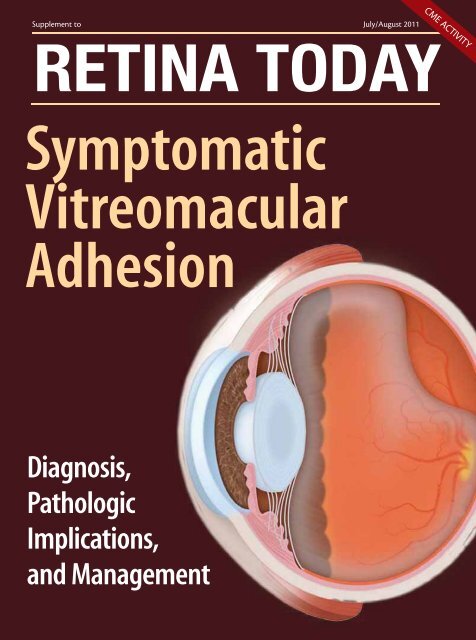
Supplement to July/August 2011<br />
RETINA TODAY<br />
Symptomatic<br />
Vitreomacular<br />
Adhesion<br />
CME ACTIVITY<br />
Diagnosis,<br />
Pathologic<br />
Implications,<br />
and Management
SYMPTOMATIC VITREOMACULAR ADHESION<br />
Release date: July 2011. Expiration date: July 2012.<br />
This continuing medical education activity is supported by an unrestricted educational grant from ThromboGenics<br />
STATEMENT OF NEED<br />
The goal of this continuing medical education (CME)<br />
supplement is to improve patient care by retina specialists<br />
and other ophthalmologists when they are managing symptomatic<br />
vitreomacular adhesion and traction.<br />
Symptomatic vitreomacular adhesion is a condition when<br />
the vitreous gel adheres in an abnormally strong manner to<br />
the retina. VMA can lead to vitreomacular traction (VMT)<br />
and subsequent loss or distortion of visual acuity.<br />
Anomalous posterior vitreous detachment PVD is linked to<br />
several retinal disorders including macular pucker, macular<br />
hole, age-related macular generation (AMD), macular<br />
edema, and retinal tears and detachment.<br />
The incidence of VMA has been reported to be as high as<br />
84% in cases of macular hole; 74% in vitreomacular traction<br />
syndrome; and 56% in idiopathic epimacular membrane. 1<br />
The incidence of VMA in macular edema appears to<br />
depend on the severity of the underlying condition. 2,3 In<br />
AMD, the rates vary 3-12 but have been reported to be as<br />
high as 59% in exudative AMD. 12<br />
Currently, pars plana vitrectomy (PPV) is used to surgically<br />
induce PVD and release the traction on the retina for selected<br />
cases. A vitrectomy procedure, however, is not without risk.<br />
Complications with standard PPV 12-15 and more recently with<br />
small-gauge PPV 16-20 have been reported and include retinal<br />
detachment, retinal tears, endophthalmitis, and postoperative<br />
cataract formation. Additionally, PPV may result in<br />
incomplete separation and it may potentially leave a nidus for<br />
vasoactive and vasoproliferative substances or it may induce<br />
development of fibrovascular membranes. Further, as is with<br />
any invasive surgical procedure, PPV introduces more trauma<br />
to the vitreous and surrounding tissues. 21,22<br />
There are data showing that pharmacological induction of<br />
PVD using ocriplasmin (formerly known as microplasmin), a<br />
proteolytic human enzyme with activity against the protein<br />
matrix that comprises the vitreoretinal interface, has the following<br />
advantages over PPV: It induces complete separation, creates<br />
a more physiologic state of the vitreomacular interface, prevents<br />
the development of fibrovascular membranes, is less traumatic<br />
to the vitreous, and is potentially prophylactic. 21,22 Additionally,<br />
vitreolysis obviates the costs associated with surgery and allows<br />
for earlier intervention, whereas surgery is reserved for more<br />
advanced cases. In two phase 3 studies, a single injection of<br />
ocriplasmin was shown to be safe and effective for PVD induction,<br />
23,24 providing further evidence that pharmacologic vitreolysis<br />
with ocriplasmin may provide an safe and effective alternative<br />
to PPV for inducing PVD.<br />
To address these gaps, retina specialists and other ophthalmologists<br />
must master insights on the pathogenesis of<br />
VMA, the role that VMA plays in various retinal pathologies,<br />
and the benefits of induced PVD vs anomalous PVD.<br />
Mastery includes knowledge of the clinical implications of<br />
VMA and the results of recent clinical trials on both surgical<br />
and pharmacologic PVD induction, an understanding of vitreolysis<br />
agents and their differences, and the ability to identify<br />
patients who may benefit from PVD induction.<br />
References<br />
1. Koerner F, Garweg J. [Diseases of the vitreo-macular interface]. Klin Monbl Augenheilkd.<br />
1999;214(5):305-310.<br />
2. Takahashi MK, Hikichi T, Akiba J, Yoshida A, Trempe CL. Role of the vitreous and macular<br />
edema in branch retinal vein occlusion. Ophthalmic Surg Lasers. 1997;28(4):294-299.<br />
3. Kado M, Jalkh AE, Yoshida A, et al. Vitreous changes and macular edema in central retinal<br />
vein occlusion. Ophthalmic Surg. 1990;21(8):544-549.<br />
4. Lambert HM, Capone A, Jr., Aaberg TM, et al. Surgical excision of subfoveal neovascular<br />
membranes in age-related macular degeneration. Am J Ophthalmol. 1992;113(3):257-262<br />
5. Weber-Krause B, Eckardt U. [Incidence of posterior vitreous detachment in eyes with and<br />
without age-related macular degeneration. An ultrasonic study]. Ophthalmologe.<br />
1996;93(6):660-665.<br />
6. Ondes F, Yilmaz G, Acar MA, et al. Role of the vitreous in age-related macular degeneration.<br />
Jpn J Ophthalmol. 2000;44(1):91-93.<br />
7. Krebs I, Brannath W, Glittenberg C, et al. Posterior vitreomacular adhesion: a potential risk<br />
factor for exudative age-related macular degeneration Am J Ophthalmol. 2007;144(5):741-<br />
746.<br />
8. Lee SJ, Lee CS, Koh HJ. Posterior vitreomacular adhesion and risk of exudative agerelated<br />
macular degeneration: paired eye study. Am J Ophthalmol. 2009;147(4):621-626 e1.<br />
9. Robison CD, Krebs I, Binder S, et al. Vitreomacular adhesion in active and end-stage agerelated<br />
macular degeneration. Am J Ophthalmol. 2009;148(1):79-82.<br />
10. Wheatley HM. Posterior vitreomacular adhesion and exudative age-related macular<br />
degeneration. Am J Ophthalmol. 2008;145(4):765; author reply -6.<br />
11. Schmidt JC, Mennel S, Meyer CH, Kroll P. Posterior vitreomacular adhesion: a potential<br />
risk factor for exudative age-related macular degeneration. Am J Ophthalmol.<br />
2008;145(6):1107; author reply -8.<br />
12. Mojana F, Cheng L, Bartsch DU, et al. The role of abnormal vitreomacular adhesion in<br />
age-related macular degeneration: spectral optical coherence tomography and surgical<br />
results. Am J Ophthalmol. 2008;146(2):218-227.<br />
13. Doft BH, Wisniewski SR, Kelsey SF, Groer-Fitzgerald S; Endophthalmitis Vitrectomy<br />
Study Group. Diabetes and postcataract extraction endophthalmitis. Curr Opin Ophthalmol.<br />
2002;13(3):147-151.<br />
14. Doft BM, Kelsey SF, Wisniewski SR. Retinal detachment in the endophthalmitis vitrectomy<br />
study. Arch Ophthalmol. 2000;118(12):1661-1665.<br />
15. Wisniewski SR, Capone A, Kelsey SF, et al. Characteristics after cataract extraction or<br />
secondary lens implantation among patients screened for the Endophthalmitis Vitrectomy<br />
Study. Ophthalmology. 2000;107(7):1274-1282.<br />
16. Gupta OP, Weichel ED, Regillo CD, et al. Postoperative complications associated with 25-<br />
gauge pars plana vitrectomy. Ophthalmic Surg Lasers Imaging. 2007;38(4):270–275.<br />
17. Liu DT, Chan CK, Fan DS, Lam SW, Lam DS, Chan WM. Choroidal folds after 25 gauge<br />
transconjunctival sutureless vitrectomy. Eye. 2005;19(7):825–827.<br />
18. Scott IU, Flynn HW Jr, Dev S, et al. Endophthalmitis after 25-gauge and 20-gauge pars<br />
plana vitrectomy: incidence and outcomes. Retina. 2008;28(1):138–142.<br />
19. Kunimoto DY, Kaiser RS; Wills Eye Retina Service. Incidence of endophthalmitis after 20-<br />
and 25-gauge vitrectomy. Ophthalmology. 2007;114(12):2133–2137.<br />
20. Kaiser RS. Complications of sutureless vitrectomy and the findings of the Micro-Surgical<br />
Safety Task Force. Paper presented at: Retina Subspecialty Day, Annual Meeting of the<br />
American Academy of Ophthalmology; November 7-8, 2008; Atlanta, GA.<br />
21. de Smet MD, Gandorfer A, Stalmans P, et al. Microplasmin intravitreal administration in<br />
patients with vitreomacular traction scheduled for vitrectomy: the MIVI I trial. Ophthalmology.<br />
2009;116(7):1349-1355.<br />
22. Goldenberg DT, Trese MT. Pharmacologic vitreodynamics and molecular flux. Dev<br />
Ophthalmol. 2009;44:31-36.<br />
23. Jumper J, Pakola S. The MIVI-007 trial. Phase 3 evaluation of single intravitreous injection<br />
of microplasmin or placebo for treatment of focal vitreomacular adhesion. Paper presented<br />
at: the American Society of Retina Specialists; August 31, 2010; Vancouver, BC.<br />
24. Packo K, Pakola S. The MIVI-006 trial. Phase 3 evaluation of single intravitreous injection<br />
of microplasmin or placebo for treatment of focal vitreomacular adhesion. Paper presented<br />
at: the American Society of Retina Specialists; August 31, 2010; Vancouver, BC.<br />
TARGET AUDIENCE<br />
This certified CME activity is designed for retina specialists<br />
and general ophthalmologists involved in the management<br />
of patients with retinal disease.<br />
LEARNING OBJECTIVES<br />
Upon completion of this activity, the participant should<br />
be able to:<br />
• Explain the process by which VMA occurs<br />
• Identify the disease states with which VMA is associated<br />
• Identify the clinical implications of anomalous PVD<br />
2 I SUPPLEMENT TO RETINA TODAY I JULY/AUGUST 2011
DIAGNOSIS, PATHOLOGIC IMPLICATIONS, AND MANAGEMENT<br />
• Identify the risks of performing vitrectomy to induce PVD<br />
• Explain the mechanism of action of pharmacologic vitreolysis<br />
• Differentiate between the various agents that can be<br />
used for pharmacologic vitreolysis in terms of their composition,<br />
advantages, and disadvantages<br />
• Discuss the available data on the safety and efficacy of<br />
vitreolysis agents for PVD induction<br />
METHOD OF INSTRUCTION<br />
Participants should read the CME activity in its entirety.<br />
After reviewing the material, please complete the selfassessment<br />
test, which consists of a series of multiplechoice questions.<br />
To answer these questions online and receive realtime<br />
results, please visit http://www.dulaneyfoundation.org<br />
and click “Online Courses.” Upon completing the activity<br />
and achieving a passing score of over 70% on the self-assessment<br />
test, you may print out a CME credit letter awarding<br />
1 AMA PRA Category 1 Credit. The estimated time to complete<br />
this activity is 1 hour.<br />
ACCREDITATION AND DESIGNATION<br />
This activity has been planned and implemented in<br />
accordance with the Essential Areas and policies of the<br />
Accreditation Council for Continuing Medical Education<br />
(ACCME) through the joint sponsorship of the Dulaney<br />
Foundation and Retina Today. The Dulaney Foundation is<br />
accredited by the ACCME to provide continuing education<br />
for physicians. The Dulaney Foundation designates this<br />
enduring material for a maximum of 1 AMA PRA Category<br />
1 Credit. Physicians should claim only the credit commensurate<br />
with the extent of their participation in the activity.<br />
DISCLOSURE<br />
In accordance with the disclosure policies of the Dulaney<br />
Foundation and to conform with ACCME and US Food and<br />
Drug Administration guidelines, anyone in a position to affect<br />
the content of a CME activity is required to disclose to the<br />
activity participants (1) the existence of any financial interest or<br />
other relationships with the manufacturers of any commercial<br />
products/devices or providers of commercial services and (2)<br />
identification of a commercial product/device that is unlabeled<br />
for use or an investigational use of a product/device not yet<br />
approved.<br />
FACULTY CREDENTIALS<br />
David M. Brown, MD, is the director of the<br />
Greater Houston Retina Research Center and<br />
practices at Retina Consultants of Houston and<br />
the Methodist Hospital in Houston, TX. He is a<br />
member of the Retina Today Editorial Board.<br />
Pravin U. Dugel, MD, is Managing Partner of<br />
Retinal Consultants of Arizona in Phoenix; Clinical<br />
Associate Professor of Ophthalmology, Doheny<br />
Eye Institute, Keck School of Medicine at the<br />
University of Southern California, Los Angeles; and<br />
Founding Member of the Spectra Eye Institute in Sun City,<br />
AZ. He is a member of the Retina Today Editorial Board.<br />
Mark S. Humayun, MD, PhD, is Professor of<br />
Ophthalmology, Biomedical Engineering, Cell<br />
and Neurobiology, Keck School of Medicine of<br />
the University of Southern California and is<br />
Associate Director of Research at the Doheny<br />
Retina Institute in Los Angeles.<br />
Peter K. Kaiser, MD is Professor of<br />
Ophthalmology at the Cleveland Clinic Lerner<br />
College of Medicine and a staff surgeon in the<br />
Vitreoretinal Department at the Cole Eye<br />
Institute, Cleveland Clinic. He is the Founding<br />
Director of the Digital Optical Coherence Tomography<br />
Reading Center (DOCTR) at the Cole Eye Institute. He is a<br />
member of the Retina Today Editorial Board.<br />
Michael T. Trese, MD, is Clinical Professor<br />
of Biomedical Sciences at The Eye Research<br />
Institute of Oakland University, Clinical<br />
Associate Professor at Wayne State University<br />
School of Medicine, and Chief of Pediatric and<br />
Adult Vitreoretinal Surgery at William Beaumont<br />
Hospital.<br />
FACULTY/STAFF DISCLOSURE DECLARATIONS<br />
David M. Brown, MD, reports that he receives financial<br />
support from Alcon, Alimera, Allergan, Eli Lilly, Genentech,<br />
Molecular Partners, Ophthotech, Paloma, Regeneron, Steba<br />
Biotech, and Thrombogenics; honoraria and/or travel reimbursement<br />
from Allergan, Genentech, and Regeneron; and is<br />
a consultant to Alcon, Alimera, Allergan, Genentech,<br />
Molecular Partners, Paloma, Regeneron, Steba Biotech, and<br />
Thrombogenics.<br />
Pravin U. Dugel, MD, reports that he is a consultant to<br />
Abbott Medical Optics, Alcon, Allergan, Arctic Dx, Bausch +<br />
Lomb, Genentech, Macusight, Neovista, ORA, QLT,<br />
Regeneron, and Santen.<br />
Mark S. Humayun, MD, PhD, reports that he is a consultant<br />
to, shareholder of, receives honoraria and/or travel reimbursement<br />
from, and holds patents with Second Sight<br />
Medical Products.<br />
Peter K. Kaiser, MD, reports that he is a consultant to<br />
Alcon, Arctic Dx, Bausch + Lomb, Bayer, Genentech, Kang<br />
Hong Biotech, Novartis, Opthotec, Oraya, and Regeneron;<br />
and is a shareholder in SKS Ocular.<br />
Michael T. Trese, MD, reports that he is an investor/shareholder<br />
with Nu-Vue Technologies; holds patents with Nu-<br />
Vue Technologies, and Thrombogenics; and is a consultant<br />
to Synergetics.<br />
All of those involved in the planning, editing, and peer<br />
review of this educational activity report no financial<br />
relationships.<br />
JULY/AUGUST 2011 I SUPPLEMENT TO RETINA TODAY I 3
SYMPTOMATIC VITREOMACULAR ADHESION<br />
Symptomatic Vitreomacular<br />
Adhesion (VMA): Diagnosis,<br />
Pathologic Implications,<br />
and Management<br />
Michael Trese, MD: Vitreomacular adhesion (VMA) is<br />
a condition when the vitreous gel adheres in an abnormally<br />
strong manner to the retina. VMA can lead to vitreomacular<br />
traction (VMT) and subsequent loss or distortion<br />
(metamorphopsia) of visual acuity, a condition<br />
known as symptomatic VMA. VMA occurs in the context<br />
of an incomplete or anomalous posterior vitreous<br />
detachment (PVD) and is linked to several retinal disorders<br />
including macular hole, epiretinal membrane (ERM),<br />
neovascular age-related macular degeneration (AMD),<br />
diabetic macular edema (DME), retina vein occlusion<br />
(RVO), and retinal tears and detachment. 1<br />
SYMPTOMATIC VMA AND ANOMALOUS PVD<br />
Dr. Trese: Dr. Brown, can you explain the process by<br />
which VMA and anomalous posterior vitreous detachment<br />
(PVD) occurs<br />
David M. Brown, MD: The simplest way to explain<br />
the process of VMA and anomalous PVD is how I<br />
explain it to my patients. I tell them that there is a gel<br />
inside the eye and that as a person gets older, the gel<br />
degenerates and clumps up creating floaters. Eventually<br />
the gel structure cannot keep its shape as a Jello ball,<br />
and it collapses in on itself. The vitreous gel is connected<br />
to the retina similar to Velcro and the adhesion is<br />
usually strongest at the optic nerve and at the fovea.<br />
When it pulls on the optic disc, it does not create much<br />
of a problem, but when it pulls on the fovea, the most<br />
delicate part of the photoreceptors and the thinnest<br />
part of the retina, it can create a macular hole, which<br />
requires surgical intervention.<br />
Peter K. Kaiser, MD: Most of us will eventually develop<br />
a posterior vitreous separation as a result of age, which<br />
involves both liquefaction of the vitreous as well as<br />
release of the vitreous from the retina. If the vitreous<br />
“Vitreomacular adhesion<br />
occurs in the context of<br />
an incomplete or anomalous<br />
posterior vitreous<br />
detachment and is linked to several<br />
retinal disorders.”<br />
— Michael T. Trese, MD<br />
liquifies and releases uniformly and in a synchronized<br />
manner, the result is a successful PVD; however, if VMA<br />
develops in the foveal region, the result can include macular<br />
hole, macular traction, vitreomacular traction syndrome<br />
(VMT), and in patients with diabetes, posterior<br />
hyaloid traction.<br />
Pravin U. Dugel, MD: We are beginning to understand<br />
that a PVD is not as simple as we once thought it<br />
was. It is important to note, as Dr. Kaiser stated, that<br />
there are two distinct steps that occur: liquefaction, or<br />
synesis, and separation of the interface, or syneresis,<br />
and if that two-step process does not occur in proper<br />
synchrony and completion, anomalous PVD can result,<br />
leading to VMA.<br />
Years ago, Jerry Sebag, MD, talked about a unifying<br />
concept in anomalous PVD. 1 A VMA resulting from<br />
anomalous PVD can go one of two ways. The vitreous<br />
can actually split to form a schisis and a partial thickness<br />
PVD, resulting in traction. If the traction goes outward,<br />
a macular hole may result. If the traction goes<br />
inward, an epiretinal membrane (ERM) or macular<br />
pucker may form. A full thickness separation of the vitreous<br />
may occur, but it may be caught up somewhere.<br />
If it is caught in the posterior pole, and posterior trac-<br />
4 I SUPPLEMENT TO RETINA TODAY I JULY/AUGUST 2011
DIAGNOSIS, PATHOLOGIC IMPLICATIONS, AND MANAGEMENT<br />
Figure 1. The implications of anomalous PVD.<br />
tion occurs with peripheral separation, macular traction<br />
may result. Macular traction can cause vitreomacular<br />
traction syndrome, but it is also linked to many<br />
other conditions such as neovascular AMD, diabetic<br />
retinopathy, DME, and RVO. If traction occurs at the<br />
optic disc, vitreopapillary syndrome, and ensuing disc<br />
edema, can occur, and if there is posterior separation<br />
but peripheral traction, retinal tears and retinal detachment<br />
can occur (Figure 1).<br />
It is interesting to see how VMA resulting from anomalous<br />
PVD is involved in so many diseases—I think that<br />
we are gaining more understanding because our imaging<br />
techniques are now married with what we know about<br />
the pathophysiology of this process.<br />
Mark Humayun, MD: In the era of optical coherence<br />
tomography (OCT), which allows for easier detection of<br />
macular adhesions, what is the definition of symptomatic<br />
VMA Perhaps we can see distortion of the retina on our<br />
OCT scans, but the patient may or may not attribute<br />
their symptoms to this condition. It is interesting, particularly<br />
in an age where we are able to see these adhesion<br />
areas much better.<br />
(Information courtesy of Jerry Sebag, MD)<br />
Dr. Trese: The diagnosis of PVD, which we all have on<br />
our diagnostic sheets, is really an exclusion of a retinal<br />
tear. How many times have each of us seen a patient<br />
who presents with flashes and floaters and diagnosed<br />
PVD, only to have the patient come back 2 years later<br />
with flashes and floaters, leading to the realization that<br />
the rest their vitreous has probably separated As previously<br />
stated, there are two separate concepts that are<br />
involved in the change in the vitreous—liquefaction<br />
and separation. Liquefaction is a core change and separation<br />
is a vitreoretinal juncture change. If the core<br />
change occurs without the juncture change, or the two<br />
do not take place relatively close in time to one another,<br />
anomalous PVD and subsequent VMA leading to traction<br />
and associated symptoms can occur.<br />
Dr Kaiser: Anomalous PVD is really any time liquefaction<br />
and separation do not occur at the same time. If the<br />
liquefaction develops without the release, symptomatic<br />
VMA or a tear in the periphery can result.<br />
Dr. Trese: If you look at the dispase study, which<br />
evaluated this agent that liquifies the vitreous without<br />
JULY/AUGUST 2011 I SUPPLEMENT TO RETINA TODAY I 5
SYMPTOMATIC VITREOMACULAR ADHESION<br />
Chemical Vitreous Manipulation<br />
May Represent New Frontier in the<br />
Treatment of Retinal Disease of Wet AMD<br />
Ocriplasmin, a bioengineered truncated form of the<br />
human plasmin molecule, has proved effective in providing<br />
resolution of vitreomacular adhesion (VMA) and improving<br />
visual acuity and function when compared to placebo injection<br />
in two large phase 3 studies.<br />
Pravin U. Dugel, MD, presented the pooled data from<br />
MIVI-6 and MIVI-7 during the Institut de Microcirurgia<br />
Ocular Trends in Surgical and Medical Retina Meeting in<br />
Barcelona. 1 Dr. Dugel said that 26.5% of patients achieved<br />
the primary endpoint of pharmacologic VMA resolution at<br />
day 28 after one injection of ocriplasmin, with approximately<br />
75% of these patients experiencing resolution within<br />
1 week of injection. Enrollment criteria for the trials did not<br />
exclude patients with epiretinal membrane (ERM). “If you<br />
look at patients who did not have an ERM with one injection<br />
alone, 37.4% achieved this end goal of resolution of<br />
VMA. If you look at patients with an ERM, 8.7% had resolution<br />
of VMA, which is also statistically significant.”<br />
Toward the secondary endpoints, 13.4% of patients<br />
achieved complete posterior vitreous detachment (PVD)<br />
and 23.7% and almost 10% gained two or three lines of<br />
vision or more, respectively. Over 40% of patients with fullthickness<br />
macular hole had closure after a single injection.<br />
For macular holes smaller than 250 µm, however, this<br />
increased to almost 60%, said Dr. Dugel. Visual function<br />
and quality of life parameters were also favorable in the<br />
groups that received ocriplasmin injection. The safety profile<br />
of ocriplasmin was favorable in both phase 3 studies,<br />
according to Dr. Dugel.<br />
Although the mechanisms of PVD appear simple, the<br />
process is complicated, requiring the steps of liquefaction<br />
and separation of the vitreous from the retina to be synchronized,<br />
said Dr. Dugel. Abnormal VMA and subsequent<br />
partial PVD can lead to vitreomacular traction (VMT) syndrome,<br />
resulting in metamorphopsia and decreased vision.<br />
Many patients with VMA, however, present with relatively<br />
good visual acuity. “For these patients, we just watch and<br />
wait, because the risk/benefit ratio of vitrectomy, currently<br />
the only treatment available, will be too large,” said<br />
Dr. Dugel. “However, these patients come to our office<br />
unhappy and leave our office unhappy.”<br />
VMA is implicated in a variety of retinal diseases, including<br />
macular hole, neovascular age-related macular degeneration<br />
(AMD), diabetic macular edema (DME), retinal<br />
vein occlusions (RVO), and vitreopapillary syndrome, said<br />
Dr. Dugel, and improved imaging may allow for earlier<br />
detection of VMA. “Chemical vitreous manipulation has<br />
the potential to have a major effect on clinical practice,”<br />
he concluded.<br />
1. Dugel PU. A single injection of ocriplasmin for the treatment of symptomatic vitreomacular<br />
adhesion (sMVA): results of phase III MIVI-TRUST program. Paper presented at:<br />
Trends in Surgical and Medical Retina. June 3-4, 2011; Barcelona.<br />
causing vitreous-retina interface separation, the retinal<br />
detachment rate was approximately 5%, which highlights<br />
the danger of inducing liquefaction without<br />
manipulation of the vitreoretinal juncture. 2<br />
Dr. Brown: Both liquefaction and syneresis occur naturally<br />
as the eye ages. Disease processes, however, such as<br />
diabetes and AMD, cause exudation and make the retinal<br />
interfaces thicker and harder to break. Thus, there may<br />
be a higher incidence of anomalous PVDs as the gel<br />
needs to pull away in these eyes, resulting in more<br />
pathology.<br />
Dr. Trese: In the case of diabetes, hyperglycemia alone<br />
increases laminin and fibronectin, two of the three<br />
components in the vitreoretinal juncture as well as the<br />
glomerular extracellular matrix, tightening the vitreoretinal<br />
juncture and reducing the filtration rate from<br />
the kidneys.<br />
Dr. Humayun: There are many different ways to think<br />
about VMA. Most of us know that the macula is the area<br />
we are trying to protect from developing neovascularization,<br />
hemorrhage, or macular edema. We also know that<br />
vitreous is adherently attached to the optic nerve and<br />
along the vessels and can create VMT. This interface is<br />
complex—it can have both fibrous and cellular components.<br />
As we better understand the retinal interface, it is<br />
becoming clear that it plays a role in many retina diseases,<br />
particularly when there is traction on the retina.<br />
It would be advantageous to use the tools we currently<br />
have to perform an optical-imaging biopsy with OCT<br />
6 I SUPPLEMENT TO RETINA TODAY I JULY/AUGUST 2011
DIAGNOSIS, PATHOLOGIC IMPLICATIONS, AND MANAGEMENT<br />
under dynamic conditions to better understand the<br />
mechanical functions.<br />
PHARMACOLOGIC VITREOLYSIS<br />
Dr. Trese: How would you define pharmacologic<br />
vitreolysis<br />
Dr. Brown: Pharmacologic vitreolysis or pharmacologic<br />
vitreodynamics is the process by which an enzyme<br />
is used to mechanically change the vitreous. This<br />
involves two distinct mechanisms. One mechanism is<br />
liquefaction of the core vitreous and the other is degradation<br />
of the vitreoretinal interface. By doing this we<br />
change not only the physical traction where the<br />
“Velcro” is trying to separate from the retina, but also<br />
changes in the milieu with the oxygenation and chemical<br />
balance that go along with this separation of the vitreous<br />
from the retina.<br />
Dr. Trese: What is the role of the vitreous in disease<br />
states such as AMD, DME, and RVO<br />
Dr. Dugel: It may be too early to tell, but there are<br />
some articles that are coming out that are compelling. A<br />
study from Susan Binder, MD’s, group showed that PVD<br />
may be protective against AMD, whereas VMA may promote<br />
exudative AMD. 3 Another earlier paper from Dr.<br />
Binder’s group demonstrated that persistent PVD may be<br />
a risk factor for exudative AMD from inflammationinduced<br />
vitreoretinal traction. 4<br />
The role of the vitreous in disease states like AMD is<br />
interesting. Obviously, we have an unsustainable model<br />
in our current monotherapy treatment of AMD with<br />
injections of anti-VEGF, and I think that we can all agree<br />
that combination therapy is an improvement on<br />
monotherapy. However, we have just recently started<br />
talking about manipulation of the vitreous and how this<br />
may play an important role in disease management.<br />
Your group published an animal study regarding the<br />
effect of bevacizumab on vitreous detachment. 5 A study<br />
such as this, which evaluates the effects of pharmacologic<br />
agents on vitreous manipulation, may have important<br />
implications.<br />
Dr. Kaiser: It is difficult to grasp the extent of the vitreous’<br />
role once a patient has developed choroidal neovascularization<br />
(CNV) and AMD. There is clearly a strong<br />
association, as evidenced in the literature. 3,4,6,7 The question<br />
that remains, however, is if CNV is already present,<br />
would inducing vitreous separation alleviate symptoms<br />
or enhance the effect of an anti-VEGF agent to decrease<br />
the number of injections needed<br />
“We have just recently started<br />
talking about manipulation<br />
of the vitreous and how this<br />
may play an important role in<br />
disease management.”<br />
— Pravin U. Dugel, MD<br />
Dr. Trese: Dr. Dugel, can you describe the enzymatic<br />
agent, ocriplasmin (ThromboGenics), formerly known as<br />
microplasmin that has been under study for pharmacologic<br />
vitreolysis for symptomatic VMA<br />
Dr. Dugel: Ocriplasmin is a truncated form of human<br />
plasmin. Ocriplasmin is not a new drug—it has been in<br />
development for many years and there are excellent<br />
phase 1 and phase 2 studies that were previously<br />
encouraging. The phase 3 studies, which were two large<br />
multicenter, prospective, randomized trials, were<br />
remarkably consistent in regard to the safety and efficacy<br />
outcomes. 8,9 As far as clinical use is concerned, this<br />
drug was tested in a diverse population of patients with<br />
diseases such as VMT, macular hole, and ERM (See “Key<br />
Published Literature”). The applicability, however, will<br />
extend far beyond these diseases. It will go to other diseases<br />
where VMT may be involved such as DME and<br />
neovascular AMD. It will also extend to the combination<br />
of ocriplasmin with other drugs such as anti-VEGF<br />
agents; there is now evidence that this drug may make<br />
anti-VEGF treatments more effective when used in combination.<br />
5 Finally, I think this drug will also be used as an<br />
adjunct to surgery. The potential of injecting this drug<br />
the day before surgery and making a very complicated<br />
case much easier is intriguing. In general, I think that we<br />
have a great deal of evidence regarding this drug but the<br />
potential clinical applications of pharmacologic vitreolysis<br />
may be enormous.<br />
POTENTIAL APPLICATIONS FOR VITREOLYSIS<br />
Dr. Brown: One of the unmet medical needs in terms<br />
of classic VMT is for patients with impending macular<br />
hole but relatively good visual acuity, placing the surgeon<br />
on the fence as to whether to operate sooner or later. To<br />
have an agent for vitreolysis would help a fair number of<br />
our patients avoid surgery completely.<br />
Dr. Kaiser: You could take it even one step further.<br />
Patients with so-called stage zero macular holes where<br />
the fellow eye has a gull-wing shaped vitreous coming<br />
JULY/AUGUST 2011 I SUPPLEMENT TO RETINA TODAY I 7
SYMPTOMATIC VITREOMACULAR ADHESION<br />
Pharmacologic Vitreolysis:<br />
Key Published Literature<br />
• Benz MS, Packo KH, Gonzalez V, et al. A placebo-controlled<br />
trial of microplasmin intravitreous injection to<br />
facilitate posterior vitreous detachment before vitrectomy.<br />
Ophthalmology. 2010;117(4):791-797.<br />
• de Smet MD, Gandorfer A, Stalmans P, et al. Microplasmin<br />
intravitreal administration in patients with vitreomacular<br />
traction scheduled for vitrectomy: the MIVI I trial.<br />
Ophthalmology. 2009;116(7):1349-1355.<br />
• de Smet MD, Valmaggia C, Zarranz-Ventura J, Willekens B.<br />
Microplasmin: ex vivo characterization of its activity in<br />
porcine vitreous. Invest Ophthalmol Vis Sci. 2009;50(2):814-<br />
819.<br />
• Gad Elkareem AM, Willekens B, Stassen JM, de Smet MD.<br />
Differential vitreous dye diffusion following microplasmin<br />
or plasmin pre-treatment. Curr Eye Res. 2010;35(3):235-<br />
241.<br />
• Gad Elkareem AM, Willekens B, Vanhove M, Noppen B,<br />
Stassen JM, de Smet MD. Characterization of a stabilized<br />
form of microplasmin for the induction of posterior vitreous<br />
detachment. Curr Eye Res. 2010;35(10):909-915.<br />
• Gandorfer A, Rohleder M, Sethi C, et al. Posterior vitreous<br />
detachment induced by microplasmin. Invest Ophthalmol<br />
Vis Sci. 2004;45(2):641-647.<br />
• Gandorfer A. Enzymatic vitreous disruption. Eye (Lond).<br />
2008;22(10):1273-1277.<br />
• Gandorfer A. Experimental evaluation of<br />
microplasmin – an alternative to vital dyes. Dev<br />
Ophthalmol. 2008;42:153-159.<br />
• Gandorfer A. Microplasmin-assisted vitrectomy. Dev<br />
Ophthalmol. 2009;44:26-30.<br />
• Gandorfer A. Pharmacological vitreolysis [in German]. Klin<br />
Monbl Augenheilkd. 2011;228(3):201-207.<br />
• Giblin FJ, Quiram PA, Leverenz VR, Baker RM, Dang L,<br />
Trese MT. Enzyme-induced posterior vitreous detachment<br />
in the rat produces increased lens nuclear pO 2 levels. Exp<br />
Eye Res. 2009;88(2):286-292.<br />
• Goldenberg DT, Giblin FJ, Cheng M, et al. Posterior vitreous<br />
detachment with microplasmin alters the retinal penetration<br />
of intravitreal bevacizumab (Avastin) in rabbit<br />
eyes. Retina. 2011;31(2):393-400.<br />
• Goldenberg DT, Trese MT. Pharmacologic vitreodynamics<br />
and molecular flux. Dev Ophthalmol. 2009;44:31-36.<br />
• Lopez-Lopez F, Rodriguez-Blanco M, Gómez-Ulla F,<br />
Marticorena J. Enzymatic vitreolysis. Curr Diabetes Rev.<br />
2009;5(1):57-62.<br />
• Quiram PA, Leverenz VR, Baker RM, Dang L, Giblin FJ,<br />
Trese MT. Microplasmin-induced posterior vitreous<br />
detachment affects vitreous oxygen levels. Retina.<br />
2007;27(8):1090-1096.<br />
• Sakuma T, Tanaka M, Mizota A, Inoue J, Pakola S. Safety of<br />
in vivo pharmacologic vitreolysis with recombinant<br />
microplasmin in rabbit eyes. Invest Ophthalmol Vis Sci.<br />
2005;46(9):3295-3299.<br />
• Sebag J, Ansari RR, Suh KI. Pharmacologic vitreolysis with<br />
microplasmin increases vitreous diffusion coefficients.<br />
Graefes Arch Clin Exp Ophthalmol. 2007;245(4):576-580.<br />
• Sebag J. Molecular biology of pharmacologic vitreolysis.<br />
Trans Am Ophthalmol Soc. 2005;103:473-94.473-494.<br />
• Stalmans P, Delaey C, de Smet MD, van Dijkman E, Pakola<br />
S. Intravitreal injection of microplasmin for treatment of<br />
vitreomacular adhesion: results of a prospective, randomized,<br />
sham-controlled phase II trial (the MIVI-IIT trial).<br />
Retina. 2010;30(7):1122-1127.<br />
• Udaondo P, Diaz-Llopis M, Garcia-Delpech S, Salom D,<br />
Arevalo JF. Microplasmin for vitreomacular traction.<br />
Ophthalmology. 2010;117(9):1859; author reply 1859-1860.<br />
8 I SUPPLEMENT TO RETINA TODAY I JULY/AUGUST 2011
DIAGNOSIS, PATHOLOGIC IMPLICATIONS, AND MANAGEMENT<br />
down to the fovea but no hole and no true traction<br />
may be a higher risk for developing a bilateral hole<br />
some time in the future. 10 Should we treat these<br />
patients with a vitreolysis agent as prophylaxis for a<br />
potential future problem<br />
Dr. Trese: Gull-shaped retinal detachments create a<br />
large amount of traction. Lois Smith, MD, PhD, told me<br />
about an individual in the lab at Children’s Hospital<br />
Boston who has demonstrated that isolated traction on<br />
Muller cells can generate VEGF. I am not sure what role<br />
vitreoretinal adhesion plays in AMD, but I agree that it is<br />
most likely early in the process. Once CNV is present, we<br />
need to study if cleaving the adhesion will help.<br />
Dr. Dugel: I also see potential for vitreolysis agents as<br />
adjuncts to surgery. Some of the hardest cases we see are<br />
complicated tractional retinal detachments in diabetics<br />
and retinal detachments in pediatric patients. If we can<br />
inject a vitreolysis agent a day prior to surgery, it might<br />
make the the surgery easier and more successful.<br />
Dr. Brown: There is postulation that schisis pockets<br />
can act as a sink for VEGF. We know that after a PVD<br />
there is more oxygenation, so a PVD would be ideal for<br />
every patient with diabetes. Even when the glycosalated<br />
hemoglobin is causing vascular damage, you want<br />
more oxygen for the retina and a better escape for any<br />
ischemic factors. I do not see any reason not to induce a<br />
PVD as long as I have an agent that is effective and safe.<br />
Dr. Kaiser: Assuming we had a drug that is effective<br />
and safe, it would be best to induce PVD prior to the<br />
development of exudation and protein release that<br />
increases adherence, because after these occur, it will<br />
become more difficult to release the junction. So I think<br />
you can argue that it is reasonable to inject with ocriplasmin<br />
when mild changes become apparent to prevent<br />
future complications.<br />
Dr. Dugel: Because we know a lot more about the<br />
role of the vitreous in diabetes than we do in AMD, we<br />
are more apt to argue for early injection. The safety<br />
profile of ocriplasmin was shown to be excellent in the<br />
phase 3 trials. 8,9 In fact, there were more cases of retinal<br />
tears and detachments in the placebo group than<br />
in the drug group, so these were most likely complications<br />
of vitrectomy.<br />
Dr. Trese: Let’s say that sometime in the future you<br />
have a patient with a 9-year history of diabetes who has a<br />
couple of dot and blot hemorrhages and no PVD on<br />
“I do not see any reason<br />
not to induce a PVD as<br />
long as I have an agent that<br />
is effective and safe.”<br />
— David M. Brown, MD<br />
OCT or ultrasound. What do you do if, after injecting<br />
ocriplasmin, a PVD does not result Peter Stalmans, MD,<br />
presented data from the phase 2 ocriplasmin study on a<br />
group of patients with diabetes. He injected the enzyme<br />
in these patients, many of whom had proliferation and<br />
had undergone laser and approximately 11% to 14%<br />
cleaved. In my opinion, this is a miracle because there<br />
was no case selection. 12<br />
If you did not have a PVD in such a patient, would you<br />
reinject<br />
Dr. Dugel: I would reinject. Regarding the data to<br />
which you refer from the phase 2 trials, there was one<br />
cohort of nine patients for whom reinjection was<br />
allowed. Although the group was small, the rate of PVD<br />
after reinjection was almost 60%. 12 Given the very strong<br />
safety profile of ocriplasmin in over 800 eyes, I would not<br />
hesitate to inject two or three times.<br />
Dr. Kaiser: The phase 3 study did not look at reinjection,<br />
but I think that it is important to consider<br />
that maybe the first injection is just not getting the<br />
drug to the right location. With a second or third<br />
injection, you will certainly increase the likelihood of<br />
getting it to the right place and being able to cleave<br />
the junction. If and when ocriplasmin is approved by<br />
the US Food and Drug Administration, a good investigator-sponsored<br />
trial might include an analysis of<br />
reinjection data.<br />
Dr. Trese: Another scenario where vitreous manipulation<br />
may be justified is in cases of capillary dropout in<br />
retinal vein occlusion. Injecting ocriplasmin could help to<br />
re-establish circulation in some of these cases.<br />
Dr. Dugel: Our whole way of thinking of neovascularization<br />
might change if we could use vitreous manipulation<br />
to affect circulation. Currently, in patients with<br />
ischemia form any cause, we have no effective treatment.<br />
It would be very valuable to have a drug that could<br />
increase circulation by chemical vitreous manipulation.<br />
This is plausible: there is good evidence, originally from<br />
JULY/AUGUST 2011 I SUPPLEMENT TO RETINA TODAY I 9
SYMPTOMATIC VITREOMACULAR ADHESION<br />
Stefansson, that mechanical vitreous removal increases<br />
oxygenation. 13<br />
Dr. Trese: Imagine that you are examining a patient<br />
who has VMT due to VMA without a membrane, a macular<br />
hole with less than a 1,500-µm attachment, and lattice<br />
degeneration in the periphery. How do you handle<br />
this case<br />
“Vitrectomy is certainly an<br />
excellent way to fix symptomatic<br />
VMAs, but if a safer,<br />
quicker way exists to do it,<br />
it is better for the patient.”<br />
— Peter K. Kaiser, MD<br />
Dr. Brown: Because the induction of a PVD might<br />
very well increase the risk of a peripheral tear in this<br />
patient with peripheral lattice, I would probably demarcate<br />
the peripheral lattice degeneration lesions with<br />
thermal laser at least 3 or 4 days prior to injecting an<br />
agent to induce pharmacologic vitreolysis. In the<br />
ocriplasmin clinical trials at our site, I did not have any<br />
cases of retinal tearing, but we did not have many<br />
patients with peripheral pathology.<br />
Dr. Trese: One of the first patients treated in the clinical<br />
trials in Europe did have a retinal tear. The clinician<br />
who was treating the patient did not see any sign of lattice<br />
degeneration in the periphery. However, after being<br />
treated with laser, they did well. Although the tear rate<br />
was not high in the clinical trials, it is something that we<br />
need to think about.<br />
Dr. Kaiser: Although I do not consider lattice degeneration<br />
a contraindication for ocriplasmin injection, it<br />
is important to discuss the risks of retinal tears with<br />
your patients. It is important to conduct a thorough<br />
exam prior to considering this type of injection, and if<br />
lattice degeneration is detected, the option of treating<br />
with laser prior to injection vs just injection should be<br />
discussed. Additionally, patients should be counseled<br />
on symptoms of retinal tear, should it occur.<br />
MAXIMIZING THE MECHANICAL AND<br />
BIOCHEMICAL ACTIVITY OF OCRIPLASMIN<br />
Dr. Dugel: The size of the VMA is directly related to<br />
how well ocriplasmin works. Additionally, the location<br />
of the injection is important. If it is injected away from<br />
the pathology, it will be hard for the drug to reach the<br />
location of the adhesion.<br />
Dr. Trese: If you look at the 250-µm adhesions from<br />
the MIVI-TRUST phase 3 clinical trials for ocriplasmin,<br />
there were approximately 60% of these adhesions that<br />
cleaved. 8,9 For those that were 1,500 µm or larger, the<br />
numbers that cleaved were small. How do you assess<br />
this clinically First, as Dr. Dugel said, delivery of the<br />
enzyme is critical to get the ocriplasmin to where it<br />
can work.<br />
We presented a poster at the Association for<br />
Research in Vision and Ophthalmology 2011 meeting<br />
that shows a technique that attempts to achieve a larger<br />
area of drug from the same bolus. We found that<br />
when injecting India ink with a 30-gauge quadraport<br />
needle, as compared to a standard 30-gauge needle to<br />
inject India ink, the ink spread more quickly and to a<br />
wider area, whereas the ink accumulated around the tip<br />
of the standard 30-gauge needle. 11<br />
In another group of animals injected with ocriplasmin<br />
and plasmin using a standard 30-gauge needle vs the<br />
quadraport 30-gauge needle, scanning electron<br />
microscopy at 1 week showed that there was no residual<br />
enzyme on the internal limiting membrane with the<br />
quadraport needle. 11<br />
Dr. Dugel: Achieving needle delivery that will allow for<br />
selective infusion of the drug to the site where it is needed<br />
is a challenging task. Approximately 14% of patients<br />
and 20% of patients without an ERM treated with<br />
ocriplasmin in the pivotal phase 3 trials achieved the secondary<br />
endpoint of total PVD induction. 8,9<br />
Dr. Humayun: Mechanically speaking, the idea of<br />
injecting in different places, lifting the attachment, and<br />
eventually making the entire sheet come off seems to<br />
make sense.<br />
Dr. Brown: So you are saying is that after injecting<br />
ocriplasmin in several locations, if you lift up one edge<br />
and you move it back and forth, this may be of benefit<br />
and a more gentle vitreolysis<br />
Dr. Dugel: Actually, sequential injections may be easier.<br />
We have some data on this from the phase 2 trials<br />
regarding these from one cohort in which this was<br />
allowed, and it was shown that sequential injections my<br />
increase the rate of total PVD without having to move<br />
the patient’s eye.<br />
Dr. Trese: When discussing how to maximize mechani-<br />
10 I SUPPLEMENT TO RETINA TODAY I JULY/AUGUST 2011
DIAGNOSIS, PATHOLOGIC IMPLICATIONS, AND MANAGEMENT<br />
cal and biochemical activity, reliable imaging may<br />
enhance the ability to target exactly where you want to<br />
inject the enzyme. The dynamic SD-OCT imaging that<br />
we have with the Spectralis (Heidelberg Engineering,<br />
Carlsbad, CA) offers a better image of the vitreous so<br />
that we can better understand its relationship to retinal<br />
disease.<br />
We have put together a procedure room to perform<br />
the injections of ocriplasmin that allows us to do the<br />
injection through an operating microscope. It would be<br />
ideal to be able to do these injections in an OCT-guided<br />
manner.<br />
Dr. Kaiser: You already have half of the system in<br />
place. In the future, microscopes with built-in OCT<br />
systems will be available. Currently, the Bioptigen<br />
Spectral Domain Ophthalmic Imaging System (SDOIS;<br />
Research Triangle Park, NC) has a mount for most<br />
microscopes, and Carl Zeiss Meditec (Dublin, CA) is<br />
working on a version of microscope with OCT that is<br />
built in. The ability to see underneath the vitreous<br />
during injection is imminent.<br />
Dr. Brown: What do we know of the diffusion of<br />
ocriplasmin<br />
Dr. Trese: The way that ocriplasmin works is that it<br />
takes one bite and then it either binds to substrate or<br />
consumes itself. So if you inject it in a large bolus through<br />
a standard needle it consumes substrate and then itself. If<br />
you use a multiport needle, however, such as the quadraport<br />
needle that was used in our study, the chances of<br />
getting more substrate are higher.<br />
CHANGING TREATMENT PARADIGMS<br />
Dr. Trese: This has been an interesting year in retina.<br />
We learned the results of the Comparison of AMD<br />
Treatments Trial, which showed that an inexpensive anti-<br />
VEGF agent, bevacizumab (Avastin, Genentech) is noninferior<br />
to a very expensive anti-VEGF agent, ranibizumab<br />
(Lucentis, Genentech). We also have results from the two<br />
large clinical trials showing the safety and efficacy of<br />
VEGF trap (Eyelea, Regeneron), although we do not<br />
know how this drug will be priced when and if it is<br />
approved.<br />
How do you think that physicians feel about something<br />
that takes away few of the surgical procedures that<br />
they enjoy performing and that may represent another<br />
expensive drug to handle in the office<br />
Dr. Kaiser: As physicians, we all have the same goal: to<br />
treat patients in the best and safest way we can.<br />
“In ophthalmology,<br />
when a noninvasive option<br />
becomes available we<br />
typically intervene earlier.”<br />
— Mark S. Humayun, MD, PhD<br />
Vitrectomy is certainly an excellent way to fix symptomatic<br />
VMAs, but if a safer, quicker way exists to do it, it is<br />
better for the patient.<br />
Dr. Dugel: I agree. However, there are many instances<br />
where surgery is still indicated. In the clinical trials for<br />
ocriplasmin, there was almost a 60% closure rate of macular<br />
hole with a single injection if the hole was 250 µm or<br />
smaller. 9,10 But if in the case of a large macular hole with<br />
no VMA, there is no reason to inject ocriplasmin; we still<br />
need to perform surgery.<br />
Another consideration is that this drug may actually<br />
enhance our surgery. I can picture using this drug for<br />
complicated traction detachment or for a young patient<br />
who requires a PVD, so I do not think that pharmacologic<br />
vitreolysis is going to take away from our surgery. Also,<br />
in patients with ERM, ocriplasmin will help to resolve<br />
part of the problem; symptoms associated with VMA<br />
leading to antereoposterior traction. We will still have to<br />
perform surgeries to relieve the tangential forces exerted<br />
by an ERM. In fact, I think ocriplasmin will be a safe and<br />
effective alternative for patients who currently have no<br />
treatment options, patients with VMA and early symptoms,<br />
and will be an effective adjuvant for difficult surgical<br />
cases, making us better surgeons. I do not see push<br />
back of this drug from surgeons. On the contrary, I predict<br />
surgeons will see the potential beneficial effect of<br />
this drug for our patients in the office and in surgery and<br />
will welcome access to it.<br />
Dr. Humayun: In ophthalmology, when a noninvasive<br />
option becomes available we typically intervene<br />
earlier. Ocriplasmin will allow us to treat patients in<br />
whom there clearly is pathology and who are symptomatic<br />
but for whom surgery is not indicated because<br />
of the poor risk/benefit ratio. I do not think that there<br />
is much overlap with surgery, particularly when you<br />
are talking about earlier disease.<br />
Dr. Trese: I think that eventually, criteria for early intervention<br />
for symptomatic VMA with ocriplasmin will be<br />
developed. I imagine that the American Academy of<br />
JULY/AUGUST 2011 I SUPPLEMENT TO RETINA TODAY I 11
SYMPTOMATIC VITREOMACULAR ADHESION<br />
Ophthalmology will produce guidelines based on highresolution<br />
OCT imaging to help physicians in using this<br />
drug effectively.<br />
SUMMARY<br />
Dr. Kaiser: The ability to treat symptomatic VMA, particularly<br />
early macular holes, with pharmacologic vitreolysis<br />
offers an efficient and safe way to improve our<br />
patients symptoms without requiring surgery or facedown<br />
positioning. Moreover, the therapy may help us if<br />
surgery is required in the future, as the vitreous is more<br />
liquefied and easier to peel.<br />
Dr. Brown: The addition of ocriplasmin will be a welcome<br />
addition to the armamentarium of vitreoretinal<br />
surgeons. The agent will be first used in patients with<br />
impending macular holes and small holes with persistent<br />
VMT at the edges. If multiple injections (or novel injection<br />
techniques) can result in reliable pharmacologic vitreolysis,<br />
I can easily see the agent being used routinely<br />
before vitreoretinal surgery and possibly as a prophylactic<br />
agent in diabetic patients prior to the development of<br />
proliferative disease.<br />
Dr. Humayun: As the field of enzymatic vitreolysis continues<br />
to advance and FDA-approved methodologies<br />
become available, there is no doubt that such an<br />
approach will be used to treat symptomatic VMA.<br />
Dr. Dugel: There are three forces that I believe will<br />
come together to make ocriplasmin a very valuable<br />
drug for the retina specialist. The first force is the<br />
advent of technological advances with OCT that allow<br />
us to easily visualize VMA. The second force is that all<br />
retina specialists are adept at performing an intravitreal<br />
injection. The third force is that we currently<br />
employ a watch-and-wait strategy for many of our<br />
patients due to the lack of options for a safe, noninvasive<br />
procedure. If ocriplasmin becomes available, I<br />
believe that retina practices are prime to adopt this<br />
agent for pharmacologic vitreolysis. ■<br />
1. Sebag J. Anomalous posterior vitreous detachment: a unifying concept in vitreo-retinal<br />
disease. Graefes Arch Clin Exp Ophthalmol. 2004;242(8):690-698.<br />
2. Tezel TH, Del Priore LV, Kaplan HJ. Posterior vitreous detachment with dispase. Retina.<br />
1998;18:7-15.<br />
3. Robison CD, Krebs I, Binder S, et al. Vitreomacular adhesion in active and end-stage<br />
age-related macular degeneration. Am J Ophthalmol. 2009;148(1):79-82.<br />
4. Krebs I, Brannath W, Glittenberg C, Zeiler F, Sebag J, Binder S. Posterior vitreomacular<br />
adhesion: a potential risk factor for exudative age-related macular degeneration Am J<br />
Ophthalmol. 2007;144(5):741-746.<br />
5. Goldenberg DT, Giblin FJ, Cheng M, et al. Posterior vitreous detachment with<br />
microplasmin alters the retinal penetration of intravitreal bevacizumab (Avastin) in rabbit<br />
eyes. Retina. 2011;31(2):393-400.<br />
6. Nomura Y, Ueta T, Iriyama A, et al. Vitreomacular interface in typical exudative agerelated<br />
macular degeneration and polypoidal choroidal vasculopathy. Ophthalmology.<br />
2011;118(5):853-859.<br />
7. Stefánsson E, Geirsdóttir A, Sigurdsson H. Metabolic physiology in age related macular<br />
degeneration.Prog Retin Eye Res. 2011;30(1):72-80.<br />
8. Jumper J, Pakola S. The MIVI-007 trial. Phase 3 evaluation of single intravitreous injection<br />
of microplasmin or placebo for treatment of focal vitreomacular adhesion. Paper presented<br />
at: the American Society of Retina Specialists; August 31, 2010; Vancouver, BC.<br />
9. Packo K, Pakola S. The MIVI-006 trial. Phase 3 evaluation of single intravitreous injection<br />
of microplasmin or placebo for treatment of focal vitreomacular adhesion. Paper presented<br />
at: the American Society of Retina Specialists; August 31, 2010; Vancouver, BC.<br />
10. Chan A, Duker JS, Schuman JS, Fujimoto JG. Stage 0 macular holes: observations by<br />
optical coherence tomography. Ophthalmology. 2004;111(11):2027-2032.<br />
11. Trese MT, Asami T. A quadraport needle gives better spread of drug during intravitreous<br />
injection. Poster presented at the Association for Research in Vision and Ophthalmology<br />
annual meeting; May 1-5, 2011; Fort Lauderdale, FL.<br />
12. Stalmans P, Delaey C, de Smet, M, van Dijkman E, Pakola S. Intravitreal injection of<br />
microplasmin for treatment of vitreomacular adhesion. Results of a prospective, randomized,<br />
sham-controlled phase II trial (The MIVI-IIT Trial). Retina. 2010.30(7):1122-1127.<br />
13. Stefánsson E. Ocular oxygenation and the treatment of diabetic retinopathy. Surv<br />
Ophthalmol. 2006;51(4)364-380.<br />
12 I SUPPLEMENT TO RETINA TODAY I JULY/AUGUST 2011
DIAGNOSIS, PATHOLOGIC IMPLICATIONS, AND MANAGEMENT<br />
Symptomatic VMA: Diagnosis, Pathologic Implications, and Management<br />
1.0 AMA PRA Category 1 Credit Expires July 2012<br />
CME credit is available electronically via www.dulaneyfoundation.org.<br />
To answer these questions online and receive real-time results, please visit www.dulaneyfoundation.org and click “Online Courses.” If<br />
you are experiencing problems with the online test, please e-mail us at support@dulaneyfoundation.org or call +1-610-619-0414.<br />
Alternatively, you can fax your exam to us at +1-610-771-4443. Certificates are issued electronically, so supply your email addresses below.<br />
Please type or print clearly or we will be unable to issue your certificate.<br />
Name ____________________________________________________________________ ❏ MD participant ❏ non-MD participant<br />
Phone (required) ________________________________________ e-mail (required) __________________________________________<br />
City _________________________________ State ________________<br />
CME QUESTIONS<br />
1. Effective posterior vitreous detachment (PVD) requires<br />
that vitreous liquefaction and separation from the retina<br />
occur __________.<br />
a. simultaneously<br />
b. in a synchronized manner<br />
c. quickly<br />
d. none of the above<br />
4. Anomalous PVD may have a role in the following<br />
diseases:<br />
a. macular pucker<br />
b. macular hole<br />
c. AMD<br />
d. DME<br />
e. RVO<br />
f. all of the above<br />
2. Symptomatic vitreomacular adhesion (VMA) can cause:<br />
a. distorted vision<br />
b. retinal tear<br />
c. macular hole<br />
d. all of the above<br />
3. A total of ___% of patients in the MIVI-6 and MIVI-7<br />
studies achieved the primary endpoint of pharmacologic<br />
resolution of VMA.<br />
a. 51.1<br />
b. 26.5<br />
c. 49.9<br />
d. 15.5<br />
e. none of the above<br />
5. In the phase 3 trials, almost 60% of patients with<br />
macular holes of what size had closure after a single<br />
injection of ocriplasmin<br />
a. 450 µm or smaller<br />
b. 150 µm or smaller<br />
c. 200 µm or smaller<br />
d. 250 µm or smaller<br />
e. none of the above<br />
6. The phase 3 clinical trials evaluated ocriplasmin in<br />
connection with the following disease states:<br />
a. AMD<br />
b. DME<br />
c. macular hole<br />
d. RVO<br />
e. all of the above<br />
Sponsored by the Dulaney Foundation<br />
Supported by an unrestricted educational grant from ThromboGenics<br />
JULY/AUGUST 2011 I SUPPLEMENT TO RETINA TODAY I 13
SYMPTOMATIC VITREOMACULAR ADHESION<br />
Symptomatic VMA: Diagnosis, Pathologic Implications, and Management<br />
Your responses to the questions below will help us evaluate this CME activity. This will provide<br />
us with evidence that improvements were made in patient care as a result of this activity as<br />
required by the Accreditation Council for Continuing Medical Education (ACCME). Please<br />
complete the following course evaluation and send it back to the Dulaney Foundation via fax<br />
at +1 610-771-4443.<br />
Name ___________________________________________________________________________________<br />
Do you feel the program was educationally sound and commercially balanced ❒ Yes ❒ No<br />
Comments regarding commercial bias:<br />
_____________________________________________________________________________________<br />
_____________________________________________________________________________________<br />
_____________________________________________________________________________________<br />
Rate your knowledge/skill level prior to participating in this course: 5 = High, 1 = Low _____<br />
Rate your knowledge/skill level after participating in this course: 5 = High, 1 = Low________<br />
Would you recommend this program to a colleague ❒ Yes ❒ No<br />
Do you feel the information presented will change your patient care ❒ Yes ❒ No<br />
If yes, please specify. We may contact you by e-mail in 1 to 2 months to see if you have made this change.<br />
_____________________________________________________________________________________<br />
_____________________________________________________________________________________<br />
_____________________________________________________________________________________<br />
If no, please identify barriers to change.<br />
_____________________________________________________________________________________<br />
_____________________________________________________________________________________<br />
_____________________________________________________________________________________<br />
List any additional topics you would like to see offered at future Dulaney Foundation programs or<br />
other suggestions or comments.<br />
_____________________________________________________________________________________<br />
_____________________________________________________________________________________<br />
_____________________________________________________________________________________<br />
Sponsored by the Dulaney Foundation<br />
July/August 2011 supplement to Retina Today<br />
14 I SUPPLEMENT TO RETINA TODAY I JULY/AUGUST 2011
RETINA TODAY