ACOG Practice Bulletin No. 76: Postpartum Hemorrhage
ACOG Practice Bulletin No. 76: Postpartum Hemorrhage
ACOG Practice Bulletin No. 76: Postpartum Hemorrhage

Create successful ePaper yourself
Turn your PDF publications into a flip-book with our unique Google optimized e-Paper software.
▲<br />
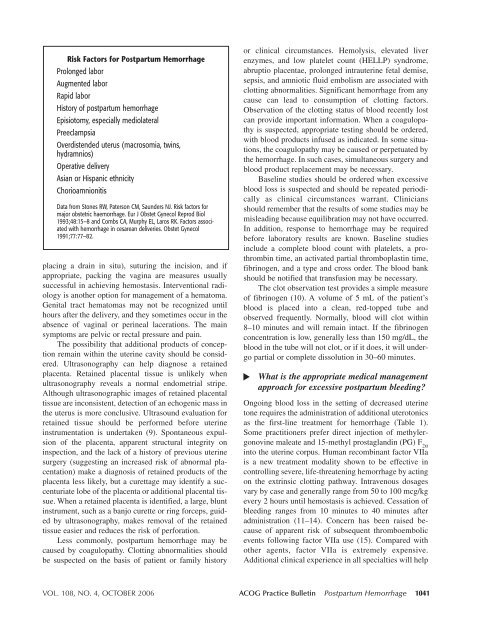
Risk Factors for <strong>Postpartum</strong> <strong>Hemorrhage</strong><br />
Prolonged labor<br />
Augmented labor<br />
Rapid labor<br />
History of postpartum hemorrhage<br />
Episiotomy, especially mediolateral<br />
Preeclampsia<br />
Overdistended uterus (macrosomia, twins,<br />
hydramnios)<br />
Operative delivery<br />
Asian or Hispanic ethnicity<br />
Chorioamnionitis<br />
Data from Stones RW, Paterson CM, Saunders NJ. Risk factors for<br />
major obstetric haemorrhage. Eur J Obstet Gynecol Reprod Biol<br />
1993;48:15–8 and Combs CA, Murphy EL, Laros RK. Factors associated<br />
with hemorrhage in cesarean deliveries. Obstet Gynecol<br />
1991;77:77–82.<br />
placing a drain in situ), suturing the incision, and if<br />
appropriate, packing the vagina are measures usually<br />
successful in achieving hemostasis. Interventional radiology<br />
is another option for management of a hematoma.<br />
Genital tract hematomas may not be recognized until<br />
hours after the delivery, and they sometimes occur in the<br />
absence of vaginal or perineal lacerations. The main<br />
symptoms are pelvic or rectal pressure and pain.<br />
The possibility that additional products of conception<br />
remain within the uterine cavity should be considered.<br />
Ultrasonography can help diagnose a retained<br />
placenta. Retained placental tissue is unlikely when<br />
ultrasonography reveals a normal endometrial stripe.<br />
Although ultrasonographic images of retained placental<br />
tissue are inconsistent, detection of an echogenic mass in<br />
the uterus is more conclusive. Ultrasound evaluation for<br />
retained tissue should be performed before uterine<br />
instrumentation is undertaken (9). Spontaneous expulsion<br />
of the placenta, apparent structural integrity on<br />
inspection, and the lack of a history of previous uterine<br />
surgery (suggesting an increased risk of abnormal placentation)<br />
make a diagnosis of retained products of the<br />
placenta less likely, but a curettage may identify a succenturiate<br />
lobe of the placenta or additional placental tissue.<br />
When a retained placenta is identified, a large, blunt<br />
instrument, such as a banjo curette or ring forceps, guided<br />
by ultrasonography, makes removal of the retained<br />
tissue easier and reduces the risk of perforation.<br />
Less commonly, postpartum hemorrhage may be<br />
caused by coagulopathy. Clotting abnormalities should<br />
be suspected on the basis of patient or family history<br />
or clinical circumstances. Hemolysis, elevated liver<br />
enzymes, and low platelet count (HELLP) syndrome,<br />
abruptio placentae, prolonged intrauterine fetal demise,<br />
sepsis, and amniotic fluid embolism are associated with<br />
clotting abnormalities. Significant hemorrhage from any<br />
cause can lead to consumption of clotting factors.<br />
Observation of the clotting status of blood recently lost<br />
can provide important information. When a coagulopathy<br />
is suspected, appropriate testing should be ordered,<br />
with blood products infused as indicated. In some situations,<br />
the coagulopathy may be caused or perpetuated by<br />
the hemorrhage. In such cases, simultaneous surgery and<br />
blood product replacement may be necessary.<br />
Baseline studies should be ordered when excessive<br />
blood loss is suspected and should be repeated periodically<br />
as clinical circumstances warrant. Clinicians<br />
should remember that the results of some studies may be<br />
misleading because equilibration may not have occurred.<br />
In addition, response to hemorrhage may be required<br />
before laboratory results are known. Baseline studies<br />
include a complete blood count with platelets, a prothrombin<br />
time, an activated partial thromboplastin time,<br />
fibrinogen, and a type and cross order. The blood bank<br />
should be notified that transfusion may be necessary.<br />
The clot observation test provides a simple measure<br />
of fibrinogen (10). A volume of 5 mL of the patient’s<br />
blood is placed into a clean, red-topped tube and<br />
observed frequently. <strong>No</strong>rmally, blood will clot within<br />
8–10 minutes and will remain intact. If the fibrinogen<br />
concentration is low, generally less than 150 mg/dL, the<br />
blood in the tube will not clot, or if it does, it will undergo<br />
partial or complete dissolution in 30–60 minutes.<br />
What is the appropriate medical management<br />
approach for excessive postpartum bleeding<br />
Ongoing blood loss in the setting of decreased uterine<br />
tone requires the administration of additional uterotonics<br />
as the first-line treatment for hemorrhage (Table 1).<br />
Some practitioners prefer direct injection of methylergonovine<br />
maleate and 15-methyl prostaglandin (PG) F 2α<br />
into the uterine corpus. Human recombinant factor VIIa<br />
is a new treatment modality shown to be effective in<br />
controlling severe, life-threatening hemorrhage by acting<br />
on the extrinsic clotting pathway. Intravenous dosages<br />
vary by case and generally range from 50 to 100 mcg/kg<br />
every 2 hours until hemostasis is achieved. Cessation of<br />
bleeding ranges from 10 minutes to 40 minutes after<br />
administration (11–14). Concern has been raised because<br />
of apparent risk of subsequent thromboembolic<br />
events following factor VIIa use (15). Compared with<br />
other agents, factor VIIa is extremely expensive.<br />
Additional clinical experience in all specialties will help<br />
VOL. 108, NO. 4, OCTOBER 2006 <strong>ACOG</strong> <strong>Practice</strong> <strong>Bulletin</strong> <strong>Postpartum</strong> <strong>Hemorrhage</strong> 1041