ACOG Practice Bulletin No. 76: Postpartum Hemorrhage
ACOG Practice Bulletin No. 76: Postpartum Hemorrhage
ACOG Practice Bulletin No. 76: Postpartum Hemorrhage
Create successful ePaper yourself
Turn your PDF publications into a flip-book with our unique Google optimized e-Paper software.
▲<br />
▲<br />
rates vary between 0.4% and 1.6% (30). Clinical judgment<br />
is an important determinant, given that estimates of<br />
blood loss often are inaccurate, determination of hematocrit<br />
or hemoglobin concentrations may not accurately<br />
reflect the current hematologic status, and symptoms and<br />
signs of hemorrhage may not occur until blood loss<br />
exceeds 15% (8). The purpose of transfusion of blood<br />
products is to replace coagulation factors and red cells for<br />
oxygen-carrying capacity, not for volume replacement.<br />
To avoid dilutional coagulopathy, concurrent replacement<br />
with coagulation factors and platelets may be necessary.<br />
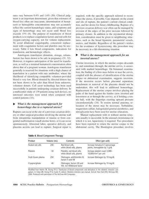
Table 4 lists blood components, indications for<br />
transfusion, and hematologic effects.<br />
Autologous transfusion (donation, storage, retransfusion)<br />
has been shown to be safe in pregnancy (31, 32).<br />
However, it requires anticipation of the need for transfusion,<br />
as well as a minimal hematocrit concentration often<br />
above that of a pregnant woman. Autologous transfusion<br />
generally is reserved for situations with a high chance of<br />
transfusion in a patient with rare antibodies, where the<br />
likelihood of identifying compatible volunteer-provided<br />
blood is very low. Blood donated by directed donors has<br />
not been shown to be safer than blood from unknown,<br />
volunteer donors. Cell saver technology has been used<br />
successfully in patients undergoing cesarean delivery. In<br />
a multicenter study of 139 patients using such devices, no<br />
untoward outcomes were noted when compared with<br />
control patients (33).<br />
What is the management approach for<br />
hemorrhage due to a ruptured uterus<br />
Rupture can occur at the site of a previous cesarean delivery<br />
or other surgical procedure involving the uterine wall<br />
from intrauterine manipulation or trauma or from congenital<br />
malformation (small uterine horn), or it can occur<br />
spontaneously. Abnormal labor, operative delivery, and<br />
placenta accreta can lead to rupture. Surgical repair is<br />
required, with the specific approach tailored to reconstruct<br />
the uterus, if possible. Care depends on the extent<br />
and site of rupture, the patient’s current clinical condition,<br />
and her desire for future childbearing. Rupture of a<br />
previous cesarean delivery scar often can be managed by<br />
revision of the edges of the prior incision followed by<br />
primary closure. In addition to the myometrial disruption,<br />
consideration must be given to neighboring structures,<br />
such as the broad ligament, parametrial vessels,<br />
ureters, and bladder. Regardless of the patient’s wishes<br />
for the avoidance of hysterectomy, this procedure may<br />
be necessary in a life-threatening situation.<br />
What is the management approach for an<br />
inverted uterus<br />
Uterine inversion, in which the uterine corpus descends<br />
to, and sometimes through, the uterine cervix, is associated<br />
with marked hemorrhage. On bimanual examination,<br />
the finding of a firm mass below or near the cervix,<br />
coupled with the absence of identification of the uterine<br />
corpus on abdominal examination, suggests inversion.<br />
If the inversion occurs before placental separation,<br />
detachment or removal of the placenta should not be<br />
undertaken; this will lead to additional hemorrhage.<br />
Replacement of the uterine corpus involves placing the<br />
palm of the hand against the fundus (now inverted and<br />
lowermost at or through the cervix), as if holding a tennis<br />
ball, with the fingertips exerting upward pressure<br />
circumferentially (34). To restore normal anatomy, relaxation<br />
of the uterus may be necessary. Terbutaline,<br />
magnesium sulfate, halogenated general anesthetics, and<br />
nitroglycerin have been used for uterine relaxation.<br />
Manual replacement with or without uterine relaxants<br />
usually is successful. In the unusual circumstance in<br />
which it is not, laparotomy is required. Two procedures<br />
have been reported to return the uterine corpus to the<br />
abdominal cavity. The Huntington procedure involves<br />
Table 4. Blood Component Therapy<br />
Product Volume (mL) Contents Effect (per unit)<br />
Packed red cells 240 Red blood cells, Increase hematocrit 3 percentage<br />
white blood cells, plasma points, hemoglobin by 1 g/dL<br />
Platelets 50 Platelets, red blood cells, Increase platelet count 5,000–<br />
white blood cells, plasma 10,000/mm 3 per unit<br />
Fresh frozen plasma 250 Fibrinogen, antithrombin III, Increase fibrinogen by 10 mg/dL<br />
factors V and VIII<br />
Cryoprecipitate 40 Fibrinogen, factors VIII and Increase fibrinogen by 10 mg/dL<br />
XIII, von Willebrand factor<br />
Modified from Martin SR, Strong TH Jr. Transfusion of blood components and derivatives in the obstetric intensive care<br />
patient. In: Foley MR, Strong TH Jr, Garite TJ, editors. Obstetric intensive care manual. 2nd ed. New York (NY): McGraw-Hill;<br />
2004. Produced with permission of The McGraw-Hill Companies.<br />
1044 <strong>ACOG</strong> <strong>Practice</strong> <strong>Bulletin</strong> <strong>Postpartum</strong> <strong>Hemorrhage</strong> OBSTETRICS & GYNECOLOGY


