ACOG Practice Bulletin No. 76: Postpartum Hemorrhage
ACOG Practice Bulletin No. 76: Postpartum Hemorrhage
ACOG Practice Bulletin No. 76: Postpartum Hemorrhage

You also want an ePaper? Increase the reach of your titles
YUMPU automatically turns print PDFs into web optimized ePapers that Google loves.
▲<br />
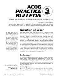
Table 3. Surgical Management of <strong>Postpartum</strong> <strong>Hemorrhage</strong><br />
Technique<br />
Uterine curettage<br />
Uterine artery ligation<br />
B-Lynch suture<br />
Hypogastric artery ligation<br />
Repair of rupture<br />
Hysterectomy<br />
Comment<br />
Bilateral; also can ligate uteroovarian<br />
vessels<br />
Less successful than earlier thought;<br />
difficult technique; generally<br />
reserved for practitioners<br />
experienced in the procedure<br />
than 1,000 B-Lynch procedures with only seven failures<br />
(21). However, because the technique is new, many clinicians<br />
have limited experience with this procedure (22).<br />
Hemostatic multiple square suturing is another new<br />
surgical technique for postpartum hemorrhage caused by<br />
uterine atony, placenta previa, or placenta accreta. The<br />
procedure eliminates space in the uterine cavity by suturing<br />
both anterior and posterior uterine walls. One study<br />
reported on this technique in 23 women after conservative<br />
treatment failed. All patients were examined after 2<br />
months, and ultrasound findings confirmed normal<br />
endometrial linings and uterine cavities (23).<br />
What are the clinical considerations for<br />
suspected placenta accreta<br />
Abnormal attachment of the placenta to the inner uterine<br />
wall (placenta accreta) can cause massive hemorrhage. In<br />
fact, accreta and uterine atony are the two most common<br />
reasons for postpartum hysterectomy (24, 25). Risk factors<br />
for placenta accreta include placenta previa with or without<br />
previous uterine surgery, prior myomectomy, prior<br />
cesarean delivery, Asherman’s syndrome, submucous<br />
leiomyomata, and maternal age older than 35 years (26).<br />
Prior cesarean delivery and the presence of placenta<br />
previa in a current pregnancy are particularly important risk<br />
factors for placenta accreta. In a multicenter study of more<br />
than 30,000 patients who had cesarean delivery without<br />
labor, the risk of placenta accreta was approximately 0.2%,<br />
0.3%, 0.6%, 2.1%, 2.3%, and 7.7% for women experiencing<br />
their first through sixth cesarean deliveries, respectively.<br />
In patients with placenta previa in the current pregnancy,<br />
the risk of accreta was 3%, 11%, 40%, 61%, and 67% for<br />
those undergoing their first through their fifth or greater<br />
cesarean deliveries, respectively (27).<br />
Women with placenta previa or placenta accreta<br />
have a higher incidence of postpartum hemorrhage and<br />
are more likely to undergo emergency hysterectomy<br />
(28). In the multicenter study cited previously, hysterectomy<br />
was required in 0.7% for the first cesarean delivery<br />
and increased with each cesarean delivery up to 9% for<br />
patients with their sixth or greater cesarean delivery.<br />
In the presence of previa or a history of cesarean<br />
delivery, the obstetric care provider must have a high<br />
clinical suspicion for placenta accreta and take appropriate<br />
precautions. Ultrasonography may be helpful in<br />
establishing the diagnosis in the antepartum period.<br />
Color Doppler technology may be an additional adjunctive<br />
tool for suspected accreta (29). Despite advances in<br />
imaging techniques, no diagnostic technique affords the<br />
clinician complete assurance of the presence or absence<br />
of placenta accreta.<br />
If the diagnosis or a strong suspicion is formed<br />
before delivery, a number of measures should be taken:<br />
• The patient should be counseled about the likelihood<br />
of hysterectomy and blood transfusion.<br />
• Blood products and clotting factors should be available.<br />
• Cell saver technology should be considered if available.<br />
• The appropriate location and timing for delivery<br />
should be considered to allow access to adequate<br />
surgical personnel and equipment.<br />
• A preoperative anesthesia assessment should beobtained.<br />
The extent (area, depth) of the abnormal attachment<br />
will determine the response—curettage, wedge resection,<br />
medical management, or hysterectomy. Uterine conserving<br />
options may work in small focal accretas, but abdominal<br />
hysterectomy usually is the most definitive treatment.<br />
▲ ▲<br />
Under what circumstances is arterial<br />
embolization indicated<br />
A patient with stable vital signs and persistent bleeding,<br />
especially if the rate of loss is not excessive, may be a candidate<br />
for arterial embolization. Radiographic identification<br />
of bleeding vessels allows embolization with Gelfoam,<br />
coils, or glue. Balloon occlusion is also a technique used in<br />
such circumstances. Embolization can be used for bleeding<br />
that continues after hysterectomy or can be used as an<br />
alternative to hysterectomy to preserve fertility.<br />
When is blood transfusion recommended<br />
Is there a role for autologous transfusions<br />
or directed donor programs<br />
Transfusion of blood products is necessary when the<br />
extent of blood loss is significant and ongoing, particularly<br />
if vital signs are unstable. <strong>Postpartum</strong> transfusion<br />
VOL. 108, NO. 4, OCTOBER 2006 <strong>ACOG</strong> <strong>Practice</strong> <strong>Bulletin</strong> <strong>Postpartum</strong> <strong>Hemorrhage</strong> 1043