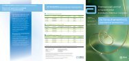
Results of Coronary Stenting using the Stents with
Results of Coronary Stenting using the Stents with
Results of Coronary Stenting using the Stents with

Create successful ePaper yourself
Turn your PDF publications into a flip-book with our unique Google optimized e-Paper software.
Miscellaneous<br />
vasculitis at <strong>the</strong> level <strong>of</strong> pulmonary arterioles. This<br />
leads to <strong>the</strong> growth and <strong>the</strong> enlargement <strong>of</strong> bronchial<br />
arteries aimed at <strong>the</strong> compensation <strong>of</strong> decreased<br />
pulmonary blood flow. Bronchial arterial blood flow in<br />
patients <strong>with</strong> bronchoectatic pulmonary disease can<br />
reach up to 30% <strong>of</strong> <strong>the</strong> cardiac output (7). The impact<br />
<strong>of</strong> increased blood pressure and bacterial agents on<br />
<strong>the</strong> bronchial arteries contributes to <strong>the</strong>ir rupture and<br />
subsequent pulmonary bleeding (2, 8).<br />
Chest X-ray, FBS and CT are <strong>the</strong> main methods<br />
for <strong>the</strong> diagnostics <strong>of</strong> pulmonary bleeding. In most<br />
cases chest X-ray examination allows to reveal some<br />
pulmonary disease, but does not detect <strong>the</strong> source<br />
<strong>of</strong> bleeding. FBS is <strong>the</strong> main diagnostic procedure in<br />
patients <strong>with</strong> pulmonary bleeding. It allows to find <strong>the</strong><br />
source <strong>of</strong> bleeding in 93% <strong>of</strong> cases. However, <strong>the</strong> diagnostic<br />
value <strong>of</strong> FBS in patients <strong>with</strong>out any pathology<br />
revealed by chest X-ray is significantly reduced<br />
(0-30%). The advantage <strong>of</strong> bronchoscopy consists<br />
in <strong>the</strong> feasibility <strong>of</strong> local application <strong>of</strong> hemostatic<br />
agents to stop <strong>the</strong> bleeding. The examination <strong>with</strong> CT<br />
allows to reveal simultaneously bronchopulmonary<br />
and vascular pathology, thus CT is <strong>the</strong> method <strong>of</strong><br />
choice for <strong>the</strong> diagnostics <strong>of</strong> pulmonary bleeding. It<br />
allows to detect <strong>the</strong> source <strong>of</strong> bleeding in 63-100%<br />
<strong>of</strong> cases (1, 9).<br />
Angiography <strong>of</strong> <strong>the</strong> bronchial arteries is <strong>the</strong> final<br />
step in <strong>the</strong> diagnostics <strong>of</strong> <strong>the</strong> source <strong>of</strong> pulmonary<br />
bleeding prior to embolization. The main distinctive<br />
angiographic features <strong>of</strong> bronchial arterial pathology<br />
in pulmonary bleeding are: <strong>the</strong> enlargement<br />
<strong>of</strong> <strong>the</strong> main trunk <strong>of</strong> <strong>the</strong> bronchial artery (>3 mm),<br />
<strong>the</strong> hypervascularization <strong>of</strong> <strong>the</strong> pathological focus<br />
area, <strong>the</strong> impregnation <strong>of</strong> parenchyma <strong>with</strong> contrast<br />
medium and <strong>the</strong> presence <strong>of</strong> bronchopulmonary<br />
shunts.<br />
Virtually in all cases bronchial arteries arise from<br />
<strong>the</strong> thoracic aorta at <strong>the</strong> level <strong>of</strong> Т4-Т7 vertebrae<br />
(10, 11), in 90% <strong>of</strong> cases <strong>the</strong>ir origin is located at <strong>the</strong><br />
level <strong>of</strong> <strong>the</strong> intervebral disc between <strong>the</strong> Т5 and Т6<br />
vertebrae. Bronchial arteries supply <strong>the</strong> trachea, <strong>the</strong><br />
bronchi, regional lymphatic nodes, <strong>the</strong> pleura and <strong>the</strong><br />
esophagus.<br />
There are 4 main types <strong>of</strong> bronchial arteries origin<br />
(2):<br />
• Type 1: two bronchial arteries arise to <strong>the</strong> left,<br />
and one — to <strong>the</strong> right <strong>of</strong> <strong>the</strong> aorta (intercostal bronchial<br />
trunk) — is seen in 40% <strong>of</strong> cases.<br />
Etiological causes <strong>of</strong> pulmonary bleeding<br />
Table 1.<br />
Infection:<br />
• Bronchoectatic disease<br />
• Pneumonia<br />
• Chronic bronchitis<br />
• Lung abscess<br />
• Aspergillosis, mycetoma<br />
• Ascariasis<br />
• Tuberculosis<br />
• Cystic fibrosis<br />
Tumors:<br />
• Adenocarcinoma<br />
• Bronchus adenoma<br />
• Bronchus carcinoid<br />
• Endometriosis<br />
• Metastases in lungs<br />
Cardiovascular diseases:<br />
• Severe left ventricular failure<br />
• Mitral stenosis<br />
• Pulmonary arterial thromboembolism<br />
• Aortic aneurysm<br />
• Pulmonary arterial aneurysm<br />
• Arteriovenous malformation<br />
• Yatrogenic causes (e.g., damage by Swan-Ganz ca<strong>the</strong>ter)<br />
Vasculites:<br />
• Wegener’s disease<br />
• Systemic lupus ery<strong>the</strong>matosis<br />
• Goodpasture’s syndrome<br />
Idiopathic pulmonary hemosiderosis<br />
Aspiration <strong>of</strong> a foreign body<br />
Lung trauma (contusion)<br />
Use <strong>of</strong> anticoagulant agents, thrombolythic <strong>the</strong>rapy<br />
60<br />
(№ 26, 2011)