Cysts of the Jaws.pdf
Cysts of the Jaws.pdf
Cysts of the Jaws.pdf

Create successful ePaper yourself
Turn your PDF publications into a flip-book with our unique Google optimized e-Paper software.
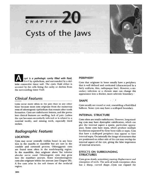
is a pathologic cavity filled with fluid,<br />
by epi<strong>the</strong>lium, and surrounded by a definite<br />
connective tissue wall. The cystic fluid ei<strong>the</strong>r is<br />
secreted by <strong>the</strong> cells lining <strong>the</strong> cavity or derives from<br />
<strong>the</strong> surrounding tissue fluid.<br />
Clinical Features<br />
<strong>Cysts</strong> occur more <strong>of</strong>ten in <strong>the</strong> jaws than in any o<strong>the</strong>r<br />
bone because most cysts originate from <strong>the</strong> numerous<br />
rests <strong>of</strong> odontogenic epi<strong>the</strong>lium that remain after tooth<br />
formation. <strong>Cysts</strong> are radiolucent lesions, and <strong>the</strong> prevalent<br />
clinical features are swelling, lack <strong>of</strong> pain (unless<br />
<strong>the</strong> cyst becomes secondarily infected or is related to a<br />
nonvital tooth), and missing teeth, especially third<br />
molars.<br />
LOCATION<br />
<strong>Cysts</strong> may occur centrally (within bone) in any location<br />
in <strong>the</strong> maxilla or mandible but are rare in <strong>the</strong><br />
condyle and coronoid process. Odont4.>genic cysts<br />
are found most <strong>of</strong>ten in <strong>the</strong> tooth~bearirig region.<br />
In <strong>the</strong> mandible, <strong>the</strong>y originate above ..<strong>the</strong>' inferior<br />
alveolar nerve canal. Odontogenic cysts n1ay grow<br />
into <strong>the</strong> maxillary antrum. Some nonodontogenic<br />
cysts also originate within <strong>the</strong> antrum (see Chapter 26).<br />
A few cysts arise in <strong>the</strong> s<strong>of</strong>t tissues <strong>of</strong> <strong>the</strong> or<strong>of</strong>acial<br />
region.<br />
PERI PH ERY<br />
<strong>Cysts</strong> that originate in bone usually have a periphery<br />
that is well defined and corticated (characterized by a<br />
fairly uniform, thin, radiopaque line). However, a secondary<br />
infection or a chronic state can change this<br />
appearance into a thicker, more sclerotic boundary. .<br />
SHAPE " ...<br />
<strong>Cysts</strong> usually are round or oval, resembling a fluid-filled<br />
balloon. Some cysts may have a scalloped boundary.<br />
INTERNAL STRUCTURE<br />
<strong>Cysts</strong> <strong>of</strong>ten are totally radiolucent. However, long-standing<br />
cysts may have dystrophic calcification, which can<br />
give <strong>the</strong> internal aspect a sparse, particulate appearance.<br />
Some cysts have septa, which produce multiple<br />
loculations separated by <strong>the</strong>se bony walls or septa. <strong>Cysts</strong><br />
that have a scalloped periphery may appear to have<br />
in ternal septa. Occasionally <strong>the</strong> image <strong>of</strong> structures that<br />
are positioned on ei<strong>the</strong>r side <strong>of</strong> <strong>the</strong> cyst may overlap <strong>the</strong><br />
internal aspect <strong>of</strong> <strong>the</strong> cyst, giving <strong>the</strong> false impression<br />
<strong>of</strong> internal structure.<br />
EFFECTS ON SURROUNDING<br />
STRUCTURES<br />
<strong>Cysts</strong> grow slowly, sometimes'.causing displacement and<br />
resorption <strong>of</strong> teeth. The aria 01toOth resorption <strong>of</strong>ten<br />
has a sharp, curved shape. ..<strong>Cysts</strong> can expand <strong>the</strong><br />
384
CHAPTER 20 CYSTS OF THE JAWS 385<br />
mandible, usually in a smooth, curved manner, and<br />
change <strong>the</strong> buccal or lingual cortical plate into a thin<br />
cortical boundary. <strong>Cysts</strong> may displace <strong>the</strong> inferior alveolar<br />
nerve canal in an inferior direction or invaginate<br />
<strong>the</strong> maxillary antrum, maintaining a thin layer <strong>of</strong> bone<br />
that separates <strong>the</strong> internal aspect <strong>of</strong> <strong>the</strong> cyst from ,t4~;.;<br />
antrum.<br />
RADICULAR CYST<br />
Synonyms<br />
Periapical cyst, apical periodontal cyst, or dental cyst<br />
Definition<br />
A radicular cyst is a cyst that most likely results when<br />
rests <strong>of</strong> epi<strong>the</strong>lial cells (Malassez) in <strong>the</strong> periodontal<br />
ligament are stimulated to proliferate and undergo<br />
cystic degeneration by inflammatory products from a<br />
nonvital tooth.<br />
Clinical Features<br />
Radicular cysts are <strong>the</strong> most common type <strong>of</strong> cyst in<br />
<strong>the</strong> jaws. They arise from nonvital teeth (i.e., teeth that<br />
have lost vitality because <strong>of</strong> extensive caries, large<br />
restorations, or previous trauma). Often radicular<br />
cysts produce no symptoms unless secondary infection<br />
occurs. A cyst that becomes large may cause<br />
swelling. On palpation <strong>the</strong> swelling may feel bony<br />
and hard if <strong>the</strong> cortex is intact, crepitant as <strong>the</strong> bone<br />
thins, and rubbery and fluctuant if <strong>the</strong> outer cortex<br />
is lost. The incidence <strong>of</strong> radicular cysts is greater in<br />
<strong>the</strong> third to sixth decades and shows a slight male<br />
predominance.<br />
Radiographic Features<br />
Location. In most cases <strong>the</strong> epicenter <strong>of</strong> a radicular cyst<br />
-is located approximately at <strong>the</strong> apex <strong>of</strong> a nonvital tooth<br />
(Fig. 20-1). Occasionally it appears on <strong>the</strong> mesial or<br />
distal surface <strong>of</strong> a tooth root, at <strong>the</strong> opening <strong>of</strong> an accessory<br />
canal, or infrequently in a deep periodontal<br />
pocket. Most radicular cysts (60%) are found in <strong>the</strong><br />
maxilla, especially around incisors and canines.<br />
Because <strong>of</strong> <strong>the</strong> distal inclination <strong>of</strong> <strong>the</strong> root, cysts that<br />
arise from <strong>the</strong> maxillary lateral incisor may invaginate<br />
<strong>the</strong> antrum. Radicular cysts may also form in relation<br />
to a nonvital deciduous molar and be positioned buccal<br />
to <strong>the</strong> developing bicuspid.<br />
Periphery and shape. The periphery usually has a welldefined<br />
cortical border (Fig. 20-2). If <strong>the</strong> cyst becomes<br />
secondarily infected, <strong>the</strong> inflammatory reaction <strong>of</strong> <strong>the</strong><br />
surrounding bone may result in loss <strong>of</strong> this cortex (see<br />
Fig. 20-1, B) or alteration <strong>of</strong> <strong>the</strong> cortex into a more sclerotic<br />
border. The outline <strong>of</strong> a radicular cyst usually is<br />
curved or circular unless it is influenced by surrounding<br />
structures such as cortical boundaries.<br />
A<br />
tj<br />
FIG. 20-1 Radicular cysts. In A, note that <strong>the</strong> epicenter is apical to <strong>the</strong> lateral incisor<br />
and <strong>the</strong> presence <strong>of</strong> a peripheral cortex (arrows). In B, note <strong>the</strong> lack <strong>of</strong> a well-defined<br />
peripheral cortex as this cyst was secondarily infected and that <strong>the</strong> root canal <strong>of</strong> <strong>the</strong> lateral<br />
incisor is abnormally wide as it is visible at <strong>the</strong> root apex.
,<br />
386 PART V RADIOGRAPHIC INTERPRETATION OF PATHOLOGY<br />
Internal structure. In most cases <strong>the</strong> internal structure<br />
<strong>of</strong> radicular cysts is radiolucent. Occasionally, dystrophic<br />
calcification may develop in long-standing cysts,<br />
appearing as sparsely distributed, small particulate<br />
radiopacities.<br />
, "FiJi<br />
Effects on surrounding structures. If a radicttlar cyst is<br />
large, displacement and resorption <strong>of</strong> <strong>the</strong> roots <strong>of</strong> adja-<br />
FIG. 20-2 A periapical tilm ot a radicular cyst reveals a<br />
lesion with a well-defined cortical boundary (arrows). Note<br />
that <strong>the</strong> presence <strong>of</strong> <strong>the</strong> inferior cortex <strong>of</strong> <strong>the</strong> mandible has<br />
influenced <strong>the</strong> circular shape <strong>of</strong> <strong>the</strong> cyst.<br />
cent teeth may occur. The resorption pattern may have<br />
a curved outline. In rare cases <strong>the</strong> cyst may resorb <strong>the</strong><br />
roots <strong>of</strong> <strong>the</strong> relatednonvital tooth. The cyst may invaginate<br />
<strong>the</strong> antrum, but <strong>the</strong>re should be evidence <strong>of</strong> a cortical<br />
boundary between <strong>the</strong> contents <strong>of</strong> <strong>the</strong> cyst and <strong>the</strong><br />
internal structure <strong>of</strong> <strong>the</strong> antrum. The outer cortical<br />
plates <strong>of</strong> <strong>the</strong> maxilla or mandible may expand in a<br />
curved or circular shape (Fig. 20-3). <strong>Cysts</strong> may displace<br />
<strong>the</strong> mandibular alveolar nerve canal in an inferior<br />
direction.<br />
Differential Diagnosis<br />
Differentiation <strong>of</strong> a small radicular cyst from an apical<br />
granuloma may be difficult and in some cases impossible.<br />
A round shape, a well-defined cortical border,<br />
and a size greater than 2 cm in diameter are more<br />
characteristic <strong>of</strong> a cyst. An early radiolucent stage <strong>of</strong><br />
periapical cemental dysplasia, a radiolucent apical<br />
scar, and a periapical surgical defect should also be<br />
considered in <strong>the</strong> differential diagnosis. The patient's<br />
history helps with <strong>the</strong> differentiation. Radicular cysts<br />
that originate from <strong>the</strong> maxillary lateral incisor and<br />
are positioned between <strong>the</strong> roots <strong>of</strong> <strong>the</strong> lateral incisor<br />
and <strong>the</strong> cuspid may be difficult to differentiate from<br />
an odontogenic keratocyst or a lateral periodontal<br />
cyst. The vitality <strong>of</strong> <strong>the</strong> involved tooth should be tested.<br />
A nonvital tooth may have a larger pulp chamber<br />
than neighboring teeth because <strong>of</strong> <strong>the</strong> lack <strong>of</strong> secondary<br />
dentin, which normally forms with time in<br />
<strong>the</strong> pulp chamber and canal <strong>of</strong> a vital tooth (see<br />
Fig. 20-1).<br />
A<br />
FIG. 20-3 A ~nd B, Two im~ges <strong>of</strong> a radicular cyst originating from a nonvital deciduous<br />
second molar show expansion <strong>of</strong> <strong>the</strong> buccal cortical plate to a circular or hydraulic<br />
shape (arrows) and displacement <strong>of</strong> <strong>the</strong> adjacent permanent teeth.<br />
D
CHAPTER 20<br />
CYSTS OF THE JAWS<br />
387<br />
FIG. 20-4 Axial (A) and coronal (8) CT images using bone algorithm <strong>of</strong> a collapsing<br />
radicular cyst within <strong>the</strong> sinus. Note <strong>the</strong> unusual shape and <strong>the</strong> fact that new bone (arrows)<br />
is being formed from <strong>the</strong> periphery (arrows) toward <strong>the</strong> center. (Courtesy <strong>of</strong> Drs. S. Ahing<br />
and T. Blight, University <strong>of</strong> Manitoba.)<br />
A large radicular cyst that has invaginated <strong>the</strong> maxillary<br />
antrum may collapse and start filling in with new<br />
bone (Fig. 20-4). With biopsy, <strong>the</strong> histologic analysis<br />
may result in an erroneous diagnosis <strong>of</strong> ossifying<br />
fibroma or a benign fibroosseous lesion. Radiographically,<br />
<strong>the</strong> important feature is that <strong>the</strong> new bone always<br />
forms first at <strong>the</strong> periphery <strong>of</strong> <strong>the</strong> cyst wall as <strong>the</strong> cyst<br />
shrinks and not in <strong>the</strong> center <strong>of</strong> <strong>the</strong> cyst; this is a different<br />
pattern <strong>of</strong> bone formation than is seen with<br />
benign fibroosseous lesions.<br />
Management<br />
Treatment <strong>of</strong> a tooth with a radicular cyst may include<br />
extraction, endodontic <strong>the</strong>rapy, and apical surgery.<br />
Treatment <strong>of</strong>a large radicular cyst usually involves surgical<br />
removal or marsupialization. The radiographic<br />
appearance <strong>of</strong> <strong>the</strong> periapical area <strong>of</strong> an endodontically<br />
treated tooth should be checked periodically to make<br />
sure that normal healing is occurring (Fig. 20-5). Characteristically,<br />
new bone grows into <strong>the</strong> defect from <strong>the</strong><br />
periphery, sometimes resulting in a radiating pattern<br />
resembling <strong>the</strong> spokes <strong>of</strong> a wheel. However, in a few<br />
cases normal bone may not fill <strong>the</strong> defect, especially if<br />
a secondary infection or a considerable amount <strong>of</strong> bone<br />
destruction occurred. Recurrence <strong>of</strong> a radicular cyst is<br />
unlikely if it has been removed completely.<br />
RESIDUAL CYST<br />
Definition<br />
A residual cyst is a cyst that remains after incomplete<br />
removal <strong>of</strong> <strong>the</strong> original cyst. The term residual is used<br />
FIG. 20-5 A radicular cyst that is healing after endodontic<br />
treatment. Arrows show <strong>the</strong> original outline <strong>of</strong> <strong>the</strong> cyst;<br />
note that <strong>the</strong> new bone grows toward <strong>the</strong> center from <strong>the</strong><br />
periphery.<br />
most <strong>of</strong>ten for a radicular cyst that may be left behind<br />
most commonly after extraction <strong>of</strong> a tooth.<br />
Clinical Features<br />
A residual cyst usually is asymptomatic and <strong>of</strong>ten is discovered<br />
on radiographic examination <strong>of</strong> an edentulous<br />
area. However, <strong>the</strong>re may be some expansion <strong>of</strong> <strong>the</strong> jaw<br />
or pain in <strong>the</strong> case <strong>of</strong> secondary infection.
388 PART V RADIOGRAPHIC INTERPRETATION OF PATHOI O~Y<br />
Radiographic Features<br />
Location. Residual cysts occur in both jaws, although<br />
<strong>the</strong>y are .found slightly more <strong>of</strong>ten in <strong>the</strong> mandible. The<br />
epicenter is positioned in a periapical location. In <strong>the</strong><br />
mandible <strong>the</strong> epicenter is always above <strong>the</strong> inferior alveolar<br />
nerve canal (Fig. 20-6).<br />
'<br />
Periphery and shape. A residual cyst has a cortical<br />
margin unless it becomes secondarily infected. Its shape<br />
i~ OV:I.1 or rirclIl:lr.<br />
Internal structure. The internal aspect <strong>of</strong> a residual cyst<br />
typically is radiolucent. Dystrophic calcifications may be<br />
present in long-standing cysts.<br />
Effects on surrounding structures. Residual cysts can<br />
cause tooth displacement or resorption. The outer cortical<br />
plates <strong>of</strong> <strong>the</strong> jaws may expand. The cyst may invaginate<br />
<strong>the</strong> maxillary antrum or depress <strong>the</strong> inferior<br />
alveolar nerve canal.<br />
Differential Diagnosis<br />
Without <strong>the</strong> patient's history and previous radiographs,<br />
<strong>the</strong> clinician may have difficulty determining whe<strong>the</strong>r<br />
a solitary cyst in <strong>the</strong> jaws is a residual cyst. O<strong>the</strong>r examples<br />
<strong>of</strong> common solitary cysts include odontogenic<br />
keratocysts. A residual cyst has greater potential for<br />
expansion compared with an odontogenic keratocyst.<br />
The epicenter <strong>of</strong> a Stafne developmental salivary gland<br />
defect is located below <strong>the</strong> mandibular canal (and thus<br />
is unlikely to be odontogenic in nature).<br />
FIG. 20-6 The epicenter <strong>of</strong> this infected residual cyst is<br />
above <strong>the</strong> inferior alveolar nerve canal and has displaced <strong>the</strong><br />
canal in an inferior direction (arrows). Note that <strong>the</strong> cortical<br />
boundary is not continuous around <strong>the</strong> whole cyst.<br />
Management<br />
The treatment for residual cysts is surgical removal or<br />
marsupialization, or both, if <strong>the</strong> cyst is large.<br />
DENTIGEROUS CYST<br />
Synonym<br />
Folli(,lll~r rv"t<br />
Definition<br />
A dentigerous cyst is a cyst that forms around <strong>the</strong> crown<br />
<strong>of</strong> an unerupted tooth. It begins when .fluid accumulates<br />
in <strong>the</strong> layers <strong>of</strong> reduced enamel epi<strong>the</strong>lium or<br />
between <strong>the</strong> epi<strong>the</strong>lium and <strong>the</strong> crown <strong>of</strong> <strong>the</strong><br />
unerupted tooth. An eruption cyst is <strong>the</strong> s<strong>of</strong>t tissue<br />
counterpart <strong>of</strong> a dentigerous cyst.<br />
Clinical Features<br />
Dentigerous cysts are <strong>the</strong> second most common type <strong>of</strong><br />
cyst. in <strong>the</strong> jaws. They develop around <strong>the</strong> crown <strong>of</strong> an<br />
unerupted or supernumerary tooth. The clinical examination<br />
reveals a missing tooth or teeth and possibly a<br />
hard swelling, occasionally resulting in facial asymmetry.<br />
The~pa6ent typically has no pain or discomfort.<br />
About 4% <strong>of</strong> individuals with at least one unerupted<br />
tooth have a dentigerous cyst. Dentigerous cysts around<br />
supernumerary teeth account for about 5% <strong>of</strong> all<br />
dentigerous cysts, most developing around a mesiodens<br />
in <strong>the</strong> anterior maxilla.<br />
Radiographic Features<br />
Location. The epicenter <strong>of</strong> a dentigerous cyst is found<br />
just above <strong>the</strong> crown <strong>of</strong> <strong>the</strong> involved tooth, which<br />
usually is tpe mandibular or maxillary third molar or<br />
<strong>the</strong> maxillary canine, <strong>the</strong> teeth most commonly affected<br />
(Fig. 20-7). An important diagnostic point is that this<br />
cyst attaches at <strong>the</strong> cementoenamel junction. Some<br />
dentigerous cysts are eccentric, developing from <strong>the</strong><br />
lateral aspect <strong>of</strong> <strong>the</strong> follicle so that <strong>the</strong>y occupy an area<br />
beside <strong>the</strong> crown instead <strong>of</strong> above <strong>the</strong> crown (see Fig.<br />
20-7, D). <strong>Cysts</strong> related to maxillary third molars <strong>of</strong>ten<br />
grow into <strong>the</strong> maxillary antrum and may become quite<br />
large before <strong>the</strong>y are discovered. <strong>Cysts</strong> attached to <strong>the</strong><br />
crown <strong>of</strong> mandibular molars may extend a considerable<br />
distance into <strong>the</strong> ramus.<br />
Periphery and shape. Dentigerous cysts typically<br />
have a well-defined cortex with a curved or circular<br />
outline. If infection is present, <strong>the</strong> cortex may be<br />
missing.<br />
Internal structure. The internal aspect is completely<br />
radiolucent except for <strong>the</strong> crown <strong>of</strong> <strong>the</strong> involved<br />
tooth.
,<br />
CHAPTER 20 CYSTS OF THE JAWS<br />
389<br />
A<br />
B
,<br />
390 PART V RADIOGRAPHIC INTERPRETATION OF PATHOLOGY<br />
Effects on surrounding structures. A dentigerous cyst<br />
has a propensity to displace and resorb adjacent,teeth<br />
(Fig. 20-8). It commonly displaces <strong>the</strong> associated tooth<br />
in an apical direction (Fig. 20-9). The degree <strong>of</strong> displacement<br />
may be considerable. For instance, maxillary<br />
third molars or cuspids may be pushed to <strong>the</strong> 'floor <strong>of</strong><br />
<strong>the</strong> orbit (see Fig. 20-8), and mandibular third molars<br />
may be moved to <strong>the</strong> condylar or coronoid regions or<br />
to <strong>the</strong> inferior cortex <strong>of</strong> <strong>the</strong> mandible (Fig. 20-10). The<br />
floor <strong>of</strong> <strong>the</strong> maxillary antrum may be displaced as <strong>the</strong>cyst<br />
invaginates <strong>the</strong> antrum, and <strong>the</strong> cyst may displace<br />
<strong>the</strong> inferior alveolar nerve canal in an inferior direction.<br />
This slow-growing cyst <strong>of</strong>ten expands <strong>the</strong> outer<br />
cortical boundary <strong>of</strong> <strong>the</strong> involved jaw.<br />
Differential Diagnosis<br />
Because <strong>the</strong> histopathologic app~arance <strong>of</strong> <strong>the</strong> lining<br />
epi<strong>the</strong>lium is not specific, <strong>the</strong> diagnosis relies on <strong>the</strong><br />
radiographic and surgical observation <strong>of</strong> <strong>the</strong> attachment<br />
<strong>of</strong> <strong>the</strong> cyst to <strong>the</strong> cementoenamel junction. A<br />
histopathologic examination must always be done to<br />
eliminate o<strong>the</strong>r possible lesions in this location.<br />
One <strong>of</strong> <strong>the</strong> most difficult differential diagnoses to<br />
make is between a small dentigerous cyst and a hyp~rplastic<br />
follicle. A cyst should be considered with anyevidence<br />
<strong>of</strong> tooth displacement or considerable expansion<br />
<strong>of</strong> <strong>the</strong> involved bone. The size <strong>of</strong> <strong>the</strong> normal follicular<br />
space is 2 to 3 mm. If <strong>the</strong> follicular space exceeds 5 mm,<br />
a dentigerous cyst is more likely. If uncertainty remains,<br />
t:S<br />
1-1\.1. ~U-B A, I nls panoramic Image reveals lne presence OT a large aenugerous CYSl<br />
associated with <strong>the</strong> left maxillary cuspid (arrow), which has been displaced. Notice <strong>the</strong> displacement<br />
and resorption <strong>of</strong> o<strong>the</strong>r teeth in <strong>the</strong> left maxilla. Band C, Coronal and axial CT<br />
images <strong>of</strong> <strong>the</strong> same case showing superior-lateral displacement <strong>of</strong> <strong>the</strong> cuspid, expansion<br />
<strong>of</strong> <strong>the</strong> anterior wall <strong>of</strong> <strong>the</strong> maxilla and expansion <strong>of</strong> <strong>the</strong> cyst into <strong>the</strong> nasal fossa.<br />
l,;
Ā<br />
--""""~ -' ""-" """"'""-"<br />
.B<br />
FIG. 20-9 A and B, These panoramic films <strong>of</strong> <strong>the</strong> same case taken several years apart<br />
demonstrate superior-posterior displacement <strong>of</strong> a maxillary third molar by a dentigerous cyst.<br />
A<br />
B<br />
c -~~-<br />
FIG. 20-10 Dentigerous cysts displacing teeth. A, The third molar has been displaced<br />
to <strong>the</strong> inferior cortex. 8, The second molar has been displaced into <strong>the</strong> ramus by a cyst<br />
associated with <strong>the</strong> first molar. Axial (C) and coronal (D) CT images using bone algorithm<br />
reveal a maxillary third molar displaced into <strong>the</strong> space occupied by <strong>the</strong> maxillary antrum.<br />
D
392 PART V RADIOGRAPHIC INTERPRETATION OF PATHOLOGY<br />
<strong>the</strong> region should be reexamined in 4 to 6 months to<br />
detect any increase in size or any influence on surrounding<br />
structures characteristic <strong>of</strong> cysts.<br />
The differential diagnosis also may include an odontogenic<br />
keratocyst, an ameloblastic fibroma, and a cystic<br />
ameloblastoma. An odontogenic keratocyst ,~ not<br />
expand <strong>the</strong> bone to <strong>the</strong> same degree as a dentigerous<br />
cyst, is less likely to resorb teeth, and may attach far<strong>the</strong>r<br />
apically on <strong>the</strong> root instead <strong>of</strong> at <strong>the</strong> cementoenamel<br />
junction. It may not be possible to differentiate a small<br />
ameloblastjc fibroma or cystic ameloblastoma from a<br />
dentigerous cyst if <strong>the</strong>re is no internal structure. O<strong>the</strong>r<br />
rare lesions that may have a similar pericoronal appearance<br />
are adenomatoid odontogenic tumors and<br />
calcified odontogenic cysts, both <strong>of</strong> which can surround<br />
<strong>the</strong> crown and root <strong>of</strong> <strong>the</strong> involved tooth.<br />
Evidence <strong>of</strong> a radiopaque internal structure should<br />
be sought in <strong>the</strong>se two lesions. Occasionally a radicular<br />
cyst at <strong>the</strong> apex <strong>of</strong> a primary tooth surrounds <strong>the</strong> crown<br />
<strong>of</strong> <strong>the</strong> developing permanent tooth positioned apical<br />
to it, giving <strong>the</strong> false impression <strong>of</strong> a dentigerous cyst<br />
associated with <strong>the</strong> permanent tooth. This occurs<br />
most <strong>of</strong>ten with <strong>the</strong> mandibular deciduous molars<br />
and <strong>the</strong> developing bicuspids. In <strong>the</strong>se cases <strong>the</strong> clinician<br />
should look for deep caries or extensive restorations<br />
in a primary tooth that would indicate a<br />
radicular cyst.<br />
Management<br />
Dentigerous cysts are treated by surgical removal, which<br />
may include <strong>the</strong> tooth as well. Large cysts may be<br />
treated by marsupialization before removal. The cyst<br />
lining should be submitted for histologic examination<br />
because ameloblastomas have been reported to occur<br />
in <strong>the</strong> cyst lining. In addition, squamous cell carcinoma<br />
has been reported to arise from <strong>the</strong> cyst lining <strong>of</strong> chronically<br />
infected cysts. Mucoepidermoid carcinoma also<br />
has been reported.<br />
BUCCAL BIFURCATION CYST<br />
Synonyms<br />
Mandibular infected buccal cyst, paradental cyst, or<br />
inflammatory collateral dental cyst<br />
Definition<br />
The source <strong>of</strong> epi<strong>the</strong>lium probably is <strong>the</strong> epi<strong>the</strong>lial<br />
cell rests in <strong>the</strong> periodontal membrane <strong>of</strong> <strong>the</strong> buccal<br />
bifurcation <strong>of</strong> mandibular molars. The histopathologic<br />
characteristics <strong>of</strong> <strong>the</strong> lining are not distinctive. The etiology<br />
<strong>of</strong> proliferation is unknown; one <strong>the</strong>ory holds<br />
that inflammation is <strong>the</strong> stimulus, but inflammation is<br />
not always present. The World Health Organization<br />
.includes <strong>the</strong>se cysts under inflammatory cysts.<br />
It is unclear whe<strong>the</strong>r <strong>the</strong> paradental cyst <strong>of</strong> <strong>the</strong> third<br />
molar and <strong>the</strong> buccal bifurcation cyst (associated with<br />
first and second molars) are <strong>the</strong> same entity. The buccal<br />
bifurcation cyst (BBC) is certainly a distinct clinical<br />
entity. An associated enamel extension into <strong>the</strong> furcation<br />
region <strong>of</strong> third molars with paradental cysts has not<br />
been documented with molars involved in a BBC. Also,<br />
<strong>the</strong> inflammatory component associated with paradental<br />
cysts is not always present with BBCs.<br />
Clinical Features<br />
A common sign is <strong>the</strong> lack <strong>of</strong> or a delay in eruption <strong>of</strong><br />
a mandibular first or second molar. On clinical examination<br />
<strong>the</strong> molar may be missing or <strong>the</strong> lingual cusp tips<br />
may be abnormally protruding through <strong>the</strong> mucosa,<br />
higher than <strong>the</strong> position <strong>of</strong> <strong>the</strong> buccal cusps. The first<br />
molar is involved more frequently than <strong>the</strong> second<br />
molar. The teeth are always vital. A hard swelling may<br />
be present buccal to <strong>the</strong> involved molar, and if it is secondarily<br />
infected, <strong>the</strong> patient has pain. The age <strong>of</strong><br />
detection is younger, within <strong>the</strong> first 2 decades for a<br />
BBC and in <strong>the</strong> third decade for a paradental cyst <strong>of</strong><br />
<strong>the</strong> third molar.<br />
Radiographic Features<br />
Location. The mandibular first molar is <strong>the</strong> most<br />
common location <strong>of</strong> a BBC, followed by <strong>the</strong> second<br />
molar. The cyst occasionally is bilateral. It is always<br />
located in <strong>the</strong> buccal furcation <strong>of</strong> <strong>the</strong> affected molar<br />
(Fig. 20-11). On periapical and panoramic films <strong>the</strong><br />
lesion may appear to be centered a littl~ distal to <strong>the</strong><br />
furcation <strong>of</strong> <strong>the</strong> involved tooth.<br />
Periphery and shape. In some cases <strong>the</strong> periphery is not<br />
readily apparent, and <strong>the</strong> lesion may be a very subtle<br />
radiolucent region superimposed over <strong>the</strong> image <strong>of</strong> <strong>the</strong><br />
roots <strong>of</strong> <strong>the</strong> molar. In o<strong>the</strong>r cases <strong>the</strong> lesion has a circular<br />
shape with a well-defined cortical border. Some<br />
cysts can become quite large before <strong>the</strong>y are detected.<br />
Internal structure. The internal structure is radiolucent.<br />
Effects on surrounding structures. The most striking<br />
diagnostic characteristic <strong>of</strong> a BBC is <strong>the</strong> tipping <strong>of</strong> <strong>the</strong><br />
involved molar so that <strong>the</strong> root tips are pushed into <strong>the</strong><br />
lingual cortical plate <strong>of</strong> <strong>the</strong> mandible (see Fig. 20-11, B<br />
and C) and <strong>the</strong> occlusal surface is tipped toward <strong>the</strong><br />
buccal aspe
CHAPTER 20 CYSTS OF THE JAWS<br />
393<br />
A<br />
B<br />
C<br />
FIG. 20-11 Bilateral buccal bifurcation cysts. A, A panoramic image showing cysts<br />
related to <strong>the</strong> mandibular first molars. Note that <strong>the</strong> occlusal surface <strong>of</strong> each tooth has<br />
been tipped in relation to <strong>the</strong> o<strong>the</strong>r teeth and that adjacent teeth have been displaced.<br />
Band C, Occlusal films <strong>of</strong> <strong>the</strong> same case. Note <strong>the</strong> circular expansion <strong>of</strong> <strong>the</strong> buccal cortex<br />
and <strong>the</strong> displacement <strong>of</strong> <strong>the</strong> roots <strong>of</strong> <strong>the</strong> first molars into <strong>the</strong> lingual cortical plate (arrows).<br />
occlusal projection, which demonstrates <strong>the</strong> abnormal<br />
position <strong>of</strong> <strong>the</strong> root apex. If <strong>the</strong> cyst is large enough, it<br />
may displace and resorb <strong>the</strong> adjacent teeth and cause a<br />
considerable amount <strong>of</strong> smooth expansion <strong>of</strong> <strong>the</strong><br />
buccal cortical plate. If <strong>the</strong> cyst is secondarily infected,<br />
periosteal new bone formation is seen on <strong>the</strong> buccal<br />
cortex adjacent to <strong>the</strong> involved tooth.<br />
Differential Diagnosis<br />
Diagnosis <strong>of</strong> a BBC relies entirely on clinical and radiographic<br />
information. The major differential diagnosis<br />
includes lesions that could elicit an inflammatory<br />
periosteal response on <strong>the</strong> buccal aspect <strong>of</strong> mandibular<br />
molars such as a periodontal abscess or Langerhans'<br />
cell histiocytosis. The fact that only a BBC tilts <strong>the</strong> molar<br />
as described helps to differentiate it from o<strong>the</strong>r lesions.<br />
Also in <strong>the</strong> differential diagnosis is <strong>the</strong> dentigerous cyst.<br />
However, <strong>the</strong> epicenter <strong>of</strong> a dentigerous cyst is different<br />
because a BBC starts near <strong>the</strong> bifurcation region <strong>of</strong><br />
<strong>the</strong> tooth and does not surround <strong>the</strong> crown, as does a<br />
dentigerous cyst.<br />
Management<br />
A BBC usually ~s removed by conservative curettage,<br />
although some c~es have resolved without intervention.<br />
The involved ~olar should not be removed. BBCs<br />
do not recur.<br />
ODONTOGENIC<br />
KERATOCYST<br />
Synonym<br />
It is a commonly held view that primordial cysts are<br />
odontogenic keratocysts. However, this view is not universally<br />
accepted.
394 I'AK I V KAUIUl.KAI'HIL INIt.KI'Kt.IAIIUN UI- I'AIHULUlJY<br />
Definition<br />
An odontogenic keratocyst (OKC) is a noninflammatory<br />
odontogenic cyst that arises from <strong>the</strong> dental<br />
lamina. Unlike o<strong>the</strong>r cysts, which are thought to grow<br />
solely by osmotic pressure, <strong>the</strong> epi<strong>the</strong>lium in an OKC<br />
appears to have innate growth potential, much as in'..,a<br />
benign tumor. This difference in <strong>the</strong> mechanism~ <strong>of</strong><br />
growth gives OKCs a different radiographic appearance.<br />
The epi<strong>the</strong>lial lining is distinctive because it is keratinized<br />
(hence <strong>the</strong> name) and thin (4 to 8 cells thick).<br />
Occasionally bvdlike proliferations <strong>of</strong> epi<strong>the</strong>lium grow<br />
from thtt basal layer into <strong>the</strong> adjacent connective tissue<br />
wall. Also, islands <strong>of</strong> epi<strong>the</strong>lium in <strong>the</strong> wall may give rise<br />
to satellite microcysts. The inside <strong>of</strong> <strong>the</strong> cyst <strong>of</strong>ten contains<br />
a viscous or cheesy material derived from <strong>the</strong><br />
epi<strong>the</strong>lial lining.<br />
Clinical Features<br />
OKCs account for about one tenth <strong>of</strong> all cysts in <strong>the</strong><br />
jaws. They occur in a wide age range, but most develop<br />
during <strong>the</strong> second and third decades, with a slight male<br />
predominance. The cysts sometimes form around an<br />
unerupted tooth. OKCs usually have no symptoms,<br />
although mild swelling may occur. Pain may occur with<br />
secondary infection. Aspiration may reveal a thick,<br />
yellow, cheesy material (keratin). It is important to note<br />
that, unlike o<strong>the</strong>r cysts, OKCs have a high propensity<br />
for recurrence, possibly because <strong>of</strong> small satellite cysts<br />
or tragments ot epi<strong>the</strong>lium left behind alter surgical<br />
removal <strong>of</strong> <strong>the</strong> cyst.<br />
Radiographic Features<br />
Location. The most common location <strong>of</strong> an OKC is <strong>the</strong><br />
posterior body <strong>of</strong> <strong>the</strong> mandible (90% occur posterior<br />
to <strong>the</strong> canines) and ramus (more than 50%) (Fig. 20-<br />
12). The epicenter is located superior to <strong>the</strong> inferior<br />
alveolar nerve canal. This type <strong>of</strong> cyst occasionally has<br />
<strong>the</strong> same pericoronal position as, and is indistinguishable<br />
from, a dentigerous cyst (see Fig. 20-12).<br />
Periphery and shape. As with o<strong>the</strong>r cysts, OKCs usually<br />
show evidence <strong>of</strong> a cortical border unless <strong>the</strong>y have<br />
become secondarily infected. The cyst may have a<br />
smooth round or oval shape identical to that <strong>of</strong> o<strong>the</strong>r<br />
cysts, or it may have a scalloped outline (a series <strong>of</strong> contiguous<br />
arcs) (see Figs. 20-12 and 20-14, C).<br />
Internal structure. The internal structure most commonly<br />
is radiolucent. The presence <strong>of</strong> internal keratin<br />
does not increase <strong>the</strong> radiopacity. In some cases curved<br />
internal septa may be present, giving <strong>the</strong> lesion a multilocular<br />
appearance (Fig. 20-13; see also Fig. 20-12).<br />
tttects on surrounding structures. An important characteristic<br />
<strong>of</strong> <strong>the</strong> OKC is its propensity to grow along <strong>the</strong><br />
internal aspect <strong>of</strong> <strong>the</strong> jaws, causing minimal expansion<br />
~ ...<br />
FIG. 20-12 A, Panoramic image shows a large keratocyst occupying <strong>the</strong> ramus and<br />
body <strong>of</strong> <strong>the</strong> mandible; note <strong>the</strong> septa (black arrow), inferiorly displaced mandibular canal<br />
(white arrow), and <strong>the</strong> root resorption. The keratocyst in B has a pericoronal position relative<br />
to <strong>the</strong> impacted third molar and <strong>the</strong> distal margin has a scalloped shape.
CHAPTER 20 CYSTS OF THE JAWS<br />
395<br />
B<br />
FIG. 20-13 A, Cropped panoramic image <strong>of</strong> a keratocyst occupying <strong>the</strong> mandibular<br />
ramus; note <strong>the</strong> septa (arrow). 8 and C, Axial CT images using bone algorithm <strong>of</strong> <strong>the</strong> same<br />
case demonstrating very little expansion in <strong>the</strong> body (8) but significant expansion in <strong>the</strong><br />
upper ramus in C (arrows).<br />
c<br />
(Fig. 20-14). This occurs throughout <strong>the</strong> mandible<br />
except for <strong>the</strong> upper ramus and coronoid process,<br />
where considerable expansion may occur (see Fig. 20-<br />
13, C). Occasionally <strong>the</strong> expansion <strong>of</strong> large cysts may<br />
exceed <strong>the</strong> ability <strong>of</strong> <strong>the</strong> periosteum to form new bone,<br />
thus allowing <strong>the</strong> cyst wall to contact s<strong>of</strong>t tissue peripheral<br />
to <strong>the</strong> outer cortex <strong>of</strong> <strong>the</strong> mandible (Fig. 20-15).<br />
The relatively slight expansion common with <strong>the</strong>se cysts<br />
probably contributes to <strong>the</strong>ir late detection, which occasionally<br />
allows <strong>the</strong>m to reach a large size. OKCs can displace<br />
and resorb teeth but to a slightly lesser degree<br />
than dentigerous cysts. The inferior alveolar nerve<br />
canal may be displaced inferiorly. In <strong>the</strong> maxilla this cyst<br />
can invaginate and occupy <strong>the</strong> entire maxillary antrum.<br />
Differential Diagnosis<br />
When in a pericoronal position, an OKC may be indistinguishable<br />
from a dentigerous cyst. The cyst is likely<br />
to be an OKC if <strong>the</strong> cyst is connected to <strong>the</strong> tooth at a<br />
point apical to <strong>the</strong> cementoenamel junction or if no<br />
expansion <strong>of</strong> <strong>the</strong> cortical plates has occurred. The<br />
typical scalloped margin and multilocular appearance<br />
<strong>of</strong> <strong>the</strong> OKC may resemble an ameloblastoma, but <strong>the</strong><br />
latter has a greater propensity to expand. An OKC may
396 PART V RADIOGRAPHIC INTERPRETATION OF PATHOLOGY<br />
A<br />
B<br />
C<br />
FIG. 20-14 A large keratocyst occupying most <strong>of</strong> <strong>the</strong> right body and ramus <strong>of</strong> <strong>the</strong><br />
mandible. A, Note that, despite <strong>the</strong> cyst's size, <strong>the</strong> buccal and lingual cortical plates <strong>of</strong> <strong>the</strong><br />
mandible have expanded only slightly, as can be seen in <strong>the</strong> occlusal film (8). C, An axial<br />
image <strong>of</strong> a keratocyst within <strong>the</strong> body <strong>of</strong> <strong>the</strong> mandible; note <strong>the</strong> lack <strong>of</strong> expansion and <strong>the</strong><br />
cyst sc~lloping between <strong>the</strong> roots <strong>of</strong> <strong>the</strong> teeth.<br />
show some similarity to an odontogenic myxoma, especially<br />
in <strong>the</strong> characteristics <strong>of</strong> mild expansion and multilocular<br />
appearance. A simple bone cyst <strong>of</strong>ten has a<br />
scalloped margin and minimal bone expansion, as with<br />
an OKC; however, <strong>the</strong> margins <strong>of</strong> a simple bone cyst<br />
usually are more delicate and <strong>of</strong>ten difficult to detect.<br />
If several OKCs are found (which occurs in 4% to 5%<br />
<strong>of</strong> cases), <strong>the</strong>se cysts may constitute part <strong>of</strong> a basal cell<br />
nevus syndrome.<br />
Management<br />
If an OKC is suspected, referral to a radiologist for a<br />
complete radiologic examination is advisable. Because<br />
this cyst has a propensity to recur, an accurate determination<br />
<strong>of</strong> <strong>the</strong> extent and location <strong>of</strong> any cortical perforations<br />
with s<strong>of</strong>t tissue extension is best achieved with<br />
computed tomography. In <strong>the</strong> case <strong>of</strong> multiple cysts and<br />
<strong>the</strong> possibility <strong>of</strong> basal cell nevus syndrome, a thorough<br />
radiologic examination is required. This allows accurate<br />
determination <strong>of</strong> <strong>the</strong> number <strong>of</strong> cysts and o<strong>the</strong>r osseous<br />
characteristics that confirm <strong>the</strong> diagnosis.<br />
Surgical treatment may vary and can include resection,<br />
curettage, or marsupialization to reduce <strong>the</strong> size<br />
<strong>of</strong> large cysts before surgical excision. More attention<br />
usually is devoted to complete removal <strong>of</strong> <strong>the</strong> walls <strong>of</strong><br />
<strong>the</strong> cyst to reduce <strong>the</strong> chance <strong>of</strong> recurrence. Mter sur-
CHAPTER 20<br />
CYSTS OF THE JAWS<br />
397<br />
A<br />
B<br />
FIG. 20-15 A, Cropped panoram1c image revealing a large keratocyst occupying most<br />
<strong>of</strong> <strong>the</strong> ramus; note <strong>the</strong> scalloping margin (arrows). 8, This axial CT using s<strong>of</strong>t tissue window<br />
<strong>of</strong> <strong>the</strong> same case showing perforation <strong>of</strong> <strong>the</strong> medial cortex and contacting <strong>the</strong> medial pterygoid<br />
muscle (arrow).<br />
gical treatment, it is important to make periodic post<br />
treatment clinical and radiographic examinations to<br />
detect any recurrence. Recurrent lesions usually<br />
develop within <strong>the</strong> first 5 years but may be delayed as<br />
long as 10 years.<br />
BASAL CEll<br />
NEVUS SYNDROME<br />
Synonyms<br />
Nevoid basal cell carcinoma syndrome or Gorlin-Goltz<br />
syndrome<br />
Definition<br />
The term basal cell nevus syndrome comprises a number<br />
<strong>of</strong> abnormalities such as multiple nevoid basal cell carcinomas<br />
<strong>of</strong> <strong>the</strong> skin, skeletal abnormalities, central<br />
nervous system abnormalities, eye abnormalities, and<br />
multiple OKCs. It is inherited as an autosomal dominant<br />
trait with variable expressivity.<br />
Clinical Features<br />
Basal cell nevus syndrome starts to appear early in life,<br />
usually after 5 years <strong>of</strong> age and before 30 years <strong>of</strong> age,<br />
with <strong>the</strong> development <strong>of</strong> jaw cysts and skin basal cell carcinomas.<br />
The lesions occur as multiple OKCs <strong>of</strong> <strong>the</strong><br />
jaws, usually appearing in multiple quadrants and<br />
earlier in life than solitary OKCs. The recurrence rate<br />
<strong>of</strong> OKCs in this syndrome appears to be higher than<br />
with <strong>the</strong> solitary variety. The skin lesions are small, flattened,<br />
flesh-
~9R DADT V RADIOC;RAPHIC INTFRPRFTATION OF PATHOlor;'<br />
falx cere brio and o<strong>the</strong>r parts <strong>of</strong> <strong>the</strong> dura occur early in<br />
I;fp<br />
Radiographic Features<br />
Location. The location is <strong>the</strong> same as that <strong>of</strong> solitary<br />
OKCs, as described previously. The multiple keratocysts<br />
may develop bilaterally and can vary in size from 1 mm<br />
to several centimeters in diameter (Fig. 20-16).<br />
O<strong>the</strong>r radiographic features. See <strong>the</strong> preceding radiographic<br />
.description <strong>of</strong> ORCs. In addition, a<br />
radiopaque line <strong>of</strong> <strong>the</strong> calcified falx cerebri may be<br />
prominent on <strong>the</strong> posteroanterior skull projection.<br />
Occasionally this calcification may appear laminated.<br />
Differential Diagnosis<br />
The presence <strong>of</strong> a cortical boundary and o<strong>the</strong>r cystic<br />
characteristics differentiate basal cell nevus syndrome<br />
from o<strong>the</strong>r abnormalities characterized by multiple<br />
radiolucencies (e.g., multiple myeloma). Cherubism<br />
appear.s as bilateral multilocular lesions but usually has<br />
significant jaw expansion, which is not characteristic<br />
<strong>of</strong> basal cell nevus syndrome. Also, cherubism pushes<br />
posterior teeth in an anterior direction, a distinctive<br />
characteristic. Occasionally patients ~with multiple<br />
dentigerous cysts may show some similarities, but<br />
denti~erous cysts are more expansile.<br />
Management<br />
The keratocysts are treated more aggressively than<br />
o<strong>the</strong>r solitary OKCs because <strong>the</strong>re appears to be an<br />
even greater propensity for recurrence. It is reasonable<br />
to examine <strong>the</strong> patient yearly for new and recurrent<br />
cysts. A panoramic film serves as an adequate<br />
screening film. Referral for genetic counseling may be<br />
appropriate.<br />
LATERAL PERIODONTAL CYST<br />
Definition<br />
Lateral periodontal cysts are thought to arise from<br />
epi<strong>the</strong>lial rests in periodontium lateral to <strong>the</strong> tooth<br />
root. This condition usually is unicystic, but it may<br />
appear as a cluster <strong>of</strong> small cysts, a condition referred<br />
to as botryoid odontogenic cysts. It has been postulated<br />
that <strong>the</strong> lateral periodontal cyst is <strong>the</strong> intrabony counterpart<br />
<strong>of</strong> <strong>the</strong> gingival cyst in <strong>the</strong> adult.<br />
Clinical Features<br />
The lesions usually are asymptomatic and less than 1 cm<br />
in diameter. The disorder has no apparent sexual<br />
predilection, and <strong>the</strong> age distribution extends from <strong>the</strong><br />
second to <strong>the</strong> ninth decades (<strong>the</strong> mean age is about 50<br />
years). If <strong>the</strong>se cysts become secondarily infected, <strong>the</strong>y<br />
will mimic a lateral periodontal abscess.<br />
Radiographic Features<br />
Location. Fifty percent to 75% <strong>of</strong> lateral periodontal<br />
cysts develop in <strong>the</strong> mandible, mostly in a region<br />
extending from <strong>the</strong> lateral incisor to <strong>the</strong> second premolar<br />
(Fig. 20-17).<br />
Occasionally <strong>the</strong>se cysts appear in <strong>the</strong> maxilla, especially<br />
between <strong>the</strong> lateral incisor and <strong>the</strong> cuspid.<br />
Periphery and shape. A lateral periodontal cyst appears<br />
as a well-defined radiolucency with a prominent cortical<br />
boundary and a round or oval shape. Rare large<br />
cvsts have a more irregular shaDe.<br />
FIG. 20-16 Multiple OKCs associated with nevoid basal cell carcinoma syndrome.<br />
A, Upper arrows point to opacified maxillary antra; smaller arrow indicates <strong>the</strong> extension<br />
<strong>of</strong> one <strong>of</strong> <strong>the</strong> mandibular cysts (lower arrows) into <strong>the</strong> bifurcation region <strong>of</strong> a mandibular<br />
molar. B, Five cysts are present, which are related to <strong>the</strong> mandibular third molars and left<br />
cuspid and to <strong>the</strong> maxillary left second premolar and third molar.
,<br />
CHAPTER 20<br />
CYSTS OF THE JAWS<br />
399<br />
A<br />
B<br />
FIG. 20-17 Lateral periodontal cysts in <strong>the</strong> mandibular premolar region. B has <strong>the</strong><br />
classic well-defined cortical border, whereas A does not.<br />
Internal structure. The internal aspect usually is radiolucent.<br />
The botryoid variety may have a multilocular<br />
appearance.<br />
Effects on surrounding structures. Small cysts may<br />
efface <strong>the</strong> lamina dura <strong>of</strong> <strong>the</strong> adjacent root. Large cysts<br />
can displace adjacent teeth and cause expansion.<br />
Differential Diagnosis<br />
Because <strong>the</strong> location and radiographic appearance <strong>of</strong> a<br />
lateral periodontal cyst are similar in o<strong>the</strong>r conditions,<br />
<strong>the</strong> following lesions should be included in <strong>the</strong> differential<br />
diagnosis: a small OKC, mental foramen, small<br />
neur<strong>of</strong>ibroma or a radicular cyst at <strong>the</strong> foramen <strong>of</strong> a<br />
lateral (accessory) pulp canal. The multiple (botryoid)<br />
cysts with a multilocular appearance may resemble a<br />
small ameloblastoma.<br />
Management<br />
Lateral periodontal cysts usually do not require sophisticated<br />
imaging because <strong>of</strong> <strong>the</strong>ir small size. Excisional<br />
biopsy or simple enucleation is <strong>the</strong> treatment <strong>of</strong> choice,<br />
since <strong>the</strong>se cysts do not tend to recur.<br />
CALCIFYING ODONTOGENIC<br />
CYST<br />
Synonyms<br />
Calcifying epi<strong>the</strong>lial odontogenic cyst or Gorlin cyst<br />
Definition<br />
Calcifying odontogenic cysts are uncommon, slowgrowing,<br />
benign lesions. They occupy a spectrum<br />
ranging from a cyst to an odontogenic tumor, with<br />
characteristics <strong>of</strong> a cyst alone or sometimes those<br />
<strong>of</strong> a solid neoplasm (epi<strong>the</strong>lial proliferation and a tendency<br />
to continue growing). The World Health Organization<br />
now categorizes calcifying odontogenic cysts as<br />
benign tumors. Tfii~ lesion may manufacture calcified<br />
tissue identified as dysplastic dentin, and in some<br />
instances <strong>the</strong> lesion is associated with an odontoma.<br />
This lesion also sometimes contains a more solid component<br />
that gives it an appearance resembling an<br />
ameloblastoma, although it does not behave like<br />
one.<br />
Clinical Features<br />
Calcifying odontogenic cysts have a wide age distribution<br />
that peaks at 10 to 19 years <strong>of</strong> age, with a mean age<br />
<strong>of</strong> 36 years. A second incidence peak occurs during <strong>the</strong><br />
seventh decade. Clinically, <strong>the</strong> lesion usually appears as<br />
a slow-growing, painless swelling <strong>of</strong> <strong>the</strong> jaw. Occasionally<br />
<strong>the</strong> patient complains <strong>of</strong> pain. In some cases <strong>the</strong><br />
expanding lesion may destroy <strong>the</strong> cortical plate, and <strong>the</strong><br />
cystic mass may become palpable as it extends into <strong>the</strong><br />
s<strong>of</strong>t tissue. The patient may report a discharge from<br />
such advanced lesions. Aspiration <strong>of</strong>ten yields a viscous,<br />
granular, yellow fluid.
400 PART V RAplOGRAPHIC INTERPRETATION OF PATHOLOGY<br />
Radiographic Features<br />
Location. At least 75% <strong>of</strong> calcifying odontogenic cysts<br />
occur in bone, with a nearly equal distribution between<br />
<strong>the</strong> jaws. Most (75%) occur anterior to <strong>the</strong> first molar,<br />
especially associated with cuspids and incisors, where<br />
<strong>the</strong> cyst sometimes manifests as a pericoronal radiolucency.<br />
Periphery and shape. The periphery can vary from well<br />
defined and corticated with a curved, cystlike shape-to<br />
ill definec! and irregular.<br />
Internal structure. The internal aspect can vary in<br />
appearance. It may be completely radiolucent; it may<br />
show evidence <strong>of</strong> small foci <strong>of</strong> calcified material that<br />
appear as white flecks or small smooth pebbles; or it<br />
may show even larger, solid, amorphous masses {Fig. 20-<br />
18). In rare cases <strong>the</strong> lesion may appear multilocular.<br />
Effects on surrounding structures. Occasionally (20%<br />
to 50% <strong>of</strong> cases) <strong>the</strong> cyst is associated with a tooth (most<br />
commonly a cuspid) and impedes its eruption. Displacement<br />
<strong>of</strong> teeth and resorption <strong>of</strong> roots may occur.<br />
Perforation <strong>of</strong> <strong>the</strong> cortical plate may be seen radiographically<br />
with enlarging lesions.<br />
Differential Diagnosis<br />
When no internal calcifications are evident and this<br />
lesion has a pericoronal position, it may be indistinguishable<br />
from a dentigerous cyst. O<strong>the</strong>r lesions that<br />
have internal calcifications to be considered include<br />
an adenomatoid odontogenic tumor, ameloblastic<br />
fibroodontoma, and calcifying epi<strong>the</strong>lial odontogenic<br />
tumor. The common position for <strong>the</strong> calcifying odontogenic<br />
cyst is not common for ei<strong>the</strong>r <strong>the</strong> fibroodontoma<br />
or <strong>the</strong> calcifying epi<strong>the</strong>lial odontogenic tumor.<br />
Finally, long-standing cysts may have dystrophic calcification,<br />
giving a similar appearance.<br />
Management<br />
Although this cyst does have some neoplastic characteristics,<br />
such as a tendency for continued growth, <strong>the</strong><br />
treatment should be enucleation and curettage.<br />
Because clinicians generally have little experience with<br />
<strong>the</strong> more solid neoplastic variants, it is wise to follow<br />
treatment with periodic radiographic evaluation for<br />
recurrence.<br />
NASOPALATINE DUCT CYST<br />
Synonyms<br />
Nasopalatine canal cyst, incisive canal cyst, nasopalatine<br />
cyst, median palatine cyst, or median anterior maxillary<br />
cyst<br />
Definition<br />
The nasopalatine canal usually contains remnants <strong>of</strong><br />
<strong>the</strong> nasopalatine duct, a primitive organ <strong>of</strong> smell, as well<br />
as <strong>the</strong> nasopalatine vessels and nerves. Occasionally a<br />
cyst forms in <strong>the</strong> nasopalatine canal when <strong>the</strong>se embryonic<br />
epi<strong>the</strong>lial remnants <strong>of</strong> <strong>the</strong> nasopalatine duct<br />
undergo proliferation and cystic degeneration.<br />
A<br />
B<br />
FIG. 20-18 A and 8, Calcifying odontogenic cyst related to <strong>the</strong> lateral incisor. Note <strong>the</strong><br />
well-defined, corticated border, internal calcifications, and resorption <strong>of</strong> part <strong>of</strong> <strong>the</strong> root <strong>of</strong><br />
<strong>the</strong> central incisor.
CHAPTER 20<br />
CYSTS OF THE JAWS<br />
401<br />
Clinical Features<br />
Nasopalatine duct cysts acco~nt for about 10% <strong>of</strong> jaw<br />
cysts. The age distribution is broad, with most cases<br />
being discovered in <strong>the</strong> fourth through sixth decades.<br />
The incidence is three times higher in males. Most <strong>of</strong><br />
<strong>the</strong>se cysts are asymptomatic or cause such mInor<br />
symptoms that <strong>the</strong>y are tolerated for long periods. The<br />
most frequent complaint is a small, well-defined swelling<br />
just posterior to <strong>the</strong> palatine papilla. This swelling<br />
usually is fluctuant and blue if <strong>the</strong> cyst is near <strong>the</strong><br />
surface. The de~per nasopalatine duct cyst is covered<br />
by normal-appearing mucosa unless it is ulcerated from<br />
masticatory trauma. If <strong>the</strong> cyst expands, it may penetrate<br />
<strong>the</strong> labial plate and produce a swelling below <strong>the</strong><br />
maxillary labial frenum or to one side. The lesion also<br />
may bulge into <strong>the</strong> nasal cavity and distort <strong>the</strong> nasal<br />
septum. Pressure from <strong>the</strong> cyst on <strong>the</strong> adjacent<br />
nasopalatine nerves that occupy <strong>the</strong> same canal may<br />
cause a burning sensation or numbness over <strong>the</strong> palatal<br />
mucosa. In some cases cystic fluid may drain into <strong>the</strong><br />
oral cavity through a sinus tract or a remnant <strong>of</strong> <strong>the</strong><br />
nasopalatine duct. The patient usually detects <strong>the</strong> fluid<br />
and reports a salty taste.<br />
Radiographic Features<br />
Location. Most nasopalatine duct cysts are found in<br />
<strong>the</strong> nasopalatine foramen or canal. However, if this<br />
cyst extends posteriorly to involve <strong>the</strong> hard palate<br />
(Fig. 20-19), it <strong>of</strong>ten is referred to as a median<br />
palatal cyst. If it expands anteriorly between <strong>the</strong><br />
central incisors, destroying or expanding <strong>the</strong> labial<br />
plate <strong>of</strong> bone and causing <strong>the</strong> teeth to diverge, it<br />
sometimes is referred to as a median anterior maxillary<br />
cyst. This cyst may not always be positioned<br />
symmetrically.<br />
Periphery and shape. The periphery usually is<br />
well defined and corticated and is circular or oval<br />
in shape. The shadow <strong>of</strong> <strong>the</strong> nasal spine sometimes<br />
is superimposed on <strong>the</strong> cyst, giving it a heart<br />
shape.<br />
Internal structure. Most nasopalatine duct cysts are<br />
totally radiolucent. Some rare cysts may have internal<br />
dystrophic calcifications, which may appear as illdefined,<br />
amorphous, scattered radiopacities..<br />
Effects on surrounding structures. Most commonly this<br />
cyst causes <strong>the</strong> roots <strong>of</strong> <strong>the</strong> central incisors to diverge,<br />
and occasionally root resorption occurs (Fig. 20-20).<br />
Seen from a lateral perspective, <strong>the</strong> cyst may expand <strong>the</strong><br />
labial cortex as well as <strong>the</strong> palatal cortex (Fig. 20-21).<br />
The floor <strong>of</strong> <strong>the</strong> nasal fossa may be displaced in a superior<br />
direction.<br />
Differential Diagnosis<br />
The most common differential diagnosis is a large<br />
incisive foramen. A foramen larger than 6 mm may<br />
simulate <strong>the</strong> appearance <strong>of</strong> a cyst. However, a clinical<br />
examination should revea,l <strong>the</strong> expansion characteristic<br />
<strong>of</strong> a cyst and o<strong>the</strong>r changes that occur with a spaceoccupying<br />
lesion, such as displacement <strong>of</strong> teeth. A<br />
lateral view <strong>of</strong> <strong>the</strong> anterior maxilla, using an occlusal<br />
film held outside <strong>the</strong> mouth and against <strong>the</strong> cheek, also<br />
can help in making <strong>the</strong> differential diagnosis, as can a<br />
cross-sectional (standard) occlusal view. If doubt still<br />
exists, comparison with previous images may be useful,<br />
or aspiration may be attempted, or ano<strong>the</strong>r image may<br />
be made in 6 months to 1 year to assess any change in<br />
size. A radicular cyst or granuloma associated with a<br />
central incisor is similar in appearance to an asymmetric<br />
nasopalatine cyst. The presence or absence <strong>of</strong> <strong>the</strong><br />
lamina dura and enlargement <strong>of</strong> <strong>the</strong> periodontal ligament<br />
space around <strong>the</strong> apex <strong>of</strong> <strong>the</strong> central incisor indicate<br />
an inflammatory lesion. A vitality test <strong>of</strong> <strong>the</strong> central<br />
incisor may be useful. A second periapical view taken at<br />
a different horizontal angulation should show an<br />
altered position <strong>of</strong> <strong>the</strong> image <strong>of</strong> a nasopalatine duct<br />
cyst, whereas a radicular cyst should remain centered<br />
about <strong>the</strong> apex <strong>of</strong> <strong>the</strong> central incisor.<br />
Management<br />
The appropriate treatment for a nasopalatine cyst is<br />
enucleation, preferably from <strong>the</strong> palate to avoid <strong>the</strong><br />
nasopalatine nerve. If <strong>the</strong> cyst is large and <strong>the</strong> danger<br />
exists <strong>of</strong> devitalizing <strong>the</strong> tooth or creating a naso-oral<br />
or antro-oral fistula, <strong>the</strong> surgeon may elect to marsupialize<br />
<strong>the</strong> cyst.<br />
NASOLABIAL CYST<br />
Synonym<br />
Nasoalveolar cyst<br />
Definition<br />
The exact origin <strong>of</strong> nasolabial cysts is unknown. They<br />
may be fissural cysts arising from <strong>the</strong> epi<strong>the</strong>lial rests in<br />
fusion lines <strong>of</strong> <strong>the</strong> globular, lateral nasal, and maxillary<br />
processes. Alternatively, <strong>the</strong> source <strong>of</strong> <strong>the</strong> epi<strong>the</strong>lium<br />
may be from <strong>the</strong> embryonic nasolacrimal duct, which<br />
initially<br />
lies on <strong>the</strong> bone surface.<br />
Clinical Features<br />
When this rare lesion is small, it may produce a very<br />
subtle, unilateral swelling <strong>of</strong> <strong>the</strong> nasolabial fold and may<br />
elicit pain or discomfort. When large, it may bulge into<br />
<strong>the</strong> floor <strong>of</strong> <strong>the</strong> nasal cavity, causing some obstruction,<br />
flaring <strong>of</strong> <strong>the</strong> alae, distortion <strong>of</strong> <strong>the</strong> nostrils, and fullness<br />
<strong>of</strong> <strong>the</strong> upper lip. If infected, it may drain into <strong>the</strong>
402 PART V<br />
RADIOGRAPHIC INTERPRETATION OF PATHOLOGY<br />
c<br />
D<br />
I<br />
FIG. 20-19 Nasopalatine duct cysts. Note <strong>the</strong> uniform periodontal membrane space<br />
around all <strong>the</strong> apices. A through D show variations in size. The differential diagnosis <strong>of</strong> a<br />
smaller cyst with a normal nasopalatine foramen may be difficult.<br />
-~
,<br />
CHAPTER 20 CYSTS OF THE JAWS<br />
403<br />
nasal cavity. It usually is unilateral, but bilateral lesions<br />
have occurred. The age <strong>of</strong> detection ranges from 12 to<br />
75 years, with a mean age 01 44 years. About 75% <strong>of</strong><br />
<strong>the</strong>se lesions occur in females.<br />
Radiographic Features<br />
Location. Nasolabial cysts are primarily s<strong>of</strong>t tissue<br />
lesions located adjacent to <strong>the</strong> alveolar process above<br />
<strong>the</strong> apices <strong>of</strong> <strong>the</strong> incisors. Because this is a s<strong>of</strong>t tissue<br />
lesion, plain radiographs may not show any detectable<br />
changes. The investigation could include ei<strong>the</strong>r<br />
computed tomography (CT) or magnetic resonance<br />
imaging (MRI), both <strong>of</strong> which can provide an image <strong>of</strong><br />
s<strong>of</strong>t tissues (Fig. 20-22).<br />
Periphery and shape. Thin axial CT images using <strong>the</strong><br />
s<strong>of</strong>t tissue algorithm with contrast reveal a circular or<br />
oval lesion with slight s<strong>of</strong>t tissue enhancement <strong>of</strong> <strong>the</strong><br />
periphery.<br />
Internal structure. In CT images using <strong>the</strong> s<strong>of</strong>t tissue<br />
algorithm <strong>the</strong> internal aspect appears homogeneous<br />
and relatively radiolucent compared with <strong>the</strong> surrounding<br />
s<strong>of</strong>t tissues.<br />
Effects on surrounding structures. Occasionally a cyst<br />
causes erosion <strong>of</strong> <strong>the</strong> underlying bone (Fig. 20-23), producing<br />
an increased radiolucency <strong>of</strong> <strong>the</strong> alveolar<br />
process beneath <strong>the</strong> cyst and apical to <strong>the</strong> incisors. Also,<br />
<strong>the</strong> usual outline <strong>of</strong> <strong>the</strong> inferior border <strong>of</strong> <strong>the</strong> nasal<br />
fossa may become distorted, resulting in a posterior<br />
bowing <strong>of</strong> this margin.<br />
FIG. 20-20 A nasopalatine canal cyst causing external<br />
root resorption <strong>of</strong> a maxillary central incisor.<br />
Differential Diagnosis<br />
The swelling caused by an infected nasolabial cyst may<br />
simulate an acute dentoalveolar abscess. It is important<br />
to establish <strong>the</strong> vitality <strong>of</strong> <strong>the</strong> adjacent teeth. This cyst<br />
may also resemble a nasal furuncle if it pushes upward<br />
into <strong>the</strong> floor <strong>of</strong> <strong>the</strong> nasal cavity. A large mucous<br />
extravasation ~yst or a cystic salivary adenoma should<br />
A<br />
B<br />
FIG. 20-21 A nasopalatine canal cyst viewed from two perspectives: (A) a standard<br />
occlusal view and (B) from <strong>the</strong> lateral aspect, which is created by placing <strong>the</strong> film outside<br />
<strong>the</strong> mouth against <strong>the</strong> cheek and directing <strong>the</strong> x-ray beam at a tangent to <strong>the</strong> labial surface<br />
<strong>of</strong> <strong>the</strong> central incisors.
404 PART V<br />
RADIOGRAPHIC INTERPRETATION OF PATHOLOG'<br />
mis and cutaneous appendages and filled with keratin<br />
or sebaceous material (and in rare cases with bone,<br />
teeth, muscle, or hair, in which case <strong>the</strong>y are properly<br />
called teratomas).<br />
FIG. 20-22 Nasolabial cyst shown in an axial CT image<br />
using a s<strong>of</strong>t tissue algorithm. Note <strong>the</strong> well-defined periphery<br />
and <strong>the</strong> erosion <strong>of</strong> <strong>the</strong> labial aspect <strong>of</strong> <strong>the</strong> alveolar process<br />
(arrow).<br />
Clinical Features<br />
Dermoid cysts may develop in <strong>the</strong> s<strong>of</strong>t tissues at any time<br />
from birth, but <strong>the</strong>y usually become clinically apparent<br />
between 12 and 25 years <strong>of</strong> age, about equally distributed<br />
between <strong>the</strong> sexes. The swelling, which is slow and<br />
painless, can grow to several centimeters in diameter,<br />
and when located in <strong>the</strong> neck or tongue, it may interfere<br />
with breathing, speaking, and eating. Depending<br />
on how deep <strong>the</strong> cyst is positioned in <strong>the</strong> neck, it can<br />
deform <strong>the</strong> submental area. On palpation <strong>the</strong>se cysts<br />
may be fluctuant or doughy, according to <strong>the</strong>ir contents.<br />
Because <strong>the</strong>y usually are in <strong>the</strong> midline, <strong>the</strong>y do<br />
not affect <strong>the</strong> teeth.<br />
Radiographic Features<br />
Because dermoid cysts are s<strong>of</strong>t tissue cysts, diagnostic<br />
imaging is best accomplished by CT or MR!.<br />
Location. A dermoid cyst is a rare developmental<br />
anomaly that may occur anywhere in <strong>the</strong> body. About<br />
10% or fewer arise in <strong>the</strong> head and neck, and only 1 %<br />
to 2% develop in <strong>the</strong> oral cavity. Of <strong>the</strong>se, about 25%<br />
occur in <strong>the</strong> floor <strong>of</strong> <strong>the</strong> mouth and on <strong>the</strong> tongue.<br />
They may be midline or lateral in location.<br />
Periphery and ,s-hape. The periphery <strong>of</strong> <strong>the</strong> lesion<br />
usually is well defined by more radiopaque s<strong>of</strong>t tissue <strong>of</strong><br />
this cyst compared with surrounding s<strong>of</strong>t tissue, as seen<br />
in CT scans.<br />
FIG. 20-23 Occlusal view <strong>of</strong>a nasolabial cyst. The radiograph<br />
shows erosion <strong>of</strong> <strong>the</strong> alveolar bone (0) and elevation<br />
<strong>of</strong> <strong>the</strong> floor <strong>of</strong> <strong>the</strong> nasal fossa (arrows). (From Montenegro<br />
Chineallato LE, Demante JH: Oral Surg 58:729, 1984.)<br />
also be considered in <strong>the</strong> differential diagnosis <strong>of</strong> an<br />
uninfected nasolabial cyst.<br />
Management<br />
The nasolabial cyst should be excised through an intraoral<br />
approach. These cysts do not tend to recur.<br />
DERMOID<br />
CYST<br />
Definition<br />
Dermoid cysts are a cystic form <strong>of</strong> a teratoma thought<br />
to be derived from trapped embryonic cells that are<br />
totipotential. The resulting cysts are lined with epider-<br />
Internal structure. Dermoid cysts seldom have any<br />
internal mineralized structures when <strong>the</strong>y occur in <strong>the</strong><br />
oral cavity; <strong>the</strong>refore <strong>the</strong>y are radiolucent on conventional<br />
radiographs. However, a CT scan <strong>of</strong> <strong>the</strong> area may<br />
reveal a s<strong>of</strong>t tissue multilocular appearance (Fig. 20-24).<br />
If teeth or bone form in <strong>the</strong> cyst, <strong>the</strong>ir radiopaque<br />
images, with characteristic shapes and densities, are<br />
apparent on <strong>the</strong> radiograph.<br />
Differential Diagnosis<br />
Lesions that are clinically similar to dermoid cysts are<br />
ranula (unilateral or bilateral blockage <strong>of</strong> Wharton's<br />
ducts), thyroglossal duct cysts, cystic hygromas,<br />
branchial cleft cysts, cellulitis, tumors (lipoma and<br />
liposarcoma), and normal fat masses in <strong>the</strong> submental<br />
area.<br />
Management<br />
Dermoid cysts do not recur afte,r surgical removal,
CHAPTER 20 CYSTS OF THE JAWS<br />
405<br />
Definition<br />
A simple bone cyst (SBC) is a cavity within bone that is<br />
lined with connective tissue. It may be empty, or it may<br />
contain fluid. However, because it has no epi<strong>the</strong>lial<br />
lining, it is not a true cyst. The etiology <strong>of</strong> SBCs is<br />
unknown, although <strong>the</strong>y may be a localized aberration<br />
in normal bone remodeling or metabolism. This <strong>the</strong>ory<br />
is supported indirectly by <strong>the</strong> fact that <strong>the</strong>se bony cavities<br />
<strong>of</strong>ten occur inside lesions <strong>of</strong> cemento-osseous dys--plasia<br />
and fibrous dysplasia. No evidence exists to<br />
support a traumatic cause.<br />
FIG. 20-24 CT scan <strong>of</strong> a dermoid cyst showing an encapsulated<br />
mass on <strong>the</strong> left and several s<strong>of</strong>t tissue loculations.<br />
(From Hunter T8 et al: Am J Roentgenol141 :1229, 1983.)<br />
FORMER CYSTS<br />
In recent years it has become clear that some names<br />
used to describe diStinct entities are no longer valid.<br />
These names include primordial cysts (now recognized<br />
largely to be odontogenic keratocysts [OKCs]), median<br />
palatal cysts (now recognized as a variant <strong>of</strong> <strong>the</strong><br />
nasopalatine duct cyst), and median mandibular and globulomaxillary<br />
cysts (because <strong>the</strong> entrapment <strong>of</strong> epi<strong>the</strong>lium<br />
<strong>the</strong>ory is no longer accepted). Globulomaxillary<br />
cysts are now recognized to be radicular or lateral periodontal<br />
cysts or OKCs.<br />
Cystlike Lesions<br />
Simple bone cysts are included in this chapter because<br />
<strong>of</strong> <strong>the</strong>ir historic classification and because <strong>the</strong> characteristics<br />
and behavior seen in diagnostic imaging are<br />
cystic in nature. However, it is important to remember<br />
that <strong>the</strong>se lesions are not true cysts.<br />
SIMPLE BONE CYST<br />
Synonyms<br />
Traumatic bone cyst, hemorrhagic bone cyst, extravasation<br />
cyst, progressive bone cavity, solitary bone cyst,<br />
or unicameral bone cyst<br />
Clinical Features<br />
SBCs are very common lesions. Most occur in <strong>the</strong> first<br />
2 decades <strong>of</strong> life, with a mean age <strong>of</strong> 17 years. The<br />
lesion shows a male predominance <strong>of</strong> approximately<br />
2: 1. Multiple SBCs can develop, especially when <strong>the</strong><br />
disorder occurs with cemento-osseous dysplasia. The<br />
occurrence <strong>of</strong> SBCs in cemento-osseous dysplasia is<br />
seen in an older population, with a mean age <strong>of</strong> 42<br />
years, and with a female predominance <strong>of</strong> 4: 1. SBCs<br />
are asymptomatic in most cases, but occasionally pain<br />
or tenderness may be present, especially if <strong>the</strong> cyst<br />
has become secondarily infected. Expansion <strong>of</strong> <strong>the</strong><br />
mandible or tooth movement is possible but unusual.<br />
The teeth in <strong>the</strong> affected region usually are vital. Most<br />
SBCs are discoyered only by chance, during radiographic<br />
examin~ons, and for this reason <strong>the</strong>y can<br />
become quite large. There is no significant incidence<br />
<strong>of</strong> pathologic fractures. When aspiration is productive,<br />
usually only a few milliliters <strong>of</strong> straw- colored or serosanguineous<br />
fluid are obtained.<br />
Radiographic Features<br />
Location. Almost all SBCs are found in <strong>the</strong> mandible<br />
(Fig. 20-25); in rare cases <strong>the</strong>y develop in <strong>the</strong> maxilla.<br />
The lesion can occur anywhere in <strong>the</strong> mandible but is<br />
seen most <strong>of</strong>ten in <strong>the</strong> ramus and posterior mandible<br />
in -older patients. SBCs also frequently occur with<br />
cemento-osseous and fibrous dysplasia.<br />
Periphery and shape. The margin may vary from a welldefined,<br />
delicate cortex to an ill-defined border that<br />
blends into <strong>the</strong> surrounding bone. The boundary<br />
usually is better defined in <strong>the</strong> alveolar process around<br />
<strong>the</strong> teeth than in <strong>the</strong> inferior aspect <strong>of</strong> <strong>the</strong> body <strong>of</strong> <strong>the</strong><br />
mandible. The shape most <strong>of</strong>ten is smooth and curved,<br />
like a cyst, with an oval or scalloped border. The lesion<br />
<strong>of</strong>ten scallops between <strong>the</strong> roots <strong>of</strong> <strong>the</strong> teeth (see Fig.<br />
20-25).<br />
Internal structure. The internal structure is totally<br />
radiolucent, but occasionally it may appear multilocular,<br />
although <strong>the</strong> lesion does not contain true septa.
406 PART V RADIOGRAPHIC INTERPRETATION OF PATHOLOGY<br />
A B C<br />
FIG. 20-25 Panoramic film demonstrating a simple bone cyst (A), an occlusal film (8),<br />
and a periapical film (C). The occlusal film shows that no expansion has occurred in <strong>the</strong><br />
buccal or lingual cortical plates. Note that, except for <strong>the</strong> superior border, <strong>the</strong> borders are<br />
ill-defined and that <strong>the</strong> lesion has scalloped around <strong>the</strong> teeth and thinned <strong>the</strong> inferior<br />
border <strong>of</strong> <strong>the</strong> mandible but <strong>the</strong> lamina dura is still present.<br />
A<br />
B<br />
FIG. 20-26 An SBC has a multilocular appearance in this lateral oblique view <strong>of</strong> <strong>the</strong><br />
mandible (A). The periapical view (B) appears to show internal septa (arrows) because <strong>of</strong><br />
<strong>the</strong> scalloping <strong>of</strong> <strong>the</strong> endosteal surface <strong>of</strong> <strong>the</strong> cortical plates, as is seen in <strong>the</strong> inferior cortex<br />
(arrows) in A and <strong>of</strong> <strong>the</strong> endosteal surface <strong>of</strong> <strong>the</strong> buccal cortex in <strong>the</strong> occlusal view (C).<br />
C<br />
This appearance is <strong>the</strong> result <strong>of</strong> pronounced scalloping<br />
<strong>of</strong> <strong>the</strong> endosteal surface <strong>of</strong> ei<strong>the</strong>r <strong>the</strong> buccal or lingual<br />
plates (Fig. 20-26). The ridges <strong>of</strong> bone produced by <strong>the</strong><br />
scalloping give <strong>the</strong> appearance <strong>of</strong> septa on a lateral view<br />
<strong>of</strong> <strong>the</strong> mandible.<br />
Effects on surrounding structures. In most cases <strong>the</strong>se<br />
lesions have no effect on <strong>the</strong> surrounding teeth,<br />
although rare cases <strong>of</strong> tooth displacement and resorption<br />
have been documented. Often <strong>the</strong> lesion involves<br />
all <strong>the</strong> bone around <strong>the</strong> roots <strong>of</strong> <strong>the</strong> teeth but leaves <strong>the</strong><br />
lamina dura intact or only partly disrupted (Fig. 20-27).<br />
Similarly, <strong>the</strong> sparing <strong>of</strong> <strong>the</strong> cortical boundary <strong>of</strong> <strong>the</strong><br />
crypt around a developing tooth is characteristic. As<br />
previously mentioned, <strong>the</strong>se lesions have a propensity<br />
to scallop <strong>the</strong> endosteal surface <strong>of</strong> <strong>the</strong> outer cortex <strong>of</strong><br />
FIG. 20-27 An SBC in which <strong>the</strong> lamina dura is maintained<br />
on most root surfaces involved with <strong>the</strong> lesion, except<br />
for <strong>the</strong> mesial surface <strong>of</strong> <strong>the</strong> distal root tip <strong>of</strong> <strong>the</strong> first molar.
CHAPTER 20 CYSTS OF THE JAWS<br />
407<br />
FIG. 20-28 A and B, An SBC extending from <strong>the</strong> first bicuspid posteriorly to <strong>the</strong> base<br />
<strong>of</strong> <strong>the</strong> ramus and occupying most <strong>of</strong> <strong>the</strong> mandible. Considering <strong>the</strong> extent <strong>of</strong> <strong>the</strong> lesion,<br />
very little expansion <strong>of</strong> <strong>the</strong> buccal or lingual cortical plates has occurred, as can be seen<br />
in <strong>the</strong> axial CT image (B) using bone algorithm.<br />
<strong>the</strong> mandible. SBCs also have a tendency to grow along<br />
<strong>the</strong> long axis <strong>of</strong> <strong>the</strong> bone, causing minimal expansion<br />
(Fig. 20-28). However, expansion <strong>of</strong> <strong>the</strong> involved bone<br />
can occur and is more common with larger lesions (Fig.<br />
20-29).<br />
Differential Diagnosis<br />
An SBC may have an appearance similar to that <strong>of</strong> a<br />
true cyst, especially an odontogenic keratocyst (OKC).<br />
This is because OKCs tend to grow along bone with very<br />
little expansion and <strong>of</strong>ten have scalloped borders<br />
similar to those <strong>of</strong> an SBC. However, OKCs usually<br />
have a more definite cortical boundary, resorb and displace<br />
teeth, and occur in an older age group. Because<br />
<strong>the</strong> SBC may remove bone around teeth without<br />
affecting <strong>the</strong> teeth, <strong>the</strong>re may be a tendency to include<br />
a malignant lesion in <strong>the</strong> differential. However,<br />
maintenance <strong>of</strong> some lamina dura and <strong>the</strong> lack <strong>of</strong><br />
an invasive periphery and bone destruction should be<br />
enough to remove this category <strong>of</strong> diseases from<br />
consideration.<br />
.,<br />
The diagnosis relif;!;S primarily on radiographic and<br />
surgical observations because <strong>the</strong> histopathologic<br />
aspects are not characteristic. These lesions occasionally<br />
heal spontaneously. A biopsy and analysis <strong>of</strong> a<br />
healing cyst may falsely indicate <strong>the</strong> presence <strong>of</strong> an<br />
ossifying fibroma or fibrous dysplasia because <strong>of</strong> <strong>the</strong><br />
formation <strong>of</strong> new immature bone (Fig. 20-30). FIG. 20-29 An SBC (arrow) positioned in <strong>the</strong> anterior <strong>of</strong><br />
<strong>the</strong> mandible. Note that <strong>the</strong> superior aspect <strong>of</strong> <strong>the</strong> peripheral<br />
Management<br />
cortex is better defined than <strong>the</strong> inferior border and that<br />
The customary treatment is a conservative opening intorettage evidence exists <strong>of</strong> some expansion <strong>of</strong> <strong>the</strong> mandible's lingual<br />
<strong>the</strong> lesion and careful cu <strong>of</strong> <strong>the</strong> lining; this cortex, which in part may due to muscle attachment at <strong>the</strong><br />
usually fuitiates bleeding and subsequent healing. genial tubercles.<br />
-,
408 PART V RADIOGRAPHIC INTERPRETATION OF PATHOLOGY<br />
Lustmann ], Bodner L: Dentigerous cysts associated with<br />
supernumerary teeth, Int] Oral Maxill<strong>of</strong>ac Surg 17 (2): 1 00,<br />
1988.<br />
Main DMG: Follicular cysts <strong>of</strong> mandibular third molar teeth:<br />
radiological evaluation <strong>of</strong> enlargement, Dentomaxill<strong>of</strong>ac<br />
RadioI18:156, 1989.<br />
Maxymiw WG, Wood RE: Carcinoma arising in a dentigerous<br />
cyst: a case report and review <strong>of</strong> <strong>the</strong> literature,] Oral Maxill<strong>of</strong>ac<br />
Surg 49(6):639, 1991.<br />
FIG. 20-30 An axial CT image using a bone algorithm<br />
displaying a small SBC in <strong>the</strong> process <strong>of</strong> healing (arrow). Note<br />
<strong>the</strong> fine internal granular bone and very slight expansion <strong>of</strong><br />
<strong>the</strong> ramus.<br />
Spontaneous healing has been reported. Periodic<br />
follow-up radiographic examinations are advisable,<br />
especially if <strong>the</strong> patient declines treatment. These<br />
lesions can recur, but it is rare.<br />
BI BLIOG RAPH Y<br />
Shear M: <strong>Cysts</strong> <strong>of</strong> <strong>the</strong> jaws: recent advances, ] Oral Pathol<br />
14:43, 1985. .<br />
Shear M: Developmental odontogenic cysts: an update,] Oral<br />
Pathol Med 23:1, 1994.<br />
ODONTOGENIC<br />
CYSTS<br />
Radicular cyst<br />
Stockdale CR, Chandler NP: The nature <strong>of</strong> <strong>the</strong> periapical<br />
lesion: a review <strong>of</strong> 1108 cases,] Dent 16(3):123,1988.<br />
SyIjauanen S et al: Radiological interpretation <strong>of</strong> <strong>the</strong> periapical<br />
cysts and granulomas, Dentomaxill<strong>of</strong>ac Radiol 11:89,<br />
1982.<br />
Toller PA: Origin and growth <strong>of</strong> cysts <strong>of</strong> <strong>the</strong> jaws, Ann R ColI<br />
Surg Engl 40:306, 1967.<br />
Wood RE et al: Radicular cysts <strong>of</strong> primary teeth mimicking<br />
premolar dentigerous cysts: report <strong>of</strong> three cases, ASDC]<br />
Dent Child 55(4):288,1988.<br />
Residual cyst<br />
High AS, Hirschmann PN: Age changes in residual cysts,<br />
J Oral PathoI15:524, 1986.<br />
Schwimmer AM et al: Squamous cell carcinoma arising in<br />
residual odontogenic cyst: report <strong>of</strong> a case and review <strong>of</strong> literature,<br />
Oral Surg 72(2) :218, 1991.<br />
Dentigerous cyst<br />
Daley TD, Wysocki GP: The small dentigerous cyst, Oral Surg<br />
Oral Med Oral Pathol Oral Radiol Endod 79:77,1995.<br />
Buccal bifurcation cyst<br />
Bohay RN, Weinberg S: The paradental cyst <strong>of</strong> <strong>the</strong> mandibular<br />
permanent first molar: report <strong>of</strong> a bilateral case, J Dent<br />
Child Sept-Oct:361, 1992.<br />
Fowler CB, Brannon RB: The paradental cyst: a clinicopathologic<br />
study <strong>of</strong> six new cases and review <strong>of</strong> <strong>the</strong> literature,<br />
J Oral Maxill<strong>of</strong>ac Surg 47:243, 1989.<br />
Packota GV et al: Paradental cysts on mandiqular first molars<br />
in children: report <strong>of</strong> five cases, Dentomaxillo(ac Radiol<br />
19:126, 1990.<br />
Shear M: <strong>Cysts</strong> <strong>of</strong> <strong>the</strong> oral regions, Bristol, UK, 1976, John<br />
Wright & Sons.<br />
Stoneman DW, Worth HM: The mandibular infected buccal<br />
cyst-molar area, Dent Radiogr Photogr 56:1','983.<br />
Odontogenic keratocyst<br />
Brannon RB: The odontogenic keratocyst: a clinicopathological<br />
study <strong>of</strong> 312 cases. I. Clinical features, Oral Surg 42:54,<br />
1976.<br />
Browne RM: The odqntokeratocyst: clinical aspects, Br Dent<br />
J 128:225, 1970.<br />
Frame JW, Wake MJC: Computerized axial tomography in <strong>the</strong><br />
assessment <strong>of</strong> mandibular keratocy~ts, Br Dent J 153:93,<br />
1982.<br />
Kakarantza-Angelopoulou E, Nicolatou 0: Odontogenic<br />
keratocysts: clinicopathologic study <strong>of</strong> 87 cases, J Oral<br />
Maxill<strong>of</strong>ac Surg 48(6):593, 1990.<br />
Kondell PA, Wiberg J: Odontogenic keratocysts: a<br />
follow-up study <strong>of</strong> 29 cases, Swed Dent J 12(1-2):57,<br />
1988.<br />
Partridge M, TowersJF: The primordial cyst (odontogenic<br />
keratocyst): its tumor-like characteristics and behavior, Br J<br />
Oral Maxill<strong>of</strong>ac Surg 25:271, 1987.<br />
Shear M: The aggressive nature <strong>of</strong> <strong>the</strong> odontogenic keratocyst:<br />
is it a benign cystic neoplasm: Part 1, Oral Oncol<br />
38:219,2002.<br />
Basal cell nevus syndrome<br />
Angelopoulou E, Angelopoulos AP: Lateral periodontal cyst:<br />
review <strong>of</strong> <strong>the</strong> literature and report <strong>of</strong> a case, J Periodontol<br />
61(2):126,1990.<br />
Donatsky 0 et al: Clinical, radiographic, and histologic features<br />
<strong>of</strong> <strong>the</strong> basal cell nevus syndrome, IntJ Oral Surg 5:19,<br />
1976.<br />
Evans DC et al: The incidence <strong>of</strong> Corlin syndrome in 173 consecutive<br />
cases <strong>of</strong> medulloblastoma, Br J Cancer 64(5):959,<br />
1991.<br />
Gorlin RJ: Nevoid basal cell carcinoma syndrome, Medicine<br />
66(2):98, 1987.
CHAPTER 20 CYSTS OF THE JAWS<br />
409<br />
lateral periodonta'l cyst<br />
RegeziJA et al: The pathology <strong>of</strong> head and neck tumors: cysts<br />
<strong>of</strong> <strong>the</strong> jaw. XII, Head Neck Surg 4:48,1981.<br />
Shear M, Pindborg.D: Microscopic features <strong>of</strong> <strong>the</strong> lateral periodontal<br />
cyst, ScandJ Dent Res 83:103, 1975.<br />
Wea<strong>the</strong>rs DR, Waldron CA: Unusual multilocular cysts <strong>of</strong> <strong>the</strong><br />
jaws (botryoid odontogenic cysts), J Periodontol 41:~49,<br />
1970.<br />
Wysocki GP et al: Histogenesis <strong>of</strong> <strong>the</strong> lateral periodontal cyst<br />
and <strong>the</strong> gingival cyst <strong>of</strong> <strong>the</strong> adult, Oral Surg 50:327, 1980.<br />
Calcifying odontCgenicyst<br />
Altini M, Farman AG: The calcifying odontogenic cyst: eight<br />
new cases and a review <strong>of</strong> <strong>the</strong> literature, Oral Surg 20:751,<br />
1975.<br />
Fejerskov 0, KroughJ: The calcifying ghost cell odontogenic<br />
tumor or <strong>the</strong> calcifying odontogenic cyst, J Oral Pathol<br />
1:272, 1972.<br />
Freedman PD et al: Calcifying odontogenic cyst, Oral Surg<br />
40:93, 1975.<br />
Hirshberg A et al: Calcifying odontogenic cyst associated with<br />
odontoma,J Oral Maxill<strong>of</strong>ac Surg 52:555,1994.<br />
Saito I et al: Calcifying odontogenic cyst: case reports, variations,<br />
and tumorous potential, J Nihon Univ Sch Dent<br />
24:69, 1982.<br />
NONODONTOGENIC CYSTS<br />
Nasopalatine duct cyst<br />
Abrams AM et al: Nasopalatine cysts, Oral Surg 16:306, 1963.<br />
Allard RHB et al: Nasopalatine duct cyst, Int ] Oral Surg<br />
10:447,1981.<br />
Hartziotis]: Median palatine cyst: report <strong>of</strong> a case,] Oral Surg<br />
24:343, 1966.<br />
Nortje C], Farman AG: Nasopalatine duct cyst: an aggressive<br />
condition in adolescent Negroes from South Africa Int]<br />
Oral Surg 7:65, 1978.<br />
Nasolabial cyst<br />
Roed-Petersen B: Nasolabial cysts, Br ] Oral Surg 7:85,<br />
1970.<br />
Seward GR: Nasolabial cysts and <strong>the</strong>ir radiology, Dent Pract<br />
12:154, 1962.<br />
Walsh-Waring GP: Nasoalveolar cysts: aetiology, presentation,<br />
and treatment,] Laryngol OtoI81:263, 1967.<br />
Dermoid cyst<br />
Seward GR: Dermoid cysts <strong>of</strong> <strong>the</strong> floor <strong>of</strong> <strong>the</strong> mouth, Br J Oral<br />
Surg 3:36, 1965.<br />
CYSTLIKE LESIONS<br />
Simple bone cyst<br />
Feinberg S et al: Recurrent "traumatic" bone cysts <strong>of</strong> <strong>the</strong><br />
mandible, Oral Surg 57:418, 1984.<br />
Kaugars GE, Cale AE: Traumatic bone cyst, Oral Surg Oral<br />
Med Oral Pathol 63:318, 1987.<br />
Saito Yet al: Simple bone cyst: a clinical and histopathologic<br />
study <strong>of</strong> fifteen cases, Oral Surg Oral Med Oral Pathol<br />
74:487,1992.<br />
Sapp PJ, Stark ML: Self-healing traumatic bone cysts, Oral<br />
Surg Oral Med Oral Pathol 69:597,1990.<br />
JH Damante JH et al: Spontaneous resolution <strong>of</strong> simple bone<br />
cysts, Dentomaxill<strong>of</strong>ac Radiol 31:182, 2002.