IVIS |Lumina II
IVIS |Lumina II
IVIS |Lumina II

Create successful ePaper yourself
Turn your PDF publications into a flip-book with our unique Google optimized e-Paper software.
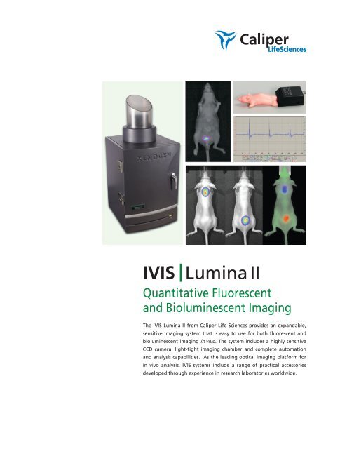
<strong>IVIS</strong> | Lumina <strong>II</strong><br />
Quantitative Fluorescent<br />
and Bioluminescent Imaging<br />
The <strong>IVIS</strong> Lumina <strong>II</strong> from Caliper Life Sciences provides an expandable,<br />
sensitive imaging system that is easy to use for both fluorescent and<br />
bioluminescent imaging in vivo. The system includes a highly sensitive<br />
CCD camera, light-tight imaging chamber and complete automation<br />
and analysis capabilities. As the leading optical imaging platform for<br />
in vivo analysis, <strong>IVIS</strong> systems include a range of practical accessories<br />
developed through experience in research laboratories worldwide.
<strong>IVIS</strong> LUMINA <strong>II</strong><br />
In Vivo Molecular Imaging<br />
Quantitative Flexible Expandable<br />
An adjustable field of view from 5 – 12.5 cm and an optional 24cm lens allows imaging of up to 5 mice or 2<br />
medium size rats. The Lumina <strong>II</strong> can also accommodate Petri dishes or micro-titer plates for in vitro imaging. The<br />
system includes premium animal handling features such as a heated stage, gas anesthesia connections and ECG<br />
monitoring.<br />
High resolution, sharp cut-off filters are simply interchangeable to achieve the highest performance, sensitivity<br />
and spectral unmixing in fluorescence imaging.<br />
Superior Imaging Results<br />
The <strong>IVIS</strong> Lumina <strong>II</strong> is capable of imaging both fluorescent and bioluminescent reporters. The system is equipped<br />
with up to 21 filter sets that can be used to image reporters that emit from green to near-infrared. Superior<br />
spectral unmixing can be achieved by Lumina <strong>II</strong>’s optional high resolution short cut off filters. Absolute calibration<br />
affords you consistent and reproducible results independent of magnification, filter selection from one instrument<br />
to any another <strong>IVIS</strong> instrument within an organization or around the world. The Living Image software yields<br />
high-quality, reproducible, quantitative results incorporating instrument calibration, background subtraction and<br />
the image algorithms.<br />
Customize the <strong>IVIS</strong> Lumina <strong>II</strong> with your own Filter Combinations<br />
Fluorophores<br />
Standard High Resolution<br />
Excitation Filter Set (Built-in)<br />
Emission Filter Options<br />
GFP, YFP and PKH26<br />
Cy 5.5, DsRed, dTomato<br />
and XenoFluor 680<br />
Indocyanine Green and<br />
XenoFluor 750<br />
Multiple Fluorophores Spanning<br />
500-900 nm Broad Imaging Solution<br />
* Median wavelength band path 20nm on emission filters<br />
430, 465, 500,<br />
535, 570, 605,<br />
640, 675, 710, 745<br />
*500 Series<br />
500, 520, 540, 560, 580, 600 and 620<br />
*600 Series<br />
580, 600, 620, 640, 660, 680 and 700<br />
*700 Series<br />
720, 740, 760, 780, 800, 820, and 840<br />
Standard Emission Filter Set<br />
515-575, 575-650, 695-770, 810-875<br />
Field of View<br />
Ex Vivo<br />
In Vivo<br />
Standard Lens FOV<br />
The <strong>IVIS</strong> Lumina <strong>II</strong> Imaging System provides 5 fields of view.<br />
New XFOV-24 Upgrade
<strong>IVIS</strong> LUMINA <strong>II</strong><br />
Imaging Results - Living Image Software with High Resolution Filters<br />
Triple Reporter Imaging<br />
Thigh Infection with Klebsiella pneumoniae expressing<br />
luxCDABE with optimized GFP or RFP. Approximately 10 8<br />
CFU per thigh. Courtesy of the University of Glasgow.<br />
Spectral Unmixing of Xenofluor 680/750<br />
Subcutaneous injections of 1014 molecules of XenoFluor 680 (scruff)<br />
and 1014 molecules of XenoFluor 750 (lower dorsal region) 605nm<br />
excitation filter.<br />
Dual Reporter Imaging - High Resolution<br />
Ex Vivo Applications<br />
Bacterial luc (500nm) and GFAP (620nm)<br />
brain imaging from mice with Pneumococcal<br />
Meningitis. Ex Vivo Kadurugamuwa et al.,<br />
Infection and Immunity, 2005.<br />
Living Image Software<br />
with <strong>IVIS</strong> Lumina <strong>II</strong> System<br />
The wide range of <strong>IVIS</strong> system instrument<br />
settings, combined with absolute<br />
calibration of each setting, allows<br />
users to track signals during longitudinal<br />
studies that vary by many orders<br />
of magnitude. In this drug study, tumor<br />
signals vary by three orders of<br />
magnitude during the course of a 35<br />
day experiment. The capability of Living<br />
Image Software makes this type of<br />
analysis simple for the user in both fluorescent<br />
and bioluminescent modes.
Inside the <strong>IVIS</strong> Lumina <strong>II</strong><br />
CCD Camera<br />
• The <strong>IVIS</strong> Lumina <strong>II</strong> CCD is 13 x 13 mm square, with 1024 x 1024 pixels 13 micron in width, yields higher imaging resolution<br />
• Back-thinned, back-illuminated grade 1 CCD provides high quantum efficiency over the entire visible to near-infrared spectrum<br />
• 16 bit digitizer delivers broad dynamic range<br />
• The CCD is thermoelectrically (Peltier) cooled to -90 ºC<br />
ensuring low dark current and low noise<br />
Imaging Chamber<br />
• Light-tight imaging chamber<br />
• High light collection lens, f /0.95 – f/16<br />
• Optional 24cm FOV lens attachment<br />
• 8 position emission filter wheels<br />
• Replaceable filter wheel upgrades for high resolution<br />
fluorescence imaging<br />
• LED lamps for photographic images<br />
• Heated stage to maintain optimum body temperature<br />
• Motor controlled stage, filter wheel, lens position, and f-stop<br />
• Optional integrated ECG monitoring system<br />
Integrated Gas Anesthesia<br />
• Gas anesthesia ports and 5 position manifold within imaging<br />
chamber allow anesthesia to be maintained during imaging sessions<br />
IMAGING SYSTEM COMPONENTS<br />
SPECIFICATIONS<br />
Camera Sensor<br />
Back-thinned, back-illuminated, cooled Grade 1 CCD<br />
CCD Size<br />
1.3 x 1.3 cm<br />
Imaging Pixels 1024 x 1024<br />
Quantum Efficiency >85% at 500 – 700 nm, >30% at 400 – 900 nm<br />
Pixel Size<br />
13 microns<br />
Min. Detectable Radiance<br />
100 photons/s/sr/cm2<br />
Min. Field of View (FOV)<br />
5 x 5 cm<br />
Max. Field of View (FOV)<br />
12.5 x 12.5 cm (optional 24 x 24 cm)<br />
Min. Image Pixel Resolution<br />
50 microns<br />
Read Noise < 3 electrons for bin=1,2, 4; < 5 electrons for bin=8, 16<br />
Dark Current (Typical)