SAB.CE insert eng rev4 0308 - Agentúra Harmony vos
SAB.CE insert eng rev4 0308 - Agentúra Harmony vos
SAB.CE insert eng rev4 0308 - Agentúra Harmony vos
Create successful ePaper yourself
Turn your PDF publications into a flip-book with our unique Google optimized e-Paper software.
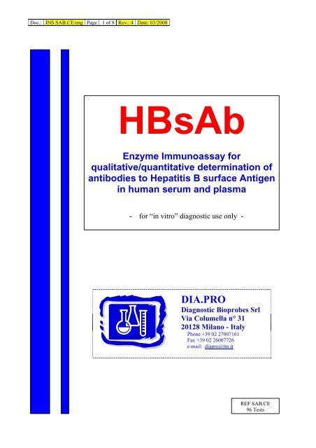
Doc.: INS <strong>SAB</strong>.<strong>CE</strong>/<strong>eng</strong> Page 1 of 8 Rev.: 4 Date: 03/2008<br />
HBsAb<br />
Enzyme Immunoassay for<br />
qualitative/quantitative determination of<br />
antibodies to Hepatitis B surface Antigen<br />
in human serum and plasma<br />
- for “in vitro” diagnostic use only -<br />
DIA.PRO<br />
Diagnostic Bioprobes Srl<br />
Via Columella n° 31<br />
20128 Milano - Italy<br />
Phone +39 02 27007161<br />
Fax +39 02 26007726<br />
e-mail: diapro@tin.it<br />
REF <strong>SAB</strong>.<strong>CE</strong><br />
96 Tests
Doc.: INS <strong>SAB</strong>.<strong>CE</strong>/<strong>eng</strong> Page 2 of 8 Rev.: 4 Date: 03/2008<br />
HBs Ab<br />
A. INTENDED USE<br />
Enzyme ImmunoAssay (ELISA) for both the quantitative and<br />
qualitative determination of antibodies to the Surface Antigen of<br />
Hepatitis B Virus in human plasma and sera.<br />
For “in vitro” diagnostic use only.<br />
B. INTRODUCTION<br />
The World Health Organization (WHO) defines Hepatitis B Virus<br />
infection as follows:<br />
“Hepatitis B is one of the major diseases of mankind and is a<br />
serious global public health problem. Hepatitis means<br />
inflammation of the liver, and the most common cause is<br />
infection with one of 5 viruses, called hepatitis A,B,C,D, and<br />
E. All of these viruses can cause an acute disease with<br />
symptoms lasting several weeks including yellowing of the<br />
skin and eyes (jaundice); dark urine; extreme fatigue;<br />
nausea; vomiting and abdominal pain. It can take several<br />
months to a year to feel fit again. Hepatitis B virus can cause<br />
chronic infection in which the patient never gets rid of the<br />
virus and many years later develops cirrhosis of the liver or<br />
liver cancer.<br />
HBV is the most serious type of viral hepatitis and the only<br />
type causing chronic hepatitis for which a vaccine is<br />
available. Hepatitis B virus is transmitted by contact with<br />
blood or body fluids of an infected person in the same way as<br />
human immunodeficiency virus (HIV), the virus that causes<br />
AIDS. However, HBV is 50 to 100 times more infectious than<br />
HIV. The main ways of getting infected with HBV are: (a)<br />
perinatal (from mother to baby at the birth); (b) child- tochild<br />
transmission; (c) unsafe injections and transfusions; (d)<br />
sexual contact.<br />
Worldwide, most infections occur from infected mother to<br />
child, from child to child contact in household settings, and<br />
from reuse of un-sterilized needles and syringes. In many<br />
developing countries, almost all children become infected<br />
with the virus. In many industrialized countries (e.g.<br />
Western Europe and North America), the pattern of<br />
transmission is different. In these countries, mother-to-infant<br />
and child-to-child transmission accounted for up to one third<br />
of chronic infections before childhood hepatitis B vaccination<br />
programmes were implemented. However, the majority of<br />
infections in these countries are acquired during young<br />
adulthood by sexual activity, and injecting drug use. In<br />
addition, hepatitis B virus is the major infectious<br />
occupational hazard of health workers, and most health care<br />
workers have received hepatitis B vaccine.<br />
Hepatitis B virus is not spread by contaminated food or<br />
water, and cannot be spread casually in the workplace. High<br />
rates of chronic HBV infection are also found in the southern<br />
parts of Eastern and Central Europe. In the Middle East and<br />
Indian sub-continent, about 5% are chronically infected.<br />
Infection is less common in Western Europe and North<br />
America, where less than 1% are chronically infected.<br />
Young children who become infected with HBV are the most<br />
likely to develop chronic infection. About 90% of infants<br />
infected during the first year of life and 30% to 50% of<br />
children infected between 1 to 4 years of age develop chronic<br />
infection. The risk of death from HBV-related liver cancer or<br />
cirrhosis is approximately 25% for persons who become<br />
chronically infected during childhood.<br />
Chronic hepatitis B in some patients is treated with drugs<br />
called interferon or lamivudine, which can help some<br />
patients. Patients with cirrhosis are sometimes given liver<br />
transplants, with varying success. It is preferable to prevent<br />
this disease with vaccine than to try and cure it.<br />
Hepatitis B vaccine has an outstanding record of safety and<br />
effectiveness. Since 1982, over one billion doses of hepatitis B<br />
vaccine have been used worldwide. The vaccine is given as a<br />
series of three intramuscular doses. Studies have shown that<br />
the vaccine is 95% effective in preventing children and adults<br />
from developing chronic infection if they have not yet been<br />
infected. In many countries where 8% to 15% of children used<br />
to become chronically infected with HBV, the rate of chronic<br />
infection has been reduced to less than 1% in immunized<br />
groups of children. Since 1991, WHO has called for all<br />
countries to add hepatitis B vaccine into their national<br />
immunization programmes.”<br />
Hepatitis B surface Antigen (HBsAg) is the major structural<br />
polypeptide of the envelope of the Hepatitis B Virus (HBV).<br />
This antigen is composed mainly of the type common<br />
determinant “a” and the type specific determinants “d” and “y”,<br />
present only on the specific serotypes.<br />
Upon infection, a strong immunological response develops firstly<br />
against the type specific determinants and in a second time<br />
against the “a” determinant.<br />
Anti “a” antibodies are however recognised to be most effective<br />
in the neutralisation of the virus, protecting the patient from<br />
other infections and leading it to convalescence.<br />
The detection of HBsAb has become important for the follow up<br />
of patients infected by HBV and the monitoring of recipients<br />
upon vaccination with synthetic and natural HBsAg.<br />
C. PRINCIPLE OF THE TEST<br />
Microplates are coated with a preparation of highly purified<br />
HBsAg that in the first incubation with sample specifically<br />
captures anti HBsAg antibodies to the solid phase.<br />
After washing, captured antibodies are detected by an HBsAg,<br />
labelled with peroxidase (HRP), that specifically binds the<br />
second available binding site of these antibodies.<br />
The enzyme specifically bound to wells, by acting on the<br />
substrate/chromogen mixture, generates an optical signal that is<br />
proportional to the amount of HBsAb in the sample and can be<br />
detected by an ELISA reader.<br />
The amount of antibodies may be quantitated by means of a<br />
standard curve calibrated against the W.H.O reference<br />
preparation.<br />
Samples are pre treated in the well with an specimen diluent<br />
able to block interference present in vaccinated individuals.<br />
D. COMPONENTS<br />
Each kit contains sufficient reagents to perform 96 tests.<br />
1. Microplate: MICROPLATE<br />
8x12 microwell strips coated with purified heat-inactivated<br />
HBsAg of both subtypes (ad and ay) from human origin and<br />
sealed into a bag with desiccant.<br />
Allow the microplate to reach room temperature before opening;<br />
reseal unused strips in the bag with desiccant and store at 4°C.
Doc.: INS <strong>SAB</strong>.<strong>CE</strong>/<strong>eng</strong> Page 3 of 8 Rev.: 4 Date: 03/2008<br />
2. Calibration Curve: CAL N° …<br />
5x2.0 ml/vial. Ready to use and colour coded standard curve,<br />
derived from HBsAb positive plasma titrated on WHO standard<br />
for anti HBsAg (1 st reference preparation 1977, lot 17-2-77),<br />
ranging: CAL1 = 0 mIU/ml // CAL2 = 10 mIU/ml // CAL3 = 50<br />
mIU/ml // CAL4 = 100 mIU/ml // CAL 5 = 250 mIU/ml.<br />
Contains human serum proteins, 5% BSA, 10 mM phosphate<br />
buffer pH 7.4+/-0.1, 0.09% sodium azide and 0.1% Kathon GC<br />
as preservatives. Standards are blue coloured.<br />
3. Wash buffer concentrate: WASHBUF 20X<br />
1x60ml/bottle. 20x concentrated solution.<br />
Once diluted, the wash solution contains 10 mM phosphate<br />
buffer pH 7.0+/-0.2, 0.05% Tween 20 and 0.1% Kathon GC.<br />
4. Enzyme conjugate : CONJ<br />
1x16.0 ml/vial. Ready-to-use solution and red color coded.<br />
It contains inactivated purified HBsAg of both subtypes ad and<br />
ay, labelled with HRP, 5% BSA, 10 mM Tris buffer pH 6.8+/-0.1,<br />
0.3 mg/ml gentamicine sulphate and 0.1% Kathon GC as<br />
preservatives.<br />
5. Chromogen/Substrate: SUBS TMB<br />
1x16ml/vial. Contains a 50 mM citrate-phosphate buffered<br />
solution at pH 3.5-3.8, 4% dimethylsulphoxide, 0.03% tetramethyl-benzidine<br />
(TMB) and 0.02% hydrogen peroxide (H2O2).<br />
Note: To be stored protected from light as sensitive to<br />
strong illumination.<br />
6. Sulphuric Acid: H2SO4 0.3 M<br />
1x15ml/vial. Contains 0.3 M H2SO4 solution.<br />
Attention: Irritant (Xi R36/38; S2/26/30)<br />
7. Specimen Diluent: DILSPE<br />
1x8ml. 10 mM Tris Buffered solution ph 7.4 +/-0.1,suggested to<br />
be used in the follow up of vaccination. It contains 0.09%<br />
sodium azide as preservatives.<br />
8. Control Serum: CONTROL …ml<br />
1 vial. Lyophilized.<br />
Contains fetal bovine serum proteins, human anti HBsAg<br />
antibodies calibrated at 50 ± 10% WHO mIU/ml. 0.3 mg/ml<br />
gentamicine sulphate and 0.1% Kathon GC as preservatives.<br />
9. Plate sealing foil n° 2<br />
10. Package <strong>insert</strong> n° 1<br />
E. MATERIALS REQUIRED BUT NOT PROVIDED<br />
1. Calibrated Micropipettes (100ul and 50ul) and disposable<br />
plastic tips.<br />
2. EIA grade water (double distilled or deionised, charcoal<br />
treated to remove oxidizing chemicals used as<br />
disinfectants).<br />
3. Timer with 60 minute range or higher.<br />
4. Absorbent paper tissues.<br />
5. Calibrated ELISA microplate thermostatic incubator (dry or<br />
wet), set at +37°C (+/-1°C tolerance)..<br />
6. Calibrated ELISA microwell reader with 450nm (reading)<br />
and possibly with 620-630nm (blanking) filters.<br />
7. Calibrated ELISA microplate washer.<br />
8. Vortex or similar mixing tools.<br />
F. WARNINGS AND PRECAUTIONS<br />
1. The kit has to be used by skilled and properly trained<br />
technical personnel only, under the supervision of a medical<br />
doctor responsible of the laboratory.<br />
2. All the personnel involved in performing the assay have to<br />
wear protective laboratory clothes, talc-free gloves and glasses.<br />
The use of any sharp (needles) or cutting (blades) devices<br />
should be avoided. All the personnel involved should be trained<br />
in biosafety procedures, as recommended by the Center for<br />
Disease Control, Atlanta, U.S. and reported in the National<br />
Institute of Health’s publication: “Biosafety in Microbiological and<br />
Biomedical Laboratories”, ed. 1984.<br />
3. All the personnel involved in sample handling should be<br />
vaccinated for HBV and HAV, for which vaccines are available,<br />
safe and effective.<br />
4. The laboratory environment should be controlled so as to<br />
avoid contaminants such as dust or air-born microbial agents,<br />
when opening kit vials and microplates and when performing the<br />
test. Protect the Chromogen (TMB) from strong light and avoid<br />
vibration of the bench surface where the test is undertaken.<br />
5. Upon receipt, store the kit at 2..8°C into a tem perature<br />
controlled refrigerator or cold room.<br />
6. Do not interchange components between different lots of<br />
the kits. It is recommended that components between two kits<br />
of the same lot should not be interchanged.<br />
7. Check that the reagents are clear and do not contain<br />
visible heavy particles or aggregates. If not, advise the<br />
laboratory supervisor to initiate the necessary procedures for kit<br />
replacement.<br />
8. Avoid cross-contamination between serum/plasma<br />
samples by using disposable tips and changing them after each<br />
sample.<br />
9. Avoid cross-contamination between kit reagents by using<br />
disposable tips and changing them between the use of each<br />
one.<br />
10. Do not use the kit after the expiration date stated on the<br />
external container and internal (vials) labels. A study conducted<br />
on an opened kit did not pointed out any relevant loss of activity<br />
up to six 6 uses of the device and up to 6 months.<br />
11. Treat all specimens as potentially infective. All human<br />
serum specimens should be handled at Biosafety Level 2, as<br />
recommended by the Center for Disease Control, Atlanta, U.S.<br />
in compliance with what reported in the Institutes of Health’s<br />
publication: “Biosafety in Microbiological and Biomedical<br />
Laboratories”, ed. 1984.<br />
12. The use of disposable plastic-ware is recommended in the<br />
preparation of the liquid components or in transferring<br />
components into automated workstations, in order to avoid<br />
cross contamination.<br />
13. Waste produced during the use of the kit has to be<br />
discarded in compliance with national directives and laws<br />
concerning laboratory waste of chemical and biological<br />
substances. In particular, liquid waste generated from the<br />
washing procedure, from residuals of controls and from samples<br />
has to be treated as potentially infective material and inactivated<br />
before waste. Suggested procedures of inactivation are<br />
treatment with a 10% final concentration of household bleach for<br />
16-18 hrs or heat inactivation by autoclave at 121°C for 20 min..<br />
14. Accidental spills from samples and operations have to be<br />
adsorbed with paper tissues soaked with household bleach and<br />
then with water. Tissues should then be discarded in proper<br />
containers designated for laboratory/hospital waste.<br />
15. The Sulphuric Acid is an irritant. In case of spills, wash the<br />
surface with plenty of water<br />
16. Other waste materials generated from the use of the kit<br />
(example: tips used for samples and controls, used microplates)<br />
should be handled as potentially infective and disposed<br />
according to national directives and laws concerning laboratory<br />
wastes.<br />
G. SPECIMEN: PREPARATION AND WARNINGS<br />
1. Blood is drawn aseptically by venepuncture and plasma or<br />
serum is prepared using standard techniques of preparation of<br />
samples for clinical laboratory analysis. No influence has been<br />
observed in the preparation of the sample with citrate, EDTA<br />
and heparin.
Doc.: INS <strong>SAB</strong>.<strong>CE</strong>/<strong>eng</strong> Page 4 of 8 Rev.: 4 Date: 03/2008<br />
2. Samples have to be clearly identified with codes or names in<br />
order to avoid misinterpretation of results. Bar code labeling and<br />
electronic reading is strongly recommended.<br />
3. Haemolysed (“red”) and visibly hyperlipemic (“milky”) samples<br />
have to be discarded as they could generate false results.<br />
Samples containing residues of fibrin or heavy particles or<br />
microbial filaments and bodies should be discarded as they<br />
could give rise to false results.<br />
4. Sera and plasma can be stored at +2°..8°C for u p to five days<br />
after collection. For longer storage periods, samples can be<br />
stored frozen at –20°C for several months. Any fr ozen samples<br />
should not be freezed/thawed more than once as this may<br />
generate particles that could affect the test result.<br />
5. If particles are present, centrifuge at 2.000 rpm for 20 min or<br />
filter using 0.2-0.8u filters to clean up the sample for testing.<br />
6. Samples whose anti-HBsAg antibody concentration is<br />
expected to be higher than 250 mIU/ml should be diluted before<br />
use either 1:10 or 1:100 in the Calibrator 0 mIU/ml. Dilutions<br />
have to be done in clean disposable tubes by diluting 50 ul of<br />
each specimen with 450 ul of Cal 0 (1:10). Then 50 ul of the<br />
1:10 dilution are diluted with 450 ul of the Cal 0 (1:100). Mix<br />
tubes thoroughly on vortex when preparing the diluted samples.<br />
H. PREPARATION OF COMPONENTS AND WARNINGS<br />
1. Microplate:<br />
Allow the microplate to reach room temperature (about 1 hr)<br />
before opening the container. Check that the desiccant has<br />
not turned green, indicating a defect in conservation.<br />
In this case, call Dia.Pro’s customer service.<br />
Unused strips have to be placed back into the aluminum pouch,<br />
with the desiccant supplied, firmly zipped and stored at +2°-8°C.<br />
After first opening, remaining strips are stable until the humidity<br />
indicator inside the desiccant bag turns from yellow to green.<br />
2. Calibration Curve<br />
Ready to use. Mix well on vortex before use.<br />
3. Control Serum<br />
Add the volume of ELISA grade water, reported on the label, to<br />
the lyophilised powder; let fully dissolve and then gently mix on<br />
vortex.<br />
Note: The control after dissolution is not stable. Store frozen in<br />
aliquots at –20°C.<br />
4. Wash buffer concentrate:<br />
The whole content of the concentrated solution has to be diluted<br />
20x with bidistilled water and mixed gently end-over-end before<br />
use. During preparation avoid foaming as the presence of<br />
bubbles could impact on the efficiency of the washing cycles.<br />
Note: Once diluted, the wash solution is stable for 1 week at<br />
+2..8° C.<br />
5. Enzyme conjugate:<br />
Ready to use. Mix well on vortex before use.<br />
Avoid contamination of the liquid with oxidising chemicals, dust<br />
or microbes. If this component has to be transferred, use only<br />
plastic, and if possible, sterile disposable containers.<br />
6. Specimen Diluent:<br />
Ready to use. Mix well on vortex before use.<br />
7. Chromogen/Substrate:<br />
Ready to use. Mix well on vortex before use.<br />
Avoid contamination of the liquid with oxidising chemicals, airdriven<br />
dust or microbes. Do not expose to strong light, oxidising<br />
agents and metallic surfaces.<br />
If this component has to be transferred use only plastic, and if<br />
possible, sterile disposable container<br />
8. Sulphuric Acid:<br />
Ready to use. Mix well on vortex before use.<br />
Attention: Irritant (Xi R36/38; S2/26/30)<br />
Legenda: R 36/38 = Irritating to eyes and skin.<br />
S 2/26/30 = In case of contact with eyes, rinse immediately with<br />
plenty of water and seek medical advice.<br />
I. INSTRUMENTS AND TOOLS USED IN COMBINATION<br />
WITH THE KIT<br />
1. Micropipettes have to be calibrated to deliver the correct<br />
volume required by the assay and must be submitted to<br />
regular decontamination (70% ethanol, 10% solution of<br />
bleach, hospital grade disinfectants) of those parts that<br />
could accidentally come in contact with the sample or the<br />
components of the kit. They should also be regularly<br />
maintained in order to show a precision of 1% and a<br />
trueness of +2%.<br />
2. The ELISA incubator has to be set at +37°C (tole rance of<br />
+1°C) and regularly checked to ensure the correct<br />
temperature is maintained. Both dry incubators and water<br />
baths are suitable for the incubations, provided that the<br />
instrument is validated for the incubation of ELISA tests.<br />
3. The ELISA washer is extremely important to the overall<br />
performances of the assay. The washer must be carefully<br />
validated and correctly optimized using the kit<br />
controls/calibrator and reference panels, before using the kit<br />
for routine laboratory tests. Usually 4-5 washing cycles<br />
(aspiration + dispensation of 350ul/well of washing solution<br />
= 1 cycle) are sufficient to ensure that the assay performs<br />
as expected. A soaking time of 20-30 seconds between<br />
cycles is suggested. In order to set correctly their number, it<br />
is recommended to run an assay with the kit<br />
controls/calibrator and well-characterized negative and<br />
positive reference samples, and check to match the values<br />
reported below in the sections “Validation of Test” and<br />
“Assay Performances”. Regular calibration of the volumes<br />
delivered and maintenance (decontamination and cleaning<br />
of needles) of the washer has to be carried out according to<br />
the instructions of the manufacturer.<br />
4. Incubation times have a tolerance of +5%.<br />
5. The ELISA microplate reader has to be equipped with a<br />
reading filter of 450nm and ideally with a second filter (620-<br />
630nm) for blanking purposes. Its standard performances<br />
should be (a) bandwidth < 10 nm; (b) absorbance range<br />
from 0 to > 2.0; (c) linearity to > 2.0; repeatability > 1%.<br />
Blanking is carried out on the well identified in the section<br />
“Assay Procedure”. The optical system of the reader has to<br />
be calibrated regularly to ensure that the correct optical<br />
density is measured. It should be regularly maintained<br />
according to the manufacturer ‘s instructions.<br />
6. When using an ELISA automated workstation, all critical<br />
steps (dispensation, incubation, washing, reading, shaking,<br />
data handling) have to be carefully set, calibrated,<br />
controlled and regularly serviced in order to match the<br />
values reported in the sections “Validation of Test” and<br />
“Assay Performances”. The assay protocol has to be<br />
installed in the operating system of the unit and validated as<br />
for the washer and the reader. In addition, the liquid<br />
handling part of the station (dispensation and washing) has<br />
to be validated and correctly set. Particular attention must<br />
be paid to avoid carry over by the needles used for<br />
dispensing samples and for washing. This must be studied<br />
and controlled to minimize the possibility of contamination of<br />
adjacent wells due to strongly reactive samples, leading to<br />
false positive results. The use of ELISA automated work<br />
stations is recommended for blood screening and when the<br />
number of samples to be tested exceed 20-30 units per run.<br />
7. Dia.Pro’s customer service offers support to the user in the<br />
setting and checking of instruments used in combination<br />
with the kit, in order to assure full compliance with the<br />
requirements described. Support is also provided for the<br />
installation of new instruments to be used with the kit.
Doc.: INS <strong>SAB</strong>.<strong>CE</strong>/<strong>eng</strong> Page 5 of 8 Rev.: 4 Date: 03/2008<br />
L. PRE ASSAY CONTROLS AND OPERATIONS<br />
1. Check the expiration date of the kit printed on the external<br />
label of the kit box. Do not use if expired.<br />
2. Check that the liquid components are not contaminated by<br />
naked-eye visible particles or aggregates. Check that the<br />
Chromogen/Substrate is colorless or pale blue by aspirating<br />
a small volume of it with a sterile transparent plastic pipette.<br />
Check that no breakage occurred in transportation and no<br />
spillage of liquid is present inside the box. Check that the<br />
aluminum pouch, containing the microplate, is not punctured<br />
or damaged.<br />
3. Dilute all the content of the 20x concentrated Wash Solution<br />
as described above.<br />
4. Dissolve the Control Serum as described above.<br />
5. Allow all the other components to reach room temperature<br />
(about 1 hr) and then mix as described.<br />
6. Set the ELISA incubator at +37°C and prepare the ELISA<br />
washer by priming with the diluted washing solution,<br />
according to the manufacturers instructions. Set the right<br />
number of washing cycles as found in the validation of the<br />
instrument for its use with the kit.<br />
7. Check that the ELISA reader has been turned on at least 20<br />
minutes before reading.<br />
8. If using an automated workstation, turn it on, check settings<br />
and be sure to use the right assay protocol.<br />
9. Check that the micropipettes are set to the required volume.<br />
10. Check that all the other equipments are available and ready<br />
to use.<br />
In case of problems, do not proceed further with the test<br />
and advise the supervisor.<br />
M. ASSAY PRO<strong>CE</strong>DURE<br />
The assay has to be carried out according to what reported<br />
below, taking care to maintain the same incubation time for all<br />
the samples in testing.<br />
Two procedures can be carried out with the device according to<br />
the request of the clinician.<br />
M.1 Quantitative analysis<br />
1. Place the required number of strips in the microplate holder.<br />
Leave A1 and B1 wells empty for the operation of blanking.<br />
Store the other strips into the bag in presence of the desiccant<br />
at 2..8°C, sealed.<br />
Then Dispense in all the wells to be used for the test, except for<br />
A1 and B1, 50µl of the Specimen Diluent.<br />
Important note: This additive is added before distributing<br />
samples and controls into specific wells and is particularly<br />
intended for blocking some substances present in people<br />
undergoing vaccination and capable to mask antibodies.<br />
2. Pipette 100µl of all the Calibrators, 100µl of Control Serum in<br />
duplicate and then 100ul of samples. The Control Serum is used<br />
to verify that the whole analytical system works as expected.<br />
Check that Calibrators, Control Serum and samples have been<br />
correctly added. Then incubate the microplate at +37°C for 60<br />
min.<br />
Important note: Strips have to be sealed with the adhesive<br />
sealing foil only when the test is performed manually. Do not<br />
cover strips when using ELISA automatic instruments.<br />
3. Wash the microplate as reported in section I.3.<br />
4. In all the wells except A1 and B1, pipette 100 µl Enzyme<br />
Conjugate. Check that the reagent has been correctly added.<br />
Incubate the microplate at +37°C for 60 minutes.<br />
Important note:<br />
1) Be careful not to touch the inner surface of the well with the<br />
pipette tip when dispensing the Enzyme Conjugate.<br />
Contamination might occur.<br />
2) Mix thoroughly the Enzyme Conjugate on vortex before use.<br />
5. Wash the microplate as described.<br />
6. Pipette 100µl TMB/H2O2 mixture in each well, the blank wells<br />
included. Check that the reagent has been correctly added.<br />
Then incubate the microplate at room temperature for 20<br />
minutes.<br />
Important note: Do not expose to strong direct light as a high<br />
background might be generated.<br />
7. Stop the enzymatic reaction by pipette 100µl Sulphuric Acid<br />
into each well and using the same pipetting sequence as in step<br />
6. Then measure the colour intensity with a microplate reader at<br />
450nm (reading) and possibly at 620-630nm (blanking),<br />
blanking the instrument on A1 and B1 wells.<br />
M.2 Qualitative analysis<br />
1. Place the required number of strips in the microplate holder.<br />
Leave A1 well empty for the operation of blanking.<br />
Store the other strips into the bag in presence of the desiccant<br />
at 2..8°C, sealed.<br />
2. Dispense 50 ul Specimen Diluent in all the wells, except for<br />
the blank A1. Then pipette 100µl of the Calibrator 0 mIU/ml in<br />
duplicate, 100µl of the Calibrator 10 mIU/ml in duplicate, 100µl<br />
of the Calibrator 250 mIU/ml in single, and then 100ul of<br />
samples. Check that Calibrators and samples have been<br />
correctly added. Then incubate the microplate at +37°C for 60<br />
min.<br />
3. Wash the microplate as reported in section I.3.<br />
4. In all the wells except A1, pipette 100 µl Enzyme Conjugate.<br />
Check that the reagent has been correctly added. Incubate the<br />
microplate at +37°C for 60 minutes.<br />
Important note:<br />
3) Be careful not to touch the inner surface of the well with the<br />
pipette tip when dispensing the Enzyme Conjugate.<br />
Contamination might occur.<br />
4) Mix thoroughly the Enzyme Conjugate on vortex before use.<br />
5. Wash the microplate as described.<br />
6. Pipette 100µl TMB/H2O2 mixture in each well, the blank wells<br />
included. Check that the reagent has been correctly added.<br />
Then incubate the microplate at room temperature for 20<br />
minutes.<br />
Important note: Do not expose to strong direct light as a high<br />
background might be generated.<br />
7. Stop the enzymatic reaction by pipette 100µl Sulphuric Acid<br />
into each well and using the same pipetting sequence as in step<br />
6. Then measure the colour intensity with a microplate reader at<br />
450nm (reading) and possibly at 620-630nm (blanking),<br />
blanking the instrument on A1 and B1 wells.<br />
Important general notes:<br />
1. If the second filter is not available, ensure that no finger<br />
prints are present on the bottom of the microwell before<br />
reading at 450nm. Finger prints could generate false<br />
positive results on reading.<br />
2. Reading has should ideally be performed immediately after<br />
the addition of the Stop Solution but definitely no longer than<br />
20 minutes afterwards. Some self oxidation of the<br />
chromogen can occur leading to a higher background.
Doc.: INS <strong>SAB</strong>.<strong>CE</strong>/<strong>eng</strong> Page 6 of 8 Rev.: 4 Date: 03/2008<br />
N. ASSAY SCHEME (standard procedure)<br />
Specimen Diluent<br />
Calibrators<br />
Control Serum<br />
Samples<br />
50 ul<br />
100 ul<br />
100 ul<br />
100 ul<br />
1 st incubation 60 min<br />
Temperature<br />
+37°C<br />
Wash step<br />
4-5 cycles<br />
Enzyme Conjugate<br />
100 ul<br />
2 nd incubation 60 min<br />
Temperature<br />
+37°C<br />
Wash step<br />
4-5 cycles<br />
TMB/H2O2 mix<br />
100 ul<br />
3 rd incubation 20 min<br />
Temperature<br />
r.t.<br />
Sulphuric Acid<br />
100 ul<br />
Reading OD<br />
450nm & 620nm<br />
An example of dispensation scheme in quantitative assays is<br />
reported below:<br />
Microplate<br />
1 2 3 4 5 6 7 8 9 10 11 12<br />
A BLK CAL4 S3<br />
B BLK CAL4 S4<br />
C CAL1 CAL5 S5<br />
D CAL1 CAL5 S6<br />
E CAL2 CS S7<br />
F CAL2 CS S8<br />
G CAL3 S1 S9<br />
H CAL3 S2 S10<br />
Legenda: BLK = Blank // CAL = Calibrators // CS = Control Serum // S =<br />
Sample<br />
An example of dispensation scheme in qualitative assays is<br />
reported below:<br />
Microplate<br />
1 2 3 4 5 6 7 8 9 10 11 12<br />
A BLK S 3 S 11<br />
B CAL1 S 4 S 12<br />
C CAL1 S 5 S 13<br />
D CAL2 S 6 S 14<br />
E CAL2 S 7 S 15<br />
F CAL5 S 8 S 16<br />
G S1 S 9 S 17<br />
H S2 S 10 S 18<br />
Legenda: BLK = Blank // CAL = Calibrators // S = Sample<br />
O. INTERNAL QUALITY CONTROL<br />
A validation check is carried out on the controls any time the kit<br />
is used in order to verify whether the performances of the assay<br />
are as qualified.<br />
Control that the following data are matched:<br />
Parameters<br />
Blank well<br />
Requirements<br />
< 0.100 OD450nm<br />
Calibrator < 0.200 OD450nm after blanking<br />
0 WHO mIU/ml<br />
Calibrator OD450nm higher than the OD450nm of the<br />
10 WHO mIU/ml Calibrator 0 mIU/ml + 0.100<br />
Calibrator > 1.500 OD450nm<br />
250 WHO mIU/ml<br />
Control Serum OD450nm = OD450nm CAL 50 mIU/ml ± 10%<br />
Coefficient of<br />
variation<br />
< 30% for the Calibrator 0 mIU/ml<br />
If the results of the test match the requirements stated above,<br />
proceed to the next section.<br />
If they do not, do not proceed any further and perform the<br />
following checks:<br />
Problem<br />
Blank well<br />
> 0.100 OD450nm<br />
Calibrator 0<br />
mIU/ml<br />
> 0.200<br />
coefficient of<br />
variation > 30%<br />
Calibrator 10<br />
mIU/ml<br />
OD450nm<br />
< Cal 0 + 0.100<br />
Calibrator 250<br />
mIU/ml<br />
< 1.500 OD450nm<br />
Control Serum<br />
Different from<br />
expected value<br />
P. RESULTS<br />
Check<br />
1. that the Chromogen/Substrate solution has<br />
not become contaminated during the assay<br />
1. that the washing procedure and the washer<br />
settings are as validated in the pre qualification<br />
study;<br />
2. that the proper washing solution has been<br />
used and the washer has been primed with it<br />
before use;<br />
3. that no mistake has been done in the assay<br />
procedure when the dispensation of standards<br />
is carried out;<br />
4. that no contamination of the Cal 0 mIU/ml or<br />
of the wells where it was dispensed has<br />
occurred due to positive samples, to spills or to<br />
the enzyme conjugate;<br />
5. that micropipettes have not become<br />
contaminated with positive samples or with the<br />
enzyme conjugate<br />
6. that the washer needles are not blocked or<br />
partially obstructed.<br />
1. that the procedure has been correctly<br />
performed;<br />
2. that no mistake has occurred during its<br />
distribution (ex.: dispensation of a wrong<br />
calibrator);<br />
3. that the washing procedure and the washer<br />
settings are as validated in the pre qualification<br />
study;<br />
4. that no external contamination of the<br />
standard has occurred.<br />
1. that the procedure has been correctly<br />
performed;<br />
2. that no mistake has occurred during its<br />
distribution;<br />
3. that the washing procedure and the washer<br />
settings are as validated in the pre qualification<br />
study;<br />
4. that no external contamination of the<br />
standard has occurred.<br />
First verify that:<br />
1. the procedure has been correctly performed;<br />
2. no mistake has occurred during its<br />
distribution (ex.: dispensation of a wrong<br />
sample);<br />
3. the washing procedure and the washer<br />
settings are correct;<br />
4. no external contamination of the standard<br />
has occurred.<br />
5. the Control Serum has been dissolved with<br />
the right volume reported on the label.<br />
If a mistake has been pointed out, the assay<br />
has to be repeated after eliminating the reason<br />
of this error.<br />
If no mistake has been found, proceed as<br />
follows:<br />
a) a value up to +/-20% is obtained: the overall<br />
Precision of the laboratory might not enable the<br />
test to match the expected value +/-10%.<br />
Report the problem to the Supervisor for<br />
acceptance or refusal of this result.<br />
b) a value higher than +/-20% is obtained: in<br />
this case the test is invalid and the DiaPro’s<br />
customer service has to be called.<br />
P.1 Quantitative method<br />
If the test turns out to be valid, use for the quantitative method<br />
an approved curve fitting program to draw the calibration curve<br />
from the values obtained by reading at 450nm (4-parameters<br />
interpolation is suggested).<br />
Then on the calibration curve calculate the concentration of anti<br />
HBsAg antibody in samples.<br />
An example of Calibration curve is reported in the next page.
Doc.: INS <strong>SAB</strong>.<strong>CE</strong>/<strong>eng</strong> Page 7 of 8 Rev.: 4 Date: 03/2008<br />
3000<br />
2500<br />
2000<br />
Example of Calibration Curve :<br />
R. PERFORMAN<strong>CE</strong>S<br />
Evaluation of Performances has been conducted in accordance<br />
to what reported in the Common Technical Specifications or<br />
CTS (art. 5, Chapter 3 of IVD Directive 98/79/EC).<br />
1. LIMIT OF DETECTION:<br />
The limit of detection of the assay has been calculated by<br />
means of the HBsAb international preparation supplied by CLB<br />
on behalf of WHO (1 st reference preparation 1977, lot 17-2-77),<br />
on which Calibration Curve has been calibrated. HBV negative<br />
serum was used as diluent, as recommended by the supplier.<br />
Results of Quality Control are given in the following table:<br />
1500<br />
1000<br />
500<br />
OD450nm<br />
WHO<br />
mIU/ml<br />
<strong>SAB</strong>.<strong>CE</strong><br />
Lot # 1002<br />
<strong>SAB</strong>.<strong>CE</strong><br />
Lot # 1001<br />
<strong>SAB</strong>.<strong>CE</strong><br />
Lot # 1002/2<br />
50 0.933 0.812 0.846<br />
10 0.219 0.192 0.194<br />
5 0.110 0.096 0.104<br />
2.5 0.057 0.058 0.067<br />
Std 0 0.021 0.015 0.023<br />
0<br />
0 50 100 150 200 250 300<br />
Important Note:<br />
Do not use the calibration curve above to make calculations.<br />
P.2 Qualitative method<br />
In the qualitative method, calculate the mean OD450nm values<br />
for the Calibrators 0 and 10 mIU/ml and then check that the<br />
assay is valid.<br />
Example of calculation:<br />
The following data must not be used instead or real figures obtained by<br />
the user.<br />
Calibrator 0 mIU/ml: 0.020 – 0.024 OD450nm<br />
Mean Value: 0.022 OD450nm<br />
Lower than 0.200 – Accepted<br />
Calibrator 10 mIU/ml: 0.250 – 0.270 OD450nm<br />
Mean Value: 0.260 OD450nm<br />
Higher than Cal 0 + 0.100 – Accepted<br />
Calibrator 250 mIU/ml: 2.845 OD450nm<br />
Higher than 1.500 – Accepted<br />
Q. INTERPRETATION OF RESULTS<br />
Samples with a concentration lower than 10 WHO mIU/ml are<br />
considered negative for anti HBsAg antibody by most of the<br />
international medical literature.<br />
Samples with a concentration higher than 10 WHO mIU/ml are<br />
considered positive for anti HBsAg antibody.<br />
In the follow up of vaccination recipients, however, the value of<br />
20 WHO mIU/ml is usually accepted by the medical literature as<br />
the minimum concentration at which the patient is considered<br />
clinically protected against HBV infection.<br />
Important notes:<br />
1. Interpretation of results should be done under the<br />
supervision of the laboratory supervisor to reduce the risk of<br />
judgement errors and misinterpretations.<br />
2. When test results are transmitted from the laboratory to<br />
another facility, attention must be paid to avoid erroneous<br />
data transfer.<br />
3. Diagnosis has to be done and released to the patient by a<br />
suitably qualified medical doctor.<br />
2. DIAGNOSTIC SPECIFICITY AND SENSITIVITY<br />
A Performance Evaluation has been conducted on a total<br />
number of more than 700 samples.<br />
2.1 Diagnostic Specificity<br />
It is defined as the probability of the assay of scoring negative in<br />
the absence of specific analyte.<br />
More than 500 negative specimens were tested, internally and<br />
externally, against a European company.<br />
A diagnostic specificity of 98.8% was assessed. .<br />
Moreover, diagnostic specificity was assessed by testing 113<br />
potentially interfering specimens (other infectious diseases,<br />
patients affected by non viral hepatic diseases, dialysis patients,<br />
pregnant women, hemolized, lipemic, etc.) against the European<br />
company. A value of specificity of 100% was assessed.<br />
Finally, both human plasma, derived with different standard<br />
techniques of preparation (citrate, EDTA and heparin), and<br />
human sera have been used to determine the specificity.<br />
No false reactivity due to the method of specimen preparation<br />
has been observed.<br />
2.2 Diagnostic Sensitivity<br />
It defined as the probability of the assay of scoring positive in<br />
the presence of specific analyte.<br />
106 vaccinated patients were evaluated providing a diagnostic<br />
sensitivity of 100%.<br />
More than 100 HBV naturally infected patients were tested,<br />
internally and externally, against the European company; a<br />
diagnostic sensitivity of 100% was found.<br />
3. PRECISION:<br />
The mean values obtained from a study conducted on three<br />
samples of different anti-HBsAg reactivity, examined in 16<br />
replicates in three separate runs is reported below:<br />
<strong>SAB</strong>.<strong>CE</strong>: lot # 1202<br />
Calibrator 0 mIU/ml (N = 16)<br />
Mean values 1st run 2nd run 3 rd run Average<br />
value<br />
OD 450nm 0.038 0.038 0.039 0.039<br />
Std.Deviation 0.003 0.004 0.005 0.004<br />
CV % 8.8 9.5 11.8 10.0<br />
Calibrator 10 mIU/ml (N = 16)<br />
Mean values 1st run 2nd run 3 rd run Average<br />
value<br />
OD 450nm 0.250 0.243 0.244 0.246<br />
Std.Deviation 0.020 0.023 0.017 0.020<br />
CV % 8.0 9.3 7.0 8.1
Doc.: INS <strong>SAB</strong>.<strong>CE</strong>/<strong>eng</strong> Page 8 of 8 Rev.: 4 Date: 03/2008<br />
Calibrator 250 mIU/ml (N = 16)<br />
Mean values 1st run 2nd run 3 rd run Average<br />
value<br />
OD 450nm 2.998 3.000 3.259 3.085<br />
Std.Deviation 0.152 0.151 0.158 0.153<br />
CV % 5.1 5.0 4.8 5.0<br />
<strong>SAB</strong>.<strong>CE</strong>: lot # 1002<br />
Calibrator 0 mIU/ml (N = 16)<br />
Mean values 1st run 2nd run 3 rd run Average<br />
value<br />
OD 450nm 0.048 0.048 0.050 0.049<br />
Std.Deviation 0.005 0.004 0.006 0.005<br />
CV % 9.4 8.4 11.5 9.8<br />
Calibrator 10 mIU/ml (N = 16)<br />
Mean values 1st run 2nd run 3 rd run Average<br />
value<br />
OD 450nm 0.249 0.252 0.242 0.248<br />
Std.Deviation 0.021 0.020 0.023 0.021<br />
CV % 8.3 7.9 9.6 8.6<br />
Calibrator 250 mIU/ml (N = 16)<br />
Mean values 1st run 2nd run 3 rd run Average<br />
value<br />
OD 450nm 3.544 3.653 3.612 3.603<br />
Std.Deviation 0.153 0.176 0.138 0.156<br />
CV % 4.3 4.8 3.8 4.3<br />
<strong>SAB</strong>.<strong>CE</strong>: lot # 1002/2<br />
Calibrator 0 mIU/ml (N = 16)<br />
Mean values 1st run 2nd run 3 rd run Average<br />
value<br />
OD 450nm 0.050 0.051 0.050 0.050<br />
Std.Deviation 0.005 0.006 0.006 0.005<br />
CV % 10.0 10.9 11.9 10.9<br />
Calibrator 10 mIU/ml (N = 16)<br />
Mean values 1st run 2nd run 3 rd run Average<br />
value<br />
OD 450nm 0.226 0.238 0.239 0.234<br />
Std.Deviation 0.015 0.017 0.018 0.016<br />
CV % 6.5 7.0 7.5 7.0<br />
Calibrator 250 mIU/ml (N = 16)<br />
Mean values 1st run 2nd run 3 rd run Average<br />
value<br />
OD 450nm 3.526 3.457 3.499 3.494<br />
Std.Deviation 0.137 0.143 0.162 0.147<br />
CV % 3.9 4.1 4.6 4.2<br />
REFEREN<strong>CE</strong>S<br />
1. Engvall E. et al., J.Immunochemistry, 8, 871-874, 1971.<br />
2. Engvall E. et al., J.Immunol. 109, 129-135, 1971.<br />
3. Remington J.S. and Klein J.O. In “Infectious diseases of<br />
the fetus and newborn infant”. Sanders, Philadelphia,<br />
London, Toronto.<br />
4. Volk W.A. In “Essential of Medical Microbiology”. 2 nd ed.,<br />
pp 729, G.B.Lippincott Company, Philadelphia, New York,<br />
S.Josè, Toronto<br />
5. Snydman D.R. et al., Ann.Int.Med., 83 : 838, 1975.<br />
6. Barker L.F., Dodd R.J., Sandler S.G.. In “viral Hepatitis:<br />
Laboratory and Clinical Science” F.Deinhardt, J. Deinhardt<br />
eds., M.Dekker Inc., New York, 215-230, 1983.<br />
7. Cossart Y., Brit.Med.Bull., 28: 156, 1972<br />
8. Lander J.J. et al., J.Immunol., 106: 1066, 1971<br />
9. Mushawar I.K. et al.. Ann.J.Clin.Pathol., 76: 773, 1981.<br />
10. Howard C.R., Immunol.Today, 5: 185, 1984<br />
11. Aach R.D.. Lancet 7874: 190-193, 1974.<br />
12. Jilg W. et al.. J.Hepatol. 9 : 201-207, 1988<br />
13. P.Crovari et al., Boll. Ist. Sieroter. Milan., 63: 14-18, 1984<br />
14. M.Davidson et al., J.Natl.Cancer Inst., 59 : 1451-1467,<br />
1977<br />
15. F.Gyorkey et al., J.Natl.Cancer Inst., 59: 1451-1467, 1977<br />
16. S.Hadler et al., N.E.J.Med., 315: 209-214, 1986<br />
17. J.H.Hoofnagle et al., Hepatology, 7: 758-763, 1987<br />
18. C.L.Howard, J.Gen.Virol., 67: 1215-1235<br />
19. W.Jilg et al. J.Hepatol., 6: 201-207, 1988<br />
20. P.Michel et al., Nephrologie, 7: 114-117, 1986<br />
21. W.Szmuness et al., N.E.J.Med., 303: 833-836, 1980<br />
22. P.Tiollais et al., Nature, 317: 489-495, 1985<br />
23. A.J.Zuckermann et al., in “Hepatitis Viruses of Man”<br />
Academic Press, London, 1979<br />
|<br />
0318<br />
Produced by<br />
Dia.Pro. Diagnostic Bioprobes Srl.<br />
via Columella n° 31 – Milano - Italy<br />
The variability shown in the tables did not result in sample<br />
misclassification.<br />
4. ACCURACY<br />
The assay accuracy has been checked by the dilution and<br />
recovery tests. Any “hook effect”, underestimation likely to<br />
happen at high doses of analyte, was ruled out up to 10.000<br />
mIU/ml.<br />
S. LIMITATIONS OF THE PRO<strong>CE</strong>DURE<br />
Bacterial contamination or heat inactivation of the specimen<br />
may affect the absorbance values of the samples with<br />
consequent alteration of the level of the analyte.<br />
This test is suitable only for testing single samples and not<br />
pooled ones.<br />
Diagnosis of an infectious disease should not be established on<br />
the basis of a single test result. The patient’s clinical history,<br />
symptomatology, as well as other diagnostic data should be<br />
considered.



![[APTT-SiL Plus]. - Agentúra Harmony vos](https://img.yumpu.com/50471461/1/184x260/aptt-sil-plus-agentara-harmony-vos.jpg?quality=85)
![[SAS-1 urine analysis]. - Agentúra Harmony vos](https://img.yumpu.com/47529787/1/185x260/sas-1-urine-analysis-agentara-harmony-vos.jpg?quality=85)

![[SAS-MX Acid Hb]. - Agentúra Harmony vos](https://img.yumpu.com/46129828/1/185x260/sas-mx-acid-hb-agentara-harmony-vos.jpg?quality=85)
