Scanning Electron Microscopy - Gbhenterprises.com
Scanning Electron Microscopy - Gbhenterprises.com
Scanning Electron Microscopy - Gbhenterprises.com

You also want an ePaper? Increase the reach of your titles
YUMPU automatically turns print PDFs into web optimized ePapers that Google loves.
Test M604: <strong>Scanning</strong> electron microscopy<br />
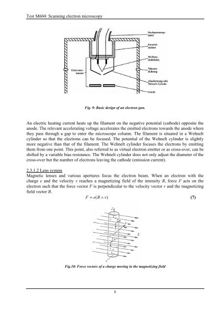
Fig. 9: Basic design of an electron gun.<br />
An electric heating current heats up the filament on the negative potential (cathode) opposite the<br />
anode. The relevant accelerating voltage accelerates the emitted electrons towards the anode where<br />
they pass through a gap to enter the microscope column. The filament is situated in a Wehnelt<br />
cylinder so that the electrons can be focused. The potential of the Wehnelt cylinder is slightly<br />
more negative than that of the filament. The Wehnelt cylinder focuses the electrons by emitting<br />
them from one point. This point, also referred to as virtual electron emitter or as cross-over, can be<br />
shifted by a variable bias resistance. The Wehnelt cylinder does not only adjust the diameter of the<br />
cross-over but the number of electrons leaving the cathode (emission current).<br />
2.3.1.2 Lens system<br />
Magnetic lenses and various apertures focus the electron beam. When an electron with the<br />
charge e and the velocity v reaches a magnetizing field of the intensity B, force F acts on the<br />
electron such that the force vector F is perpendicular to the velocity vector v and the magnetizing<br />
field vector B.<br />
F = e( B ∧ v)<br />
(7)<br />
Fig.10: Force vectors of a charge moving in the magnetizing field<br />
8