Scanning Electron Microscopy - Gbhenterprises.com
Scanning Electron Microscopy - Gbhenterprises.com
Scanning Electron Microscopy - Gbhenterprises.com

You also want an ePaper? Increase the reach of your titles
YUMPU automatically turns print PDFs into web optimized ePapers that Google loves.
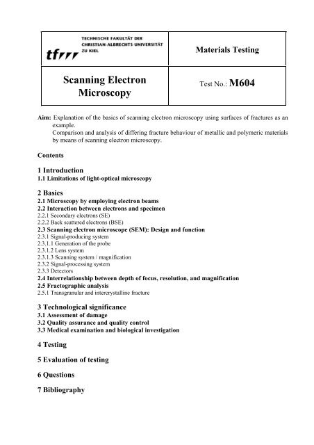
Materials Testing<br />
<strong>Scanning</strong> <strong>Electron</strong><br />
<strong>Microscopy</strong><br />
Test No.: M604<br />
Aim: Explanation of the basics of scanning electron microscopy using surfaces of fractures as an<br />
example.<br />
Comparison and analysis of differing fracture behaviour of metallic and polymeric materials<br />
by means of scanning electron microscopy.<br />
Contents<br />
1 Introduction<br />
1.1 Limitations of light-optical microscopy<br />
2 Basics<br />
2.1 <strong>Microscopy</strong> by employing electron beams<br />
2.2 Interaction between electrons and specimen<br />
2.2.1 Secondary electrons (SE)<br />
2.2.2 Back scattered electrons (BSE)<br />
2.3 <strong>Scanning</strong> electron microscope (SEM): Design and function<br />
2.3.1 Signal-producing system<br />
2.3.1.1 Generation of the probe<br />
2.3.1.2 Lens system<br />
2.3.1.3 <strong>Scanning</strong> system / magnification<br />
2.3.2 Signal-processing system<br />
2.3.3 Detectors<br />
2.4 Interrelationship between depth of focus, resolution, and magnification<br />
2.5 Fractographic analysis<br />
2.5.1 Transgranular and intercrystalline fracture<br />
3 Technological significance<br />
3.1 Assessment of damage<br />
3.2 Quality assurance and quality control<br />
3.3 Medical examination and biological investigation<br />
4 Testing<br />
5 Evaluation of testing<br />
6 Questions<br />
7 Bibliography
Test M604: <strong>Scanning</strong> electron microscopy<br />
1 Introduction<br />
Minor defects often result in considerable damage. Small fractures or cracks in materials can have<br />
disastrous effects on the stability of buildings, tools, etc. Once an accident has happened, its<br />
causes have to be found. A microscope examination of the fracture surface shows whether a<br />
material defect or a processing defect has caused the fracture. Light-optical and electron-optical<br />
microscopes are used for this purpose. <strong>Electron</strong> microscopes are advantageous in that a high<br />
degree of magnification as well as an excellent depth of focus (Fig.1) can be achieved. As a rule,<br />
surfaces of fracture are very rough so that a light-optical microscope often cannot produce a<br />
sufficiently clear enlargement of the relevant image section.<br />
Fig. 1: Photo of blood corpuscles taken by means of a) a light-optical microscope and b) an electron-optical<br />
microscope (same magnification).<br />
1.1 Limitations of light-optical microscopy<br />
The amount of information a micrograph can provide is dependent on resolution. The maximum<br />
resolution that can be achieved using a microscope means the smallest interval distinguishable<br />
between two adjacent points. Any magnification exceeding such maximum would not make sense<br />
since further information cannot be provided.<br />
The maximum resolution mainly depends on the wavelength of the radiation selected for the<br />
image. Beams entering the lens- and aperture system of the microscope produce overlapping<br />
diffraction patterns for each object point. The distance r 1<br />
between two diffraction maxima must<br />
exceed full width half maximum (FWHM), otherwise the diffraction maxima cannot be discerned<br />
as being separate (Fig. 2). According to a simple rule found by Rayleigh, distinction is possible<br />
when the maximum of the zero order coincides with the first minimum of the second diffraction<br />
pattern. The distance between the two first minima d 1<br />
is inversely proportional to the diameter of<br />
the aperture.<br />
2
Test M604: <strong>Scanning</strong> electron microscopy<br />
Fig. 2: Minimal distance between two diffraction maxima still projected separately<br />
Diffraction patterns are dependent on the wavelength λ, on the index of refraction of the<br />
surrounding medium µ, and on the angle α formed by the optical axis and the edge beam, which<br />
can only just pass through the aperture. For r 1<br />
results:<br />
1<br />
0,61λ<br />
r<br />
1<br />
= d =<br />
(1)<br />
2 µ sinα<br />
The product µ sin α is referred to as numeric aperture.<br />
Thus, high resolution can be achieved by a short wavelength, a high index of refraction of the<br />
surrounding medium, and a short distance to the sample (hereinafter also referred to as<br />
"specimen") (wide angle α). When normal light-optical microscopes are used, the surrounding<br />
medium is air (µ = 1) and the distance between sample and lens cannot be decreased at discretion.<br />
For this reason, the maximum resolution with regard to wavelengths of visible light (400 - 700<br />
nm) is limited to about 200 nm, and any degree of magnification beyond 1000 would not make<br />
sense.<br />
2 Basics<br />
2.1 <strong>Microscopy</strong> by employing electron beams<br />
(Hereinafter the term "electron beam is also referred to as "probe"). If electrons are used instead<br />
of optical waves, much smaller wavelengths can be achieved. The wavelength can be varied<br />
depending on the voltage set to accelerate the electrons towards the sample. The velocity v of a<br />
single electron can almost reach the velocity of light c. In that case, relativistic corrections be<strong>com</strong>e<br />
necessary. The electron mass changes according to the following equation:<br />
me<br />
m =<br />
, ( 2 )<br />
1<br />
2 2<br />
⎡ ⎛ v ⎞ ⎤<br />
⎢1<br />
− ⎜ ⎟ ⎥<br />
⎢⎣<br />
⎝ c ⎠ ⎥⎦<br />
m e<br />
is the rest mass of the electron.<br />
The deBroglie relation determines the interrelationship between wavelength and momentum.<br />
h h λ = = , ( 3 )<br />
p mv<br />
h is Planck’s quantum (constant of action). The energy transmitted to an electron eV can be<br />
equated with the energy of relativistic mass changes:<br />
eV = m − m c<br />
(4)<br />
( ) 2<br />
e<br />
By means of these three equations the dependence of wavelength on accelerating voltage can be<br />
derived:<br />
3
Test M604: <strong>Scanning</strong> electron microscopy<br />
2<br />
2 h<br />
λ =<br />
(5)<br />
2 2<br />
⎛ 2eVm<br />
+ ⎞<br />
e<br />
e V<br />
⎜<br />
⎟<br />
2<br />
⎝ c ⎠<br />
⎡ 1,5 ⎤<br />
λ = ⎢<br />
nm<br />
6 2 ⎥<br />
(6)<br />
−<br />
⎣( V + 10 V ) ⎦<br />
An accelerating voltage of e.g. 20 kV results in 8.6E-3 nm = 8.6 pm, whereas at 500 kV only<br />
1.4E-3 nm are reached.<br />
Since electrons would be too strongly scattered in air, a high vacuum is required in an electron<br />
microscope. In addition, the samples to be tested have to be electrically conductive, otherwise they<br />
would be overcharged with electrons during irradiation. For this reason, conductors and insulators<br />
of inferior quality have to be coated with a conductive layer of metal or carbon prior to<br />
microscopic investigation.<br />
2.2 Interaction between electrons and specimen<br />
<strong>Electron</strong>s in scanning electron microscopes are accelerated at voltages in the range of 2 to 40 kV.<br />
An electron beam < 0.01µm in diameter is focused on the specimen. These fast primary electrons<br />
(PE) interact in various ways with the surface layers of the specimen. The zone, in which such<br />
interaction occurs, and in which different signals are produced, is called "interaction volume" or<br />
"electron – diffusion cloud". The size of the interaction volume is proportional to the energy of<br />
primary electrons, its shape is determined dependent upon scattering processes by the mean atomic<br />
number. Secondary electrons (SE), back scattered electrons (BSE), and absorbed electrons are<br />
produced, flowing off as specimen current. In addition, X-rays, Auger electrons, and<br />
cathodoluminescence are produced (Fig. 3).<br />
1<br />
2<br />
Fig. 3: Interaction volume<br />
R: The range of primary electrons (PE);<br />
T: Escape level for back scattered electrons (BSE)<br />
Resolution limit of BSE ≈ ½ R<br />
Resolution limit of X-radiation ≈ interaction volume<br />
Resolution limit of secondary fluorescence > interaction volume<br />
4
Test M604: <strong>Scanning</strong> electron microscopy<br />
2.2.1 Secondary electrons (SE)<br />
Although secondary electrons are produced in the entire interaction volume, they can only escape<br />
from surface layers (metals: max. 5mm, insulators: max. 50 mm, Fig. 4: Escape level t).<br />
Secondary electrons are very slow, their escape energy is ≤ 50 eV. Approximately half of all SE<br />
are produced very near to the point of impact of PE (SE1). Owing to back scattered electrons<br />
(BSE) diffusing in the specimen material, SE are also produced at a distance in the range of 0.1 to<br />
some µm to the point of impact (SE2). Back scattered electrons reacting with the wall of the<br />
specimen chamber are the third source of SE. This reaction process causes background radiation<br />
and thus a smaller degree of contrast, which, however, can partly be increased again electronically.<br />
(Fig. 4)<br />
Fig. 4: Production of SE and BSE<br />
The best lateral point resolution can be achieved by means of SE1. The signal can be intensified<br />
when the primary beam hits the samples at an angle of < 90°; this is referred to as inclination<br />
contrast. If radiation can penetrate specimen structures such as tips, fibres, or edges, the images of<br />
these structures will be very bright (edge contrast) owing to a high SE yield.<br />
The SE signal, <strong>com</strong>prising all essential information on topography, produces electron-micrographs<br />
of high resolution.<br />
Fig. 5: SE yield δ is dependent on the atomic number Z<br />
2.2.2 Back scattered electrons (BSE)<br />
The electrons escaping from the surface of the sample and having an energy of ≥ 50 eV are<br />
referred to as back scattered electrons (BSE). BSE are produced in the entire interaction volume at<br />
a larger distance to the point of impact of PE (Fig. 4). When atomic numbers are low, the escape<br />
5
Test M604: <strong>Scanning</strong> electron microscopy<br />
level T is approx. half the range R; at accelerating voltages > 20kV and when atomic numbers are<br />
high, the escape level T is lower. The higher the PE energy and the smaller the atomic number of<br />
the specimen material, the more extends the area of production of BSE and the lower the<br />
achievable resolution. However, the dependence on the atomic number of the sample material is<br />
an advantage in that, apart from the topography contrast, a material contrast can be made visible.<br />
Moreover, owing to higher energy charging occurs less frequently than in case of SE.<br />
Fig. 6: RE yield η is dependent on the atomic number Z<br />
2.3 <strong>Scanning</strong> electron microscope (SEM): Design and function<br />
The surface of a specimen is brought into the focus of electron beams. The signals produced<br />
control the brightness of a screen tube such that an image of the surface of the sample appears.<br />
Fig. 7 illustrates the basic design of a scanning electron microscope.<br />
Fig. 7: Basic design of an SEM<br />
In a scanning electron microscope the signal-producing system and the signal-processing system<br />
operate independently.<br />
6
Test M604: <strong>Scanning</strong> electron microscopy<br />
2.3.1 Signal-producing system<br />
The signal-producing system (see Fig. 7 to the left and Fig. 8) is to generate a probe of the<br />
smallest diameter possible and of maximum brightness when hitting the surface of the specimen. It<br />
consists of an electron gun, (cathode – Wehnelt cylinder – anode), lens system (lenses, apertures,<br />
beam deflection coils and stigmator coils) and the specimen chamber.<br />
Fig. 8: Course of the probe in the signal-producing system<br />
At least two pumps are required to reduce pressure to a vacuum. A vane-type rotary pump<br />
produces a pre-vacuum of approx. 10 -3<br />
mbar. Either a turbomolecular pump or an oil diffusion<br />
pump maintain the operation vacuum of at least 10 -5<br />
mbar in the column and in the chamber.<br />
Dependent upon the type of cathode used, a third pump, the ion getter pump, may be operated.<br />
For further information please refer to technical literature!<br />
2.3.1.1 Generation of the probe<br />
In the field of electron microscopy free electrons are usually produced by thermal emission. Other<br />
microscopes operate by means of field emission (> 10 9 V/m). Mostly, tungsten filaments or - as<br />
described here - LaB 6<br />
-crystals serve as cathode. The electron emitter consists of a three-electrode<br />
arrangement (Fig. 9).<br />
7
Test M604: <strong>Scanning</strong> electron microscopy<br />
Fig. 9: Basic design of an electron gun.<br />
An electric heating current heats up the filament on the negative potential (cathode) opposite the<br />
anode. The relevant accelerating voltage accelerates the emitted electrons towards the anode where<br />
they pass through a gap to enter the microscope column. The filament is situated in a Wehnelt<br />
cylinder so that the electrons can be focused. The potential of the Wehnelt cylinder is slightly<br />
more negative than that of the filament. The Wehnelt cylinder focuses the electrons by emitting<br />
them from one point. This point, also referred to as virtual electron emitter or as cross-over, can be<br />
shifted by a variable bias resistance. The Wehnelt cylinder does not only adjust the diameter of the<br />
cross-over but the number of electrons leaving the cathode (emission current).<br />
2.3.1.2 Lens system<br />
Magnetic lenses and various apertures focus the electron beam. When an electron with the<br />
charge e and the velocity v reaches a magnetizing field of the intensity B, force F acts on the<br />
electron such that the force vector F is perpendicular to the velocity vector v and the magnetizing<br />
field vector B.<br />
F = e( B ∧ v)<br />
(7)<br />
Fig.10: Force vectors of a charge moving in the magnetizing field<br />
8
Test M604: <strong>Scanning</strong> electron microscopy<br />
The magnetizing field of an electromagnetic lens can be divided into an axial and a radial part.<br />
The axial part, running in parallel to the direction of movement of the electron, does not influence<br />
the electron. The radial part, however, forces the electron to take a helix-path by the force<br />
(B rad<br />
e v). Thus, due to such circular <strong>com</strong>ponent the velocity vector is influenced by the axial<br />
magnetizing field (B ax<br />
e v zirk<br />
). As a result the radius of the helix-path is be<strong>com</strong>ing ever smaller.<br />
The electromagnetic lenses of an SEM produce an image reduced in diameter of the cross-over in<br />
the gun on the surface of the specimen. Two condenser lenses (Fig. 8) reduce the diameter of the<br />
electron beam (the diameter of the electron beam is also referred to as "probe size") from d 0<br />
to d 2<br />
.<br />
The higher the lens current, the smaller the diameter (Fig. 11).<br />
Fig. 11: Schematic illustration of the probe a) low, b) high lens current<br />
The smaller the probe size, the smaller the portion of electrons reaching the specimen since not all<br />
electrons leaving lens 1 can pass through lens 2. (Fig. 11): α<br />
2<br />
< α1<br />
. Increasing noise results,<br />
limiting the resolving power of the SEM.<br />
The third lens, i.e. the objective lens, focuses the probe towards the specimen.<br />
Lens holes which are not absolutely symmetrical mechanically, whose magnetizing fields are<br />
inhomogeneous, and whose pole piece holes are contaminated, and contaminated apertures in<br />
particular, will result in an elliptical probe producing "axial astigmatism". The surface of the<br />
specimen cannot be brought into focus accurately since an elliptical probe will produce a distorted<br />
image of specimen structures during the focusing process. A corrective magnetic field, required to<br />
recover the rotational symmetry of the probe, is to be produced by a stigmator. A stigmator<br />
consists of 2 times 4 coils arranged centrically towards the optical axis.<br />
2.3.1.3 <strong>Scanning</strong> system / magnification<br />
Beam deflection coils in a scanning generator (Fig. 7) scan the specimen surface by means of the<br />
primary electron beam for a certain period of time; beam deflection coils are installed in the pole<br />
piece duct of the objective lens. Simultaneously a cathode ray scans the screen of a monitor.<br />
Due to the principle of scanning, an SEM lineagraph consists of many spots. The beam deflection<br />
coil can be used to produce horizontal and vertical deflections by means of the electron beam.<br />
Horizontal deflection generates a line whose position is determined by vertical deflection.<br />
9
Test M604: <strong>Scanning</strong> electron microscopy<br />
<strong>Scanning</strong> speed depends on the time set for the scanning of one line and on the number of lines per<br />
scanning process ("frame").<br />
In order to increase magnification the current in the beam deflection coils must be increased. This<br />
involves a reduction of the scanning pattern produced on the specimen, whereas the size of the<br />
image displayed remains unchanged. Thus, magnification results from the ratio between the edge<br />
length of the screen and the edge length of the zone scanned on the specimen (Fig. 7). If, e.g., a<br />
zone of 1mm x 1mm is scanned, while the edge length of the screen is 30 cm, the degree of<br />
magnification will be 300-fold.<br />
2.3.2 Signal-processing system<br />
Fig.12: System of signal processing<br />
Due to the principle of scanning, signals - e.g. secondary electrons - are successively produced by<br />
each object point. After registration by means of a detector an electrical signal, the video signal, is<br />
generated and amplified by a preamplifier and by video amplification. The video signal, such<br />
amplified, modulates the cathode ray deflected simultaneously to the primary electron beam such<br />
that an image appears on the monitor. In this way, there is a spot-by-spot-correlation between the<br />
signal level of an object point and the brightness of the corresponding display spot.<br />
The amplitudes of the signal can be displayed as Y-modulation.<br />
The modulation of object signals to successive electrical signals is advantageous in that the latter<br />
can be modified in order to optimise image information (brightness, contrast etc.).<br />
2.3.3 Detectors<br />
Detectors connect the signal-producing and the signal-processing system of an SEM. They convert<br />
the signals produced (electrons) into electrical signals. As a rule, each signal (secondary electrons,<br />
back scattered electrons, X-rays) requires a special detector.<br />
10
Test M604: <strong>Scanning</strong> electron microscopy<br />
Fig. 13: Everhart - Thornley – Detector<br />
K: Collector, S: Scintillator, LL: Optical fibre, V: Preamplifier, PM: Photo multiplier<br />
The most widely used detector of secondary electrons is the Everhart-Thornley-Detector (Fig. 13).<br />
A driving potential of e.g. +300 to 400 V is applied between the specimen and the collector for the<br />
intake of secondary electrons of low energy. Between collector and scintillator, high voltage of 10<br />
kV is applied, accelerating the SE to <strong>com</strong>e forcibly into contact with the scintillator. The<br />
scintillator consists either of a glass plate coated with luminescent powder (phosphor <strong>com</strong>pound)<br />
or of a YAG- or YAP- monocrystal. The photons produced pass via the optical fibre to the photo<br />
multiplier. The photons release electrons at the photocathode of the multiplier. The multiplier<br />
voltage accelerates these electrons towards the dynodes where they produce cumulatively a<br />
multiple of electrons.<br />
BSE are also detected. If an image is to be produced by BSE only, no SE must be present; the<br />
collector must be switched off or a negative voltage must be applied to repulse the SE.<br />
Scintillator detectors (Robinson detector) or semiconductor detectors are especially in use to detect<br />
BSE.<br />
2.4 Interrelationship between depth of focus, resolution, and magnification<br />
Great depth of focus is required for an analysis of fracture surfaces. The term "depth of focus"<br />
describes that zone of object positions, in which a change in focus cannot be perceived through the<br />
sight. Fig. 14 shows the interrelationship between the depth of focus and the point resolution X or<br />
magnification.<br />
At a 1000-fold magnification, the light-optical microscope can only project a depth of approx.<br />
0.2 µm, whereas 100 µm can be achieved by means of an electron microscope.<br />
11
Test M604: <strong>Scanning</strong> electron microscopy<br />
Fig. 14: Interrelationship between depth of focus, point resolution, and magnification: Light-optical microscope<br />
and scanning electron microscope.<br />
2.5 Fractographic analysis<br />
Any fracture of a body starts with the formation and propagation of cracks in submicroscopic,<br />
microscopic, and eventually macroscopic dimensions. The structure of the fracture surface varies<br />
depending on the <strong>com</strong>position and microstructure of the material in question as well as on other<br />
conditions given during the process of breaking, such as temperature and stress state. Thus an<br />
analysis of the fracture surface can provide essential information on the cause of fracture.<br />
2.5.1 Transgranular and intercrystalline fracture<br />
Metals are <strong>com</strong>posed of a multitude of small crystallites formed when the melt is cooling down.<br />
Atoms are very regularly arranged in the crystallites. At the boundary between two crystallites the<br />
order of the crystal lattice is disarranged. These crystal boundaries show two-dimensional lattice<br />
defects. As the atoms at the crystal boundaries are not in an equilibrium state, the crystal<br />
boundaries in engineering materials are in general of higher strength than those of regular<br />
crystallites. They form a barrier to the propagation of small cracks so that - at room temperature<br />
and at lower temperatures - cracks normally run through the grains. This process is referred to as<br />
transgranular fracture (Figs. 15 a and c).<br />
12
Test M604: <strong>Scanning</strong> electron microscopy<br />
Fig. 15: a) Transgranular cleavage fracture, b) intercrystalline cleavage fracture c) dimple fracture<br />
(transgranular), d) fatigue fracture<br />
Various types of separation occur in brittle and tough material. In the case of transgranular brittle<br />
fracture, crystallites are split without deformation (Fig. 15a). If the material is tough, sliding<br />
processes occur in crystallographically preferred planes; microvoids and cavities form themselves.<br />
The cavities widen, any metal remaining in between propagates and narrow edges are formed. The<br />
resulting microstructure is called dimple fracture, see Fig.15c).<br />
Cyclic straining (cf. Test M512) leads to transgranular cracks showing fracture paths and fatigue<br />
striation (Fig. 15d).<br />
At higher temperatures atoms move more easily, and the strength of crystal boundaries is reduced.<br />
The path of fractures that have occurred after a long time of load at high temperatures runs along<br />
crystal boundaries. Such fractures are referred to as intercrystalline fractures (Fig. 15b); they do<br />
not occur at room temperature unless crystal boundaries have been weakened or embrittled due to<br />
precipitation or impurities. In particular the influence of hydrogen can also lead to intercrystalline<br />
fractures.<br />
3 Technological significance<br />
3.1 Assessment of damage<br />
As has been mentioned in the introduction, scanning electron microscopy is essential to an<br />
assessment of causes of damage due to fracture. Microscopic analysis has made it possible to<br />
distinguish between material defects and processing defects. Thus, considerable legal<br />
consequences may result with regard to liability for damage.<br />
Slag inclusion in welding seams, e.g., cannot be clearly identified using a light-optical microscope<br />
whereas, owing to the fact that the conductivity of metal differs considerably from that of slag,<br />
13
Test M604: <strong>Scanning</strong> electron microscopy<br />
contrasts be<strong>com</strong>e clearly visible when an electron microscope is used. In connection with X-ray<br />
analysis, such slag inclusion can be clearly identified.<br />
Heavy expenses occur to insurance <strong>com</strong>panies, both in the <strong>com</strong>mercial and in the private field, for<br />
the evaluation of damage due to corrosion of water pipes etc. The cause of corrosion can be<br />
determined by electron-microscopic investigation such that e.g. defective connections between<br />
different metals can be located.<br />
3.2 Quality assurance and quality control<br />
<strong>Electron</strong> microscopes are well suitable for controlling and ensuring e.g. a constant surface quality<br />
or a defined roughness of workpieces. However, some disadvantages must be mentioned here, too.<br />
In practice, electron microscopes cannot be integrated directly in a production line as they require<br />
high-vacuum for operation so that usually investigation can only be made by taking samples.<br />
Apart from vacuum resistance, the electric conductivity of the specimen surface is of utmost<br />
importance. Although electric conductivity is easy to achieve by coating even relatively sensitive<br />
organic material with metal or carbon, there are high expenses involved so that a wide application<br />
of this method would be disadvantageous. Meanwhile the development of atomic force<br />
microscopy has be<strong>com</strong>e a <strong>com</strong>petitive alternative as far as topographic investigation is concerned:<br />
The forces of attraction acting between the specimen surface and the measuring prod are<br />
determined in atomic dimensions.<br />
3.3 Medical examination and biological investigation<br />
Particularly in the field of medical and biologic research, electron microscopy has enormously<br />
contributed to improve examination and investigation. Here, low-vacuum units have been<br />
developed, enabling the investigation of non-conducting, hydrous, organic preparations. The great<br />
depth of focus is not as important as the fact that the 1000-fold magnification achieved by an<br />
optical microscope can be exceeded.<br />
4 Testing<br />
In the course of this test you are to analyse and <strong>com</strong>pare the differing fracture behaviour of<br />
metallic and polymeric materials in order to give an example of scanning electron microscopy as<br />
frequently used in practice. Use the surfaces of fracture obtained as a result of other tests<br />
conducted in this laboratory course, e.g. the tensile test or the notched-bar impact test. As far as<br />
metals are concerned use the samples cut to adequate size (no further preparation necessary). In<br />
case of polymeric materials, insulators are usually used. Prior to testing, coat the specimen<br />
surfaces with a thin film of precious metal to prevent charging. For this purpose a low-vacuum<br />
cathode sputtering unit is available<br />
Do not operate the electron microscope and the cathode sputtering unit unless the adviser is<br />
present. Follow the instructions to the letter!<br />
For purposes of documentation and later evaluation store and print typical images of the specimen<br />
you have tested.<br />
Carefully note down in writing information obtained and experience gained during the laboratory<br />
course!<br />
Specimen<br />
The adviser will hand over the specimen to you.<br />
Testing equipment<br />
<strong>Scanning</strong> electron microscope SEM XL30 (Philips)<br />
Cathode sputtering unit SCD 050 (Baltec)<br />
14
Test M604: <strong>Scanning</strong> electron microscopy<br />
5 Evaluation of testing<br />
Prior to giving your results, describe in detail the theory of the electron microscope. Describe<br />
experience gained and information obtained from the specimen, referring to theory. If necessary,<br />
consult technical literature.<br />
Describe the individual fracture behaviour of each specimen. Determine - to the extent possible -<br />
the average dimensions of characteristic features such as size of crystallite, size of dimple, fatigue<br />
striation. Briefly describe the differing material properties or conditions of fracture, respectively,<br />
that have caused the surfaces of fracture you observed.<br />
6 Questions<br />
• How can the range of usage of a light-optical microscope be extended<br />
• Which are the advantages/ disadvantages of transmission electron microscopy (TEM) in<br />
<strong>com</strong>parison to scanning electron microscopy (SEM)<br />
• Which are the scattering types that can occur at atoms in case of accelerated electrons Give<br />
some examples!<br />
• Field emission SEM: Describe the principle! Which are the advantages of this method<br />
7 Bibliography<br />
• Macherauch, Eckard: Praktikum in Werkstoffkunde (Laboratory course in<br />
metallography); Publishing house: F. Vieweg & Sohn,<br />
Braunschweig / Wiesbaden 1992<br />
• Flegler, Heckman, Klomparens: Elektronenmikroskopie (<strong>Electron</strong> microscopy);<br />
Publishing house: Spektrum Akademischer Verlag,<br />
Heidelberg 1995<br />
• L. Reimer, G. Pfefferkorn: Rasterelektronenmikroskopie (<strong>Scanning</strong> electron<br />
microscopy); Publishing house: Springer-Verlag,<br />
Berlin 1977<br />
• L. Engel, H. Klingele: Rasterelektronenmikroskopische Untersuchungen von<br />
Metallschäden (<strong>Scanning</strong> electron microscopy used for<br />
the inspection of damage to metal), Publishing house:<br />
Gerling, Köln 1982<br />
• W. Schatt.: Einführung in die Werkstoffwissenschaft (Introduction to<br />
materials science), Publishing house: Deutscher Verlag<br />
für Grundstoffindustrie, Leipzig 1972<br />
• E. Hornbogen, B. Skrotzki Werkstoffmikroskopie (Materials microscopy),<br />
Publishing house: Springer Verlag, Berlin 1993<br />
15
Test M604: <strong>Scanning</strong> electron microscopy<br />
Abbildungen des Versuchs M604<br />
Abb. 3<br />
Primärelektronenstrahl<br />
primary electron beam<br />
1 mm Auger Elektronen Auger electrons, 1 mm<br />
Rückstreuelektronen<br />
back scattered electrons<br />
Charakterische Röntgenstrahlung<br />
characteristic X-radiation<br />
Röntgenstrahlung: Kontinuum<br />
X-radiation: continuum<br />
Sekundäre Fluoreszenz (Kontinuum und<br />
charakteristische Röntgenstrahlung)<br />
Secondary fluorescence (continuum and<br />
characteristic X-radiation)<br />
RE-Auflösung<br />
BSE resolution<br />
Röntgenstrahlung, Auflösung<br />
X-radiation, resolution<br />
Abb. 4<br />
Wandung<br />
wall<br />
zum Detektor<br />
towards detector<br />
Probenoberfläche<br />
specimen surface<br />
Austrittstiefe<br />
escape level<br />
Reichweite<br />
range<br />
Elektronendiffusionswolke<br />
electron diffusion cloud<br />
Abb. 5<br />
Ordnungszahl<br />
atomic number<br />
Abb. 7<br />
Elektronenstrahl<br />
electron beam<br />
Kondensorlinsen<br />
condenser lenses<br />
Ablenkspulen<br />
beam deflection coils<br />
Objektivlinse<br />
objective lens<br />
Probe<br />
specimen<br />
Verstärker<br />
amplifier<br />
Rastereinheit<br />
scanning unit<br />
Signaldetektor<br />
signal detector<br />
Sichtbildschirm<br />
screen<br />
Abb. 8<br />
Kathode<br />
cathode<br />
Wehneltzylinder<br />
Wehnelt cylinder<br />
Anode<br />
anode<br />
Sprayblende<br />
dispersion aperture<br />
Kondensorlinse<br />
condenser lens<br />
Stigmator<br />
stigmator<br />
Objektiv<br />
objective<br />
Bildfeinverschiebung<br />
fine-adjustment of micrograph<br />
Aperturblende<br />
aperture diaphragm<br />
Probe<br />
specimen<br />
Abb. 9<br />
Elektronenkanone<br />
electron gun<br />
Hochspannungskabel<br />
high-voltage cable<br />
Keramikisolator<br />
ceramic insulator<br />
Wolframdrahtfaden<br />
tungsten filament<br />
16
Test M604: <strong>Scanning</strong> electron microscopy<br />
Vakuumdichtung<br />
Abschirmung oder Wehneltzylinder<br />
Anode<br />
Abb. 11<br />
Linse<br />
Abb. 12<br />
Elektronen-optische Parameter<br />
Vorverstärker<br />
Videoverstärker<br />
Kontrast<br />
Helligkeit<br />
Differenzierung<br />
Inversion<br />
Oszillograph<br />
weiß<br />
schwarz<br />
Photobildschirm<br />
Beobachtungsbildschirm<br />
Abb. 14<br />
Punktauflösung<br />
Schärfentiefe<br />
REM<br />
Förderliche Vergrößerung<br />
Betr. Querverweis auf M512<br />
transkristallin<br />
(in M512 engl. "transcrystalline")<br />
interkristallin<br />
Bruchbahnen<br />
(in M512 eng. "slip planes")<br />
Schwingungsstreifen<br />
(in M512 engl. "nodal lines")<br />
vacuum seal<br />
shielding or Wehnelt cylinder<br />
anode<br />
lens<br />
electron-optical parameters<br />
preamplifier<br />
video amplification<br />
contrast<br />
brightness<br />
differentiation<br />
inversion<br />
oscillograph<br />
white<br />
black<br />
screen / micrographs<br />
screen<br />
point resolution<br />
depth of focus<br />
SEM<br />
useful magnification<br />
M604<br />
transgranular<br />
intercrystalline<br />
fracture paths<br />
fatigue striation<br />
17