Article Comparison of open and closed methods for ... - Irvine Scientific
Article Comparison of open and closed methods for ... - Irvine Scientific
Article Comparison of open and closed methods for ... - Irvine Scientific

Create successful ePaper yourself
Turn your PDF publications into a flip-book with our unique Google optimized e-Paper software.
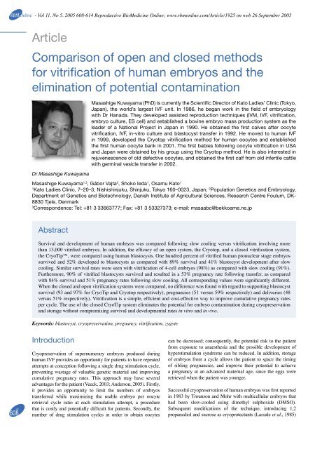
RBMOnline - Vol 11. No 5. 2005 608-614 Reproductive BioMedicine Online; www.rbmonline.com/<strong>Article</strong>/1925 on web 26 September 2005<br />
<strong>Article</strong><br />
<strong>Comparison</strong> <strong>of</strong> <strong>open</strong> <strong>and</strong> <strong>closed</strong> <strong>methods</strong><br />
<strong>for</strong> vitrification <strong>of</strong> human embryos <strong>and</strong> the<br />
elimination <strong>of</strong> potential contamination<br />
Dr Masashige Kuwayama<br />
Masashige Kuwayama (PhD) is currently the <strong>Scientific</strong> Director <strong>of</strong> Kato Ladies’ Clinic (Tokyo,<br />
Japan), the world’s largest IVF unit. In 1986, he began work in the field <strong>of</strong> embryology<br />
with Dr Hanada. They developed assisted reproduction techniques (IVM, IVF, vitrification,<br />
embryo culture, ES cell) <strong>and</strong> established a bovine embryo mass production system as the<br />
leader <strong>of</strong> a National Project in Japan in 1990. He obtained the first calves after oocyte<br />
vitrification, IVF, in-vitro culture <strong>and</strong> blastocyst transfer in 1992. He moved to human IVF<br />
in 1999, developed the Cryotop vitrification method <strong>for</strong> human oocytes <strong>and</strong> established<br />
the first human oocyte bank in 2001. The first babies following oocyte vitrification in USA<br />
<strong>and</strong> Japan were obtained by his group using the Cryotop method. He is also interested in<br />
rejuvenescence <strong>of</strong> old defective oocytes, <strong>and</strong> obtained the first calf from old infertile cattle<br />
with germinal vesicle transfer in 2002.<br />
Masashige Kuwayama 1,3 , Gábor Vajta 2 , Shoko Ieda 1 , Osamu Kato 1<br />
1<br />
Kato Ladies Clinic, 7–20–3, Nishishinjuku, Shinjuku, Tokyo 160–0023, Japan; 2 Population Genetics <strong>and</strong> Embryology,<br />
Department <strong>of</strong> Genetics <strong>and</strong> Biotechnology, Danish Institute <strong>of</strong> Agricultural Sciences, Research Centre Foulum, DK-<br />
8830 Tjele, Denmark<br />
3<br />
Correspondence: Tel: +81 3 33663777; Fax: +81 3 53327373; e-mail: masaabc@bekkoame.ne.jp<br />
Abstract<br />
Survival <strong>and</strong> development <strong>of</strong> human embryos was compared following slow cooling versus vitrification involving more<br />
than 13,000 vitrified embryos. In addition, the efficacy <strong>of</strong> an <strong>open</strong> system, the Cryotop, <strong>and</strong> a <strong>closed</strong> vitrification system,<br />
the CryoTip, were compared using human blastocysts. One hundred percent <strong>of</strong> vitrified human pronuclear stage embryos<br />
survived <strong>and</strong> 52% developed to blastocysts as compared with 89% survival <strong>and</strong> 41% blastocyst development after slow<br />
cooling. Similar survival rates were seen with vitrification <strong>of</strong> 4-cell embryos (98%) as compared with slow cooling (91%).<br />
Furthermore, 90% <strong>of</strong> vitrified blastocysts survived <strong>and</strong> resulted in a 53% pregnancy rate following transfer, as compared<br />
with 84% survival <strong>and</strong> 51% pregnancy rates following slow cooling. All corresponding values were significantly different.<br />
When the <strong>closed</strong> <strong>and</strong> <strong>open</strong> vitrification systems were compared, no difference was found with regard to supporting blastocyst<br />
survival (93 <strong>and</strong> 97% <strong>for</strong> CryoTip <strong>and</strong> Cryotop respectively), pregnancies (51 versus 59% respectively) <strong>and</strong> deliveries (48<br />
versus 51% respectively). Vitrification is a simple, efficient <strong>and</strong> cost-effective way to improve cumulative pregnancy rates<br />
per cycle. The use <strong>of</strong> the <strong>closed</strong> CryoTip system eliminates the potential <strong>for</strong> embryo contamination during cryopreservation<br />
<strong>and</strong> storage without compromising survival <strong>and</strong> developmental rates in vitro <strong>and</strong> in vivo.<br />
Keywords: blastocyst, cryopreservation, pregnancy, vitrification, zygote<br />
608<br />
Introduction<br />
Cryopreservation <strong>of</strong> supernumerary embryos produced during<br />
human IVF provides an opportunity <strong>for</strong> patients to have repeated<br />
attempts at conception following a single drug stimulation cycle,<br />
preventing wastage <strong>of</strong> valuable genetic material <strong>and</strong> improving<br />
cumulative pregnancy rates. This approach may have several<br />
advantages <strong>for</strong> the patient (Veeck, 2003; Anderson, 2005). Firstly,<br />
it provides an opportunity to limit the numbers <strong>of</strong> embryos<br />
transferred while maximizing the usable embryo per oocyte<br />
retrieval cycle ratio at each stimulation attempt, a procedure<br />
that is costly <strong>and</strong> potentially difficult <strong>for</strong> patients. Secondly, the<br />
number <strong>of</strong> drug stimulation cycles in order to obtain oocytes<br />
can be decreased; consequently, the potential risk to the patient<br />
from exposure to anaesthesia <strong>and</strong> the possible development <strong>of</strong><br />
hyperstimulation syndrome can be reduced. In addition, storage<br />
<strong>of</strong> embryos from a cycle allows the patient to space the timing<br />
<strong>of</strong> sibling pregnancies, <strong>and</strong> improve their potential to achieve<br />
a pregnancy at an advanced maternal age, since the eggs were<br />
retrieved when the patient was younger.<br />
Successful cryopreservation <strong>of</strong> human embryos was first reported<br />
in 1983 by Trounson <strong>and</strong> Mohr with multicellular embryos that<br />
had been slow-cooled using dimethyl sulphoxide (DMSO).<br />
Subsequent modifications <strong>of</strong> the technique, introducing 1,2<br />
propanediol <strong>and</strong> sucrose as cryoprotectants (Lassale et al., 1985)
<strong>Article</strong> - Vitrification <strong>of</strong> human embryos - M Kuwayama et al.<br />
<strong>and</strong> slow cooling to –30°C prior to plunging into liquid nitrogen,<br />
resulted in the introduction <strong>of</strong> cryopreservation as a st<strong>and</strong>ard<br />
method <strong>of</strong>fered by virtually every full-service IVF programme<br />
world-wide (Anderson et al., 2003).<br />
The primary disadvantages to slow cooling <strong>for</strong> human embryo<br />
cryopreservation are the requirement <strong>for</strong> an expensive<br />
programmable freezing machine <strong>and</strong> the time-consuming<br />
procedure. The introduction <strong>of</strong> a technique that could be per<strong>for</strong>med<br />
without the use <strong>of</strong> costly equipment <strong>and</strong> could be completed by<br />
one cryopreservation specialist within minutes would provide<br />
significant benefits <strong>for</strong> any busy IVF programme.<br />
Vitrification <strong>of</strong> embryos <strong>and</strong> oocytes may <strong>of</strong>fer a solution <strong>for</strong> this<br />
problem. Vitrification can be defined as an extreme elevation<br />
in viscosity, i.e. solidification <strong>of</strong> solutions without ice crystal<br />
<strong>for</strong>mation at low temperature (Liebermann et al., 2002a; Fuller<br />
<strong>and</strong> Paynter, 2004; Kasai, 2004; Liebermann <strong>and</strong> Tucker, 2004).<br />
This phenomenon can be induced by either applying an extreme<br />
cooling rate or by using high concentrations <strong>of</strong> cryoprotectant<br />
solutions. Methods developed <strong>for</strong> vitrification in embryology<br />
use a combination <strong>of</strong> these two possibilities (Liebermann et al.,<br />
2003). The advantages <strong>of</strong> vitrification in embryology may be<br />
considerable. Oocytes <strong>and</strong> embryos are sensitive to ice crystal<br />
<strong>for</strong>mation; consequently, the elimination <strong>of</strong> this type <strong>of</strong> injury<br />
may increase their chances <strong>for</strong> survival. The required high<br />
cooling rate can be achieved by simple <strong>methods</strong> including, <strong>for</strong><br />
example, direct plunging into liquid nitrogen, thus the need<br />
<strong>for</strong> expensive equipment is eliminated. Additionally, the time<br />
required <strong>for</strong> equilibration <strong>and</strong> cooling is considerably reduced. On<br />
the other h<strong>and</strong>, disadvantages <strong>of</strong> vitrification are the required high<br />
cryoprotectant concentration, <strong>and</strong> consequently the increased risk<br />
<strong>of</strong> toxic <strong>and</strong> osmotic damage, <strong>and</strong> the need to use special tools<br />
permitting high cooling rate by reducing radically the volume <strong>of</strong><br />
solutions containing the embryos.<br />
Successful vitrification <strong>of</strong> mammalian embryos was first reported<br />
by Rall <strong>and</strong> Fahy in 1985. Since then, a number <strong>of</strong> cryoprotectant<br />
solutions have been investigated <strong>for</strong> human use including the use<br />
<strong>of</strong> DMSO, glycerol, ethylene glycol, propanediol <strong>and</strong> sugars in<br />
various combinations (Chen et al., 2000; M<strong>and</strong>elbaum, 2000;<br />
Shaw et al., 2000; Wright et al., 2004). In addition, numerous<br />
carrier systems have been tried, including electron microscope<br />
grids, <strong>open</strong> pulled straws, denuding pipettes, <strong>open</strong> hemi-straws,<br />
<strong>and</strong> cryoloops (Martino et al., 1996; Arav <strong>and</strong> Zeron, 1997; Vajta<br />
et al., 1998a,b; Lane et al., 1999; Kuleshova <strong>and</strong> Shaw, 2000; Park<br />
et al., 2000; V<strong>and</strong>erzwalmen et al., 2000; V<strong>and</strong>ervorst et al., 2001;<br />
Yeoman et al., 2001; Liebermann <strong>and</strong> Tucker, 2002; Liebermann<br />
et al., 2002a,b; Mukaida et al., 2002, 2003; Selman <strong>and</strong> El-<br />
Danasouri, 2002; V<strong>and</strong>erzwalment et al., 2002, 2003; Isachenko<br />
et al., 2003; Son et al., 2003, 2005; Cremades et al., 2004; Walker<br />
et al., 2004). While all <strong>of</strong> these systems have their advantages<br />
<strong>and</strong> disadvantages, the primary concern <strong>for</strong> many authorities <strong>and</strong><br />
scientists is the potential risk <strong>of</strong> contamination <strong>for</strong> patients. As<br />
the rapid cooling in all these systems requires direct contact <strong>of</strong><br />
the embryo containing solution <strong>and</strong> liquid nitrogen, there is a<br />
potential risk <strong>of</strong> disease transmission through contaminated liquid<br />
nitrogen during cooling <strong>and</strong> storage (Bielanski et al., 2000).<br />
The purposes <strong>of</strong> this study were: (i) to compare the efficacy <strong>of</strong><br />
vitrification versus traditional slow cooling <strong>for</strong> cryopreservation <strong>of</strong><br />
human embryos, <strong>and</strong> (ii) to investigate the possibility <strong>of</strong> replacing<br />
an earlier <strong>open</strong> system (Cryotop) with a newly introduced<br />
<strong>closed</strong> method (Cryotip TM ) to eliminate the potential danger <strong>of</strong><br />
contamination.<br />
Materials <strong>and</strong> <strong>methods</strong><br />
Patient treatment <strong>and</strong> embryo culture<br />
Experiments were conducted in patients following in<strong>for</strong>med<br />
consent <strong>and</strong> IRB approval. Patients were stimulated during an<br />
IVF cycle by the use <strong>of</strong> clomiphene citrate (Clomid; Shionogi<br />
Co. Ltd, Osaka, Japan). Clomiphene administration (50 mg/<br />
day) was initiated on day 3 <strong>of</strong> the cycle <strong>and</strong> continued until<br />
the rise in LH, caused by the nasal administration <strong>of</strong> 300 μg <strong>of</strong><br />
gonadotrophin-releasing hormone agonist (GnRHa) (Suprecur;<br />
Mochida Pharmaceutical Co. Ltd, Tokyo, Japan). Human<br />
menopausal gonadotrophin (HMG) (Humegon; Organon Co. Ltd,<br />
Netherl<strong>and</strong>s) was initiated on day 8 (150 IU) <strong>and</strong> continued every<br />
other day until the administration <strong>of</strong> GnRHa.<br />
Oocytes were recovered by using a transvaginal ultrasoundguided<br />
device (GM07M05V110; Mochida Pharmaceutical). After<br />
retrieval, oocytes were cultured in TCM199 medium buffered<br />
with 11 mmol/l Hepes, 9 mmol/l Na-Hepes <strong>and</strong> 5 mmol/l NaHCO 3<br />
(referred further as TCM 199) supplemented with 10% Synthetic<br />
Serum Substitute (SSS; <strong>Irvine</strong> <strong>Scientific</strong>, Santa Ana, CA, USA)<br />
in 5% CO 2<br />
in air at 37°C. After 2 h, insemination was per<strong>for</strong>med<br />
by intracytoplasmic sperm injection (ICSI). The following day,<br />
fertilized oocytes with 2 pronuclei <strong>and</strong> two polar bodies were<br />
transferred into 0.5 ml droplets <strong>of</strong> Quinn’s Advantage Cleavage<br />
Medium (Sage BioPharma, USA) supplemented with 10% SSS<br />
<strong>and</strong> cultured further as described above. Embryos that developed<br />
beyond the 4-cell stage on day 5 were transferred into 100 μl<br />
droplets <strong>of</strong> blastocyst medium (<strong>Irvine</strong> <strong>Scientific</strong>) <strong>and</strong> cultured<br />
under identical conditions to day 6 after ICSI.<br />
Vitrification <strong>of</strong> embryos<br />
Equilibration, vitrification, thawing, dilution <strong>and</strong> washing solutions<br />
were equivalents to those in Vit Kit (<strong>Irvine</strong> <strong>Scientific</strong>). At the<br />
time <strong>of</strong> vitrification embryos were transferred into equilibration<br />
solution (ES) consisting <strong>of</strong> 7.5% (v/v) ethylene glycol <strong>and</strong> 7.5%<br />
(v/v) DMSO dissolved in TCM199 supplemented with 20%<br />
SSS at 27°C <strong>for</strong> 5–15 min. After an initial shrinkage, embryos<br />
regained their original volume, <strong>and</strong> were transferred into three 20<br />
µl drops <strong>of</strong> vitrification solution (VS) consisting <strong>of</strong> 15% (v/v) EG<br />
<strong>and</strong> 15%(v/v) DMSO <strong>and</strong> 0.5 mol/l sucrose dissolved in TCM199<br />
supplemented with 20% SSS, After incubation <strong>for</strong> 20 s in each<br />
drop respectively, embryos were loaded into CryoTips (<strong>Irvine</strong><br />
<strong>Scientific</strong>) or on Cryotops <strong>and</strong> plunged into liquid nitrogen.<br />
CryoTip consists <strong>of</strong> a plastic straw with a thin part (250 μm inner<br />
diameter, 20 μm wall thickness <strong>and</strong> 3 cm length) connected to a<br />
thick part (2000 μm inner diameter <strong>and</strong> 150 μm wall thickness,<br />
4.5 cm length) <strong>and</strong> equipped with a movable protective metal<br />
sleeve (Figure 1). Embryos were loaded in approximately 1<br />
μl solution into the narrow part <strong>of</strong> the CryoTips without any<br />
air bubbles by aspiration <strong>of</strong> medium, embryo <strong>and</strong> medium, <strong>and</strong><br />
medium by a connected syringe. Subsequently, the straw was<br />
heat-sealed at both ends, the protective sleeve was pulled over<br />
the narrow part <strong>and</strong> the device was plunged into liquid nitrogen.<br />
The time required <strong>for</strong> loading, sealing, adjustment <strong>of</strong> the sleeve<br />
609
<strong>Article</strong> - Vitrification <strong>of</strong> human embryos - M Kuwayama et al.<br />
<strong>and</strong> plunging did not exceed 90 s.<br />
For warming, CryoTip was removed from liquid nitrogen,<br />
plunged into a 37°C water bath <strong>for</strong> 3 s, wiped with 70%<br />
ethanol <strong>and</strong> a paper towel <strong>and</strong> the sealed ends were cut with a<br />
sterile scissor. By using a syringe adjusted to the thick end <strong>of</strong><br />
the straw, the contents were expelled onto a sterile Petri dish.<br />
Solutions used <strong>for</strong> further manipulations were kept at 27°C. A<br />
1 μl aliquot <strong>of</strong> thawing solution (TS) consisting <strong>of</strong> 1.0 mol/l<br />
sucrose in TCM199 plus 20% SSS was placed adjacent to the<br />
expelled drop <strong>and</strong> merged subsequently with the other drop<br />
containing the embryos. After 1 min, embryos were retrieved<br />
<strong>and</strong> transferred to a second drop <strong>of</strong> TS <strong>for</strong> 1 min, then transferred<br />
to two 20 μl drops <strong>of</strong> dilution solution (DS) consisting <strong>of</strong> 0.5<br />
mol/l sucrose in TCM199 plus 20% SSS <strong>for</strong> 2 min each. After<br />
three subsequent washings through three successive 20 μl<br />
drops <strong>of</strong> washing solution (WS; TCM199 supplemented with<br />
20% SSS) <strong>for</strong> 3 min each, embryos were transferred into 100 μl<br />
droplets <strong>of</strong> Blastocyst medium (<strong>Irvine</strong> <strong>Scientific</strong>) <strong>and</strong> cultured<br />
under conditions described earlier.<br />
Cryotop (Kitazato Supply Co., Fujinomiya, Japan), consists <strong>of</strong> a<br />
0.4 mm wide × 20 mm long × 0.1 mm thick polypropylene strips<br />
attached to a plastic h<strong>and</strong>le <strong>and</strong> equipped with a cover straw<br />
(Figure 2; Kuwayama et al., 2005). When using the Cryotops,<br />
after equilibration as described above, individual embryos<br />
Figure 1. The CryoTip TM vitrification tool. (A)<br />
For loading, sealing, warming <strong>and</strong> expelling, the<br />
metal sleeve (m) is positioned over the wide part<br />
(w) <strong>of</strong> the straw. (B) For safe storage in liquid<br />
nitrogen, the sleeve is pulled over the narrow part<br />
(n) <strong>of</strong> the straw.<br />
c<br />
a<br />
b<br />
610<br />
d<br />
Figure 2. The Cryotop vitrification tool. The<br />
polypropylene strip (a) is attached to a hard<br />
plastic h<strong>and</strong>le (b). After vitrification, a hard<br />
plastic cover (c) is attached to protect the strip<br />
during storage in liquid nitrogen (d). From:<br />
Kuwayama et al. (2005), by permission <strong>of</strong><br />
the Editor.
<strong>Article</strong> - Vitrification <strong>of</strong> human embryos - M Kuwayama et al.<br />
were picked up in an extremely small volume (
<strong>Article</strong> - Vitrification <strong>of</strong> human embryos - M Kuwayama et al.<br />
Table 2. Survival <strong>and</strong> pregnancy rates with human 4-cell embryos<br />
cryopreserved by either slow cooling or vitrification using the<br />
Cryotop method.<br />
Slow cooling<br />
Vitrification<br />
Survived/cryopreserved rate (%) 857/942 (91) a 879/897 (98) b<br />
Pregnancy/transfer rate (%) 172/536 (32) a 136/504 (27) a<br />
a,b<br />
Values within rows with different superscripts are significantly different (P < 0.01).<br />
Table 3. Survival <strong>and</strong> pregnancy rates with human blastocysts<br />
cryopreserved by either slow cooling as compared with vitrification<br />
using the Cryotop method.<br />
Slow cooling<br />
Vitrification<br />
Survived/vitrified rate (%) 131/156 (84) a 5695/6328 (90) b<br />
Number <strong>of</strong> blastocysts transferred 127 5659<br />
Pregnancy/transfer rate (%) 50/98 (51) a 2516/4745 (53) a<br />
Live birth/transfer rate (%) 40/98 (41) a 2138/4745 (45) a<br />
a,b<br />
Values within rows with different superscripts are significantly different (P < 0.05).<br />
Table 4. Survival, pregnancy <strong>and</strong> delivery rates after single<br />
embryo transfer <strong>of</strong> human blastocysts vitrified with either the<br />
Cryotop or the CryoTip method.<br />
Cryotop<br />
CryoTip<br />
Survived/vitrified rate (%) 221/227 (97) 82/88 (93)<br />
Pregnancy/transfer rate (%) 131/221 (59) 42/82 (51)<br />
Delivery/transfer rate (%) 113/221 (51) 39/82 (48)<br />
No significant differences between corresponding values were found.<br />
612<br />
Discussion<br />
Cryopreservation <strong>of</strong> embryos is a critical step in maximizing<br />
the efficiency <strong>of</strong> an IVF cycle <strong>for</strong> patients. Clinical success with<br />
cryopreservation seems to be highly variable from laboratory to<br />
laboratory, <strong>and</strong> may depend on many factors including patient<br />
age <strong>and</strong> stimulation protocol, quality <strong>of</strong> embryos selected <strong>for</strong><br />
freezing, developmental stage at freezing, media <strong>for</strong>mulation<br />
including type <strong>of</strong> cryoprotectants used, parameters <strong>of</strong> cooling<br />
<strong>and</strong> warming, <strong>and</strong> type <strong>and</strong> quality control <strong>of</strong> programmable<br />
freezing unit employed. The latter problem, however,<br />
occurs only at slow cooling, as vitrification does not require<br />
sophisticated equipment <strong>of</strong> questionable reliability at certain<br />
makes <strong>and</strong> types. Variations caused by different media <strong>and</strong><br />
cryoprotectants can also be minimized by using commercially<br />
available kits, recently also produced <strong>for</strong> vitrification (by,<br />
<strong>for</strong> example, <strong>Irvine</strong> <strong>Scientific</strong>, Kitazato). On the other h<strong>and</strong>,<br />
according to previous publications, <strong>and</strong> also confirmed by<br />
the present work, results achieved by vitrification are at least<br />
equal or significantly better than those obtained with traditional<br />
slow cooling <strong>for</strong> cryopreservation <strong>of</strong> mammalian oocytes <strong>and</strong><br />
embryos including humans (Kuleshowa <strong>and</strong> Lopata, 2002;<br />
Liebermann et al., 2003; Smith <strong>and</strong> Silva, 2004; Walker et al.,<br />
2004; Kuwayama et al., 2005).<br />
Moreover, a considerable number <strong>of</strong> publications reported<br />
normal in-vitro <strong>and</strong> in-vivo development following human<br />
oocyte, early embryo <strong>and</strong> blastocyst vitrification (Yokota et al.,<br />
2000; Jelinkova et al., 2002; Selman et al., 2002; Isachenko<br />
et al., 2003, 2004a,b; Katayama et al., 2003; Liebermann et<br />
al., 2003b; Son et al., 2003; Huang et al., 2004; Rienzi et al.,<br />
2004; Smith et al., 2004; Son et al., 2005), as well as deliveries
<strong>Article</strong> - Vitrification <strong>of</strong> human embryos - M Kuwayama et al.<br />
<strong>of</strong> healthy births (El-Danasouri <strong>and</strong> Selman, 2001; Yokota<br />
et al., 2001a,b; V<strong>and</strong>erzwalmen et al., 2002; Mukaida et al.,<br />
2003; Kuwayama et al., 2005). The extensive reports to date<br />
exhibit ongoing improvements in vitrification <strong>methods</strong>, <strong>and</strong><br />
further support the overall efficacy <strong>of</strong> vitrification as a viable<br />
alternative to cryopreservation by slow cooling <strong>methods</strong>. The<br />
comprehensive results provided in this report further extend the<br />
efficacy <strong>and</strong> safety <strong>of</strong> vitrification <strong>for</strong> clinical human use.<br />
However, one <strong>of</strong> the major unresolved issues associated with<br />
the vitrification process is the choice <strong>of</strong> vessel, vial or straw<br />
to hold the embryo during vitrification. As overviewed in the<br />
Introduction, numerous types <strong>of</strong> vessels have been described<br />
in the literature. While all <strong>of</strong> these devices have been shown to<br />
work as carriers <strong>for</strong> embryos during vitrification, a number <strong>of</strong><br />
concerns have been raised regarding the potential risk to human<br />
embryos from exposure to contaminants already present in liquid<br />
nitrogen at the time <strong>of</strong> vitrification, or potentially introduced<br />
to the embryos during storage in <strong>open</strong> containers. While no<br />
studies have demonstrated unintentional uptake by a human<br />
or mammalian embryo <strong>of</strong> any pathogen during vitrification<br />
or storage, under experimental conditions such contamination<br />
may occur (Bielanski et al., 2000). Accordingly, a number <strong>of</strong><br />
governing bodies worldwide have expressed concern about<br />
the potential risk. Previous attempts to eliminate this danger<br />
included cooling per<strong>for</strong>med in liquid nitrogen filtered through<br />
a 0.2 μm pore-size filter <strong>and</strong> placing the carrier tool into a<br />
container that partially or completely isolates it from the liquid<br />
nitrogen during storage (Vajta et al., 1998; Kuwayama et al.,<br />
2005).<br />
As proven by the present work, the newly developed CryoTip<br />
method may provide a simple <strong>and</strong> practical solution to the<br />
problem. After loading, the plastic straw can be heat-sealed on<br />
both ends, <strong>and</strong> consequently the solution containing the embryo<br />
<strong>and</strong> the liquid nitrogen is hermetical isolated during cooling<br />
<strong>and</strong> storage. On the other h<strong>and</strong>, although the isolation slightly<br />
decreased the rate <strong>of</strong> cooling, the values that could be obtained<br />
with the CryoTip were still high enough to obtain appropriate<br />
vitrification. Consequently, embryo survival, as well as in-vitro<br />
<strong>and</strong> in-vivo developmental data, was identical to those achieved<br />
by the <strong>open</strong> Cryotop vitrification system. An additional benefit<br />
<strong>of</strong> the CryoTip vitrification is that any embryologist familiar<br />
with the usual method <strong>of</strong> loading <strong>and</strong> sealing 0.25 ml cryostraws<br />
will easily accommodate to using the smaller volume,<br />
but similar approach in h<strong>and</strong>ling, <strong>of</strong> the CryoTip. Moreover, the<br />
protective metal sleeve ensures safe storage <strong>of</strong> the CryoTip in<br />
a relatively small space, <strong>and</strong> the sleeve as well as the wide part<br />
<strong>of</strong> the plastic tube <strong>of</strong>fers enough space <strong>for</strong> proper marking <strong>of</strong><br />
straws <strong>for</strong> safe identification.<br />
Recently, Isachenko et al. (2005) published a method <strong>for</strong> aseptic<br />
vitrification <strong>of</strong> human zygotes by using <strong>open</strong> pulled straws<br />
(Vajta et al., 1998) hermetically isolated from liquid nitrogen<br />
be<strong>for</strong>e cooling. According to the authors, the considerably<br />
decreased cooling rate achievable by this technique did<br />
not compromise survival <strong>and</strong> further development. This<br />
observation is in contrast to unpublished observations, <strong>and</strong><br />
seems to contradict the principles <strong>of</strong> many previous ultrarapid<br />
vitrification <strong>methods</strong> (Martino et al., 1996; Vajta et al., 1998;<br />
Lane et al., 1999; Park et al., 2000; V<strong>and</strong>erzwalmen et al., 2000;<br />
Liebermann <strong>and</strong> Tucker, 2002; Katayama et al., 2003; Mukaida<br />
et al., 2003; Kuwayama et al., 2005). Moreover, it may also<br />
be questioned if the physical phenomenon <strong>of</strong> vitrification could<br />
be obtained by using the applied cryoprotectant concentrations<br />
<strong>and</strong> the achievable cooling rate in the method <strong>of</strong> Isachenko<br />
et al. (2005). Based on these concerns, further comparative<br />
investigations per<strong>for</strong>med by independent groups might be<br />
required to determine the difference in efficiency <strong>and</strong> practical<br />
value <strong>of</strong> the two approaches.<br />
In conclusion, the present results, based on more than 16,000<br />
human embryo cryopreservations, prove that vitrification is<br />
a simple, inexpensive <strong>and</strong> safe alternative <strong>of</strong> traditional slow<br />
cooing resulting in higher survival <strong>and</strong> in-vitro developmental<br />
rates <strong>for</strong> PN, multicellular <strong>and</strong> blastocyst stage human embryos.<br />
Pregnancy <strong>and</strong> live birth dates did not differ between the<br />
two <strong>methods</strong>. Additionally, the newly developed CryoTip TM<br />
technology eliminates the danger <strong>of</strong> contamination while<br />
maintaining the high efficiency <strong>of</strong> the procedure.<br />
References<br />
Anderson AR 2005 Reduction <strong>of</strong> high order multiples in frozen<br />
embryo transfers. Reproductive BioMedicine Online 10, 402–405.<br />
Bielanski A, Nadin-Davis S, Sapp T et al. 2000 Viral contamination<br />
<strong>of</strong> embryos cryopreserved in liquid nitrogen. Cryobiology 40,<br />
110–116.<br />
Chen SU, Lien YR, Chao KH et al. 2000 Cryopreservation <strong>of</strong> mature<br />
human oocytes by vitrification with ethylene glycol in straws.<br />
Fertility <strong>and</strong> Sterility 74, 804–808.<br />
Cremades N, Sousa M, Silva J et al. 2004 Experimental vitrification<br />
<strong>of</strong> human compacted morulae <strong>and</strong> early blastocysts using fine<br />
diameter plastic micropipettes. Human Reproduction 19, 300–305.<br />
El-Danasouri I, Selman H 2001 Successful pregnancies <strong>and</strong> deliveries<br />
after a simple vitrification protocol <strong>for</strong> day 3 human embryos.<br />
Fertility <strong>and</strong> Sterility 76, 400–402.<br />
Fuller B, Paynter S, 2004 Fundamentals <strong>of</strong> cryobiology in<br />
reproductive medicine. Reproductive BioMedicine Online 9,<br />
680–691.<br />
Huang CC, Lee TH, Chen SU et al. 2004 Successful pregnancy<br />
following blastocyst cryopreservation using super-cooling ultrarapid<br />
vitrification. Human Reproduction 20, 122–128.<br />
Isachenko V, Montag M, Isachenko E et al. 2005 Aseptic technology<br />
<strong>of</strong> vitrification <strong>of</strong> human pronuclear oocytes using <strong>open</strong>-pulled<br />
straws. Human Reproduction 20, 492–496.<br />
Isachenko V, Montag M, Isachenko E et al. 2004a Developmental rate<br />
<strong>and</strong> ultrastructure <strong>of</strong> vitrified human pronuclear oocytes after stepwise<br />
versus direct rehydration. Human Reproduction 19, 660–665.<br />
Isachenko V, Selman H, Isachenko E et al. 2004b Effect <strong>of</strong><br />
cryoprotectants on the ultrastructure <strong>of</strong> cooled human pronuclear<br />
oocytes. Fertility <strong>and</strong> Sterility 81, 720–722.<br />
Isachenko V, Selman H, Isachenko E et al. 2003 Modified vitrification<br />
<strong>of</strong> human pronuclear oocytes: efficacy <strong>and</strong> effect on ultrastructure.<br />
Reproductive BioMedicine Online 7, 211–216.<br />
Jelinkova L, Selman H, Arav A et al. 2002 Twin pregnancy after<br />
vitrification <strong>of</strong> 2-pronuclei human embryos. Fertility <strong>and</strong> Sterility<br />
77, 412–414.<br />
Kasai M 2004 Cryopreservation <strong>of</strong> animal <strong>and</strong> human embryos by<br />
vitrification. Reproductive BioMedicine Online 9, 164–170.<br />
Katayama KP, Stehlik J, Kuwayama M et al. 2003 High survival rate<br />
<strong>of</strong> vitrified human oocytes results in clinical pregnancy. Fertility<br />
<strong>and</strong> Sterility 80, 223–224.<br />
Kuleshova LL, Lopata A 2002 Vitrification can be more favorable than<br />
slow cooling. Fertility <strong>and</strong> Sterility 78, 449–454.<br />
Kuleshova LL, Shaw JM 2000 A strategy <strong>for</strong> rapid cooling <strong>of</strong><br />
mouse embryos within a double straw to eliminate the risk<br />
<strong>of</strong> contamination during storage in liquid nitrogen. Human<br />
Reproduction 15, 2604–2609.<br />
Kuwayama M, Vajta G, Kato L, Leibo SP Highly efficient vitrification<br />
method <strong>for</strong> cryopreservation <strong>of</strong> human oocytes. Reproductive<br />
613
<strong>Article</strong> - Vitrification <strong>of</strong> human embryos - M Kuwayama et al.<br />
614<br />
BioMedicine Online, in press.<br />
Lane M, Schoolcraft WB, Gardner DK 1999 Vitrification <strong>of</strong> mouse<br />
<strong>and</strong> human blastocysts using a novel cryoloop container-less<br />
technique. Fertility <strong>and</strong> Sterility 72, 1073–1078.<br />
Lasalle B, Testart J, Renard JP 1985 Human embryo features that<br />
influence the success <strong>of</strong> cryopreservation with the use <strong>of</strong> 1,2<br />
propanediol. Fertility <strong>and</strong> Sterility 44, 645–651.<br />
Liebermann J, Tucker M 2004 Vitrifying <strong>and</strong> warming <strong>of</strong> human<br />
oocytes, embryos, <strong>and</strong> blastocysts. Methods in Molecular Biology,<br />
254, 345–364.<br />
Liebermann J, Tucker MJ 2002 Effect <strong>of</strong> carrier system on the yield<br />
<strong>of</strong> human oocytes <strong>and</strong> embryos as assessed by survival <strong>and</strong><br />
developmental potential after vitrification. Reproduction 124,<br />
483–489.<br />
Liebermann J, Dietl J, V<strong>and</strong>erzwalmen P et al. 2003a Recent<br />
developments in human oocyte, embryo <strong>and</strong> blastocyst<br />
vitrification: where are we now Reproductive BioMedicine Online<br />
7, 623–633.<br />
Liebermann J, Nawroth F, Isachenko V et al. 2002a Potential<br />
importance <strong>of</strong> vitrification in reproductive medicine. Biology <strong>of</strong><br />
Reproduction 67, 1671–1680.<br />
Liebermann J, Tucker MJ, Graham JR et al. 2002b Blastocyst<br />
development after vitrification <strong>of</strong> multipronuclear zygotes using<br />
the Flexipet denuding pipette. Reproductive BioMedicine Online<br />
4, 146–150.<br />
M<strong>and</strong>elbaum J 2000 Embryo <strong>and</strong> oocyte cryopreservation. Human<br />
Reproduction 15, 43–47.<br />
Martino A, Songsasen N, Leibo SP 1996 Development into blastocysts<br />
<strong>of</strong> bovine oocytes cryopreserved by ultra-rapid cooling. Biology <strong>of</strong><br />
Reproduction 54, 1059–1069.<br />
Mukaida T, Nakamura S, Tomiyama T et al. 2003 Vitrification <strong>of</strong><br />
human blastocysts using cryoloops: clinical outcome <strong>of</strong> 223<br />
cycles. Human Reproduction 18, 384–391.<br />
Mukaida T, Takahashi K, Kasai M 2002 Blastocyst cryopreservation:<br />
ultrarapid vitrification using cryoloop technique. Reproductive<br />
BioMedicine Online 6, 221–225.<br />
Park SP, Kim EY, Oh JH et al. 2000 Ultra-rapid freezing <strong>of</strong> human<br />
multipronuclear zygotes using electron microscope grids. Human<br />
Reproduction 15, 1787–1790.<br />
Rall WF, Fahy GM 1985 Ice-free cryopreservation <strong>of</strong> mouse embryos<br />
at -196°C by vitrification. Nature 313, 573–575.<br />
Rienzi L, Martinez F, Ubaldi F et al. 2004 Polscope analysis <strong>of</strong><br />
meiotic spindle changes in living metaphase II human oocytes<br />
during the freezing <strong>and</strong> thawing procedures. Human Reproduction<br />
19, 655–659.<br />
Selman HA, El-Danasouri I 2002 Pregnancies derived from vitrified<br />
human zygotes. Fertility <strong>and</strong> Sterility 77, 422–423.<br />
Shaw JM, Oranratnachai A, Trounson AO 2000 Cryopreservation <strong>of</strong><br />
oocytes <strong>and</strong> embryos. In: H<strong>and</strong>book <strong>of</strong> In Vitro Fertilization, 2nd<br />
edn. CRC Press, Boca Raton, pp. 373–412.<br />
Smith GD, Silva CA 2004 Developmental consequences <strong>of</strong><br />
cryopreservation <strong>of</strong> mammalian oocytes <strong>and</strong> embryos.<br />
Reproductive BioMedicine Online 9, 171–178.<br />
Son WY, Lee SY, Chang MJ et al. 2005 Pregnancy resulting from<br />
transfer <strong>of</strong> repeat vitrified blastocysts produced by in-vitro matured<br />
oocytes in patient with polycystic ovary syndrome. Reproductive<br />
BioMedicine Online 10, 398–401.<br />
Son WY, Yoon SH, Yoon HJ et al. 2003 Pregnancy outcome<br />
following transfer <strong>of</strong> human blastocysts vitrified on electron<br />
microscopy grids after induced collapse <strong>of</strong> the blastocoele. Human<br />
Reproduction 18, 137–139.<br />
Trounson A, Mohr L 1983 Human pregnancy following<br />
cryopreservation, thawing <strong>and</strong> transfer <strong>of</strong> an eight-cell embryo.<br />
Nature 305, 707–709.<br />
Vajta G, Holm P, Kuwayama M et al. 1998a Open pulled straw (OPS)<br />
vitrification: a new way to reduce cryoinjuries <strong>of</strong> bovine ova <strong>and</strong><br />
embryos. Molecular Reproduction <strong>and</strong> Development 51, 53–58.<br />
Vajta G, Lewis IM, Kuwayama M et al. 1998b Sterile application <strong>of</strong><br />
the Open Pulled Straw (OPS) vitrification method. Cryo-Letters<br />
19, 389–392.<br />
V<strong>and</strong>ervorst M, V<strong>and</strong>erzwalmen P, St<strong>and</strong>aart V et al. 2001 Blastocyst<br />
transfer after vitrification in a hemi-straw (HS) system. Human<br />
Reproduction 16 (suppl. 1), 153 (Abstract P-133).<br />
V<strong>and</strong>erzwalmen P, Bertin G, Debauche C et al. 2003 Vitrification<br />
<strong>of</strong> human blastocysts with the Hemi-straw carrier: application<br />
<strong>of</strong> assisted hatching after thawing. Human Reproduction 18,<br />
1504–1511.<br />
V<strong>and</strong>erzwalmen P, Bertin G, Debauche C et al. 2002 Births after<br />
vitrification at morula <strong>and</strong> blastocyst stages: effect <strong>of</strong> artificial<br />
reduction <strong>of</strong> the blastocoelic cavity be<strong>for</strong>e vitrification. Human<br />
Reproduction 17, 744–751.<br />
V<strong>and</strong>erzwalmen P, Bertin G, Debauche V et al. 2000 In vitro survival<br />
<strong>of</strong> metaphase II oocytes (MII) <strong>and</strong> blastocysts after vitrification<br />
in an Hemi-Straw (HS) system. Fertility <strong>and</strong> Sterility 74 (suppl.),<br />
S215 (Abstract P-385).<br />
Walker D, Tummon IS, Hammitt DG et al. 2004 Vitrification versus<br />
programmable rate freezing <strong>of</strong> late stage embryos: a r<strong>and</strong>omized<br />
comparison prior to application in clinical IVF. Reproductive<br />
BioMedicine Online 8, 558–568.<br />
Wright DL, Eroglu Ali, Toner M et al. 2004 Use <strong>of</strong> sugars in<br />
cryopreserving human oocytes. Reproductive BioMedicine Online<br />
9, 179–186.<br />
Yeoman RR, Gerami-Naini B, Mitalipov S et al. 2001 Cryoloop<br />
vitrification yields superior survival <strong>of</strong> Rhesus monkey blastocysts.<br />
Human Reproduction 16, 1965–1969.<br />
Yokota Y, Sato S, Yokota M et al. 2001a Birth <strong>of</strong> a healthy baby<br />
following vitrification <strong>of</strong> human blastocysts. Fertility <strong>and</strong> Sterility<br />
75, 1027–1029.<br />
Yokota Y, Yokota H, Yokota M et al. 2001b Birth <strong>of</strong> healthy twins<br />
from in vitro development <strong>of</strong> human refrozen embryos. Fertility<br />
<strong>and</strong> Sterility 76, 1063–1065.<br />
Yokota Y, Sato S, Yokota M et al. 2000 Successful pregnancy<br />
following blastocyst vitrification. Human Reproduction 15,<br />
1802–1803.<br />
Received 23 June 2005; refereed 1 July 2005; accepted 1 August<br />
2005.