

10 - World Journal of Gastroenterology
10 - World Journal of Gastroenterology
10 - World Journal of Gastroenterology

You also want an ePaper? Increase the reach of your titles
YUMPU automatically turns print PDFs into web optimized ePapers that Google loves.
Zou J et al . Proteome <strong>of</strong> colon cancer stem cells<br />
B<br />
Numbers <strong>of</strong> colony<br />
C<br />
Cell number (×<strong>10</strong> 4 )<br />
A<br />
<strong>10</strong>0<br />
50<br />
0<br />
12<br />
<strong>10</strong><br />
8<br />
6<br />
4<br />
2<br />
0<br />
SW1116csc<br />
SW1116CSC<br />
SW1116<br />
t/wk<br />
SW1116<br />
1 2 3 4 5 6 7<br />
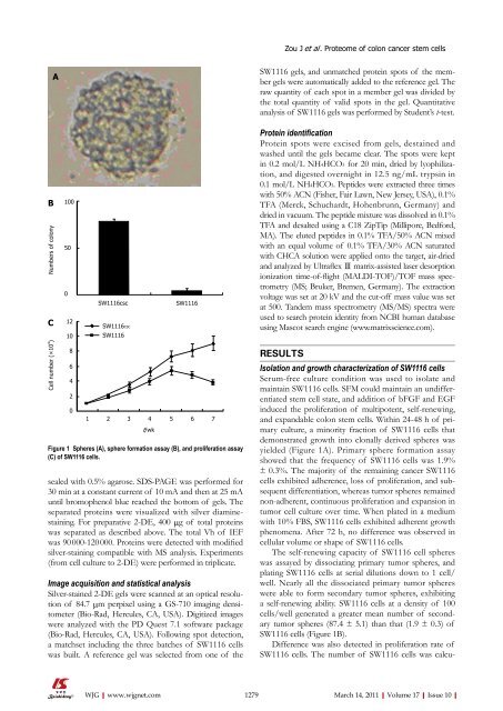
Figure 1 Spheres (A), sphere formation assay (B), and proliferation assay<br />
(C) <strong>of</strong> SW1116 cells.<br />
sealed with 0.5% agarose. SDS-PAGE was performed for<br />
30 min at a constant current <strong>of</strong> <strong>10</strong> mA and then at 25 mA<br />
until bromophenol blue reached the bottom <strong>of</strong> gels. The<br />
separated proteins were visualized with silver diaminestaining.<br />
For preparative 2-DE, 400 μg <strong>of</strong> total proteins<br />
was separated as described above. The total Vh <strong>of</strong> IEF<br />
was 90 000-120 000. Proteins were detected with modified<br />
silver-staining compatible with MS analysis. Experiments<br />
(from cell culture to 2-DE) were performed in triplicate.<br />
Image acquisition and statistical analysis<br />
Silver-stained 2-DE gels were scanned at an optical resolution<br />
<strong>of</strong> 84.7 μm perpixel using a GS-7<strong>10</strong> imaging densitometer<br />
(Bio-Rad, Hercules, CA, USA). Digitized images<br />
were analyzed with the PD Quest 7.1 s<strong>of</strong>tware package<br />
(Bio-Rad, Hercules, CA, USA). Following spot detection,<br />
a matchset including the three batches <strong>of</strong> SW1116 cells<br />
was built. A reference gel was selected from one <strong>of</strong> the<br />
SW1116 gels, and unmatched protein spots <strong>of</strong> the member<br />
gels were automatically added to the reference gel. The<br />
raw quantity <strong>of</strong> each spot in a member gel was divided by<br />
the total quantity <strong>of</strong> valid spots in the gel. Quantitative<br />
analysis <strong>of</strong> SW1116 gels was performed by Student’s t-test.<br />
Protein identification<br />
Protein spots were excised from gels, destained and<br />
washed until the gels became clear. The spots were kept<br />
in 0.2 mol/L NH4HCO3 for 20 min, dried by lyophilization,<br />
and digested overnight in 12.5 ng/mL trypsin in<br />
0.1 mol/L NH4HCO3. Peptides were extracted three times<br />
with 50% ACN (Fisher, Fair Lawn, New Jersey, USA), 0.1%<br />
TFA (Merck, Schuchardt, Hohenbrunn, Germany) and<br />
dried in vacuum. The peptide mixture was dissolved in 0.1%<br />
TFA and desalted using a C18 ZipTip (Millipore, Bedford,<br />
MA). The eluted peptides in 0.1% TFA/50% ACN mixed<br />
with an equal volume <strong>of</strong> 0.1% TFA/30% ACN saturated<br />
with CHCA solution were applied onto the target, air-dried<br />
and analyzed by Ultraflex Ⅲ matrix-assisted laser desorption<br />
ionization time-<strong>of</strong>-flight (MALDI-TOF)/TOF mass spectrometry<br />
(MS; Bruker, Bremen, Germany). The extraction<br />
voltage was set at 20 kV and the cut-<strong>of</strong>f mass value was set<br />
at 500. Tandem mass spectrometry (MS/MS) spectra were<br />
used to search protein identity from NCBI human database<br />
using Mascot search engine (www.matrixscience.com).<br />
RESULTS<br />
Isolation and growth characterization <strong>of</strong> SW1116 cells<br />
Serum-free culture condition was used to isolate and<br />
maintain SW1116 cells. SFM could maintain an undifferentiated<br />
stem cell state, and addition <strong>of</strong> bFGF and EGF<br />
induced the proliferation <strong>of</strong> multipotent, self-renewing,<br />
and expandable colon stem cells. Within 24-48 h <strong>of</strong> primary<br />
culture, a minority fraction <strong>of</strong> SW1116 cells that<br />
demonstrated growth into clonally derived spheres was<br />
yielded (Figure 1A). Primary sphere formation assay<br />
showed that the frequency <strong>of</strong> SW1116 cells was 1.9%<br />
± 0.3%. The majority <strong>of</strong> the remaining cancer SW1116<br />
cells exhibited adherence, loss <strong>of</strong> proliferation, and subsequent<br />
differentiation, whereas tumor spheres remained<br />
non-adherent, continuous proliferation and expansion in<br />
tumor cell culture over time. When plated in a medium<br />
with <strong>10</strong>% FBS, SW1116 cells exhibited adherent growth<br />
phenomena. After 72 h, no difference was observed in<br />
cellular volume or shape <strong>of</strong> SW1116 cells.<br />
The self-renewing capacity <strong>of</strong> SW1116 cell spheres<br />
was assayed by dissociating primary tumor spheres, and<br />
plating SW1116 cells at serial dilutions down to 1 cell/<br />
well. Nearly all the dissociated primary tumor spheres<br />
were able to form secondary tumor spheres, exhibiting<br />
a self-renewing ability. SW1116 cells at a density <strong>of</strong> <strong>10</strong>0<br />
cells/well generated a greater mean number <strong>of</strong> secondary<br />
tumor spheres (87.4 ± 5.1) than that (1.9 ± 0.3) <strong>of</strong><br />
SW1116 cells (Figure 1B).<br />
Difference was also detected in proliferation rate <strong>of</strong><br />
SW1116 cells. The number <strong>of</strong> SW1116 cells was calcu-<br />
WJG|www.wjgnet.com<br />
1279 March 14, 2011|Volume 17|Issue <strong>10</strong>|


