

10 - World Journal of Gastroenterology
10 - World Journal of Gastroenterology
10 - World Journal of Gastroenterology

You also want an ePaper? Increase the reach of your titles
YUMPU automatically turns print PDFs into web optimized ePapers that Google loves.
Holzner PA et al . Remote ischemic preconditioning in anastomoses<br />
paper on IPC. In the control group, the infrarenal aorta<br />
was prepared but not branched <strong>of</strong>f. The arginine group<br />
received 200 μg/kg L-arginine (L-Arginin-Hydrochlorid;<br />
Fresenius) intraoperatively without preparing the abdominal<br />
aorta.<br />
Afterwards, an approximately 1-cm segment, about<br />
15 cm oral to the ileocecal valve was resected. Ileal continuity<br />
was restored by eight inverting interrupted sutures<br />
(Prolene 8/0; Ethicon, Germany). We used a silicon<br />
catheter (Heidelberger Verlängerung; Braun, Melsungen,<br />
Germany; diameter 5 mm) to standardize and simplify the<br />
suture technique. The silicon catheter was inserted into<br />
both lumina <strong>of</strong> the small intestine. At first, front-wall sutures<br />
were made, then we turned around the anastomosis<br />
to perform the back-wall sutures, removing the catheter<br />
just before the last two sutures. The distance between the<br />
single sutures and the stitches to the resection margin was<br />
1-2 mm. The abdominal cavity was closed using a twolayer<br />
technique: musculoperitoneal layer (Monocryl 4/0<br />
SHplus; Ethicon), and fasciocutaneous layer (Vicryl 4/0<br />
SHplus; Ethicon).<br />
On postoperative day 4, the rats were re-laparotomized<br />
and sacrificed by cardiac puncture with injection <strong>of</strong> a lethal<br />
dose <strong>of</strong> potassium. The abdomen was opened by a<br />
complete midline incision combined with a transverse incision<br />
to gain maximum exposure. Careful exploration was<br />
carried out to look for signs <strong>of</strong> inflammation, adhesions,<br />
anastomotic insufficiency, and abscesses. Without dissecting<br />
the directly adjacent tissue around the anastomosis, an<br />
approximately 4-6-cm segment bearing the anastomosis<br />
was harvested for further analysis.<br />
Bursting pressure<br />
The bowel segment was water-tight and connected to<br />
an infusion pump (Perfusor fm; Braun) filled with isoosmolar<br />
saline solution (0.9% NaCl; Braun) via a 14 G<br />
silicon catheter (Vas<strong>of</strong>ix Safety; Braun) and to a digital<br />
pressure transducer (Codman ICP Express; Ethicon).<br />
Intraluminal pressure was increased by an infusion rate<br />
<strong>of</strong> 60 mL/h. Monitoring included the bursting pressure<br />
recorded just before sudden loss <strong>of</strong> tension and the site<br />
<strong>of</strong> rupture (mesenterial vs anti-mesenterial). Subsequently,<br />
the bowel wall was released from all adhering tissue and<br />
the complete suture line was excised within a total length<br />
<strong>of</strong> the bowel <strong>of</strong> 1 cm. The anastomosis was opened at the<br />
mesenterial site and gently washed using a saline solution<br />
(0.9% NaCl, Braun). The anastomosis was divided at the<br />
anti-mesenterial site into two parts <strong>of</strong> the same length for<br />
paraffin embedding and measurement <strong>of</strong> hydroxyproline<br />
concentration. The former was achieved by immediately<br />
fixing the specimen in 4% phosphate-buffered formaldehyde<br />
(pH 7.3), and the latter by preservation <strong>of</strong> the anastomotic<br />
strip in an Eppendorf tube at -80℃ until spectrophotometric<br />
measurement.<br />
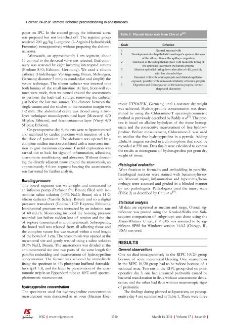
Table 2 Mucosal injury scale from Chiu et al [23]<br />
Grade<br />
Hydroxyproline concentration<br />
The specimens used for hydroxyproline concentration<br />
measurement were desiccated in an oven (Heraeus Electronic<br />
UT5042EK, Germany) until a constant dry weight<br />
was achieved. Hydroxyproline concentration was determined<br />
by using the Chloramine-T spectrophotometric<br />
method as previously described by Reddy et al [22] . The practice<br />
is based on alkaline hydrolysis <strong>of</strong> the tissue homogenate<br />
and the consecutive measurement <strong>of</strong> free hydroxyproline.<br />
Before measurement, Chloramine-T was used<br />
to oxidize the free hydroxyproline in a pyrrole. Adding<br />
Ehrlich’s reagent resulted in a chromophore that could be<br />
recorded at 550 nm. Data finally were calculated to express<br />
the results as micrograms <strong>of</strong> hydroxyproline per gram dry<br />
weight <strong>of</strong> tissue.<br />
Histological evaluation<br />
After fixation in formalin and embedding in paraffin,<br />
histological sections were stained with hematoxylin-eosin.<br />
Mucosal injury, inflammation and hyperemia/hemorrhage<br />
were assessed and graded in a blinded manner<br />
by two pathologists. Pathologists used the injury scale<br />
(Table 2) as described by Chiu et al [23] .<br />
Statistical analysis<br />
All data are expressed as median and range. Overall significance<br />
was proved using the Kruskal-Wallis test. Subsequent<br />
comparison <strong>of</strong> subgroups was done using the<br />
Mann-Whitney U test; P < 0.05 was assumed to be significant.<br />
SPSS for Windows version 14.0.2 (Chicago, IL,<br />
USA) was used.<br />
RESULTS<br />
Definition<br />
0 Normal mucosal villi<br />
1 Development <strong>of</strong> subepithelial Gruenhagen’s space at the apex<br />
<strong>of</strong> the villus, <strong>of</strong>ten with capillary congestion<br />
2 Extension <strong>of</strong> the subepithelial space with moderate lifting <strong>of</strong><br />
the epithelial layer from the lamina propria<br />
3 Massive epithelial lifting down the sides <strong>of</strong> villi, possibly<br />
with few denuded tips<br />
4 Denuded villi with lamina propria and dilated capillaries<br />
exposed, possibly with increased cellularity <strong>of</strong> lamina propria<br />
5 Digestion and disintegration <strong>of</strong> the lamina propria, hemorrhage<br />
and ulceration<br />
General observations<br />
One rat died intraoperatively in the RIPC <strong>10</strong>/20 group<br />
because <strong>of</strong> acute mesenterial bleeding. One anastomosis<br />
in the RIPC <strong>10</strong>/20 group had to be redone because <strong>of</strong> a<br />
technical issue. Two rats in the RIPC group died on postoperative<br />
day 3: one had advanced peritonitis caused by<br />
bacterial translocation in ileus without anastomotic dehiscence;<br />
and the other had ileus without macroscopic signs<br />
<strong>of</strong> peritonitis.<br />
The findings during planned re-laparotomy on postoperative<br />
day 4 are summarized in Table 1. There were three<br />
WJG|www.wjgnet.com<br />
13<strong>10</strong> March 14, 2011|Volume 17|Issue <strong>10</strong>|


