MicroImaging Newsletter Sept 09:Layout 1 - Carl Zeiss
MicroImaging Newsletter Sept 09:Layout 1 - Carl Zeiss
MicroImaging Newsletter Sept 09:Layout 1 - Carl Zeiss

You also want an ePaper? Increase the reach of your titles
YUMPU automatically turns print PDFs into web optimized ePapers that Google loves.
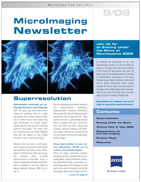
Microscopy from <strong>Carl</strong> <strong>Zeiss</strong><br />
<strong>MicroImaging</strong><br />
<strong>Newsletter</strong><br />
9/<strong>09</strong><br />
Join Us for<br />
an Evening Under<br />
the Stars at<br />
Neuroscience 20<strong>09</strong><br />
Superresolution<br />
Superresolution microscopes put cutting<br />
edge discoveries at your fingertips<br />
Over 100 years ago, Ernst Abbe determined<br />
the resolution limit of a light<br />
microscope. This minimum value of about<br />
220 nm holds true for such modern day<br />
light microscopes as simple camera<br />
systems through high end laser scanning<br />
confocal microscopes. This means that<br />
two structures that are closer together<br />
than 220 nm appear as one unified<br />
structure in your current microscope.<br />
We are developing instruments based on<br />
two Superresolution techniques:<br />
Superresolution Structured Illumination<br />
Microscopy (SR-SIM) and Photoactivation<br />
Localization Microscopy (PAL-M). These<br />
systems promise to yield images beyond<br />
what is capable with your current system.<br />
And with <strong>Carl</strong> <strong>Zeiss</strong>’ easy-to-use<br />
software, hands-on training, and extensive<br />
support network, you can be sure to<br />
hit the ground running with publication<br />
quality images.<br />
To celebrate the introduction of our new<br />
Superresolution systems, we will be hosting an<br />
evening in Chicago during the annual meeting<br />
of the Society for Neuroscience. The event will<br />
feature talks by two leading scientists in the field<br />
of Superresolution microscopy: Dr. Eric Betzig<br />
(Howard Hughes Medical Institute, Janelia Farm)<br />
and Dr. Rainer Heintzmann (Kings College<br />
London). Prior to the talks, relax and socialize in<br />
the gallery with complimentary drinks and appetizers<br />
to the sound of live jazz music. The event<br />
begins at 6 pm on Tuesday, October 20th.<br />
Reservations are required and can be<br />
made at www.zeiss.com/neuroscience.<br />
Contents<br />
Superresolution<br />
Evening Under the Stars<br />
Xtreme Field of View SEM<br />
Requirements for<br />
Live Cell Imaging<br />
Product News<br />
Recently, there has been a small explosion<br />
of papers as researchers have broken<br />
that resolution limit with various modifications<br />
of fluorescence light microscopy.<br />
The potential these so-called<br />
Superresolution microscopes bring to<br />
research caused the technique to be identified<br />
as Method of the Year 20<strong>09</strong> by<br />
Nature Methods (January 20<strong>09</strong>, Vol 6,<br />
No. 1).<br />
Bring Superresolution to your current<br />
experiments: SR-SIM can be<br />
used on conventional samples<br />
When an image containing regularly<br />
spaced structures is overlayed with a structured<br />
pattern, simple interference pattern,<br />
also called Moiré fringes, can be seen. In a<br />
microscope system, if the frequency of the<br />
grid is at or near the resolution limit of the<br />
microscope, these Moiré fringes will con-<br />
…more on page 2<br />
Resources
Xtreme Field of<br />
View Scanning<br />
Electron<br />
Microscopy<br />
The ZEISS SEM Visualization Engine (SEM-<br />
VE) enables the acquisition of a variety of<br />
scanning electron microscopy images (SE,<br />
BSE, STEM, etc.) over large fields of view<br />
(>1 gigapixel) at nanometer scale resolution.<br />
In the neuroscience field, researchers<br />
are using SEM-VE to map large neural networks<br />
including synaptic junctions -<br />
requiring nanometer-resolution – over<br />
cubic millimeter volumes.<br />
10 µm<br />
A single acquisition extreme<br />
field-of-view low-voltage STEM image<br />
(2 nm pixel size) of a rat hippocampus<br />
acquired in a field-emission SEM.<br />
(Sample courtesy of J. Mendenhall,<br />
Univ. of Texas at Austin)<br />
The SEM-VE adapts to any standard<br />
ZEISS SmartSEM platform, and can<br />
extend the number of pixels acquired<br />
in an image by 150 times, without<br />
sacrificing ultimate resolution. This<br />
dramatically improves automation and<br />
throughput by reducing stage motion<br />
requirements that can be critical to laborious<br />
image stitching routines.<br />
<strong>MicroImaging</strong> <strong>Newsletter</strong><br />
Superresolution<br />
…continued from page 1<br />
tain high frequency information that is<br />
normally irresolvable. By analyzing these<br />
Moiré fringes in Fourier space, this higher<br />
frequency information can be decoded and<br />
unveiled in the final image. Using this<br />
technique of Superresolution Structured<br />
Illumination Microscopy (SR-SIM), the resolution<br />
can be doubled in both XY and Z.<br />
As SR-SIM works with any fluorophore (eg.<br />
Alexa dyes, DAPI, Cy dyes) or fluorescent<br />
protein (eg. GFP, YFP, mCherry, mTomato)<br />
and does not require special sample<br />
preparation, SR-SIM can easily be incorporated<br />
into your current experiments to produce<br />
4-color, Superresolution images in<br />
3D. In addition, with acquisition speeds at<br />
1-2 frames per second, Superresolution<br />
imaging of live cells undergoing slower<br />
movements is possible.<br />
Experience resolution approaching<br />
that of electron microscopy with PAL-M<br />
As stated, two points of light appear as<br />
one if they are closer than 220 nm in a<br />
traditional fluorescent microscope. The<br />
idea behind PAL-M is to image these two<br />
points separately, determine their exact<br />
XY locations, and then overlay them in a<br />
post-processing step. This technique is<br />
possible with photoactivable or photoconvertible<br />
fluorescent highlighters<br />
whereby low powers of activation light<br />
affect a very small population of the<br />
sample. By repeating a process of activating<br />
a few molecules, imaging these<br />
molecules, then bleaching or switching<br />
them off, a pointillist image is constructed<br />
over time with resolution limits down<br />
to 20 nm. Z-resolution down to 100 nm<br />
is achieved with TIRF illumination using<br />
our new PAL-M 100x/1.57 NA objective<br />
lens. With a <strong>Carl</strong> <strong>Zeiss</strong> Superresolution<br />
specialist available to train you and your<br />
lab on the logistics of fluorophores, sample<br />
preparation, imaging, and analysis,<br />
you can expect images approaching EMresolution<br />
while still using relatively easy<br />
sample preps and fluorophore labels.<br />
Accurately correlate confocal, SR-<br />
SIM, and PAL-M images from the<br />
same structures Only <strong>Carl</strong> <strong>Zeiss</strong> can<br />
offer you both SIM and PAL-M on the<br />
same microscope stand as a confocal<br />
microscope. With the same software, you<br />
can easily switch between confocal or<br />
either of the Superresolution techniques.<br />
Compare and contrast the same structures<br />
with all three techniques to cross-examine<br />
your data and build more convincing scientific<br />
models. Additionally, LSM 710s can<br />
be integrated with a Superresolution system,<br />
allowing existing owners to extend<br />
their imaging capabilities.<br />
www.zeiss.com/superresolution<br />
2<br />
Learn more about SEM-VE and brain<br />
mapping at: www.zeiss.com/nts
Since the 1970s scientists have tried to<br />
capture life on screen. Such attempts<br />
range from observing cultured cell lines to<br />
time-lapse photography of developing<br />
early embryos to deep brain imaging in<br />
waking-state small mammals. Although<br />
imaging of living specimens introduces<br />
many challenges, only living samples can<br />
accurately portray biology’s natural<br />
state. Although the number and sophistication<br />
of the tools utilized for live cell<br />
microscopy has increased enormously,<br />
there are many parameters which must be<br />
carefully considered when planning such<br />
an experiment.<br />
Five basic requirements must be satisfied<br />
in order to conduct a successful live cell<br />
experiment:<br />
1 Although the goal in live cell imaging<br />
is to observe the natural state of the<br />
cells or organisms, most experiments<br />
require artificial introduction of a<br />
label for proteins, sub-cellular components,<br />
or particular cell types. The<br />
development of an array of fluorescent<br />
proteins and dyes has provided<br />
biomedical research with enormous<br />
flexibility of markers that extend now<br />
to photo-activatable and -switchable<br />
versions that can be used, for example,<br />
in cell lineage experiments.<br />
2 Having introduced specific marker(s)<br />
into the cells or organism, the second<br />
critical step is to excite or activate<br />
them with minimal illumination.<br />
This reduces the potential for photoxicity<br />
and resulting damage or<br />
death to the sample. Examples of<br />
this include high speed LED illumination<br />
systems for widefield imaging<br />
and AOTF controlled or directly modulated<br />
solid-state lasers employed in<br />
confocal microscopes, which both<br />
work well to image cells with the<br />
gentlest light possible.<br />
<strong>MicroImaging</strong> <strong>Newsletter</strong><br />
Live Cell Imaging:<br />
Five Basic Requirements<br />
3 Use of a detection device with the<br />
highest possible efficiency ensures that<br />
the lowest illumination intensity can be<br />
used. CCD cameras have improved<br />
remarkably over the last decade with<br />
quantum efficiencies greater than 85%<br />
whereas EMCCDs allow for very high<br />
acquisition rates combined with high<br />
efficiency. Unitary detectors, such as<br />
the PMTs used in point scanning confocal<br />
microscopes, have not improved to<br />
the same extent as CCDs, but the use<br />
of materials such as gallium arsenide<br />
phosphide (GaAsP) and significant<br />
reductions in dark current have<br />
enabled these systems to be much<br />
more applicable to live cell imaging.<br />
4 A live cell experiment only works if<br />
the cells are kept alive - often for<br />
days. To achieve this, an incubation<br />
system may be required to control<br />
temperature, pH, CO 2 , O 2 , and<br />
humidity levels. The most refined systems<br />
include computer control and<br />
recording of these parameters.<br />
5 Last, but not least, the sample has to<br />
be held in focus over the term of the<br />
experiment. Two techniques are now<br />
commonly used. Patterns formed with<br />
infra-red light are projected onto the<br />
underside of the coverglass and a<br />
small camera linked to the microscope’s<br />
focus mechanism is used to<br />
hold the pattern in focus, thereby holding<br />
the cells in focus. Alternatively, a<br />
laser (typically in a laser scanning<br />
microscope) can be used to image the<br />
undersurface of the coverglass in<br />
reflected light mode, thereby providing<br />
an absolute reference from which the<br />
focus can be returned to the sample.<br />
If you have any questions on getting<br />
started with live cell imaging or<br />
with your current experiments, send<br />
an email to Support@zeiss.com.<br />
Products<br />
VivaTome: Fast<br />
optical sectioning<br />
made easy<br />
and affordable<br />
With the new VivaTome, <strong>Carl</strong> <strong>Zeiss</strong><br />
introduces an innovative concept for fast<br />
acquisition of optical<br />
sections. Using the<br />
technique of aperture<br />
correlation, VivaTome<br />
combines the light<br />
efficiency of structured<br />
illumination<br />
with the speed of a<br />
spinning disk. A grid<br />
pattern with high<br />
transmission efficiency<br />
is used to modulate<br />
the excitation light, therefore,<br />
VivaTome can be operated with a standard<br />
white light source. All information necessary<br />
to calculate an optical section is captured<br />
simultaneously in one single image,<br />
efficiently excluding motion artifacts.<br />
Furthermore, a standard widefield image<br />
can be calculated from the same data –<br />
so all the information from your sample<br />
is accessible in a single shot. Two different<br />
grid patterns allow the use of a wide<br />
variety of different<br />
objectives with optimal<br />
sectioning performance.<br />
VivaTome<br />
allows image capture<br />
at high frame<br />
rates suitable for<br />
imaging of living<br />
cells and for observation<br />
of dynamic<br />
events in developmental<br />
processes.<br />
The simple and compact<br />
instrument can be mounted on the<br />
camera port of a variety of upright and<br />
inverted ZEISS microscopes. Field<br />
upgrades with current systems are also<br />
possible. It is fully integrated in the powerful<br />
AxioVision Software package, opening<br />
up new possibilities in imaging of<br />
dynamic events. 3
<strong>MicroImaging</strong> Resources<br />
<strong>MicroImaging</strong> <strong>Newsletter</strong><br />
<strong>Zeiss</strong> on<br />
Your Campus:<br />
Join us for<br />
a new lecture<br />
series on live<br />
cell microscopy<br />
With over 1000 participants in 47 locations,<br />
the recently completed North American<br />
<strong>Zeiss</strong> on Your Campus (ZOYC) Tour 20<strong>09</strong> on<br />
3D Imaging Techniques was a great success.<br />
Our goal in these workshops was to<br />
enable scientists to fully optimize the imaging<br />
parameters using their current systems,<br />
as well as understand the various available<br />
3D techniques in order to make informed<br />
decisions when selecting a microscope system<br />
for a given experiment. This was<br />
achieved through a short series of informative<br />
lectures and hands-on sessions.<br />
Based on the positive feedback from this<br />
endeavor, we will launch another educational<br />
tour in 2010. This new tour will focus on imaging<br />
of living cells and animals, a topic which<br />
has been strongly requested by the scientific<br />
community. Our lectures and hands-on sessions<br />
with living samples will include topics<br />
such as balancing resolution and speed during<br />
image acquisition, cell viability and photo-toxicity,<br />
and presenting 4D data sets. Some of the<br />
locations we will be visiting in 2010 include:<br />
• Columbia University<br />
• McGill University<br />
• Stanford University<br />
• St. Jude’s Children’s Research Hospital<br />
• University of California at Irvine<br />
• University of Michigan<br />
• University of Pennsylvania<br />
• And more…<br />
Please visit www.zeiss.com/zoyc to<br />
view the full list of locations with dates and<br />
to reserve your spot.<br />
<strong>Zeiss</strong> Online Campus<br />
Wondering how to correctly analyze<br />
your images for colocalization Curious<br />
how FRET Biosensors work Do you<br />
have questions about the latest<br />
developments in fluorescent proteins<br />
<strong>Carl</strong> <strong>Zeiss</strong> answers such questions at<br />
www.zeiss.com/campus. Developed<br />
in collaboration with Mike Davidson of<br />
Service & Support<br />
Our in-house service and support teams<br />
work mainly behind the scenes; but if<br />
something goes wrong, our people make<br />
the difference. We are pleased to introduce<br />
team members in this, as well as<br />
upcoming newsletters.<br />
Working in the application support<br />
group, Tim Pratt answers customer questions<br />
about image analysis and Laser<br />
Microdissection systems. Having done his<br />
PhD in Molecular Genetics, he has a solid<br />
background in laboratory benchwork and<br />
experimental design. Tim has been with<br />
<strong>Carl</strong> <strong>Zeiss</strong> for over four years and participates<br />
in many internal training sessions<br />
and external scientific meetings to keep<br />
his knowledge of microscopy and related<br />
techniques current.<br />
Tim and his peers in the support group<br />
use remote access tools which enable<br />
them to connect to any system and troubleshoot<br />
most questions immediately.<br />
They answer questions ranging from<br />
Florida State University, our site is an<br />
online reference guide for microscopy,<br />
digital imaging, and related applications.<br />
The combination of interactive tutorials,<br />
review articles, and comprehensive lists<br />
of published references make this site the<br />
best place to learn more about leading<br />
edge techniques in microscopy.<br />
“Why is my camera image in the wrong<br />
color” to “How do I correct shading<br />
in my image” to “Can I image a particular<br />
fluorophore with my current system”<br />
Tim and his colleagues can be<br />
reached Mon – Fri from 9 am – 8 pm<br />
Eastern time on the <strong>Carl</strong> <strong>Zeiss</strong><br />
support line at 1-800-5<strong>09</strong>-3905.<br />
They can also be reached by email at<br />
support@zeiss.com.<br />
<strong>Carl</strong> <strong>Zeiss</strong> <strong>MicroImaging</strong>, Inc.<br />
One <strong>Zeiss</strong> Drive<br />
Thornwood, NY 10594<br />
1-800-233-2343<br />
micro@zeiss.com<br />
www.zeiss.com/micro<br />
Customer Service: 1-800-233-2343<br />
Systems Service: 1-800-633-6610<br />
Application Support: 1-800-5<strong>09</strong>-3905<br />
Editors: Duncan McMillan, Dr. Maya Everett,<br />
and Dr. Jochen Tham<br />
Production:<br />
<strong>Carl</strong> <strong>Zeiss</strong> <strong>MicroImaging</strong>, Inc., Thornwood, NY<br />
For more information, to request literature<br />
on our products, or to remove your<br />
name from our mailing list, please email<br />
micro@zeiss.com