Correction of Congenitally Missing Lateral Incisors with Porcelain
Correction of Congenitally Missing Lateral Incisors with Porcelain
Correction of Congenitally Missing Lateral Incisors with Porcelain

You also want an ePaper? Increase the reach of your titles
YUMPU automatically turns print PDFs into web optimized ePapers that Google loves.
CONTINUING EDUCATION 1 6<br />
CORRECTION OF CONGENITALLY<br />
MISSING LATERAL INCISORS WITH<br />
PORCELAIN VENEERS<br />
Jack D. Griffin, Jr., DMD*<br />
GRIFFIN<br />
18<br />
8<br />
SEPTEMBER<br />
The ability <strong>of</strong> the dental pr<strong>of</strong>essional to improve a patient’s smile has become a<br />
benchmark in modern aesthetic dentistry. When one type <strong>of</strong> tooth is transformed<br />
into another through the restorative process, special care must be taken to ensure<br />
correct preparation and gingival contours to create the illusion that the teeth are<br />
in their correct position. With accurate digital photographic planning, active patient<br />
consultation, detailed tooth preparation, and a meticulous laboratory sequence,<br />
porcelain laminate veneers can be functional and can achieve the aesthetic expectations<br />
<strong>of</strong> the patient.<br />
Learning Objectives:<br />
This article discusses the options for correcting congenitally missing lateral incisors<br />
and the aesthetic complications when managing repositioned canines. Upon reading<br />
this article, the reader should:<br />
• Understand the aesthetic requisites that must be satisfied when transforming<br />
canines into lateral incisors and premolars into canines.<br />
• Recognize the need for communication <strong>with</strong> the orthodontist to coordinate<br />
tooth movement <strong>with</strong> aesthetic solutions and treatment options.<br />
Key Words: veneers, mockup, porcelain, congenitally missing incisors<br />
*Private practice, Eureka, MO.<br />
Jack D. Griffin, Jr., DMD, 18 Hilltop Village Center Drive, Eureka, MO 63025<br />
Tel: 636-938-4141 • E-mail: Esmilecenter@aol.com<br />
Pract Proced Aesthet Dent 2006;18(8):475-480 475
Practical Procedures & AESTHETIC DENTISTRY<br />
For over 20 years, bonded porcelain restorations have<br />
been used successfully to enhance the aesthetic<br />
appearance <strong>of</strong> teeth compromised by form, color, or position.<br />
1 <strong>Porcelain</strong> veneers, which have been accepted for<br />
long-term augmentation <strong>of</strong> tooth and smile deficiencies<br />
<strong>with</strong> congenitally missing teeth, allow clinicians to create<br />
this aesthetic ruse. 2,3 There are many challenges to consider<br />
when canines are used to replace congenitally missing<br />
lateral incisors. From an aesthetic standpoint, not only<br />
must the canines mimic the lateral incisors, but the first<br />
premolars must be made to appear as canines. In such<br />
cases, the anatomical and functional challenges must<br />
be incorporated into a thorough treatment plan, s<strong>of</strong>t tissues<br />
must be enhanced, and the preparation design must<br />
be visualized to provide the needed contours. 4,5<br />
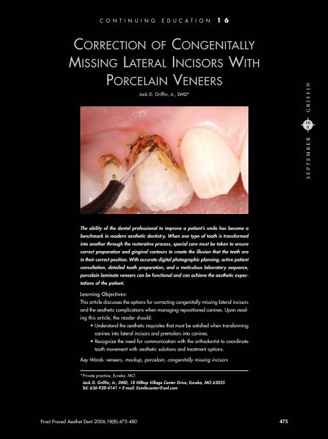
Figure 1. The preoperative smile was unnaturally<br />
dark due to the mesially repositioned.<br />
Orthodontic Considerations<br />
With congenitally missing incisors, the orthodontic practitioner<br />
must weigh many factors when deciding if the<br />
canines should be placed laterally or if gaps should be<br />
opened and treatment planned for fixed partial dentures<br />
(FPDs) or dental implants. 6 When the canine is<br />
brought mesially into the position <strong>of</strong> the lateral incisor,<br />
all posterior teeth must also be mesialized to close the<br />
resulting spaces. Selection <strong>of</strong> the ideal treatment option<br />
depends upon many factors, including occlusion and<br />
patient desires. 7<br />
Since the canines are normally darker, broader buccally,<br />
and have s<strong>of</strong>t tissue shape <strong>of</strong>ten inconsistent <strong>with</strong><br />
lateral incisors, spaces are commonly created to allow<br />
FPD or implant placement. 8 This allows the first premolars<br />
to stay in their posterior position, where the more<br />
occlusal tissue heights and lingual cusps can function<br />
and appear more natural.<br />
In the author’s experience, teeth being moved<br />
mesially <strong>of</strong>ten compromise buccal corridor development<br />
leading to shadow because <strong>of</strong> a loss <strong>of</strong> total tooth volume.<br />
The reduction in tooth volume and arch length can<br />
cause a narrowing effect that leads to shadowing inside<br />
the cheek from a frontal smile perspective. As a result <strong>of</strong><br />
these and other factors, the orthodontist and restorative<br />
dentist should be in agreement <strong>with</strong> final treatment goals<br />
and tooth positioning prior to finalizing orthodontic treatment.<br />
This article discusses a technique using porcelain<br />
veneers to provide corrective treatment to patients <strong>with</strong><br />
congenitally missing lateral incisors.<br />
Figure 2. Since the canines were wider than the<br />
lateral incisors they would replace, a compensating<br />
reduction in width was required.<br />
Figure 3. The tissues <strong>of</strong> the first premolar<br />
required gingivoplasty to move tissues apically<br />
and to widen the emergence from the gingiva.<br />
Case Presentation<br />
A 20-year-old female patient presented after having<br />
undergone orthodontic treatment to improve her smile<br />
(Figure 1). Orthodontic therapy was completed <strong>with</strong>out<br />
Figure 4. Preoperative lingual view <strong>of</strong> the<br />
patient’s maxillary arch.<br />
476 Vol. 18, No. 8
Griffin<br />
Figure 5. The calipers were then held over the<br />
canine in the lateral position to verify a correct<br />
lateral to central ratio.<br />
consulting a restorative clinician, and all posterior teeth<br />
were brought mesial to compensate for the congenitally<br />
missing lateral incisors. This resulted in a darker, unnatural<br />
looking result.<br />
The treatment plan included a frenectomy to<br />
decrease the likelihood <strong>of</strong> recession around the central<br />
incisors, gingivoplasty around the first premolars, and<br />
gingival crown lengthening around the premolars (Figures<br />
2 through 4). 9 Diode lasers can be used predictably for<br />
these tissue enhancements when care is shown in the<br />
proper use <strong>of</strong> settings, when measuring pocket depths,<br />
and when there is an understanding <strong>of</strong> biologic width. 10,11<br />
Since natural canine gingival heights are more apical<br />
and broader than those <strong>of</strong> the premolars, tissue<br />
reshaping was planned to increase the emergence pr<strong>of</strong>ile<br />
to a level more <strong>of</strong>ten seen in canines. Canines have<br />
a buccal prominence not evident in lateral incisors; care<br />
would thus be taken to provide the laboratory technician<br />
<strong>with</strong> adequate space to cover the tooth and maintain<br />
new incisor contours.<br />
Figure 6. The mockup was evaluated and any<br />
required adjustments were made.<br />
Figure 7. Preparations were first completed as a<br />
0.5-mm to 1-mm porcelain veneer reduction and<br />
margins were placed at the gingival levels.<br />
Treatment Mockup<br />
A mockup was critical for fabrication <strong>of</strong> the provisional<br />
restorations, enabling the patient and clinician to evaluate<br />
aesthetics and phonetics, and to preview the anticipated<br />
result. 12 Before anesthesia was delivered, retractors<br />
were placed and the canines and premolars were<br />
etched. A microhybrid composite (eg, Esthet-X, Dentsply<br />
Caulk, Milford, DE; Filtek Supreme, 3M Espe, St. Paul,<br />
MN) that approximated the final shade was placed on<br />
the canines and premolars. The width <strong>of</strong> the lateral<br />
incisors was determined by measuring the central incisors<br />
<strong>with</strong> calipers. After composite resin was added, the<br />
calipers were held over the canines so that the width<br />
approximated 75% <strong>of</strong> the central width (Figure 5).<br />
After composite resin had been applied to the facial<br />
surfaces <strong>of</strong> the premolars, the contours and phonetics<br />
were evaluated (Figure 6). Tooth proportion was critical<br />
for a pleasing outcome and, following the principles <strong>of</strong><br />
the “Golden Proportion,” central dominance and a heightto-width<br />
ratio <strong>of</strong> about 75%, served as a basis for the<br />
mockup. 13 From this, an alginate impression was made,<br />
poured up, and a vacuum-formed matrix made for subsequent<br />
fabrication <strong>of</strong> the provisional restorations. This<br />
mockup was also forwarded to the laboratory.<br />
Figure 8. The s<strong>of</strong>t tissue margins were moved<br />
apically and shaped on the mesial side to widen<br />
the emergence pr<strong>of</strong>ile <strong>of</strong> the first premolar.<br />
Intraoral Preparations<br />
There are many choices for aesthetic materials, each<br />
<strong>with</strong> its own preparation protocol. 14 With these systems<br />
it is critical to understand the reduction needed, optimal<br />
margin placement, and preparation shape for proper<br />
PPAD 477
Practical Procedures & AESTHETIC DENTISTRY<br />
restoration aesthetics and long-term function. 15,16 A coarse<br />
reduction depth diamond and a tapered diamond were<br />
used to provide sufficient reduction for the desired feldspathic<br />
porcelain. 17 A standard 0.5-mm to 1-mm reduction<br />
was performed on all teeth <strong>with</strong> rounded chamfer<br />
margins, as is performed for a standard porcelain veneer<br />
case <strong>with</strong>out regard to tooth proportion (Figure 7).<br />
Since excessive frenal tissue between the incisors<br />
can “pull” on the gingiva and the potential for recession<br />
existed, 9 a frenectomy was performed <strong>with</strong> a diode laser<br />
(ie, Odyssey, Ivoclar Vivadent, Amherst, NY) (Figure 9).<br />
The laser was set on a low power setting <strong>of</strong> 2.0 watts<br />
and the frenum was removed <strong>with</strong> “brushing” strokes to<br />
remove all loose tissue (Figure 8).<br />
Gingival enhancements were then performed on the<br />
premolars. The pocket depths were measured at 2 mm<br />
to 3.5 mm and recontoured, preserving biologic width. 18<br />
To mimic a canine, tissue was removed on the mesial<br />
side to widen the emergence pr<strong>of</strong>ile (Figure 9). This reduction<br />
was blended onto the facial aspect, where 1 mm<br />
to 2 mm <strong>of</strong> tissue height was removed to make the premolars<br />
appear as long as the teeth being replaced.<br />
This reshaping was accomplished as the clinician preserved<br />
a gradual decrease in clinical crown length from<br />
mesial to distal.<br />
Impressions and Provisionalization<br />
The canine margins were then refined and moved approximately<br />
1 mm subgingivally, as needed, in areas <strong>of</strong> s<strong>of</strong>t<br />
tissue sculpting. A smooth transition <strong>of</strong> the gingival heights<br />
from the central incisors back to the second premolars<br />
and margins placed just apical to those points was<br />
ensured. The conversion <strong>of</strong> the canines into lateral incisors<br />
included flattening the faciogingival aspects and distal<br />
reduction to a knife-edge margin to reduce the tooth<br />
width as planned (Figure 10).<br />
Conversion <strong>of</strong> the premolars to canines involved carrying<br />
the mesial margin toward the lingual side to provide<br />
sufficient space for the increased width <strong>of</strong> the<br />
restorations. Although the lingual aspect <strong>of</strong> the canine<br />
was wider than a lateral incisor would be, the illusion<br />
<strong>of</strong> its harmony in the arch was created by narrowing <strong>of</strong><br />
the distal facial line angle. This angle was also moved<br />
mesially and prepared <strong>with</strong> a tapering feather-edge margin<br />
lingually. This provided the ceramist space to make<br />
porcelain <strong>of</strong> sufficient thickness <strong>with</strong>out reducing the entire<br />
tooth for a full-coverage crown.<br />
Braided cord was used for retraction along <strong>with</strong> a<br />
gingival retraction paste (ie, Expa-Syl, Kerr/Sybron,<br />
Orange, CA). The cord provided gingival deflection<br />
in areas that measured more than 1 mm subgingival.<br />
The gingival retraction paste effectively controlled<br />
Figure 9. Gingival heights were examined to verify<br />
a smooth transition from central incisor to molar.<br />
Figure 10. The facial aspects <strong>of</strong> the canines were<br />
flattened, and the distal side was reduced to a<br />
width 1 mm less than the desired width <strong>of</strong> the<br />
intended lateral incisors.<br />
Figure 11. Retraction paste was placed primarily<br />
in areas <strong>of</strong> bleeding; braided retraction cord<br />
was placed to increase tissue displacement.<br />
Figure 12. Self-cured provisional restorations were<br />
made using a 0.020” vacuum-formed matrix.<br />
478 Vol. 18, No. 8
Griffin<br />
Figure 13. Tissue adaptation was positive at two<br />
weeks, <strong>with</strong> minor irritation on the distal side <strong>of</strong><br />
the restored lateral.<br />
Figure 14. The small amount <strong>of</strong> cement left<br />
between the lateral and the canine was cleaned<br />
up using scaler and composite knife.<br />
bleeding and rinsed cleanly from the preparation, thereby<br />
reducing the potential <strong>of</strong> “black margins” later from hemostatic<br />
contaminants (Figure 11). Two full-arch vinyl<br />
polysiloxane impressions were made, a lower model<br />
was made from the alginate, and a bite registration<br />
was taken.<br />
Immediate dentin sealing <strong>with</strong> dentin adhesives has<br />
been noted to increase dentin bond strengths, reduce<br />
bacterial microleakage, and cause less dentin sensitivity<br />
in comparison to provisional restorations placed on<br />
unbonded teeth. 19 The teeth never leave the author’s practice<br />
in an unsealed state, which may reduce the potential<br />
<strong>of</strong> bacterial invasion from microleakage and may<br />
lead to postoperative sensitivity. 20<br />
The teeth were etched and subsequently coated <strong>with</strong><br />
two coats <strong>of</strong> a dual-cure bonding agent (eg, Cabrio,<br />
Discus Dental, Culver City, CA; Optibond Solo Plus,<br />
Kerr/Sybron, Orange, CA). This was followed by a glycerin<br />
separating medium to keep the temporary material<br />
from bonding to the teeth. The provisional restorations<br />
were then fabricated from a composite material (eg,<br />
PerfecTemp II, Discus Dental, Culver City, CA; Luxatemp,<br />
DMG/Zenith, Englewood, NJ) inside a 0.020” suck<br />
down splint (Figure 12). The contours were then refined<br />
and polished. An adjustment was made at a one-week<br />
follow-up visit. An alginate impression <strong>of</strong> the corrected<br />
provisional restorations was made and forwarded to the<br />
dental laboratory, where eight feldspathic porcelain<br />
veneers <strong>with</strong> incisal characterization were fabricated. A<br />
moderate amount <strong>of</strong> translucency to create a natural<br />
appearance was necessary to meet the patient’s expectations.<br />
The liquid-powder-stacked porcelain veneers<br />
would yield excellent vitality while providing sufficient<br />
strength for the anterior region. 21<br />
Figure 15. The smile was more proportional,<br />
while the color differences in the original canines<br />
have been masked <strong>with</strong>out the appearance <strong>of</strong><br />
thick, bulky, or opaque teeth.<br />
Figure 16. <strong>Lateral</strong> view demonstrated the<br />
improved proportions <strong>of</strong> the lateral incisors,<br />
canines and first premolar teeth.<br />
Insertion and Evaluation<br />
The provisional restorations were removed, and the teeth<br />
were cleaned <strong>with</strong> a hand scaler and pumice, and were<br />
rinsed thoroughly. After securing patient approval, the<br />
teeth were etched according to the total-etch technique<br />
and recoated <strong>with</strong> a bonding agent to ensure coverage<br />
<strong>of</strong> all enamel and dentin surfaces. The veneers were luted<br />
in place <strong>with</strong> a light-cured resin cement, starting from the<br />
midline and working distally. The restorations for the<br />
incisors were placed, cured, and cleaned. The posterior<br />
teeth were treated similarly, and verification <strong>of</strong> proper<br />
occlusion followed.<br />
Two weeks postoperatively, tissue response was very<br />
good <strong>with</strong> slight redness in the areas where recontouring<br />
was performed. The areas around the new “canines”<br />
responded well, and a natural emergence pr<strong>of</strong>ile was<br />
created (Figures 13 and 14). Despite the loss <strong>of</strong> tooth<br />
PPAD 479
Practical Procedures & AESTHETIC DENTISTRY<br />
preparation, and communication <strong>with</strong> the patient and<br />
laboratory technician enabled the author to meet the<br />
patient’s expectations. It is important for the clinician and<br />
patient to preview the teeth <strong>with</strong> direct composite, and<br />
then prepare to give the technician a chance to properly<br />
contour the case.<br />
Figure 17. Emergence pr<strong>of</strong>ile <strong>of</strong> the canines and<br />
first premolars achieved the patient’s expectations.<br />
Figure 18. Postoperative lingual view demonstrated<br />
a more natural morphology and width.<br />
volume, mesial movement <strong>of</strong> teeth, and the narrowing<br />
<strong>of</strong> the arch that accompanies it, there was sufficient development<br />
<strong>of</strong> the buccal corridor (Figure 15). Tooth proportions<br />
were achieved to create a proportional smile<br />
while preserving gingival health.<br />
Eight weeks after preparation and gingival enhancements,<br />
the healing was complete and physiologically<br />
acceptable (Figures 16 and 17). Had the teeth remained<br />
in their normal position and FPDs or implants placed in<br />
the position <strong>of</strong> the missing lateral incisors, the aesthetics<br />
might have been even further enhanced. This would have<br />
allowed for broader arch development in length and<br />
width in the canine eminence area. At the conclusion <strong>of</strong><br />
treatment, however, contours and function were clinically<br />
acceptable and a more ideal shape and width to the<br />
incisors was achieved <strong>with</strong> a relatively minor amount <strong>of</strong><br />
tooth reductions and gingival refinement (Figure 18).<br />
Conclusion<br />
<strong>Porcelain</strong> laminate veneers can meet the aesthetic and<br />
functional desires <strong>of</strong> a patient <strong>with</strong> congenitally missing<br />
lateral incisors. Veneers are a great choice for transforming<br />
canines into lateral incisors and cuspids from first<br />
bicuspids while providing long term clinical success <strong>with</strong><br />
relatively minor tooth preparation. Diligent visualization,<br />
Acknowledgment<br />
The author mentions his gratitude to Adriam Jurim, CDT,<br />
for his contributions to the case presented herein.<br />
References<br />
1. Swift EJ Jr, Friedman MJ. <strong>Porcelain</strong> veneer outcomes, Part II. J Esthet<br />
Restor Dent 2006;18(2):110-113.<br />
2. Tipton PA. Aesthetic tooth alignment using etched porcelain restorations.<br />
Pract Proced Asthet Dent 2001;13(7):551-555.<br />
3. Dumfahrt H. <strong>Porcelain</strong> laminate veneers. A retrospective evaluation<br />
after 1 to 10 years <strong>of</strong> service: Part 1–Clinical procedure. Int<br />
J Prosthodont 1999;12(6):505-513.<br />
4. Peumans M, Van Meerbeek B, Lambrechts P, Vanherle G. <strong>Porcelain</strong><br />
veneers: A review <strong>of</strong> the literature. J Dent 2000;28(3):163-177.<br />
5. Bassett JL. Replacement <strong>of</strong> missing mandibular lateral incisors <strong>with</strong><br />
a single pontic all-ceramic prosthesis: A case report. Prac Periodont<br />
Aesthet Dent 1997;9(4):455-461.<br />
6. Trushkowsky RD. Replacement <strong>of</strong> congenitally missing lateral incisors<br />
<strong>with</strong> ceramic resin-bonded fixed partial dentures. J Prosthet Dent<br />
1995;73(1):12-16.<br />
7. Kinzer GA, Kokich VO Jr. Managing congenitally missing lateral<br />
incisors. Part II: Tooth-supported restorations. J Esthet Rest Dent<br />
2005;17(2):76-84.<br />
8. Kinzer GA, Kokich VO Jr. Managing congenitally missing lateral<br />
incisors. Part I: Canine substitution. J Esthet Rest Dent 2005;17(1):<br />
5-10.<br />
9. Epstein SR. The frenectomy: A comparison <strong>of</strong> classic versus laser<br />
technique. Pract Periodont Aesthet Dent 1991;3(5):27-30.<br />
10. Rice JH. Laser use in fixed, removable, and implant dentistry.<br />
Dent Clin North Am 2000;44(4):767-777.<br />
11. Adams TC, Pang PK. Lasers in aesthetic dentistry. Dent Clin North<br />
Am 2004;48(4):833-860.<br />
12. Mizrahi B. Visualization before finalization: A predictable procedure<br />
for porcelain laminate veneers. Prac Proced Aesthet Dent<br />
2005;17(8):513-518.<br />
13. Blitz N, Steel C, Wilhite C. Principles <strong>of</strong> proportion and central<br />
dominance. In: Diagnosis and treatment evaluation in cosmetic<br />
dentistry. A guide to Accreditation Criteria. American Academy<br />
<strong>of</strong> Cosmetic Dentistry. 2000:16-17.<br />
14. Hornbrook DS. Invisible beauty: The ultimate in esthetic restorations.<br />
Contemp Esthet Rest Prac 2005;9(12):14-21.<br />
15. Rosenthal L. The art <strong>of</strong> tooth preparation and recontouring. Dent<br />
Today 1997;16(4):48-55.<br />
16. Sim C, Ibbetson R. Comparison <strong>of</strong> fit <strong>of</strong> porcelain veneers fabricated<br />
using different techniques. Int J Prothodont 1993;6(1):<br />
36-42.<br />
17. Gurel G. Predictable, precise, and repeatable tooth preparation<br />
for porcelain laminate veneers. Prac Proced Aesthet Dent 2003;<br />
15(1):17-24.<br />
18. Robbins JW. Esthetic gingival recontouring–A plea for honesty.<br />
Quintessence Int 2000;31(8):553-556.<br />
19. Mange P. Immediate dentin sealing: A fundamental procedure<br />
for indirect bonded restorations. J Esthet Rest Dent 2005;17(3):<br />
144-154.<br />
20. Hiatt WH. Incomplete crown root fracture in pulpal-periodontal<br />
disease. J Periodont 1973;44(6):369-379.<br />
21. Peumans M, De Munck J, Fiews S,et al. A prospective ten-year<br />
clinical trial <strong>of</strong> porcelain veneers. J Adhes Dent 2004;6(1):<br />
65-76.<br />
480 Vol. 18, No. 8
CONTINUING EDUCATION<br />
(CE) EXERCISE NO. 16<br />
16<br />
CONTINUING CE<br />
EDUCATION<br />
To submit your CE Exercise answers, please use the answer sheet found <strong>with</strong>in the CE Editorial Section <strong>of</strong> this issue and<br />
complete as follows: 1) Identify the article; 2) Place an X in the appropriate box for each question <strong>of</strong> each exercise; 3) Clip<br />
answer sheet from the page and mail it to the CE Department at Montage Media Corporation. For further instructions,<br />
please refer to the CE Editorial Section.<br />
The 10 multiple-choice questions for this Continuing Education (CE) exercise are based on the article “<strong>Correction</strong> <strong>of</strong><br />
congenitally missing lateral incisors <strong>with</strong> porcelain veneers,” by Jack D. Griffin, Jr., DMD. This article is on Pages<br />
475-480.<br />
1. Treatment planning involving orthodontics and<br />
restorative dentistry is best planned by whom<br />
a. Periodontist.<br />
b. Orthodontist.<br />
c. Restorative dentist.<br />
d. Plan devised by restorative dentist<br />
and orthodontist.<br />
2. Which <strong>of</strong> the following is NOT a problem <strong>of</strong>ten<br />
associated <strong>with</strong> bringing canines and premolars<br />
mesial to close a lateral incisor space<br />
a. Buccal corridor deficiencies.<br />
b. Increased sensitivity to cold.<br />
c. Proper proportion in final tooth contours.<br />
d. Development <strong>of</strong> aesthetic gingival morphology.<br />
3. Restorative choices for congenitally missing lateral<br />
incisors include which <strong>of</strong> the following<br />
a. Implant and fixed crown.<br />
b. All porcelain fixed partial dentures.<br />
c. Veneers on teeth moved orthodontically to<br />
close spaces.<br />
d. All <strong>of</strong> the above.<br />
4. Which <strong>of</strong> the following is NOT a factor influencing<br />
a satisfactory outcome<br />
a. Type <strong>of</strong> composite used in mockup.<br />
b. Overcontoured buccal aspect <strong>of</strong> canines.<br />
c. Emergence pr<strong>of</strong>ile <strong>of</strong> first premolar tissues.<br />
d. Darkness <strong>of</strong>ten found in canines that will<br />
become lateral incisors.<br />
5. Which <strong>of</strong> the following is true <strong>of</strong> the mockup<br />
a. It is not important.<br />
b. It should be done after anesthesia.<br />
c. It is made <strong>of</strong> composite and done after tooth<br />
preparation to verify adequate reduction.<br />
d. It is only an approximation <strong>of</strong> final tooth shape<br />
and contour.<br />
6. Why is the mockup an important tool<br />
a. It aids in fabrication <strong>of</strong> the temporaries.<br />
b. It helps in planning tooth reduction.<br />
c. It allows the patient to see a preview <strong>of</strong> the<br />
anticipated results.<br />
d. All <strong>of</strong> the above.<br />
7. Which principle must NOT be violated when<br />
recontouring the gingival<br />
a. Antes law.<br />
b. The golden proportion.<br />
c. Biologic width.<br />
d. Reduction <strong>of</strong> excessive frenum.<br />
8. Which side <strong>of</strong> the tooth is reduced when the<br />
width <strong>of</strong> the canines is excessive compared to<br />
the lateral being replaced<br />
a. Distal.<br />
b. Mesial.<br />
c. Lingual.<br />
d. Occlusal.<br />
9. Why is it important to send a full series <strong>of</strong><br />
digital images to the laboratory<br />
a. To be used in the laboratory portfolio.<br />
b. They are used for final shade matching.<br />
c. To serve as tooth proportion measurement<br />
guides.<br />
d. To communicate the goals <strong>of</strong> treatment.<br />
10. Which is not a key to success in the case<br />
presented in this article<br />
a. Visualization <strong>of</strong> the outcome.<br />
b. Preparation to allow ceramist room to make<br />
correct teeth.<br />
c. Communication <strong>with</strong> the patient to ensure<br />
expectations are met.<br />
d. Convincing patient <strong>of</strong> a treatment plan.<br />
481 Vol. 18, No. 8<br />
PPAD 481