March 2012 - Institute of Physics and Engineering in Medicine
March 2012 - Institute of Physics and Engineering in Medicine
March 2012 - Institute of Physics and Engineering in Medicine

Create successful ePaper yourself
Turn your PDF publications into a flip-book with our unique Google optimized e-Paper software.
SCOPE | FEATURE<br />
▼<br />
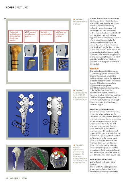
FIGURE 1.<br />
Description <strong>of</strong> the<br />
method.<br />
▼<br />
FIGURE 2.<br />
Reference po<strong>in</strong>ts<br />
for the PHILOS<br />
plate <strong>and</strong> the nonfixated<br />
humerus.<br />
▼<br />
m<strong>in</strong>eral density, bone tissue m<strong>in</strong>eral<br />
density <strong>and</strong> bone volume fraction,<br />
while BMA is def<strong>in</strong>ed by trabecular<br />
thickness, trabecular number,<br />
trabecular separation, degree <strong>of</strong><br />
anisotropy <strong>and</strong> structural model<br />
<strong>in</strong>dex. This method assesses the BMD<br />
<strong>and</strong> BMA <strong>in</strong> the cancellous bone<br />
regions where the anchor<strong>in</strong>g elements<br />
<strong>of</strong> an implant (<strong>in</strong> our study, the<br />
lock<strong>in</strong>g screws) are to be placed,<br />
before the actual fixation is carried<br />
out. By identify<strong>in</strong>g the directions <strong>in</strong><br />
bone where the best purchase can be<br />
achieved, the implant design can be<br />
optimised. The method is applicable<br />
to any lock<strong>in</strong>g implant, <strong>and</strong> we have<br />
tested its feasibility on a lock<strong>in</strong>g<br />
proximal humeral plate available on<br />
the market.<br />
METHOD<br />
The method consists <strong>of</strong> four steps:<br />
(1) temporary, partial fixation <strong>of</strong> the<br />
plate to the humeral shaft us<strong>in</strong>g<br />
lock<strong>in</strong>g screws (outside the region <strong>of</strong><br />
<strong>in</strong>terest) <strong>in</strong> order to def<strong>in</strong>e a reference<br />
system, (2) implant removal <strong>and</strong><br />
high-resolution peripheral<br />
quantitative computed tomography<br />
(HR-pQCT) <strong>of</strong> the bone, (3)<br />
determ<strong>in</strong>ation <strong>of</strong> BMD <strong>and</strong> BMA<br />
along the implant-anchor<strong>in</strong>g locations<br />
(with<strong>in</strong> the region <strong>of</strong> <strong>in</strong>terest) <strong>and</strong> (4)<br />
evaluation <strong>of</strong> alternative, optimised<br />
directions for implant-anchor<strong>in</strong>g<br />
locations (figure 1).<br />
FIGURE 3.<br />
Implant<br />
optimisation<br />
procedure by<br />
assess<strong>in</strong>g the<br />
local bone<br />
properties along<br />
alternative paths<br />
for each screw.<br />
▼<br />
Reference system def<strong>in</strong>ition<br />
We def<strong>in</strong>ed two co-ord<strong>in</strong>ate systems,<br />
one for the plate <strong>and</strong> one for the<br />
specimen. Two sets <strong>of</strong> three unaligned<br />
reference po<strong>in</strong>ts on the correspond<strong>in</strong>g<br />
3D reconstruction were retrieved<br />
(figure 2). For the plate, the first<br />
reference po<strong>in</strong>t (A) was the most<br />
distal lock<strong>in</strong>g hole, the second<br />
reference po<strong>in</strong>t (B) was the second<br />
most distal lock<strong>in</strong>g hole <strong>and</strong> the third<br />
reference (C) po<strong>in</strong>t was the tip <strong>of</strong> the<br />
lock<strong>in</strong>g screw <strong>in</strong> the second most<br />
distal hole. For the bone, the first<br />
reference po<strong>in</strong>t (A) was the most<br />
distal hole on its medial side, the<br />
second reference po<strong>in</strong>t (B) was the<br />
second most distal hole on its lateral<br />
side <strong>and</strong> the third reference po<strong>in</strong>t (C)<br />
was this same hole on its medial side.<br />
Virtual screw position <strong>and</strong><br />
evaluation <strong>of</strong> peri-screw bone<br />
quality<br />
The co-ord<strong>in</strong>ates <strong>of</strong> the proximal<br />
screws were collected from the<br />
absolute co-ord<strong>in</strong>ate system <strong>of</strong> the<br />
10 | MARCH <strong>2012</strong> | SCOPE