Application note: White Light Laser - Department of Cell Biology and ...
Application note: White Light Laser - Department of Cell Biology and ...
Application note: White Light Laser - Department of Cell Biology and ...

Create successful ePaper yourself
Turn your PDF publications into a flip-book with our unique Google optimized e-Paper software.
Fluorescence 1833 to 2008+<br />
In 1833, D. Brewster found a blood-red light in a<br />
solution <strong>of</strong> Chlorophyll at the edge <strong>of</strong> a focusing<br />
cone <strong>and</strong> in 1845, J. Herschel discovered a bluish<br />
shimmer when a quinine solution was observed<br />
in the bright sun. G. Stokes repeated those experiments<br />
<strong>and</strong> raised the first theory on the phenomenon<br />
<strong>and</strong> also coined the term Fluorescence<br />
(G. Stokes, 1852) 1 . At that time, the sun was the<br />
best light source for short wavelength, <strong>and</strong> the<br />
excitation filter was a piece <strong>of</strong> blue church glass.<br />
Emission could be separated in “transmitted fluorescence<br />
mode”, as we would call it today, when<br />
the object was looked at through a glass <strong>of</strong> dark<br />
yellow wine – the barrier filter. The fluorescent<br />
dye they observed was quinine, the working ingredient<br />
in tonic water that prevents <strong>and</strong> cures<br />
malaria fever. Quinine has a broad absorption in<br />
the UV-blue range <strong>and</strong> emits green-yellow light.<br />
Commercial Tonic Water is therefore a very popular<br />
specimen to introduce beginners into the<br />
world <strong>of</strong> fluorescence effects.<br />
Fluorescence was introduced as a new method<br />
in microscopy in the beginning <strong>of</strong> the 20th century,<br />
<strong>and</strong> the first commercially available fluorescence<br />
microscope was produced by Reichert in<br />
Vienna 1911. At that time, Fluorescence usually<br />
was done only with UV-illumination, using Cobaltglass<br />
in front <strong>of</strong> bright arc-lamps as excitation<br />
source. The samples were stained materials, that<br />
empirically were found to fluoresce, <strong>and</strong> mineral<br />
samples. At this time, the most famous biological<br />
stain available was Feulgen-stain for DNA.<br />
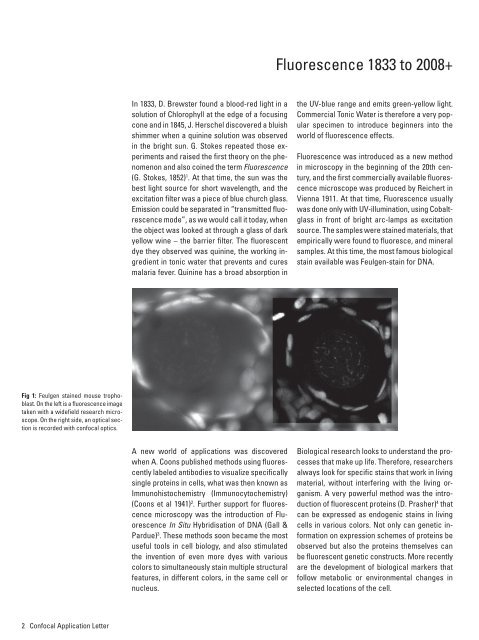
Fig 1: Feulgen stained mouse trophoblast.<br />
On the left is a fluorescence image<br />
taken with a widefield research microscope.<br />
On the right side, an optical section<br />
is recorded with confocal optics.<br />
A new world <strong>of</strong> applications was discovered<br />
when A. Coons published methods using fluorescently<br />
labeled antibodies to visualize specifically<br />
single proteins in cells, what was then known as<br />
Immunohistochemistry (Immunocytochemistry)<br />
(Coons et al 1941) 2 . Further support for fluorescence<br />
microscopy was the introduction <strong>of</strong> Fluorescence<br />
In Situ Hybridisation <strong>of</strong> DNA (Gall &<br />
Pardue) 3 . These methods soon became the most<br />
useful tools in cell biology, <strong>and</strong> also stimulated<br />
the invention <strong>of</strong> even more dyes with various<br />
colors to simultaneously stain multiple structural<br />
features, in different colors, in the same cell or<br />
nucleus.<br />
Biological research looks to underst<strong>and</strong> the processes<br />
that make up life. Therefore, researchers<br />
always look for specific stains that work in living<br />
material, without interfering with the living organism.<br />
A very powerful method was the introduction<br />
<strong>of</strong> fluorescent proteins (D. Prasher) 4 that<br />
can be expressed as endogenic stains in living<br />
cells in various colors. Not only can genetic information<br />
on expression schemes <strong>of</strong> proteins be<br />
observed but also the proteins themselves can<br />
be fluorescent genetic constructs. More recently<br />
are the development <strong>of</strong> biological markers that<br />
follow metabolic or environmental changes in<br />
selected locations <strong>of</strong> the cell.<br />
2 Confocal <strong>Application</strong> Letter