Application note: White Light Laser - Department of Cell Biology and ...
Application note: White Light Laser - Department of Cell Biology and ...
Application note: White Light Laser - Department of Cell Biology and ...

You also want an ePaper? Increase the reach of your titles
YUMPU automatically turns print PDFs into web optimized ePapers that Google loves.
D y e S e p a r a t i o n<br />
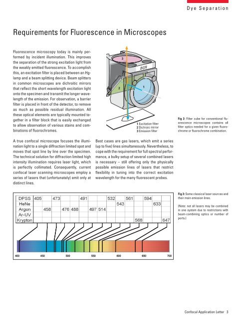
Requirements for Fluorescence in Microscopes<br />
Fluorescence microscopy today is mainly performed<br />
by incident illumination. This improves<br />
the separation <strong>of</strong> the strong excitation light from<br />
the weakly emitted fluorescence. To accomplish<br />
this, an excitation filter is placed between an Hglamp<br />
<strong>and</strong> a beam splitting device. Beam splitters<br />
in common microscopes are dichroitic mirrors<br />
that reflect the short wavelength excitation light<br />
onto the specimen <strong>and</strong> transmit the longer wavelength<br />
<strong>of</strong> the emission. For observation, a barrier<br />
filter is placed in front <strong>of</strong> the detector, to remove<br />
as much as possible residual illumination. All<br />
these optical elements are typically mounted together<br />
in a filter block that is easily exchanged<br />
to allow observation <strong>of</strong> various stains <strong>and</strong> combinations<br />
<strong>of</strong> fluorochromes.<br />
2<br />
3<br />
1<br />
1 Excitation filter<br />
2 Dichroic mirror<br />
3 Emission filter<br />
Fig 2: Filter cube for conventional fluorescence<br />
microscopes contains all<br />
filter optics needed for a given fluorochrome<br />
or fluorochrome combination.<br />
A true confocal microscope focuses the illumination<br />
light to a single diffraction limited spot <strong>and</strong><br />
moves that spot line by line over the specimen.<br />
The technical solution for diffraction limited high<br />
intensity illumination requires laser light, which<br />
is perfectly collimated. Consequently, current<br />
confocal laser scanning microscopes employ a<br />
series <strong>of</strong> lasers that (unfortunately) emit only at<br />
distinct lines.<br />
Best cases are gas lasers, which emit a series<br />
(up to five) lines simultaneously. Nevertheless, to<br />
cope with the requirement for full spectral performance,<br />
a bulky setup <strong>of</strong> several combined lasers<br />
is necessary – still <strong>of</strong>fering only the physically<br />
possible emission lines <strong>of</strong> lasers that restrict<br />
flexibility in tuning into the correct excitation<br />
wavelength for the many fluorescent probes.<br />
Fig 3: Some classical laser sources <strong>and</strong><br />
their main emission lines.<br />
(Note: not all lasers may be combined<br />
in one system due to restrictions with<br />
beam-combining optics or number <strong>of</strong><br />
ports.)<br />
400 450 500 550 600 650 700<br />
Confocal <strong>Application</strong> Letter 3