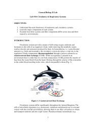
The Microscope & Cell Structure and Function
The Microscope & Cell Structure and Function
The Microscope & Cell Structure and Function

You also want an ePaper? Increase the reach of your titles
YUMPU automatically turns print PDFs into web optimized ePapers that Google loves.
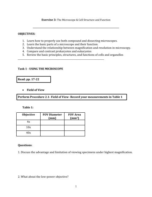
OBJECTIVES:<br />
Exercise 3: <strong>The</strong> <strong>Microscope</strong> & <strong>Cell</strong> <strong>Structure</strong> <strong>and</strong> <strong>Function</strong><br />
______________________________________________________________________________<br />
1. Learn how to properly use both compound <strong>and</strong> dissecting microscopes.<br />
2. Learn the basic parts of a microscope <strong>and</strong> their function.<br />
3. Underst<strong>and</strong> the relationship between magnification <strong>and</strong> resolution in microscopy.<br />
4. Compare <strong>and</strong> contrast prokaryotes <strong>and</strong> eukaryotes<br />
5. Review the basic principles, structures, <strong>and</strong> functions of cells <strong>and</strong> organelles<br />
______________________________________________________________________________<br />
Task 1 - USING THE MICROSCOPE<br />
Read: pp. 17-22<br />
<br />
Field of View<br />
Perform Procedure 2.1- Field of View- Record your measurements in Table 1<br />
Table 1:<br />
Objective<br />
4x<br />
10x<br />
40x<br />
FOV Diameter<br />
(mm)<br />
FOV Area<br />
(mm 2 )<br />
Questions:<br />
1. Discuss the advantage <strong>and</strong> limitation of viewing specimens under highest magnification.<br />
2. What about the low-power objective<br />
1
3. Which magnification provides the largest FOV Which provides the smallest<br />
<br />
DEPTH OF FIELD<br />
Perform depth of field procedure on pp.20<br />
Questions:<br />
4. How does depth of field affect viewing biological phenomena that are thick<br />
5. Are all three threads visible under the low power Can they all be seen at the same time<br />
under higher power<br />
6. Which objective provides the greatest depth of field<br />
<br />
Preparing Wet Mounts of Biological Specimens<br />
Perform the wet mount preparation using periphyton procedure on pp.20<br />
Sketch your scientific drawing in the space below<br />
2
Periphyton wet mount<br />
Magnification: _________<br />
Figure 1. MICROSCOPE TIPS:<br />
This is an air bubble, NOT your specimen<br />
<strong>The</strong>se are cotton fibers, not your specimen<br />
Procedure: Estimating the size of an object under the microscope.<br />
1. Use the diameter of the field of view for the power of magnification you are<br />
using.<br />
2. Look through the ocular lens; estimate how many times the object will fit across<br />
the field of view. Pick two organisms in the FOV to perform this procedure.<br />
3. Calculate the size of the object using the formula below<br />
3
Organism #1 Organism #2<br />
Magnification: _________<br />
Magnification: ________<br />
<br />
F. Dissecting <strong>Microscope</strong><br />
Perform the dissecting microscope procedure on pp.21<br />
Ocular<br />
Lens<br />
Zoom<br />
Magnification<br />
Adjustment<br />
Arm<br />
Focus<br />
Adjustment<br />
Transmitted<br />
Light Source<br />
Stage<br />
Base<br />
Figure 2. Major parts of a dissecting microscope<br />
4
Periphyton<br />
Magnification: ________<br />
7. Use a ruler to measure the FOV diameter at the lowest <strong>and</strong> the highest<br />
magnification.<br />
FOV diameter low power = _____________<br />
FOV diameter high power = _____________<br />
8. Now calculate the FOV area for both magnifications.<br />
FOV area low power = _____________<br />
FOV area high power = _____________<br />
9. Now calculate the size of an organism viewed under the microscope:<br />
10. Looking through the lens, move the Petri dish containing the “pond water”<br />
backwards <strong>and</strong> forwards, then left <strong>and</strong> right. Is the direction noted through the lens<br />
the same as when observed with the naked eye<br />
11. How does the image move when the slide is moved to the left or right<br />
5
G. Comparison of Compound <strong>and</strong> Dissecting <strong>Microscope</strong>s<br />
Compare the two types of microscopes we examined today in Table 10.<br />
Table 10:<br />
Characteristic<br />
Magnification<br />
Resolution<br />
Size of field of view<br />
Depth of field<br />
Dissecting<br />
<strong>Microscope</strong><br />
Light <strong>Microscope</strong><br />
Task 2: <strong>Cell</strong>ular <strong>Structure</strong>s & <strong>Function</strong>s<br />
READ: pp.23-27<br />
Examine Cyanobacteria:<br />
1. Examine a prepared slide of Oscillatoria <strong>and</strong> one of Gloeocapsa. Sketch each organism in<br />
the space provided below. Note the magnification at which you viewed your specimens.<br />
Oscillatoria<br />
Gloeocapsa<br />
Mag: ________<br />
Mag: ________<br />
6
2. On clean slides, prepare a wet mount of Oscillatoria <strong>and</strong> another of Gloeocapsa.<br />
Compare your observations to those previously visualized using prepared slides.<br />
OBSERVATIONS OF CYANOBACTERIA WET MOUNTS:<br />
Questions:<br />
12. Were you able to locate nuclei in either species Did you expect to Explain.<br />
13. Is Oscillatoria a prokaryote or eukaryote Gloeocapsa Explain your reasoning.<br />
14. Are there chloroplasts present in Oscillatoria Gloeocapsa<br />
15. Where are the pigments located in the cyanobacteria that you examined Are they<br />
present throughout the entire organism or only in certain locations<br />
16. How does the cell morphology differ between the two species of cyanobacteria<br />
17. How many cells are held together within one sheath of Gloeocapsa<br />
7
_____________________________________________________________________________<br />
Task 3 - EUKARYOTIC CELLS<br />
Questions:<br />
18. Based on the endosymbiotic theory, some of the eukaryotic organelles have<br />
prokaryotic origins. Which organelles do you think these are <strong>and</strong> why<br />
19. What evidence would you need to gather to support the endosymbiotic theory<br />
Explain how each piece of evidence would support the theory. (Hint: consider the<br />
structure of the modern eukaryotic cell)<br />
A. Plant cells<br />
<br />
Perform Procedure 3.1: Examine Elodea cells<br />
Mag: _______<br />
8
Questions:<br />
20. What shape are the Elodea cells Are they round or do they have distinct sides<br />
21. Try to determine how many cells thick your leaf is by focusing up <strong>and</strong> down through<br />
the layers of cells.<br />
22. What cell structures are visible What are the functions of these structures Make<br />
sure to label these structures in your scientific drawing above.<br />
23. Why do you think plants have cell walls Why are cell walls absent in animal cells<br />
24. Locate the chloroplasts within the cells. Try to estimate of the number present in<br />
one cell. Where in the cell do you generally find the chloroplasts<br />
25. Locate the central vacuole. Since it contains water, what should you see within it<br />
Should there be any shapes or colors This information should help you to locate it.<br />
26. Locate the nucleus. It may be pressed against the edge of the cell by the vacuole <strong>and</strong><br />
may appear gray compared to the surrounding chloroplasts which are green. To<br />
enhance visibility, add a drop of iodine to the slide.<br />
9
II. Perform Procedure 3.2 -Examine onion cells:<br />
27. For mitochondria to stain well, the onion cells must be healthy <strong>and</strong> metabolically<br />
active. Why would this have to be the case<br />
Are there any visible chloroplast Explain your reasoning.<br />
28. Draw what you see in the space provided make sure to label all visible structures:<br />
Janus B Green Stain<br />
Mag: ______<br />
Neutral Red Stain<br />
Mag: ______<br />
B. Animal cells<br />
I. Perform Procedure 3.3- Examine human epithelial cells<br />
1. Sketch a few of the cells in the space provided. Label any visible cellular structures<br />
on your scientific drawing.<br />
10
Mag: _______<br />
Question:<br />
29. How did the cheek cells differ from the onion or Elodea cells How were they<br />
similar<br />
11