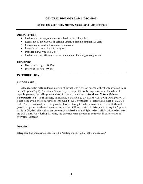
Lab #6: The Cell Cycle, Mitosis, Meiosis and Gametogenesis ...
Lab #6: The Cell Cycle, Mitosis, Meiosis and Gametogenesis ...
Lab #6: The Cell Cycle, Mitosis, Meiosis and Gametogenesis ...

Create successful ePaper yourself
Turn your PDF publications into a flip-book with our unique Google optimized e-Paper software.
GENERAL BIOLOGY LAB 1 (BSC1010L)<br />
<strong>Lab</strong> <strong>#6</strong>: <strong>The</strong> <strong>Cell</strong> <strong>Cycle</strong>, <strong>Mitosis</strong>, <strong>Meiosis</strong> <strong>and</strong> <strong>Gametogenesis</strong><br />
OBJECTIVES:<br />
• Underst<strong>and</strong> the major events involved in the cell cycle<br />
• Learn about the process of cellular division in plant <strong>and</strong> animal cells<br />
• Compare <strong>and</strong> contrast mitosis <strong>and</strong> meiosis<br />
• Learn how to examine a karyogram<br />
• Perform karyotype analysis<br />
• Underst<strong>and</strong> the difference between male <strong>and</strong> female gametogenesis<br />
READINGS:<br />
• Exercise 14: pgs 149-156<br />
• Exercise 15: pgs 159-165<br />
______________________________________________________________________________<br />
INTRODUCTION:<br />
<strong>The</strong> <strong>Cell</strong> <strong>Cycle</strong>:<br />
All eukaryotic cells undergo a series of growth <strong>and</strong> division events, collectively referred to as<br />
the cell cycle (Fig 1). Duration of the cell cycle is specific to the organism as well as the cell<br />
type. In general, the cell cycle consists of three main phases: Interphase, <strong>Mitosis</strong> (M) <strong>and</strong><br />
Cytokenesis (C). <strong>The</strong> first stage, Interphase, is considered the non-dividing or growth portion of<br />
a cell’s life cycle <strong>and</strong> is subdivided into Gap 1 (G1), Synthesis (S) phase, <strong>and</strong> Gap 2 (G2). G1<br />
<strong>and</strong> G2 are considered the main growth phases. During G1 (the normal state of a cell), the cell<br />
grows <strong>and</strong> generates the enzymes necessary for DNA replication to take place during the S phase<br />
while in G2, the cell synthesizes proteins, carbohydrates <strong>and</strong> lipids which all function to increase<br />
the cell’s size. Also during this time, the chromosomes prepare to condense in anticipation of<br />
entry into M phase.<br />
Question:<br />
Interphase has sometimes been called a “resting stage.” Why is this inaccurate<br />
1
<strong>The</strong> cell cycle is controlled by a series of checkpoints (Fig 1), namely the G1/S, G2/M<br />
<strong>and</strong> spindle checkpoints. <strong>The</strong> G1/S checkpoint, determines if the cell should continue into the S<br />
phase or if it should enter a resting state (G 0 = Gap 0 phase), which is important for some cell<br />
types that divide very infrequently <strong>and</strong>/or for cells that are already terminally differentiated (e.g.<br />
nerve cells). This checkpoint is followed by the G2/M checkpoint which serves as a control<br />
mechanism to prevent damaged cells from entering the M phase. Once the cells are committed to<br />
mitosis, the role of the spindle checkpoint is to ensure that all chromosomes are attached to the<br />
mitotic spindle during metaphase; if any chromosome is not attached, the cell will not be able to<br />
proceed into anaphase. In addition, DNA damage checkpoints located in G1, S <strong>and</strong> G2 ensure<br />
that the DNA is not damaged before allowing the cell to proceed into mitosis. If any of these<br />
checkpoints are nonfunctional or mutated, control of the cell cycle is lost <strong>and</strong> cancer develops.<br />
G1/S Checkpoint<br />
G2/M Checkpoint<br />
Spindle Checkpoint<br />
Figure 1. <strong>The</strong> cell cycle <strong>and</strong> its associated checkpoints<br />
2
Question:<br />
How might you use the knowledge of the cell cycle checkpoints to prevent, diagnose, <strong>and</strong> treat<br />
cancer<br />
<strong>Cell</strong>ular Division: <strong>Mitosis</strong> vs. <strong>Meiosis</strong><br />
<strong>The</strong> genetic material (DNA) of all eukaryotic organisms is housed within the cell’s<br />
nucleus <strong>and</strong> is passed on from generation to generation. While a cell is in interphase, the DNA<br />
exists in an extended form called chromatin (Fig 2). However, when the cell is ready to divide<br />
(i.e., it enters the M phase of the cell cycle), the DNA repeatedly folds on top of itself,<br />
condensing into visible chromosomes. <strong>The</strong> chromosomes exist in pairs <strong>and</strong> are called<br />
homologous chromosomes. Each homologue within the pair is called a sister chromatid <strong>and</strong> is<br />
joined to the other by the centromere (Fig 3). In eukaryotic organisms, the number of<br />
chromosomes present differs between species but most eukaryotes are diploid (2n), i.e., they<br />
have 2 sets of chromosomes. For example, human cells possess a total of 46 chromosomes (23<br />
chromosomes per set) while canine cells possess 78 chromosomes (39 chromosomes per set).<br />
Figure 2. <strong>Cell</strong> as it appears during Interphase<br />
3
Figure 3. A Pair of Sister Chromatids<br />
Eukaryotic cells, depending on the type (somatic vs. germ cell), divide by either mitosis<br />
or meiosis. <strong>Mitosis</strong> is the process in which a diploid (2n) parental cell is divided into 2 identical<br />
daughter cells, also diploid in number. In contrast, meiosis involves the division of a diploid<br />
parental cell into 4 daughter cells, all of which are haploid (n). <strong>Mitosis</strong> occurs in somatic cells,<br />
which are all cells of the body excluding the reproductive cells (i.e. eggs <strong>and</strong> sperm). <strong>Meiosis</strong>, on<br />
the other h<strong>and</strong>, only occurs in the germ cells, which are cells of the reproductive organs (i.e.<br />
testes <strong>and</strong> ovaries).<br />
<strong>Mitosis</strong> is comprised of 4 stages, Prophase, Metaphase, Anaphase <strong>and</strong> Telophase<br />
(although some authors describe Prometaphase as a distinct phase). Following nuclear division,<br />
cytokenesis (division of the cytoplasm) begins. <strong>The</strong> primary function of this process is to<br />
completely separate the 2 newly generated daughter cells from each other. In animal cells a<br />
cleavage furrow or indentation in the middle area of the cell develops <strong>and</strong> divides the cell into<br />
two parts. Plant cells, in contrast, are unable to divide using the cleavage furrow since they<br />
possess a cell wall. Instead they generate a cell plate at the center of the cell that splits the cell<br />
into two.<br />
<strong>Meiosis</strong>, on the other h<strong>and</strong>, occurs only in germ cells, i.e., those destined to become the<br />
gametes. This process is referred to as a reduction division since the 4 daughter cells generated<br />
from the division of the diploid parental cell are haploid. <strong>The</strong> stages of <strong>Meiosis</strong> I are Prophase I,<br />
Metaphase I, Anaphase I <strong>and</strong> Telophase I <strong>and</strong> of <strong>Meiosis</strong> II are Prophase II, Metaphase II,<br />
Anaphase II <strong>and</strong> Telophase II (Fig 4a <strong>and</strong> b). <strong>Meiosis</strong> I involves the separation of homologous<br />
pairs of chromosomes that are then separated into sister chromatids during <strong>Meiosis</strong> II.<br />
4
MEIOSIS I<br />
Figure 4a. <strong>Meiosis</strong> I<br />
5
MEIOSIS II<br />
Figure 4b. <strong>Meiosis</strong> II<br />
6
In general, <strong>Meiosis</strong> I is very similar to <strong>Mitosis</strong> except that (1) Prophase I involves<br />
synapsis (forms a tetrad) <strong>and</strong> crossing over (Fig 5) occurs between homologous pairs of<br />
chromosomes <strong>and</strong> (2) the homologous pairs of chromosomes are separated during Anaphase I.<br />
Figure 5. Crossing Over between Homologous Pairs of Chromosomes<br />
<strong>Gametogenesis</strong>:<br />
Gametes are reproductive cells with haploid nuclei that result from meiosis <strong>and</strong> are<br />
formed by gametogenesis. In mammals <strong>and</strong> many other vertebrates, gametes <strong>and</strong> gametogenesis<br />
differ between males <strong>and</strong> females. Males produce sperm through the process of<br />
spermatogenesis (Fig 6), while females produce eggs via oogenesis (Fig 7).<br />
Sperm is produced in the seminiferous tubules of the testes. Within the seminiferous<br />
tubules spermatogonia constantly replicate mitotically throughout the life cycle of males. Some<br />
of these spermatogonia move inward towards the lumen of the tubule <strong>and</strong> begin meiosis. <strong>The</strong>se<br />
spermatogonia are called primary spermatocytes. <strong>Meiosis</strong> I of a primary spermatocyte<br />
produces two secondary spermatocytes, each with a haploid set of double-str<strong>and</strong>ed<br />
7
chromosomes. <strong>Meiosis</strong> II separates the str<strong>and</strong>s of each chromosome <strong>and</strong> produces two haploid<br />
spermatids that mature <strong>and</strong> differentiate into sperm cells.<br />
Figure 6. Spermatogenesis<br />
In females, oogenesis occurs in the oocytes of the ovaries. Unlike spermatogonia,<br />
oocytes are not produced continuously. Oogonia, which are produced during early fetal<br />
development, reproduce mitotically to produce primary oocytes. In humans, the ovaries of a<br />
newborn female contain all the primary oocytes that she will ever have. At birth, primary oocytes<br />
begin meiosis I, but are arrested in prophase I. At puberty, circulating hormones will stimulate<br />
growth of the primary oocytes in the follicles (surrounding tissue) each month. Just before<br />
ovulation, the oocyte completes meiosis I producing a Graafian follicle which consists of the<br />
secondary oocyte. Each secondary oocyte contains a haploid set of double-str<strong>and</strong>ed<br />
chromosomes. <strong>Meiosis</strong> II proceeds but is not completed until fertilization occurs.<br />
8
Figure 7. Oogenesis<br />
Review the basic stages of spermatogenesis <strong>and</strong> oogenesis on pages 162-165 <strong>and</strong> answer the<br />
questions that follow.<br />
Questions:<br />
1. Why do gametes only have half the number of chromosomes as the original parent cell<br />
Why is this important<br />
2. Would evolution occur without the events of meiosis <strong>and</strong> sexual reproduction Why or<br />
why not<br />
9
In today’s lab, we will examine the cell cycle, mitosis <strong>and</strong> meiosis. We will then consider<br />
the role of the different phases in the cell cycle to better underst<strong>and</strong> the significance of each step<br />
in the production of healthy cells without DNA damage. We will also consider the consequences<br />
when specific aspects of cell division fail to function properly. Finally, we will learn how to<br />
examine karyotypes which are used to determine the number of chromosomes in a species as<br />
well as for the diagnosis of birth defects or genetic abnormalities.<br />
TASK 1: Cycling Through the <strong>Cell</strong> <strong>Cycle</strong><br />
A) Stages of <strong>Mitosis</strong><br />
1. Examine a prepared slide of the whitefish blastula on high power.<br />
2. Complete Table 1, making sure to draw examples of each phase of mitosis.<br />
Table 1:<br />
Stage of <strong>Mitosis</strong> Description of Events Drawings of Stages<br />
Prophase<br />
Metaphase<br />
Anaphase<br />
Telophase<br />
10
Questions:<br />
a. Why are cells from a blastula used to examine mitosis<br />
b. How fast do you think cells divide when an embryo is forming compared to the normal<br />
growth of an animal<br />
c. How does mitosis differ between plant <strong>and</strong> animal cells<br />
B) Time for <strong>Cell</strong>ular Replication<br />
1. Using the prepared onion root tip slide, count the number of cells in each phase of the cell<br />
cycle (i.e., interphase <strong>and</strong> mitosis) in the high power field of view. Repeat 3 times for an<br />
approximate total of 100 cells <strong>and</strong> record your results in Table 2.<br />
2. Assuming that an onion root tip cell takes 14 hours (840 minutes) to complete the cell cycle,<br />
the time that an onion cell spends in each stage of the cell cycle can be calculated using the<br />
following formula:<br />
Time for each stage = Number of cells at each stage x 840 minutes<br />
Total number of cells counted<br />
11
Stage of <strong>Cell</strong><br />
<strong>Cycle</strong><br />
Number of <strong>Cell</strong>s<br />
FOV 1 FOV 2 FOV 3 FOV 4 Total<br />
Time Spent in Each Stage<br />
Interphase<br />
Prophase<br />
Metaphase<br />
Anaphase<br />
Telophase<br />
TASK 2: Effect of Colchicine on <strong>Mitosis</strong><br />
Colchicine, a product of the plant Colchicum autumnale (common name = Meadow<br />
saffron), is well documented for its use in the treatment of gout, cirrhosis, <strong>and</strong> psoriasis, among<br />
other disorders (Ben-Chetrit <strong>and</strong> Levy, 1998). This compound is known to interact with tubulin,<br />
a component of the spindle fibers. During metaphase, the chromosomes attach to the mitotic<br />
spindle via their kinetochore <strong>and</strong> oscillate at the equatorial region under high tension. Colchicine<br />
decreases this tension, therefore suspending the chromosomes in metaphase (Jordan <strong>and</strong> Wilson,<br />
2004).<br />
Based on the information above, propose a null <strong>and</strong> alternate hypotheses about the stages<br />
of mitosis that you expect to see in the onion cells treated with colchicine.<br />
Ho:<br />
Ha:<br />
Procedure:<br />
1. Each student in the group will prepare an onion root tip slide as follows:<br />
a. To a watch glass add 5-10 drops 1M HCl.<br />
b. Using a scalpel cut the terminal 4mm of an onion root tip grown in a dilute<br />
(0.05%) colchicine solution <strong>and</strong> add it to the acid to soften the tip. Leave the tip in<br />
the acid for about 5 minutes.<br />
12
c. Add 1 drop of acetocarmine stain to a clean slide.<br />
d. Once softened, cut the root tip in half <strong>and</strong> add one half (2mm) to the slide with the<br />
acetocarmine stain.<br />
e. Use the scalpel to chop the tip into numerous pieces <strong>and</strong> then crush the pieces<br />
with a glass rod.<br />
f. Apply a cover slide to the slide <strong>and</strong> then gently warm by passing the slide over the<br />
ethanol lamp. DO NOT BOIL!!!<br />
g. Invert the slide onto a clean piece of tissue/paper towel <strong>and</strong> push down firmly<br />
with the thumb to flatten <strong>and</strong> disperse the cells.<br />
13
h. Examine the slide using the high power objective<br />
i. Count the number of cells observed in metaphase in 1 high power field of view.<br />
Repeat this step for an additional 2 high power field of views.<br />
j. Seal the cover slip to the slide by coating the edges with clear nail polish.<br />
k. Examine chromosome morphology using the oil immersion lens (See Procedure<br />
below) by selecting cells in anaphase. Count the number of chromosomes that you<br />
see at one pole since this represents the diploid number for the species. Record the<br />
number from each cell in the Table 2. Make sure to move the focus from the top layer<br />
of the cell downwards as you scan for the chromosomes.<br />
Working with the Oil immersion lens: (Adopted from Dolphin, 2005)<br />
CAUTION: Oil immersion lenses allow you to approach 1000X magnifications with an<br />
increase in resolution. However, the distance from the lens surface to the slide is very<br />
small, <strong>and</strong> it is quite easy to push the lens through the slide, possibly breaking the slide <strong>and</strong><br />
ruining the lens.<br />
Procedure:<br />
1. Focus first on the object on the slide by proceeding from the scanning (4X) to<br />
high-power (40X) objectives as you have done before. Now you are ready to<br />
try the oil immersion lens.<br />
2. Do NOT touch the focus knobs or the stage knobs. Turn the turret to swing the<br />
high-power (40X) objective out of the way. Place a single drop of immersion<br />
oil on the slide right over where the light is coming through the stage, <strong>and</strong><br />
rotate the oil immersion lens into place. <strong>The</strong> lens will actually contact the oil<br />
drop, making a column of oil from the slide surface to the lens surface. This<br />
column of oil prevents light scattering <strong>and</strong> improves resolution.<br />
3. Now look through the oculars <strong>and</strong> open the substage diaphragm to increase the<br />
light. <strong>The</strong> object on the slide should still be in the field of vision but will<br />
probably be out of focus. Use the fine-adjustment knob to focus clearly.<br />
Never use the coarse-adjustment knob!!!!<br />
4. Once you have an oil immersion lens in place, do NOT swing the 40X<br />
objective back into place. Because the objective focuses close to the slide, the<br />
40X objective will get oil on it <strong>and</strong> it is difficult to clean the oil from the lens<br />
surface.<br />
5. When you have finished using the oil immersion objective, you must clean the<br />
oil from its surface, as well as from the slide, using lens cleaner <strong>and</strong> lens paper.<br />
Because oil immersion lenses require extra cleanup <strong>and</strong> the danger of breaking<br />
slides is great, this is the only time during the semester that we will use them.<br />
14
Table 2:<br />
<strong>Cell</strong> # # of Chromosomes <strong>Cell</strong> # # of Chromosomes<br />
1 6<br />
2 7<br />
3 8<br />
4 9<br />
5 10<br />
Questions:<br />
1. Are any mitotic stages present that were not observed in the preserved onion root tip slide<br />
Are there any mitotic stages that are completely absent<br />
2. Compare the stages of mitosis that you observed in the colchicine-treated <strong>and</strong> untreated<br />
onion cells (preserved slide). What do your results suggest about your hypothesis<br />
3. Can you discern how many chromosomes are present in one cell at anaphase If so, how<br />
many<br />
4. Given colchicine’s properties, could this compound be used to prevent cancer Explain.<br />
15
TASK 3: Karyotype Analysis<br />
Karyotyping refers to the process by which scientists are able to microscopically<br />
visualize the complete set of chromosomes in an organism. Karyotype analysis is performed<br />
when the chromosomes are the most highly condensed, i.e. in metaphase (halted by the addition<br />
of colichicine), in order to determine the number of chromosomes present in the individual as<br />
well as to detect the presence of any chromosomal abnormalities such as deletions, translocations<br />
or the insertion of extra copies. A normal human karyotype should consist of 22 autosome pairs,<br />
listed from largest (chromosome 1) to smallest (chromosome 22), <strong>and</strong> 1 pair of sex<br />
chromosomes; XX if female <strong>and</strong> XY if male (See Fig 8). Known abnormalities that result from<br />
variations in normal chromosome structure or number in humans include:<br />
a. Downs Syndrome: Three copies of chromosome 21<br />
b. Turner syndrome (in females one gains reduction in female characteristics, e.g.<br />
ovaries don’t produce eggs): One copy of the X chromosome<br />
c. Cri du chat (disease that affects larynx <strong>and</strong> nervous system, infants cry like a<br />
cat): Chromosome 5 is truncated<br />
d. Edwards syndrome (severe birth defects such as intestine growing outside the<br />
body cavity): Three copies of chromosome 18<br />
e. Patau Syndrome (severe birth defects including heart <strong>and</strong> nervous dysfunction):<br />
Three copies of chromosome 13<br />
Figure 8. Normal Human Karyotype<br />
16
Procedure:<br />
A fellow scientist of BCBB Cytogeneics was assigned the task of performing karyotype<br />
analysis for 2 newborn babies, but he needs a second opinion of the results before informing the<br />
parents. <strong>The</strong> karyotype for each baby is presented below. It is your task to examine both<br />
karyotypes (#1 <strong>and</strong> #2), record your findings in the tables provided <strong>and</strong> report them to your<br />
colleague.<br />
Karyotype #1:<br />
http://www.ratsteachgenetics.com/Genetics_quizzes/Lecture%207/7q4.jpg<br />
CH # Remarks CH# Remarks CH# Remarks CH# Remarks<br />
1 7 13 19<br />
2 8 14 20<br />
3 9 15 21<br />
4 10 16 22<br />
5 11 17 23<br />
6 12 18 24<br />
CH = Chromosome<br />
17
Karyotype #2:<br />
Chart for Infant Number Two:<br />
https://ccr.coriell.org/images/karyotype/gm18241-xyy.jpg<br />
CH # Remarks CH# Remarks CH# Remarks CH# Remarks<br />
1 7 13 19<br />
2 8 14 20<br />
3 9 15 21<br />
4 10 16 22<br />
5 11 17 23<br />
6 12 18 24<br />
CH = Chromosome<br />
Questions:<br />
1. Based on the karyotypes provided, do these babies have detectable problems in their<br />
chromosomes If yes, use that information to diagnose what disease/genetic abnormality the<br />
child has.<br />
Infant Number One Diagnosis: ________________<br />
Infant Number Two Diagnosis: ________________<br />
18
TASK 4: <strong>Meiosis</strong> <strong>and</strong> <strong>Gametogenesis</strong><br />
Procedure:<br />
1. Examine prepared slides of sperm from humans, rats, <strong>and</strong> guinea pigs. How do the sperm<br />
from the three species compare<br />
2. Examine a cross section of a monkey’s seminiferous tubules <strong>and</strong> draw what you see in the<br />
space provided below. Try to locate the spermatogonia, primary spermatocytes, secondary<br />
spermatocytes, spermatids <strong>and</strong> mature sperm.<br />
3. Examine a cross section of cat ovary <strong>and</strong> draw what you see in the space provided<br />
below. Try to locate the developing follicle with the egg inside.<br />
19
4. Examine the slide of a mature follicle (Graafian follicle) <strong>and</strong> draw what you see in the space<br />
provided below.<br />
5. Complete the table below:<br />
<strong>Mitosis</strong><br />
<strong>Meiosis</strong><br />
Purpose of process<br />
Location<br />
Number of cells generated per<br />
cycle<br />
Number of nuclear divisions per<br />
cycle<br />
Ploidy (n or 2n) of daughter<br />
cells<br />
Daughter cells genetically<br />
identical to parent<br />
Pairing of homologues<br />
Occurrence of crossing over<br />
20
Questions:<br />
1. Why is meiosis referred to as reduction division<br />
2. If a species has 24 chromosomes in the nucleus prior to meiosis, what number will each cell<br />
have after meiosis is complete<br />
3. How do the sizes of the oocytes differ as they move from the follicle stage towards the<br />
mature Graffian follicle<br />
4. How do sperm <strong>and</strong> eggs differ in size Why do you think this happens Why do you think<br />
there is such a difference in the number of each produced What would happen if females<br />
produced 100’s or 1000’s of eggs during each cycle What if males were born with a limited<br />
number of sperm<br />
21
HOMEWORK:<br />
Before coming to lab next week, make sure to read the Mendelian Genetics task sheet as well as<br />
Chapter 17 (pgs. 177-185) in your lab manual.<br />
References:<br />
Ben-Chetrit, E <strong>and</strong> Levy, M (1998) Colchicine: 1998 Update. Seminars in arthritis <strong>and</strong><br />
rheumatism 28: 48-59.<br />
Jordan, MA <strong>and</strong> Wilson, L (2004) Microtubules as a target for anticancer drugs. Nature reviews<br />
4: 253- 265.<br />
22