Modeling the human PTC bitter-taste receptor interactions with bitter ...
Modeling the human PTC bitter-taste receptor interactions with bitter ...
Modeling the human PTC bitter-taste receptor interactions with bitter ...

Create successful ePaper yourself
Turn your PDF publications into a flip-book with our unique Google optimized e-Paper software.
J Mol Model (2006) 12: 931–941<br />
DOI 10.1007/s00894-006-0102-6<br />
ORIGINAL PAPER<br />
Wely B. Floriano . Spencer Hall . Nagarajan Vaidehi .<br />
Unkyung Kim . Dennis Drayna .<br />
William A. Goddard III<br />
<strong>Modeling</strong> <strong>the</strong> <strong>human</strong> <strong>PTC</strong> <strong>bitter</strong>-<strong>taste</strong> <strong>receptor</strong> <strong>interactions</strong><br />
<strong>with</strong> <strong>bitter</strong> tastants<br />
Received: 27 September 2005 / Accepted: 16 January 2006 / Published online: 11 April 2006<br />
# Springer-Verlag 2006<br />
Abstract We employed <strong>the</strong> first principles computational<br />
method MembStruk and homology modeling techniques to<br />
predict <strong>the</strong> 3D structures of <strong>the</strong> <strong>human</strong> phenylthiocarbamide<br />
(<strong>PTC</strong>) <strong>taste</strong> <strong>receptor</strong>. This protein is a seven-transmembrane-domain<br />
G protein-coupled <strong>receptor</strong> that exists<br />
in two main forms worldwide, designated <strong>taste</strong>r and<br />
non<strong>taste</strong>r, which differ from each o<strong>the</strong>r at three aminoacid<br />
positions. 3D models were generated <strong>with</strong> and <strong>with</strong>out<br />
structural similarity comparison to bovine rhodopsin. We<br />
used computational tools (HierDock and ScanBindSite) to<br />
generate models of <strong>the</strong> <strong>receptor</strong> bound to <strong>PTC</strong> ligand to<br />
estimate binding sites and binding energies. In <strong>the</strong>se<br />
models, <strong>PTC</strong> binds at a site distant from <strong>the</strong> variant amino<br />
acids, and <strong>PTC</strong> binding energy was equivalent for both <strong>the</strong><br />
<strong>taste</strong>r and non<strong>taste</strong>r forms of <strong>the</strong> protein. These models<br />
suggest that <strong>the</strong> inability of <strong>human</strong>s to <strong>taste</strong> <strong>PTC</strong> is due to a<br />
failure of G protein activation ra<strong>the</strong>r than decreased<br />
binding affinity of <strong>the</strong> <strong>receptor</strong> for <strong>PTC</strong>. Amino-acid<br />
substitutions in <strong>the</strong> sixth and seventh transmembrane<br />
domains of <strong>the</strong> non<strong>taste</strong>r form of <strong>the</strong> protein may produce<br />
W. B. Floriano<br />
Biological Sciences Department,<br />
California State Polytechnic University Pomona,<br />
Pomona, CA 91768, USA<br />
S. Hall . N. Vaidehi . W. A. Goddard III<br />
Materials and Process Simulation Center (MSC),<br />
California Institute of Technology,<br />
Pasadena, CA 91125, USA<br />
e-mail: wag@wag.caltech.edu<br />
U. Kim<br />
Department of Biology, Kyungpook National University,<br />
Daegu 702-701, Republic of Korea<br />
D. Drayna (*)<br />
National Institute on Deafness and O<strong>the</strong>r Communication<br />
Disorders, National Institutes of Health,<br />
5 Research Court,<br />
Rockville, MD 20850, USA<br />
e-mail: drayna@nidcd.nih.gov<br />
Tel.: +1-301-4024930<br />
Fax: +1-301-8279637<br />
increased steric hindrance between <strong>the</strong>se two α-helices and<br />
reduce <strong>the</strong> motion of <strong>the</strong> sixth helix required for G protein<br />
activation.<br />
Keywords Phenylthiocarbamide . Bitter . Protein<br />
structure . G Protein-coupled <strong>receptor</strong> . Taste perception<br />
Introduction<br />
The T2R mammalian <strong>taste</strong> <strong>receptor</strong>s are seven-transmembrane<br />
(TM) domain G protein-coupled <strong>receptor</strong>s (GPCRs)<br />
[1, 2]. Extracellular <strong>bitter</strong> substances bind to <strong>the</strong>se<br />
<strong>receptor</strong>s and initiate neural signaling via activation of<br />
intracellular heterotrimeric G proteins [3]. Despite numerous<br />
studies on <strong>taste</strong> <strong>receptor</strong>s, <strong>the</strong>re remains a great deal of<br />
uncertainty in what characteristics of <strong>the</strong> molecules govern<br />
<strong>the</strong> perception of <strong>taste</strong>. Particularly interesting clues are<br />
given by <strong>the</strong> <strong>human</strong> <strong>taste</strong> sensitivity to <strong>the</strong> <strong>bitter</strong> compound<br />
phenylthiocarbamide (<strong>PTC</strong>), which for some individuals is<br />
intensely <strong>bitter</strong>, but for o<strong>the</strong>rs is largely <strong>taste</strong>less. Intense<br />
studies performed since this phenomenon was discovered<br />
in 1932 [4] have determined that this difference in<br />
perception is genetically determined. In particular, three<br />
single-nucleotide polymorphisms (SNPs) in a TAS2R/T2R<br />
<strong>bitter</strong> <strong>receptor</strong> (<strong>PTC</strong>R) [5] are responsible. These SNPs<br />
result in <strong>the</strong> mutations Pro49 to Ala (P49A), Ala262 to Val<br />
(A262V), and Val296 to Ile (V296I). Two predominant<br />
haplotypes of <strong>the</strong> gene encode two major forms of <strong>the</strong><br />
protein, and <strong>the</strong>se PAV (<strong>taste</strong>r) and AVI (non<strong>taste</strong>r) forms<br />
account for <strong>the</strong> majority of <strong>PTC</strong> <strong>taste</strong>r and non<strong>taste</strong>r<br />
individuals in <strong>the</strong> worldwide population [5, 6].<br />
The bimodal distribution of <strong>taste</strong>rs/non<strong>taste</strong>rs observed<br />
for <strong>PTC</strong> is also observed for <strong>the</strong> structurally related<br />
compound 6-n-propylthiouracil (PROP) [4, 7, 8], and<br />
hence both compounds are commonly used in psychophysical<br />
studies. In addition, a large number of different<br />
compounds that share <strong>the</strong> N─C=S moiety <strong>with</strong> <strong>PTC</strong> and<br />
PROP also show that polymorphic sensitivity in <strong>human</strong>s’,<br />
and <strong>with</strong>in individuals’, <strong>taste</strong> sensitivity to all of <strong>the</strong>se<br />
compounds is strongly correlated [9, 10].
932<br />
Extensive studies have been performed on <strong>the</strong> sensory<br />
physiology of <strong>bitter</strong> <strong>taste</strong>, and variation in sensitivity to a<br />
wide variety of o<strong>the</strong>r <strong>bitter</strong> substances has been noted,<br />
including quinine, epicatechin, tetralone, urea, sucrose<br />
octaacetate, denatonium benzoate, caffeine, L-phenylalanine,<br />
and L-tryptophan [11]. The degree to which differences<br />
in <strong>the</strong> <strong>PTC</strong> <strong>receptor</strong> might affect <strong>the</strong>se o<strong>the</strong>r <strong>taste</strong>s is<br />
not understood. Psychophysical studies have demonstrated<br />
that smokers and coffee or tea drinkers tend to be non<strong>taste</strong>rs<br />
[12, 13], suggesting that <strong>bitter</strong> compounds present in<br />
cigarettes, coffee, and tea may compete <strong>with</strong> <strong>PTC</strong> for <strong>the</strong><br />
same <strong>bitter</strong> <strong>receptor</strong>s. Such data suggest that <strong>PTC</strong> <strong>taste</strong>r<br />
status correlates <strong>with</strong> a number of behaviors <strong>with</strong> important<br />
health implications. In fact, <strong>the</strong> ability to <strong>taste</strong> <strong>the</strong> <strong>bitter</strong><br />
compounds <strong>PTC</strong> and PROP was found to be a protective<br />
factor against cigarette smoking [14]. On <strong>the</strong> o<strong>the</strong>r hand,<br />
<strong>PTC</strong> <strong>taste</strong>rs may perceive vegetables such as cauliflower,<br />
cabbage, broccoli, and Brussels sprouts as unpleasantly<br />
<strong>bitter</strong>. These vegetables contain isothiocyanates, chemical<br />
compounds closely related to <strong>PTC</strong>, found to have medicinal<br />
and pharmacological properties as antitumor and antiinflammatory<br />
agents [15]. While non<strong>taste</strong>rs may be more<br />
susceptible to smoking, <strong>taste</strong>rs may avoid consuming<br />
beneficial nutrients, both <strong>with</strong> negative impact on <strong>the</strong><br />
individual’s health. <strong>PTC</strong> <strong>taste</strong>rs may also find some<br />
medicinal drugs too <strong>bitter</strong> and <strong>the</strong>y may resist taking<br />
<strong>the</strong>m, which is potentially an issue <strong>with</strong> very young<br />
children.<br />
In comparison to o<strong>the</strong>r GPCRs, <strong>the</strong> variant amino acids<br />
in <strong>the</strong> two forms of <strong>the</strong> <strong>PTC</strong> <strong>receptor</strong> were predicted to<br />
reside in <strong>the</strong> first intracellular loop, <strong>the</strong> sixth TM, and <strong>the</strong><br />
seventh TM of this protein. However, <strong>the</strong> precise location<br />
of <strong>the</strong>se variants in <strong>the</strong> protein structure and <strong>the</strong> molecular<br />
details of how <strong>the</strong>se alterations cause changes in <strong>receptor</strong><br />
function have remained unknown.<br />
As a first step in obtaining a better understanding of how<br />
<strong>the</strong> differences in molecular structure of <strong>the</strong> tastants and<br />
mutations in <strong>the</strong> <strong>PTC</strong> affect signal transduction, we used<br />
homology modeling and <strong>the</strong> recently developed Memb-<br />
Struk [16–18] methods to predict <strong>the</strong> 3D tertiary structures<br />
for <strong>the</strong> <strong>PTC</strong> <strong>bitter</strong> <strong>receptor</strong> haplotypes PAV (<strong>taste</strong>r) and AVI<br />
(non<strong>taste</strong>r).<br />
We <strong>the</strong>n used <strong>the</strong> HierDock first principles method [16,<br />
17, 19] to predict <strong>the</strong> binding site and binding affinity for<br />
12 <strong>bitter</strong> compounds (<strong>PTC</strong>, atropine, brucine, chloroquine,<br />
naringin, quinacrine, quinine, salicin, caffeine, nicotine,<br />
epicatechin, and cycloheximide) to <strong>the</strong> 3D structure of each<br />
form of <strong>the</strong> <strong>receptor</strong>. Based on <strong>the</strong> analysis of <strong>the</strong> 3D<br />
structures for <strong>the</strong> <strong>bitter</strong> compounds bound to <strong>the</strong> <strong>PTC</strong><br />
<strong>receptor</strong> and <strong>the</strong>ir binding profiles, we propose an<br />
explanation for <strong>the</strong> observed correlation between <strong>taste</strong><br />
ability and haplotypes.<br />
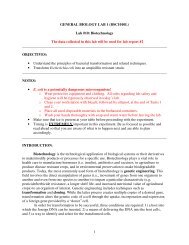
Fig. 1 MembStruk 3.5 secondary structure prediction (shaded<br />
segments) and sequence alignment against bovine rhodopsin (rhod)<br />
for phenylthiocarbamide <strong>receptor</strong> variants <strong>PTC</strong>R01 (<strong>taste</strong>r) and<br />
<strong>PTC</strong>R02 (non<strong>taste</strong>r). The secondary structure assignment for<br />
rhodopsin was taken from <strong>the</strong> pdb file (1L9H). To predict <strong>the</strong><br />
MembStruk TMs, we used a set of 12 <strong>taste</strong> <strong>receptor</strong>s (UniProt<br />
entries Q9NYV8, Q9NYV9, Q9NYW1, Q9NYW3, Q9NYW2,<br />
Q9NYW0, Q9NYW6, Q9NYW7, Q9NYW4, Q9NYW5, Q9NYV7,<br />
Q9P1R1) plus <strong>PTC</strong>R01 and <strong>PTC</strong>R02 (sequence alignment not<br />
shown). The TMs predicted by MembStruk were used to build 3D<br />
structures <strong>PTC</strong>R01a, <strong>PTC</strong>R01b, and <strong>PTC</strong>R01d, as described in <strong>the</strong><br />
“Materials and methods”. The bovine rhodopsin structure 1L9H was<br />
used to build <strong>the</strong> homology-based 3D model (<strong>PTC</strong>R01c)
Materials and methods<br />
3D structures for <strong>bitter</strong> compounds<br />
The chemical structures of <strong>the</strong> <strong>bitter</strong> compounds (<strong>PTC</strong>,<br />
atropine, brucine, chloroquine, naringin, quinacrine, quinine,<br />
salicin, caffeine, nicotine, epicatechin, and cycloheximide)<br />
were drawn using ISIS DRAW [20] and saved as a<br />
“mol” file format. We used <strong>the</strong> program Concord [21],<br />
which reads <strong>the</strong> mol file and generates 3D coordinates, to<br />
add hydrogen atoms and assign Gasteiger atomic charges<br />
[22]. The Concord-generated ligands were energy-minimized<br />
to a root mean square deviation (RMS) force of<br />
0.2 kcal mol −1 Å −1 in gas phase using conjugate-gradient<br />
minimization and <strong>the</strong> Dreiding force field [23]. These<br />
minimized structures were <strong>the</strong>n used for docking.<br />
3D structures for <strong>the</strong> <strong>PTC</strong> <strong>receptor</strong><br />
MembStruk [16–18] version 3.5 was used for de novo<br />
protein structure prediction. The homology-based models<br />
were generated using <strong>the</strong> programs Quanta [24] and Whatif<br />
[25] and <strong>the</strong> sequence alignment shown in Fig. 1. All<br />
<strong>receptor</strong> models were energy-minimized to an RMS force<br />
of 0.2 kcal mol −1 Å −1 using <strong>the</strong> Dreiding force-field [23]<br />
and CHARMM22 [26] charges.<br />
Scanning for possible binding site(s)<br />
For each of <strong>the</strong> eight 3D structures described in <strong>the</strong><br />
“Results” section (four for <strong>the</strong> <strong>taste</strong>r variant and four for <strong>the</strong><br />
non<strong>taste</strong>r variant), we used <strong>the</strong> ScanBindSite protocol to<br />
scan independently for potential binding sites using <strong>PTC</strong> as<br />
probe. The main steps of this protocol are as follows:<br />
1. Find <strong>the</strong> centers for empty pockets in <strong>the</strong> entire<br />
available volume of <strong>the</strong> <strong>receptor</strong> using <strong>the</strong> program<br />
Pass [27].<br />
2. Define <strong>the</strong> scanning regions as 10 Å around those<br />
centers.<br />
3. Generate and score multiple bound configurations of<br />
<strong>the</strong> probe ligand(s) into each scanning region.<br />
4. Eliminate configurations that have less than 90% of <strong>the</strong><br />
ligand molecular surface buried by <strong>the</strong> protein surface.<br />
5. Select based on energy <strong>the</strong> best configuration among<br />
all regions for each probe ligand. These will define <strong>the</strong><br />
location of <strong>the</strong> putative binding site to be used for<br />
predicting binding modes and binding affinities.<br />
Predicting binding modes and binding affinities<br />
We used <strong>the</strong> HierDock [16, 17, 19] first principles method<br />
technique to predict <strong>the</strong> binding site and binding energy of<br />
each ligand to <strong>the</strong> <strong>PTC</strong>R 3D models. HierDock has been<br />
used previously to predict <strong>the</strong> binding site for epinephrine<br />
to <strong>the</strong> β1 and β2 adrenergic <strong>receptor</strong>s [17, 28], for alcohols<br />
to <strong>the</strong> S25 mouse olfactory <strong>receptor</strong> (OR) [16], and for a<br />
series of odorants to several o<strong>the</strong>r ORs [29].<br />
Results<br />
Predictions of <strong>the</strong> tertiary structure<br />
933<br />
We used four different strategies to build computational 3D<br />
models for <strong>the</strong> two variants of <strong>the</strong> <strong>PTC</strong> <strong>receptor</strong>. In each<br />
case, <strong>the</strong> <strong>taste</strong>r variant was built first and <strong>the</strong>n mutated to<br />
<strong>the</strong> non<strong>taste</strong>r variant. The four strategies used were as<br />
follows:<br />
(a) We used <strong>the</strong> MembStruk protocol version 3.5 [18] to<br />
predict <strong>the</strong> TM domains and <strong>the</strong> 3D structure of <strong>the</strong><br />
<strong>receptor</strong>s. The TM domains were predicted using an<br />
alignment of 12 <strong>human</strong> <strong>taste</strong> <strong>receptor</strong>s (UniProt<br />
entries Q9NYV8, Q9NYV9, Q9NYW1, Q9NYW3,<br />
Q9NYW2, Q9NYW0, Q9NYW6, Q9NYW7,<br />
Q9NYW4, Q9NYW5, Q9NYV7, Q9P1R1) plus <strong>the</strong><br />
two <strong>PTC</strong> haplotypes. The resulting structures are<br />
identified as <strong>PTC</strong>R01a and <strong>PTC</strong>R02a.<br />
The amino-acid sequence and 3D structure of bovine<br />
rhodopsin were not used in this strategy for predicting<br />
<strong>the</strong> <strong>PTC</strong>R structures.<br />
(b) Starting <strong>with</strong> <strong>the</strong> structures obtained in (a), we<br />
optimized <strong>the</strong> rotations and translations of <strong>the</strong> helices<br />
<strong>with</strong> respect to each o<strong>the</strong>r by searching for Cys bridges<br />
that could be formed <strong>with</strong> small rotations and/or<br />
translations of <strong>the</strong> TMs carrying <strong>the</strong> potential Cys<br />
pairs. The Cys pairs <strong>with</strong>in a reasonable Cys–Cys<br />
distance in <strong>the</strong> 3D models from (a) found in this step<br />
are: C59(TM2)–C112(TM3), C254(TM6)–C289<br />
(TM7), and C261(TM6)–C282(TM7). These resulting<br />
structures are identified as structures <strong>PTC</strong>01b and<br />
<strong>PTC</strong>02b.<br />
(c) To determine how well <strong>the</strong> first principles MembStruk<br />
approach compares to standard homology modeling<br />
for determining <strong>the</strong> 3D structure of a GPCR, we built a<br />
homology-based 3D model for both <strong>receptor</strong>s using <strong>the</strong><br />
crystal structure of bovine rhodopsin (PDB code<br />
1L9H, GenBank accession K00506) using <strong>the</strong> programs<br />
Quanta [24] and Whatif [25]. The ClustalW [30]<br />
alignment used to generate <strong>the</strong> model is shown in<br />
Fig. 1. The sequence similarity and identity between<br />
bovine rhodopsin and <strong>PTC</strong>01 are 25 and 10%,<br />
respectively. These homology-based structures are<br />
referred to as <strong>PTC</strong>01c and <strong>PTC</strong>02c throughout <strong>the</strong> text.<br />
(d) Finally, we built a hybrid structure that combines <strong>the</strong><br />
TM segments from 1L9H (<strong>with</strong>out loops and <strong>with</strong>out<br />
<strong>the</strong> eight rhodopsin helices) <strong>with</strong> <strong>the</strong> TM predictions<br />
from (a) and <strong>the</strong> rotation/translation approach from (b).<br />
This leads to <strong>the</strong> Cys154(TM4)–Cys198(TM5) pair in<br />
addition to <strong>the</strong> three pairs listed in (b). We shortened or<br />
leng<strong>the</strong>ned <strong>the</strong> rhodopsin TMs to fit <strong>the</strong> TM predictions<br />
obtained through MembStruk, and <strong>the</strong>n added<br />
loops and optimized <strong>the</strong> structures using <strong>the</strong> proce-
934<br />
dures in <strong>the</strong> MembStruk protocol. The resulting<br />
structures are denoted as <strong>PTC</strong>R01d and <strong>PTC</strong>R02d.<br />
Figure 1 shows <strong>the</strong> sequence alignment between<br />
<strong>PTC</strong>R01, <strong>PTC</strong>R02, and rhodopsin (used for <strong>the</strong> homology<br />
modeling). The TM domains defined in <strong>the</strong> crystal<br />
structure are shaded in <strong>the</strong> rhodopsin’s sequence. The<br />
TMs predicted by MembStruk 3.5 (shaded segments in <strong>the</strong><br />
<strong>PTC</strong>Rs sequence) were used to build <strong>the</strong> 3D structures (a),<br />
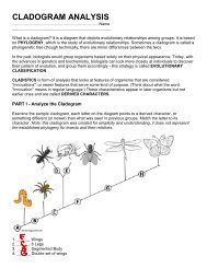
(b), and (d). Figure 2 shows <strong>the</strong> four 3D model types <strong>with</strong><br />
<strong>the</strong> Cys bridges differentiating structures (a) and (b), and<br />
<strong>the</strong> Cys bridges for model (d). Table 1 lists <strong>the</strong> RMS in<br />
coordinates for <strong>the</strong> alpha Carbon atoms (CRMS) in TM<br />
segments among <strong>the</strong> 3D models. The CRMS between each<br />
pair of <strong>taste</strong>r and non<strong>taste</strong>r structures was less than 0.15 Å<br />
in all four cases.<br />
General location of <strong>the</strong> putative binding site(s)<br />
To determine <strong>the</strong> location of <strong>the</strong> potential binding sites for<br />
<strong>the</strong> various tastants, we used <strong>the</strong> ScanBindSite computational<br />
procedure [16, 17] for each of eight (four <strong>taste</strong>r<br />
variants and four non<strong>taste</strong>r variants to each of <strong>the</strong> eight) 3D<br />
structures of <strong>PTC</strong>R. The results are shown in Fig. 2.<br />
Structures (a) (MembStruk 3.5) and (c) (homology to 1L9H<br />
rhodopsin crystal structure) have <strong>the</strong> binding site in<br />
equivalent regions, between TMs 3, 4, 5, and 6. This<br />
location is consistent <strong>with</strong> <strong>the</strong> binding site for retinal in<br />
bovine rhodopsin [31]. Structures (b) and (d) [<strong>with</strong> helices<br />
rotated, respectively, from (a) and (b)] have <strong>the</strong>ir binding<br />
sites shifted towards TMs 1 and 7 [structure (b)], and TM4<br />
[structure (d)]. Figure 3 shows, as solid sphere clusters, <strong>the</strong><br />
various scanning regions found in <strong>PTC</strong>R01 structures (a) to<br />
Fig. 2 Predicted 3D structures<br />
for phenylthiocarbamide <strong>receptor</strong><br />
(<strong>PTC</strong>R). Variant 02 (non<strong>taste</strong>r)<br />
was homology modeled<br />
from variant 01 (<strong>taste</strong>r) because<br />
<strong>the</strong>y differ in only three amino<br />
acids (P49A, A262V, and<br />
V296I). The 3D structure<br />
(<strong>PTC</strong>R01a) was built using<br />
MembStruk version 3.5, while<br />
structure <strong>PTC</strong>R01b corresponds<br />
to <strong>PTC</strong>R01a, <strong>with</strong> <strong>the</strong> helical<br />
rotations/translations adjusted to<br />
allow for optimal Cys bridges as<br />
shown. The homology-derived<br />
model (<strong>PTC</strong>R01c) was built<br />
using <strong>the</strong> experimentally determined<br />
structure for rhodopsin<br />
(1L9H). 3D model (<strong>PTC</strong>R01d)<br />
is a hybrid homology/<br />
MembStruk model (see “Results”<br />
for details)
Table 1 Root mean square deviation in coordinates for <strong>the</strong> alpha<br />
Carbon atoms in TM segments among <strong>the</strong> 3D models built for<br />
<strong>PTC</strong>R<br />
Calpha, 171 aa,<br />
TMS, rms (A)<br />
<strong>PTC</strong>R01a<br />
<strong>taste</strong>r<br />
<strong>PTC</strong>R01b<br />
<strong>taste</strong>r<br />
<strong>PTC</strong>R01c<br />
<strong>taste</strong>r<br />
<strong>PTC</strong>R01a <strong>taste</strong>r 0<br />
<strong>PTC</strong>R01b <strong>taste</strong>r 3.2 0<br />
<strong>PTC</strong>R01c <strong>taste</strong>r 6.9 7.1 0<br />
<strong>PTC</strong>R01d <strong>taste</strong>r 6.5 5.8 5.5 0<br />
<strong>PTC</strong>R01d<br />
<strong>taste</strong>r<br />
(d) (<strong>PTC</strong>R02 are identical). The binding region found to be<br />
most energetically favorable is circled in each structure.<br />
Although <strong>the</strong> 3D structures obtained using <strong>the</strong> four<br />
approaches are considerably different, two residues were<br />
found <strong>with</strong>in 4 Å of <strong>the</strong> bound ligand for all eight 3D<br />
structures: Trp99 (TM3) and Asn103 (TM3). The differences<br />
between <strong>the</strong> structures arise primarily from rotations<br />
and translations of some helices. However, <strong>the</strong> position of<br />
TM3, particularly <strong>the</strong> face <strong>with</strong> Trp99 and Asn103, seems<br />
to determine where <strong>PTC</strong> binds in all 3D structures.<br />
Location of <strong>the</strong> SNPs and <strong>the</strong>ir distances to bound <strong>PTC</strong><br />
935<br />
Figure 4 shows <strong>the</strong> TM segments <strong>with</strong> <strong>the</strong> SNPs characteristic<br />
of <strong>PTC</strong> <strong>taste</strong>rs and non<strong>taste</strong>rs displayed as van der<br />
Waals spheres for <strong>the</strong> four sets of 3D structures described<br />
above. The structural analysis of <strong>the</strong> SNPs in <strong>the</strong>se 3D<br />
models shows that<br />
1. Structures (a) (MembStruk 3.5) and (c) (homology to<br />
1L9H rhodopsin) have positions 262-TM6 facing helix<br />
7, and position 296-TM7 facing helix 6. Therefore,<br />
both 3D models suggest that <strong>the</strong>se SNPs are involved<br />
in <strong>the</strong> packing of TMs 6 and 7, and not in <strong>the</strong> direct<br />
binding of ligands.<br />
2. Structures (b) and (c) have 262-TM6 pointing towards<br />
<strong>the</strong> interior of <strong>the</strong> helical barrel, while 296-(TM7) is<br />
pointing towards TM6. For <strong>the</strong>se structures <strong>the</strong>n, 262-<br />
TM6 could potentially be directly involved in binding<br />
<strong>PTC</strong>.<br />
The closest SNP to <strong>the</strong> bound <strong>PTC</strong> is at position 262 in<br />
TM6. The distances between <strong>the</strong> carbon beta of 262-TM6<br />
and <strong>the</strong> C1 in <strong>PTC</strong> are: 8 Å in 3D model (a), 9 Å in 3D<br />
model (c), 14 Å in 3D model (b), and 13 Å in 3D model (d).<br />
Thus, in all predicted protein–ligand complexes, position<br />
Fig. 3 Location of <strong>the</strong> predicted<br />
binding site for <strong>PTC</strong> in <strong>the</strong><br />
<strong>PTC</strong>R 3D structures. The program<br />
ScanBindSite docks and<br />
scores multiple configurations<br />
of <strong>PTC</strong> in each of several empty<br />
pockets (represented as sphere<br />
clusters <strong>with</strong> different colors)<br />
throughout <strong>the</strong> <strong>receptor</strong>. The<br />
probable binding site (circled<br />
sphere cluster) is defined by <strong>the</strong><br />
locations of <strong>the</strong> most energetically<br />
favorable <strong>receptor</strong>. Both<br />
variants were independently<br />
scanned but only <strong>the</strong> sites for <strong>the</strong><br />
<strong>taste</strong>r variant (<strong>PTC</strong>R01 structures)<br />
are shown. The non<strong>taste</strong>r<br />
variant (<strong>PTC</strong>R02 structures) has<br />
<strong>the</strong> exact same binding site<br />
location as <strong>PTC</strong>R01 for each of<br />
<strong>the</strong> four structure types (a)−(d)
936<br />
Fig. 4 TM segments <strong>with</strong> <strong>the</strong> SNPs characteristic of <strong>PTC</strong> <strong>taste</strong>rs and non<strong>taste</strong>rs displayed as van der Waals spheres for <strong>the</strong> four types of 3D<br />
models built in this work<br />
262-TM6 is too far from <strong>PTC</strong> to have any significant<br />
contribution to <strong>the</strong> <strong>PTC</strong> binding energy, and <strong>the</strong>refore<br />
cannot explain <strong>the</strong> <strong>taste</strong>r/non<strong>taste</strong>r difference. That is<br />
particularly true in <strong>the</strong> case of structures (b) and (d), not<br />
only because of <strong>the</strong> longer distances but also because <strong>the</strong>se<br />
are <strong>the</strong> only structures that could have an SNP in direct<br />
contact <strong>with</strong> <strong>the</strong> ligand. Fur<strong>the</strong>rmore, <strong>the</strong> calculated<br />
binding energies for <strong>the</strong> pairs <strong>PTC</strong>R01 and <strong>PTC</strong>R02 are<br />
nearly <strong>the</strong> same for each 3D-structure type (a) to (d).<br />
Because structures (b) and (d) have one of <strong>the</strong> three SNPs<br />
pointing inside <strong>the</strong> TM barrel but not <strong>with</strong>in reasonable<br />
distance for atomic interaction <strong>with</strong> <strong>PTC</strong>, we conclude <strong>the</strong>y<br />
are inadequate representations of <strong>the</strong> <strong>PTC</strong> <strong>receptor</strong>. Hence,<br />
<strong>the</strong> rest of our analysis will concern structures<br />
(a) (MembStruk 3.5) and (c) (homology-based) only.<br />
The binding mode for <strong>PTC</strong> in <strong>PTC</strong>R01a<br />
Residues <strong>with</strong>in 4 Å of <strong>the</strong> bound ligand in <strong>the</strong> predicted<br />
complexes are shown in Fig. 5. These are Trp99, Met100,<br />
Asn103, Gln104, and Leu107 in TM3; Ala263, Phe264,<br />
and Val267 in TM6; and Cys282 and Met286 in TM7.<br />
Binding of o<strong>the</strong>r <strong>bitter</strong> ligands to <strong>the</strong> <strong>PTC</strong> <strong>receptor</strong><br />
We used <strong>the</strong> calculated binding energies for <strong>PTC</strong> and 11<br />
additional <strong>bitter</strong> compounds (atropine, brucine, chloroquine,<br />
naringin, quinacrine, quinine, salicin, caffeine,<br />
nicotine, epicatechin, and cycloheximide) docked to our<br />
3D models to investigate <strong>PTC</strong>R01 and <strong>PTC</strong>R02 fur<strong>the</strong>r.<br />
These predicted binding energies are reported in Table 2
937<br />
Fig. 5 Predicted 3D structures<br />
for phenylthiocarbamide bound<br />
to <strong>bitter</strong> <strong>receptor</strong> variants<br />
<strong>PTC</strong>R01a (<strong>taste</strong>r) and <strong>PTC</strong>R02a<br />
(non<strong>taste</strong>r). The figure shows<br />
a top view from extracellular<br />
end and b binding mode <strong>with</strong><br />
residues <strong>with</strong>in 3.5 Å of <strong>the</strong><br />
bound ligand in licorice<br />
representation<br />
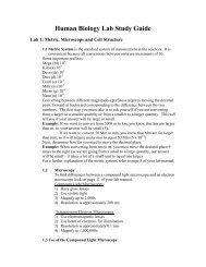
and Fig. 6. The calculated binding affinities for <strong>PTC</strong> bound<br />
to <strong>PTC</strong>R01 and <strong>PTC</strong>R02 show no significant difference in<br />
binding between <strong>the</strong>se <strong>receptor</strong> variants. In addition, no<br />
difference in binding affinity is found for <strong>the</strong> o<strong>the</strong>r <strong>bitter</strong><br />
compounds docked to <strong>PTC</strong>R01 and <strong>PTC</strong>R02, as seen in<br />
Table 2 and Fig. 6. Fur<strong>the</strong>rmore, <strong>taste</strong> sensitivity to some of<br />
<strong>the</strong> compounds <strong>with</strong> similar or higher calculated binding<br />
affinity to <strong>PTC</strong>R than <strong>PTC</strong> has been linked to <strong>PTC</strong> <strong>taste</strong>r<br />
status [9, 11, 12, 32–34].<br />
Discussion<br />
Analysis of <strong>PTC</strong> sensitivity<br />
These modeling studies emphasize <strong>the</strong> role of TM6 and<br />
TM7 in <strong>PTC</strong> <strong>receptor</strong> function. We can now consider how<br />
changes in <strong>the</strong> <strong>interactions</strong> between TMs 6 and 7 might<br />
affect <strong>taste</strong> sensitivity to <strong>PTC</strong>. Movement of TM6 has been<br />
proposed as part of <strong>the</strong> structural changes leading to<br />
Table 2 HierDock binding energies (kcal mol −1 )<br />
Ligand HierDock binding energy (kcal mol −1 )<br />
<strong>PTC</strong>R01a <strong>PTC</strong>R02a<br />
Naringin −68.9 −73.5<br />
Salicin −51.9 −52.0<br />
Epicatechin −41.1 −37.1<br />
Chloroquine −35.9 −32.2<br />
Quinacrine −31.2 −30.3<br />
Quinine −28.7 −28.0<br />
Atropine −25.5 −27.1<br />
Phenylthiocarbamide −22.1 −24.5<br />
Nicotine −20.3 −22.0<br />
Caffeine −14.6 −13.7<br />
Brucine −4.6 −8.6<br />
Cycloheximide −0.7 −3.8<br />
Binding energies were calculated as: BindE=E(bound_ligand_in_<br />
protein)−E(free_ligand_in_water). These binding energies do not<br />
include explicit entropic terms or enthalpic temperature corrections<br />
Values for <strong>the</strong> known ligand <strong>PTC</strong> in bold
938<br />
signaling in GPCRs [35, 36–39]. As shown in Fig. 7, <strong>the</strong><br />
introduction of bulkier side chains in <strong>the</strong> non<strong>taste</strong>r variant<br />
alters <strong>the</strong> packing of TMs 6 and 7, which may render <strong>the</strong><br />
movement of TM6 (necessary for signaling) more difficult<br />
or unattainable. According to our 3D structures, it is<br />
plausible that <strong>the</strong> mutations A262V and V296I would lead<br />
to ei<strong>the</strong>r an inactive <strong>receptor</strong> or to a reduced function, thus<br />
rationalizing <strong>the</strong> observed <strong>taste</strong>r/non<strong>taste</strong>r effect.<br />
Our studies lead to three main predictions:<br />
1. The binding affinities of both <strong>receptor</strong> variants for <strong>PTC</strong><br />
might be expected to have close or similar energies<br />
because <strong>the</strong> mutations are not directly involved in<br />
binding.<br />
2. Non<strong>taste</strong>r individuals for <strong>PTC</strong> would also have null or<br />
reduced (because one compound may activate multiple<br />
<strong>bitter</strong> <strong>receptor</strong> types) <strong>taste</strong> sensitivity for o<strong>the</strong>r <strong>bitter</strong><br />
compounds recognized by <strong>the</strong> same <strong>receptor</strong> and <strong>with</strong><br />
binding affinity similar to <strong>PTC</strong>. This is supported by<br />
psychophysical evidence that <strong>PTC</strong> non<strong>taste</strong>rs have<br />
reduced sensitivity to naringin [12, 33], caffeine [11,<br />
Cycloheximide<br />
O<br />
N<br />
O<br />
O<br />
O<br />
O<br />
O<br />
Epicatechin<br />
O<br />
O<br />
O<br />
O<br />
Caffeine<br />
O<br />
N<br />
O N<br />
N<br />
N<br />
Nicotine<br />
N<br />
N<br />
Salicin<br />
O<br />
O<br />
O<br />
O<br />
O<br />
O<br />
O<br />
Quinine<br />
H<br />
N<br />
H<br />
O<br />
O<br />
N<br />
O<br />
Quinacrine<br />
N<br />
N<br />
N<br />
Cl<br />
Phenylthiocarbamide<br />
__________________<br />
N<br />
N<br />
Naringin<br />
S<br />
O<br />
O<br />
O O<br />
O<br />
O<br />
O<br />
O<br />
O<br />
O<br />
O<br />
O<br />
Chloroquine<br />
Cl<br />
N<br />
O<br />
O<br />
N<br />
N<br />
Brucine<br />
O<br />
O<br />
N<br />
N<br />
O<br />
O<br />
Atropine<br />
N<br />
O<br />
O<br />
O<br />
-80 -60 -40 -20 0<br />
HierDock Binding Energy (kcal mol<br />
-1)<br />
<strong>PTC</strong>R01a <strong>taste</strong>r<br />
<strong>PTC</strong>R02a non-<strong>taste</strong>r<br />
Fig. 6 HierDock-calculated binding affinities (kcal mol −1 ) for phenylthiocarbamide (<strong>PTC</strong>), and o<strong>the</strong>r tastants docked to <strong>PTC</strong> <strong>receptor</strong><br />
variants <strong>taste</strong>r (<strong>PTC</strong>R01a) and non<strong>taste</strong>r (<strong>PTC</strong>R02a)
939<br />
32, 34], quinine [9, 11], epicatechin, tetralone, urea,<br />
sucrose octaacetate, denatonium benzoate, L-phenylalanine,<br />
and L-tryptophan [11].<br />
3. Compensatory mutations in TMs 6 and 7 described<br />
below could restore non<strong>taste</strong>r <strong>PTC</strong>R02 response to<br />
<strong>PTC</strong>.<br />
Considering <strong>the</strong> close contacts predicted to occur<br />
between residues 262-TM6 and 296-TM7 and residues in<br />
opposite helices, we suggest mutations that could validate<br />
our proposed explanation for <strong>the</strong> <strong>taste</strong>r/non<strong>taste</strong>r status of<br />
<strong>the</strong> <strong>PTC</strong>R variants studied here.<br />
1. The MembStruk model (a) has I285 (TM7) in contact<br />
<strong>with</strong> 262-TM6, and L246 (TM6) and F252 (TM6)<br />
opposite to 296-TM7. The mutation of I285 to V, L246<br />
to V, and F252 to I, would change contact to <strong>the</strong> variant<br />
amino acids <strong>with</strong>out causing significant changes in <strong>the</strong><br />
overall structure of <strong>the</strong> <strong>receptor</strong>.<br />
2. The homology-based model (c) points to residues<br />
V279 (TM7), which is opposite to 262-TM6), and<br />
S248 (TM6) and K247 (TM6), which are both opposite<br />
to 296-TM7, as candidates for compensatory mutations.<br />
Mutation of V279 to A should improve <strong>the</strong><br />
packing of I296 (non<strong>taste</strong>r variant). As for S248 and<br />
K247, finding <strong>the</strong>se residues packed against TM7<br />
indicates that <strong>the</strong> homology-derived 3D structure may<br />
have TM6 inappropriately rotated. Polar and charged<br />
residues are expected to be ei<strong>the</strong>r packed against<br />
complementary residues or pointing towards <strong>the</strong> wateraccessible<br />
center of <strong>the</strong> barrel.<br />
From <strong>the</strong> above analysis we conclude that <strong>the</strong> 3D<br />
structures <strong>PTC</strong>R01a and <strong>PTC</strong>R02a generated by Memb-<br />
Struk best represent <strong>the</strong> <strong>PTC</strong> <strong>receptor</strong> <strong>taste</strong>r/non<strong>taste</strong>r<br />
variants, and <strong>the</strong>refore, we will discuss only <strong>the</strong>se<br />
structures below.<br />
The involvement of TMs 3, 5, 6, and 7 in GPCR<br />
signaling has been documented extensively in <strong>the</strong> literature<br />
[35–42]. It has been suggested [41–43] that <strong>interactions</strong><br />
between TMs 3, 6, and 7 propagate <strong>the</strong> signal from <strong>the</strong><br />
ligand binding site at <strong>the</strong> extracellular side to <strong>the</strong> G protein<br />
binding site at <strong>the</strong> intracellular side of <strong>the</strong> <strong>receptor</strong>. For<br />
constitutively active rhodopsin mutants, <strong>the</strong> release of<br />
interhelical constraints <strong>with</strong>in TMs 3, 6, and 7 produces a<br />
structural conformation of <strong>the</strong> <strong>receptor</strong> that is more easily<br />
activated [44]. The same effect was reported in a recent<br />
random mutagenesis study on <strong>the</strong> opioid <strong>receptor</strong> [45]<br />
showing that certain mutations in TMs 3, 6, and 7 lead to<br />
constitutively active <strong>receptor</strong>s. In particular, Decaillot et al.<br />
(2003) suggested that <strong>the</strong> mutations at <strong>the</strong> intracellular end<br />
of TMs 6 and 7 weaken helical packing, allowing <strong>the</strong><br />
separation of TMs 6 and 7 at <strong>the</strong> intracellular side.<br />
Our proposed explanation for <strong>the</strong> <strong>PTC</strong> <strong>taste</strong>r/non<strong>taste</strong>r<br />
effect associated <strong>with</strong> <strong>the</strong> mutations A262V and V296I<br />
agrees <strong>with</strong> <strong>the</strong> experimental data on constitutively active<br />
GPCRs and <strong>the</strong> signaling model involving TMs 3, 6, and 7.<br />
However, instead of mutations causing <strong>the</strong> activation of<br />
<strong>receptor</strong>s <strong>with</strong>out bound ligands, <strong>the</strong> <strong>human</strong> <strong>PTC</strong> <strong>receptor</strong><br />
appears to have mutations acting in an opposite fashion. In<br />
<strong>the</strong> case of <strong>PTC</strong>R <strong>taste</strong>r/non<strong>taste</strong>r variants, <strong>the</strong> introduction<br />
of bulkier side chains at both ends of <strong>the</strong> TM6/TM7 ladder<br />
may increase <strong>the</strong> attraction between TMs 6 and 7 so that <strong>the</strong><br />
ligand binding has to provide an additional intermolecular<br />
force to activate <strong>the</strong> <strong>receptor</strong>. This would probably have <strong>the</strong><br />
effect of turning off sensitivity to relatively low-affinity<br />
ligands like <strong>PTC</strong>. We note that <strong>the</strong> proposed steric effects<br />
caused by bulkier residues in positions 262-TM6 and 296-<br />
TM7 of <strong>the</strong> non<strong>taste</strong>r variant may involve residues that<br />
Fig. 7 TMs 6 and 7 <strong>with</strong><br />
positions 262-TM6 and 296-<br />
TM7 shown as van der Waals<br />
spheres. Amino acids present in<br />
<strong>the</strong> non<strong>taste</strong>r variant are shown<br />
as gray shadows. Side chains<br />
opposite to <strong>the</strong> SNP’s positions<br />
are shown in licorice. We propose<br />
that compensative mutations<br />
in <strong>the</strong>se positions could<br />
restore <strong>PTC</strong> response in <strong>the</strong><br />
non<strong>taste</strong>r variant. Note that <strong>the</strong><br />
residues in close contact to 296-<br />
TM7 in <strong>the</strong> homology-based 3D<br />
model <strong>PTC</strong>R01c are inconsistent<br />
<strong>with</strong> helix–helix packing.<br />
The 3D model derived using<br />
MembStruk 3.5 seems to be <strong>the</strong><br />
best representative of <strong>the</strong> system<br />
studied here
940<br />
come in contact <strong>with</strong> those positions at some stage of <strong>the</strong><br />
activation process o<strong>the</strong>r than <strong>the</strong> one represented by our<br />
predicted 3D structures.<br />
Bimodal vs modulated <strong>taste</strong> response<br />
A number of studies support <strong>the</strong> view that a particular <strong>bitter</strong><br />
compound may elicit response from one or more <strong>bitter</strong><br />
<strong>receptor</strong>s present in <strong>the</strong> same cell, <strong>with</strong> individual <strong>receptor</strong>s<br />
responding to multiple ligands. Covariation patterns found<br />
in <strong>the</strong> sensitivity to certain <strong>bitter</strong> compounds [11, 12, 31–<br />
33] also support a single-ligand/multiple-<strong>receptor</strong>s model<br />
which states that a single ligand can activate multiple<br />
<strong>receptor</strong>s, as well as a single-<strong>receptor</strong>/multiple-ligands<br />
model. A correlation was found for sensitivity to L-<br />
tryptophan, L-phenylalanine, and, to a lesser extent, urea<br />
[11, 46], but not between <strong>the</strong>se and PROP. No correlation<br />
was found between <strong>the</strong> ability to <strong>taste</strong> PROP and <strong>taste</strong><br />
sensitivity to quinineHCL [47]. Under this view, <strong>the</strong><br />
bimodal distribution of sensitivity to <strong>PTC</strong> implies a single<br />
<strong>receptor</strong> type for <strong>PTC</strong>, while <strong>the</strong> decreased sensitivity to<br />
naringin, caffeine, and o<strong>the</strong>r compounds associated <strong>with</strong><br />
<strong>the</strong> <strong>PTC</strong> non<strong>taste</strong>r variant imply additional <strong>receptor</strong> types<br />
responding to <strong>the</strong>se compounds.<br />
According to our predicted binding profile in Fig. 6, <strong>the</strong><br />
weak (less negative) binding affinity of cycloheximide and<br />
brucine to both <strong>PTC</strong>R variants suggests that <strong>the</strong> <strong>bitter</strong>ness<br />
of <strong>the</strong>se compounds is not affected by <strong>the</strong> <strong>PTC</strong> <strong>receptor</strong>. On<br />
<strong>the</strong> o<strong>the</strong>r hand, compounds predicted as having similar<br />
binding affinity to <strong>PTC</strong> (nicotine, atropine, quinine,<br />
quinacrine, and chloroquine) may be perceived as being<br />
less <strong>bitter</strong> by <strong>PTC</strong> non<strong>taste</strong>rs. However, because <strong>the</strong>se<br />
compounds may activate o<strong>the</strong>r <strong>receptor</strong> types, no bimodal<br />
response is expected. Compounds we predict as having<br />
higher affinity to <strong>PTC</strong>R (naringin, salicin, and epicatechin)<br />
may bind strongly enough so that <strong>the</strong> resistance to<br />
signaling caused by <strong>the</strong> non<strong>taste</strong>r SNPs may be overcome.<br />
However, sensitivity to naringin, <strong>the</strong> compound <strong>with</strong> <strong>the</strong><br />
strongest affinity to <strong>PTC</strong>R in our set, has been linked to<br />
<strong>PTC</strong> <strong>taste</strong>r status [8, 12], which suggests that none of <strong>the</strong> 12<br />
compounds studied here are strong agonists for this<br />
<strong>receptor</strong>.<br />
Conclusion<br />
The MembStruk predicted structure combined <strong>with</strong> <strong>the</strong><br />
HierDock predicted binding site provides insight into <strong>the</strong><br />
activity of <strong>the</strong> <strong>PTC</strong> <strong>receptor</strong>. The small differences between<br />
binding affinities calculated for <strong>the</strong> same compound bound<br />
to <strong>taste</strong>r and non<strong>taste</strong>r variants of <strong>PTC</strong>R observed for all<br />
<strong>the</strong> docked compounds are consistent <strong>with</strong> our view that<br />
<strong>the</strong> SNP mutations are not directly involved in ligand<br />
binding, but ra<strong>the</strong>r in G protein activation. These predicted<br />
structures also suggest a number of experiments that can be<br />
used to validate <strong>the</strong> proposed explanation for <strong>PTC</strong> <strong>taste</strong><br />
status. We propose that compensatory mutations to<br />
<strong>PTC</strong>R02 that could restore sensitivity to <strong>PTC</strong> are I285 to<br />
V, L246 to V, and F252 to I.<br />
The molecular-level details of <strong>the</strong> <strong>interactions</strong> between<br />
<strong>PTC</strong> and its <strong>receptor</strong> could be fur<strong>the</strong>r refined by <strong>the</strong><br />
introduction of algorithms for side-chain rotamer placement<br />
and hydrogen bond network optimization during<br />
docking. However, even if some details in <strong>the</strong> predicted<br />
structures might benefit from fur<strong>the</strong>r computational<br />
refinement, <strong>the</strong> comparison to experimental results predicted<br />
from <strong>the</strong> structures would allow corrections in <strong>the</strong><br />
structures and perhaps improvements in <strong>the</strong> methods in <strong>the</strong><br />
most effective way.<br />
Acknowledgements We thank Dr. Susan Sullivan and Dr. John<br />
Northup for helpful comments on <strong>the</strong> manuscript. This work was<br />
supported by NIDCD Z01-000046-04, and by NIH-BRGRO1-<br />
GM625523, NIH-R29AI40567, and NIH-HD36385. The computational<br />
facilities at <strong>the</strong> Materials and Process Simulation Center<br />
(MSC) were provided by a Shared University Research grant from<br />
International Business Machines and Defense University Research<br />
Instrumentation Program grants from <strong>the</strong> Army Research Office<br />
(ARO) and <strong>the</strong> Office of Naval Research (ONR). The facilities of <strong>the</strong><br />
MSC are also supported by <strong>the</strong> Department of Energy-Advanced<br />
Simulation and Computing Program, National Science Foundation,<br />
Multidisciplinary Research Initiative - Army Research Office,<br />
Multidisciplinary Research Initiative - Office of Naval Research,<br />
General Motors, ChevronTexaco, Seiko-Epson, Beckman Institute,<br />
and Asahi Kasei.<br />
References<br />
1. Adler E, Hoon MA, Mueller KL, Chandrashekar J, Ryba NJ,<br />
Zuker CS (2000) Cell 100:93–702<br />
2. Chandrashekar J, Mueller KL, Hoon MA, Adler E, Feng L,<br />
Guo W, Zuker CS, Ryba NJ (2000) Cell 100:703–711<br />
3. Lindemann B (2001) Nature 413:219–225<br />
4. Fox AL (1932) Proc Natl Acad Sci USA 18:115–120<br />
5. Kim UK, Jorgenson E, Coon H, Leppert M, Risch N, Drayna D<br />
(2003) Science 299:1221–1225<br />
6. Wooding S, Kim UK, Bamshad MJ, Larsen J, Jorde LB,<br />
Drayna D (2004) Am J Hum Genet 74:637–746<br />
7. Fischer R, Griffin F, England S, Garn SM (1961) Nature<br />
191:1328<br />
8. Drewnowski A, Henderson SA, Barratt-Fornell A (2001) Drug<br />
Metab Dispos 29:535–538<br />
9. Harris H, Kalmus H (1949) Ann Eugen 15:32–45<br />
10. Barnicot N, Harris H, Kalmus H (1951) Ann Eugen 16:119–128<br />
11. Delwiche JF, Buletic Z, Breslin PA (2001) Percept Psychophys<br />
63:761–776<br />
12. Kameswaran L, Gopalakrishnan S, Sukumar M (1974) Ind J<br />
Pharmac 6:134–140<br />
13. Peterson DI, Lonergan LH, Hardinge MG (1968) Arch Environ<br />
Health 16:219–222<br />
14. Cannon D, Baker T, Piper M, Scholand MB, Lawrence D,<br />
Drayna D, McMahon W, Villegas GM, Caton T, Coon H,<br />
Leppert M (2005) Nicotine Tob Res 7:853–858<br />
15. Sultana T, Savage GP, Porter NG (2002) Proc Nutr Soc N Z<br />
27:86–91<br />
16. Floriano WB, Vaidehi N, Singer MS, Goddard WA III,<br />
Shepherd GM (2000) Proc Natl Acad Sci USA 97:10712–<br />
10716<br />
17. Vaidehi N, Floriano WB, Trabanino R, Hall SE, Freddolino P,<br />
Choi EJ, Zamanakos G, Goddard WA III (2002) Proc Natl<br />
Acad Sci USA 99:12622–12627<br />
18. Trabanino RJ, Hall S, Vaidehi N, Floriano WB, Goddard WA<br />
III (2004) Biophys J 84:1904–1921
941<br />
19. Floriano WB, Nagarajan V, Zamanakos G, Goddard WA III<br />
(2004) J Med Chem 47:56–71<br />
20. MDL Information Systems Inc, http://www.mdli.com<br />
21. Tripos Inc, http://www.tripos.com<br />
22. Gasteiger J, Marsili M (1980) Tetrahedron 36:3219–3228<br />
23. Mayo SL, Olafson BD, Goddard WA III (1990) J Phys Chem<br />
94:8897–8909<br />
24. Accelrys Inc, http://www.accelrys.com<br />
25. Vriend G (1990) J Mol Graph 8:52–56<br />
26. MacKerell AD, Bashford D, Bellott M, Dunbrack RL,<br />
Evanseck JD, Field MJ, Fischer S, Gao J, Guo H, Ha S,<br />
Joseph-McCarthy D, Kuchnir L, Kuczera K, Lau FTK,<br />
Schlenkrich M, Smith JC, Stote R, Straub J, Watanabe M,<br />
Wiorkiewicz-Kuczera J, Yin D, Karplus M (1998) J Phys Chem<br />
B 102:3586–3616<br />
27. Brady GP Jr, Stouten PFW (2000) J Comput Aided Mol Des<br />
14:383–401<br />
28. Freddolino PL, Yashar M, Kalani S, Vaidehi N, Floriano WB,<br />
Hall SE, Trabanino RJ, Kam VW, Goddard WA III (2004) Proc<br />
Natl Acad Sci USA 101:2736–2741<br />
29. Floriano WB, Vaidehi N, Goddard WA III (2004) Chem Senses<br />
29:269–290<br />
30. Thompson JD, Higgins DG, Gibson TJ (1994) Nucleic Acids<br />
Res 22:4673–4680<br />
31. Palczewski K, Kumasaka T, Hori T, Behnke CA, Motoshima H,<br />
Fox BA, Le Trong I, Teller DC, Okada T, Stenkamp RF,<br />
Yamamoto M, Miyano M (2000) Science 289:739–745<br />
32. Hall MJ, Bartoshuk LM, Cain WS, Stevens JC (1975) Nature<br />
253:442–443<br />
33. Drewnowski A, Henderson SA, Shore AB (1997) Am J Clin<br />
Nutr 66:391–397<br />
34. Bartoshuk LM (2000) Chem Senses 25:447–460<br />
35. Farrens DL, Altenbach C, Yang K, Hubbell WL, Khorana HG<br />
(1996) Science 274:768–770<br />
36. Sheikh SP, Zvyaga TA, Lichtarge O, Sakmar TP, Bourne HR<br />
(1996) Nature 383:347–350<br />
37. Ge<strong>the</strong>r U, Lin S, Ghanouni P, Ballesteros JA, Weinstein H,<br />
Kobilka BK (1997) EMBO J 16:6737–6747<br />
38. Dunham TD, Farrens DL (1999) J Biol Chem 274:1683–1690<br />
39. Medkova M, Preininger AM, Yu NJ, Hubbell WL, Hamm HE<br />
(2002) Biochemistry 41:9962–9972<br />
40. Schmidt C, Li B, Bloodworth L, Erlenbach I, Zeng FY, Wess J<br />
(2003) J Biol Chem 278:30248–30260<br />
41. Pauwels PJ, Wurch T (1998) Mol Neurobiol 17:109–135<br />
42. Ge<strong>the</strong>r U (2000) Endocr Rev 21:90–113<br />
43. Okada T, Ernst OP, Palczewski K, Hofmann KP (2001) Trends<br />
Biochem Sci 26:318–324<br />
44. Rao VR, Oprian DD (1996) Annu Rev Biophys Biomol Struct<br />
25:287–314<br />
45. Decaillot FM, Befort K, Filliol D, Yue S, Walker P, Kieffer BL<br />
(2003) Nat Struct Biol 10:629–636<br />
46. Keast RS, Breslin PA (2002) Chem Senses 27:123–131<br />
47. Schifferstein HNJ, Frijters JER (1991) Chem Senses 16:303–317