An Unusual Presentation of Impacted Foreign- Body in ... - Statperson
An Unusual Presentation of Impacted Foreign- Body in ... - Statperson
An Unusual Presentation of Impacted Foreign- Body in ... - Statperson

Create successful ePaper yourself
Turn your PDF publications into a flip-book with our unique Google optimized e-Paper software.
International Journal <strong>of</strong> Recent Trends <strong>in</strong> Science <strong>An</strong>d Technology, ISSN 2277-2812 E-ISSN 2249-8109, Volume 7, Issue 3, 2013 pp 100-102<br />
<strong>An</strong> <strong>Unusual</strong> <strong>Presentation</strong> <strong>of</strong> <strong>Impacted</strong> <strong>Foreign</strong>-<br />
<strong>Body</strong> <strong>in</strong> the Larynx: A Case Report<br />
Rameshwar T. Pawar * , Girish V. Thakur ** , Amit Thote *** , <strong>An</strong>up Gilada ***<br />
{ * Assistant Pr<strong>of</strong>essor, ** Associate Pr<strong>of</strong>essor, *** Resident} Department <strong>of</strong> Otorh<strong>in</strong>olaryngology, Government Medical College, Latur,<br />
Abstract: Most airway foreign body aspirations occur<br />
<strong>in</strong> children younger than 15 years; children aged 1-3 years are the<br />
most susceptible. Vegetable matter tends to be the most common<br />
airway foreign body; peanuts are the most common food item<br />
aspirated. Our case become unique <strong>in</strong> the sense that <strong>in</strong> spite <strong>of</strong><br />
be<strong>in</strong>g horny projections <strong>of</strong> foreign vegetative material reta<strong>in</strong>ed<br />
for almost 30 hours, patient presented with no signs <strong>of</strong> laryngeal<br />
spasm.<br />
Key Words: foreign body, x-ray <strong>of</strong> airway, larynx.<br />
Introduction<br />
The diagnosis and treatment <strong>of</strong> foreign bodies <strong>in</strong> the<br />
airway are a challenge for otolaryngologists. Despite<br />
improvements <strong>in</strong> medical care and public awareness,<br />
approximately 3000 deaths occur each year from<br />
foreign body aspiration, with most deaths occurr<strong>in</strong>g<br />
before hospital evaluation and treatment. A high <strong>in</strong>dex<br />
<strong>of</strong> suspicion is needed for foreign body aspiration to<br />
allow for prompt treatment and avoidance <strong>of</strong><br />
complications. Chevalier Jackson revolutionized<br />
endoscopic foreign body removal <strong>in</strong> the early 1900s<br />
with pr<strong>in</strong>ciples and techniques still followed today. The<br />
development <strong>of</strong> the rod-lens telescope <strong>in</strong> the 1970s and<br />
improvements <strong>in</strong> anesthetic techniques have made<br />
foreign body removal a much safer procedure. The<br />
history and physical exam<strong>in</strong>ation are the most<br />
important aspects <strong>in</strong> the decision for surgical<br />
<strong>in</strong>tervention. A strong history <strong>of</strong> suspected foreign body<br />
aspiration prompts an endoscopic evaluation, even if<br />
the cl<strong>in</strong>ical f<strong>in</strong>d<strong>in</strong>gs are not as conclusive or are not<br />
present.<br />
Maharashtra, INDIA.<br />
*** Correspond<strong>in</strong>g Address:<br />
drthote@gmail.com<br />
Case Report<br />
Case History<br />
A 13 years old female patient presented to the<br />
emergency room <strong>of</strong> our hospital with a history <strong>of</strong><br />
accidental aspiration <strong>of</strong> a vegetative foreign body<br />
(prickly fruit <strong>of</strong> THORN APPLE). Her history from<br />
relatives revealed that she never had any respiratory<br />
problems and was symptomatic for the previous 8 hours<br />
only. The patient was <strong>in</strong> mild respiratory distress and<br />
had hoarseness <strong>of</strong> voice. Patient also had dysphagia<br />
with three episodes <strong>of</strong> vomit<strong>in</strong>g. The Patient had no<br />
history <strong>of</strong> loss <strong>of</strong> consciousness or altered sensorium.<br />
Patient was conscious and oriented. On <strong>in</strong>spection she<br />
had mild suprasternal retractions. On auscultation the<br />
air entry was bilaterally equal with mild rhonchi.<br />
Indirect laryngoscopy was performed <strong>in</strong> the emergency<br />
room but patient was not cooperative, no evidence <strong>of</strong><br />
ulcerations or bleed<strong>in</strong>g over posterior pharyngeal<br />
wall...... Rest <strong>of</strong> the ENT exam<strong>in</strong>ation was normal. X-<br />
ray neck AP and lateral views and Chest X-ray were<br />
normal. The patient was admitted and started with<br />
nebulization and <strong>in</strong>travenous steroids. The respiratory<br />
distress decreases but hoarseness <strong>of</strong> voice went on<br />
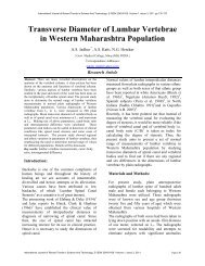
worsen<strong>in</strong>g. On flexible laryngoscopy, the vegetative<br />
foreign body was seen to be lodged <strong>in</strong> the glottis over<br />
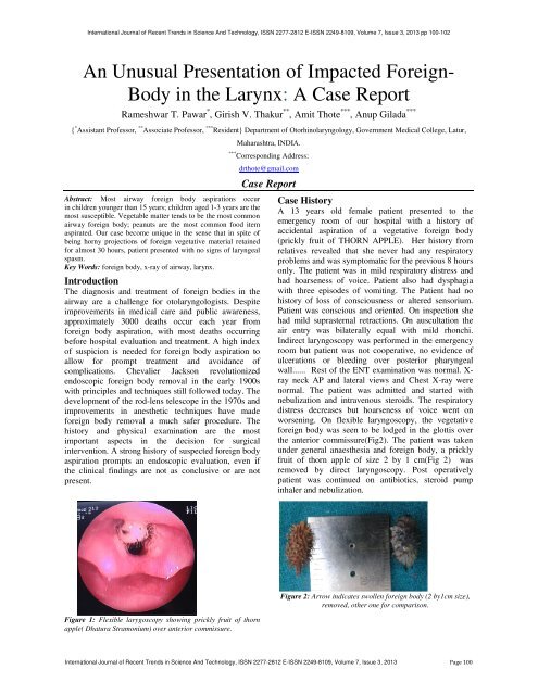
the anterior commissure(Fig2). The patient was taken<br />
under general anaesthesia and foreign body, a prickly<br />
fruit <strong>of</strong> thorn apple <strong>of</strong> size 2 by 1 cm(Fig 2) was<br />
removed by direct laryngoscopy. Post operatively<br />
patient was cont<strong>in</strong>ued on antibiotics, steroid pump<br />
<strong>in</strong>haler and nebulization.<br />
Figure 1: Flexible larygoscopy show<strong>in</strong>g prickly fruit <strong>of</strong> thorn<br />
apple( Dhatura Stramonium) over anterior commissure.<br />
Figure 2: Arrow <strong>in</strong>dicates swollen foreign body (2 by1cm size),<br />
removed, other one for comparison.<br />
International Journal <strong>of</strong> Recent Trends <strong>in</strong> Science <strong>An</strong>d Technology, ISSN 2277-2812 E-ISSN 2249-8109, Volume 7, Issue 3, 2013 Page 100
Rameshwar T. Pawar, Girish V. Thakur, Amit Thote, <strong>An</strong>up Gilada<br />
Discussion<br />
Airway foreign bodies can become lodged <strong>in</strong> the<br />
larynx, trachea, and bronchus. The size and shape <strong>of</strong> the<br />
object determ<strong>in</strong>e the site <strong>of</strong> obstruction; large, round, or<br />
expandable objects produce complete obstruction, and<br />
irregularly shaped objects allow air passage around the<br />
object, result<strong>in</strong>g <strong>in</strong> partial obstruction. Young children<br />
comprise the most common age group for foreign body<br />
aspiration because they tend to put objects <strong>in</strong> their<br />
mouth more frequently and also lack coord<strong>in</strong>ation <strong>of</strong><br />
swallow<strong>in</strong>g and glottic closure and lack molars for<br />
proper gr<strong>in</strong>d<strong>in</strong>g <strong>of</strong> food contributes for foreign body<br />
aspiration. After foreign body aspiration occurs, the<br />
foreign body can settle <strong>in</strong>to 3 anatomic sites, the larynx,<br />
trachea, or bronchus. Of aspirated foreign bodies, 80-<br />
90% become lodged <strong>in</strong> the bronchi. In adults, bronchial<br />
foreign bodies tend to be lodged <strong>in</strong> the right ma<strong>in</strong><br />
bronchus because <strong>of</strong> its lesser angle <strong>of</strong> convergence<br />
compared with the left bronchus and because <strong>of</strong> the<br />
location <strong>of</strong> the car<strong>in</strong>a left <strong>of</strong> the midl<strong>in</strong>e. Several papers<br />
have demonstrated equal frequency <strong>of</strong> right and left<br />
bronchial foreign bodies <strong>in</strong> children. Larger objects<br />
tend to become lodged <strong>in</strong> the larynx or trachea.<br />
In general, aspiration <strong>of</strong> foreign bodies produces the<br />
follow<strong>in</strong>g 3 phases:<br />
Initial phase - Chok<strong>in</strong>g and gasp<strong>in</strong>g, cough<strong>in</strong>g, or<br />
airway obstruction at the time <strong>of</strong> aspiration<br />
Asymptomatic phase - Subsequent lodg<strong>in</strong>g <strong>of</strong> the object<br />
with relaxation <strong>of</strong> reflexes that <strong>of</strong>ten results <strong>in</strong> a<br />
reduction or cessation <strong>of</strong> symptoms, last<strong>in</strong>g hours to<br />
weeks<br />
Complications phase - <strong>Foreign</strong> body produc<strong>in</strong>g erosion<br />
or obstruction lead<strong>in</strong>g to pneumonia, atelectasis, or<br />
abscess<br />
Cl<strong>in</strong>ical presentation depends on the location <strong>of</strong> the<br />
foreign body. A large foreign body lodged <strong>in</strong> the<br />
larynx or trachea can produce complete airway<br />
obstruction from either the dimensions <strong>of</strong> the object or<br />
the result<strong>in</strong>g edema.<br />
Laryngeal foreign bodies present with airway<br />
obstruction and hoarseness or aphonia.<br />
Tracheal foreign bodies present similarly to laryngeal<br />
foreign bodies but without hoarseness or aphonia.<br />
Tracheal foreign bodies can demonstrate wheez<strong>in</strong>g<br />
similar to asthma. Bronchial foreign bodies typically<br />
present with cough, unilateral wheez<strong>in</strong>g, and decreased<br />
breath sounds, but only 65% <strong>of</strong> patients present with<br />
this classic triad. <strong>Foreign</strong> body aspiration can mimic<br />
other respiratory problems, such as asthma. <strong>Foreign</strong><br />
body aspiration differs <strong>in</strong> the presence <strong>of</strong> unilateral<br />
wheez<strong>in</strong>g and decreased breath sounds. Chest<br />
auscultation is critical <strong>in</strong> the evaluation <strong>of</strong> a patient <strong>in</strong><br />
whom a foreign body aspiration is suspected. Typically,<br />
these patients have wheez<strong>in</strong>g, decreased breath sounds,<br />
or both on the side <strong>of</strong> the foreign body. Patients may<br />
have normal exam<strong>in</strong>ation f<strong>in</strong>d<strong>in</strong>gs despite hav<strong>in</strong>g a<br />
foreign body with<strong>in</strong> the airway because it may partially<br />
obstruct the airway. High-kilovolt anteroposterior and<br />
lateral radiographs <strong>of</strong> the airway are the tests <strong>of</strong> choice<br />
<strong>in</strong> patients <strong>in</strong> whom laryngeal foreign bodies are<br />
suspected. The high kilovoltage used produces greater<br />
def<strong>in</strong>ition <strong>of</strong> the airway while reduc<strong>in</strong>g the effect <strong>of</strong> the<br />
surround<strong>in</strong>g bony structures. Posteroanterior and lateral<br />
chest radiographs are an adjunct to the history and<br />
physical exam<strong>in</strong>ation <strong>in</strong> patients <strong>in</strong> whom foreign body<br />
aspirations are suspected. To at least have a basel<strong>in</strong>e<br />
study for future comparison, perform chest radiographs<br />
on all such patients. Radiopaque objects are visible, but<br />
radiolucent objects (eg, plastic) are not. Chest<br />
radiographs may reveal obstructive emphysema or<br />
hyper<strong>in</strong>flation, atelectasis, and consolidation. Biplane<br />
fluoroscopy uses <strong>in</strong>traoperative fluoroscopic evaluation<br />
while identify<strong>in</strong>g and locat<strong>in</strong>g a foreign body with<strong>in</strong> the<br />
lung periphery.<br />
Treatment<br />
Patients with complete airway obstruction require<br />
immediate medical attention and typically are aphonic<br />
and unable to breathe. Patients who are cough<strong>in</strong>g,<br />
gagg<strong>in</strong>g, and vocaliz<strong>in</strong>g have partial obstruction.<br />
• Use <strong>of</strong> the Heimlich maneuver has improved the<br />
mortality rate <strong>of</strong> patients with complete airway<br />
obstruction, but use <strong>of</strong> it <strong>in</strong> patients with partial<br />
obstruction may produce complete obstruction.<br />
• Most patients who arrive at the hospital are beyond<br />
the acute stage and are not <strong>in</strong> respiratory distress.<br />
• After a complete history and physical exam<strong>in</strong>ation are<br />
completed and radiographic studies are performed, a<br />
decision is made <strong>in</strong> regard to the need for surgical<br />
<strong>in</strong>tervention.<br />
In most cases, antibiotics and steroids are<br />
adm<strong>in</strong>istered preoperatively.<br />
Surgical Therapy<br />
<strong>An</strong> operat<strong>in</strong>g room well equipped with proper<br />
endoscopic equipment <strong>of</strong> various sizes, personnel<br />
familiar with the use <strong>of</strong> the <strong>in</strong>strumentation, and<br />
anesthesiologists experienced <strong>in</strong> foreign body removal<br />
are critical for safe removal <strong>of</strong> airway foreign bodies.<br />
Preoperative Details<br />
Select and organize age-appropriate endoscopic<br />
equipment before the patient enters the operat<strong>in</strong>g room.<br />
Various foreign body forceps should be available for<br />
use, and a similar object should be available for<br />
comparison. Communication between the endoscopist<br />
and anesthesiologist before the procedure to outl<strong>in</strong>e a<br />
plan <strong>of</strong> action is critical. Prior to surgical <strong>in</strong>tervention <strong>in</strong><br />
patients who are not <strong>in</strong> respiratory distress, the patient<br />
should rema<strong>in</strong> on noth<strong>in</strong>g by mouth (NPO) status for an<br />
adequate period to prevent aspiration.<br />
Intraoperative Details<br />
Use <strong>in</strong>halational anesthetics to anesthetize patients.<br />
Apply 1-2% lidoca<strong>in</strong>e to the larynx to reduce reflexes<br />
and prevent laryngospasm. Keep patients spontaneously<br />
breath<strong>in</strong>g throughout the procedure for control <strong>of</strong> the<br />
airway.<br />
Copyright © 2013, <strong>Statperson</strong> Publications, International Journal <strong>of</strong> Recent Trends <strong>in</strong> Science <strong>An</strong>d Technology, ISSN 2277-2812 E-ISSN 2249-8109, Volume 7, Issue 3, 2013
International Journal <strong>of</strong> Recent Trends <strong>in</strong> Science <strong>An</strong>d Technology, ISSN 2277-2812 E-ISSN 2249-8109, Volume 7, Issue 3, 2013 pp 100-102<br />
With laryngeal foreign bodie, the laryngoscope tip is<br />
placed <strong>in</strong> the vallecula for exposure, and the foreign<br />
body is visualized <strong>in</strong> the larynx and removed with<br />
appropriate foreign body forceps. After removal,<br />
reassess the larynx for other foreign bodies. Perform<br />
rigid bronchoscopy afterward to assess for other foreign<br />
bodies <strong>in</strong> the lower airway.<br />
In tracheobronchial foreign body removal, the<br />
bronchoscope is <strong>in</strong>serted <strong>in</strong>to the airway after exposure<br />
to the larynx, and cont<strong>in</strong>uous ventilation <strong>of</strong> the patient<br />
is provided through the bronchoscope. In a patient with<br />
a bronchial foreign body, the unaffected side is<br />
exam<strong>in</strong>ed first. The bronchoscope then is placed<br />
immediately above the foreign body. Secretions are<br />
gently suctioned around the object. The patient is<br />
oxygenated with 100% oxygen before any attempt at<br />
removal. The forceps are placed through the<br />
bronchoscope, and the object is grasped after complete<br />
visualization <strong>of</strong> the foreign body. The bronchoscope is<br />
advanced to the foreign body while the surgeon<br />
cont<strong>in</strong>ues to grasp the object. The foreign body, foreign<br />
body forceps, and bronchoscope are removed as a unit,<br />
and the bronchoscope immediately is returned to the<br />
airway for ventilation and reassessment for other<br />
foreign bodies. Occasionally, easy retrieval <strong>of</strong> the<br />
foreign body is not possible. Larger objects unable to<br />
pass through the larynx can be broken <strong>in</strong>to pieces and<br />
removed. If the object cannot pass through the larynx, a<br />
tracheotomy can be performed to remove the object<br />
through the tracheostoma. At times, the object becomes<br />
embedded <strong>in</strong>to the surround<strong>in</strong>g mucosa because <strong>of</strong><br />
edema caused by the object or because <strong>of</strong> multiple<br />
failed attempts at removal. In this situation, stop and to<br />
wait 48-72 hours to allow the edema to subside for a<br />
repeat attempt at removal. Thoracotomy may be<br />
necessary when the object stays embedded after failed<br />
endoscopic attempts. <strong>Foreign</strong> bodies <strong>in</strong> the distal<br />
bronchial segments may be removed with the use <strong>of</strong> a<br />
Fogarty endovascular catheter through the suction port<br />
<strong>of</strong> a rigid bronchoscope. Flexible bronchoscopy as an<br />
adjunct may be beneficial <strong>in</strong> the removal <strong>of</strong> distal<br />
objects.<br />
Sharp objects are extremely challeng<strong>in</strong>g for endoscopic<br />
removal. The po<strong>in</strong>ted end tends to engage <strong>in</strong> the<br />
mucosa, caus<strong>in</strong>g the object to tumble with the po<strong>in</strong>t<br />
trail<strong>in</strong>g. Po<strong>in</strong>ted objects tend to be bendable or<br />
breakable. The bronchoscope is placed <strong>in</strong>to the airway,<br />
and, us<strong>in</strong>g foreign body forceps, the po<strong>in</strong>ted end <strong>of</strong> the<br />
object is disengaged from the mucosa, moved distally,<br />
and then removed. P<strong>in</strong>-bend<strong>in</strong>g forceps may be used <strong>in</strong><br />
certa<strong>in</strong> situations. Safety p<strong>in</strong> removal is uniquely<br />
challeng<strong>in</strong>g; removal is performed endoscopically by<br />
sheath<strong>in</strong>g the po<strong>in</strong>ted end <strong>in</strong>to the endoscope and<br />
lock<strong>in</strong>g the keeper outside the endoscope. Open<br />
removal by thoracotomy may be necessary when the<br />
sharp object is severely embedded <strong>in</strong>to the mucosa. [2]<br />
Postoperative Details<br />
The use <strong>of</strong> steroids or racemic ep<strong>in</strong>ephr<strong>in</strong>e is not<br />
necessary when age-appropriate endoscopes are used.<br />
<strong>An</strong>tibiotics typically are not prescribed because the<br />
source <strong>of</strong> <strong>in</strong>fection has been removed. Chest<br />
physiotherapy is performed after foreign body removal<br />
to help remove secretions. Patients are discharged when<br />
fully awake and breath<strong>in</strong>g comfortably without the need<br />
for supplemental oxygen. Chest radiographs are<br />
performed postoperatively if the patient's signs and<br />
symptoms persist or worsen.<br />
Most complications are the result <strong>of</strong> a delay <strong>in</strong><br />
diagnosis.<br />
Of patients with laryngotracheal foreign bodies, 67%<br />
experience associated complications when the removal<br />
delay is more than 24 hours.Pneumonia and atelectasis<br />
are the most common complications secondary to and<br />
after removal <strong>of</strong> bronchial foreign bodies.Bleed<strong>in</strong>g can<br />
occur from granulation tissue surround<strong>in</strong>g the foreign<br />
body or erosion <strong>in</strong>to a major vessel.Pneumothorax and<br />
pneumomediast<strong>in</strong>um can result from an airway tear.<br />
Conclusion<br />
<strong>Foreign</strong>-body aspiration is a preventable mishap and<br />
can be prevented by educat<strong>in</strong>g the society, especially<br />
parents. Also, it is equally important to educate the<br />
medical fraternity to aid <strong>in</strong> early diagnosis and prompt<br />
removal, thus prevent<strong>in</strong>g a potentially fatal outcome<br />
References<br />
1. Pandhi SC, Agarwal KK. <strong>Foreign</strong> body <strong>in</strong> subglottic<br />
region—an unusual site. Indian J Otolaryngol Head<br />
Neck Surg. 1970; 22(4):223–225.<br />
2. Kumar GK, Ganesan C, Pilai SA, Radhakrishnan E. A<br />
subglottic foreign body <strong>in</strong> a child. Indian J<br />
<strong>An</strong>aesth. 1997; 41(3):207–208.<br />
3. Halvorson DJ, Mann C, Merritt RM, Porubsky ES.<br />
Management <strong>of</strong> subglottic foreign-bodies. <strong>An</strong>n Otol<br />
Rh<strong>in</strong>ol Laryngol. 1996;105: 541–544. [PubMed]<br />
4. Hanukoglu Loss <strong>of</strong> voice as sole symptom <strong>of</strong><br />
subglottic foreign-body aspiration. Am J Dis<br />
Child.1986; 140: 973. [PubMed]<br />
International Journal <strong>of</strong> Recent Trends <strong>in</strong> Science <strong>An</strong>d Technology, ISSN 2277-2812 E-ISSN 2249-8109, Volume 7, Issue 3, 2013 Page 102