Primary Malignant Melanoma of the Cervical Spinal ... - E-kjs.org
Primary Malignant Melanoma of the Cervical Spinal ... - E-kjs.org
Primary Malignant Melanoma of the Cervical Spinal ... - E-kjs.org

You also want an ePaper? Increase the reach of your titles
YUMPU automatically turns print PDFs into web optimized ePapers that Google loves.
<strong>Primary</strong> <strong>Malignant</strong> <strong>Melanoma</strong> <strong>of</strong> <strong>the</strong> <strong>Cervical</strong> <strong>Spinal</strong> Nerve Root<br />
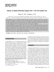
Fig. 2. <strong>Malignant</strong> melanoma composed <strong>of</strong> highly anaplastic cells with hyperchromaic nuclei, abundant<br />
eosinophilic cytoplasm, and melanin pigments (×200) (A). Positive findings for vimentin, s-100 protein,<br />
and HMB-45 (×200) (B).<br />
scattered low signal intensity lesions within <strong>the</strong> mass were<br />
identified on <strong>the</strong> T2 weighted image; <strong>the</strong>se were thought to be<br />
due to <strong>the</strong> presence <strong>of</strong> melanin, later (Fig. 1). Preoperative clini<br />
cal and radiographic diagnosis was nerve sheath tumor such as<br />
neur<strong>of</strong>ibroma or schwannoma. An operation using <strong>the</strong> anterolateral<br />
approach at left supraclavicular area between <strong>the</strong> carotid<br />
sheath and sternocleidomastoid muscle was performed. The<br />
bulk <strong>of</strong> <strong>the</strong> mass was removed. At surgery an encapsulated pitch<br />
black mass was found. It was large, firmly attached to brachial<br />
plexus and C6-7 neural foramen. Pathological examination confirmed<br />
<strong>the</strong> diagnosis <strong>of</strong> malignant melanoma. It is composed <strong>of</strong><br />
highly anaplastic cells with hyperchromaic nuclei, abundant<br />
eosinophilic cytoplasm, and melanin pigments. And it presented<br />
positive findings for vimentin, s-100 protein, and HMB-45<br />
(Fig. 2). The tumor had a high Ki-67 labeling index (35%). The<br />
patient’s symptoms improved totally after <strong>the</strong> operation. The<br />
patient wanted to transfer to o<strong>the</strong>r hospital for more thorough<br />
evaluation on post-op 11th day. So we could not proceed adjuvant<br />
<strong>the</strong>rapy and follow-up observation.<br />
DISCUSSION<br />
The cell <strong>of</strong> origin <strong>of</strong> melanotic tumors involving <strong>the</strong> spinal<br />
nerve root can be ei<strong>the</strong>r a melanocyte, a Schwann cell, or an<br />
uncharacterized cell type 11) . Melanocytes are melanin-producing<br />
cells that arise from <strong>the</strong> neural crest during embryogenesis, and<br />
migrate to <strong>the</strong> skin, mucous membranes, and central nervous<br />
system. Tumors arising from melanocytic cells (primary and<br />
metastatic melanoma) and Schwann cells (melanotic nerve sheath<br />
tumors) have a common embryological origin in <strong>the</strong> neural<br />
crest 8,11) .<br />
Because <strong>of</strong> rarity <strong>of</strong> primary melanoma in spinal nerve root,<br />
<strong>the</strong> diagnosis can be difficult. Radiological studies may not be<br />
specific. Magnetic resonance imaging may <strong>of</strong>fer some indication<br />
<strong>of</strong> a melanotic lesion, melanin can show a hyperintense<br />
or isointense signal on T1 weighted images and decreased signal<br />
on T2 weighted images 7,9,12) .<br />
The pathological differential diagnosis <strong>of</strong> primary melanoma<br />
includes metastatic melanoma, melanocytoma, melanotic schwannoma,<br />
and melanotic clear-cell sarcoma. Melanocytoma lacks<br />
anaplastic features such as necrosis, significant mitotic activity,<br />
and pleomorphism, in contrast with primary melanoma. The<br />
characteristics <strong>of</strong> primary malignant melanomas are considerable,<br />
including nuclear pleomorphisms leading to large and bizarreshaped<br />
tumor cells with multinucleated giant cells 13) . A high<br />
mitotic rate, coagulative necrosis and hemorrhage are also commonly<br />
found in primary malignant melanoma 1,13) . Immunohistochemical<br />
staining is also helpful for differentiation. Significant<br />
s-100 protein expression without <strong>the</strong> expression <strong>of</strong> epi<strong>the</strong>lial<br />
membrane antigen supports <strong>the</strong> diagnosis <strong>of</strong> a schwannoma<br />
or melanoma 5,13) . Negative immunoreactivity for Leu-7<br />
rules out schwannoma whereas <strong>the</strong> detection <strong>of</strong> HMB-45 favors<br />
<strong>the</strong> diagnosis <strong>of</strong> a melanocytic tumor 5,13) .<br />
Schneider et al. 11) reported a case <strong>of</strong> primary melanoma arising<br />
from <strong>the</strong> L3 spinal nerve root that was treated by wide<br />
en-bloc resection. Skarli et al. 12) reported a similar case <strong>of</strong> primary<br />
malignant melanoma arising in a cervical spinal nerve<br />
root that did not recur or metastasize over 3 years without adjuvant<br />
chemo<strong>the</strong>rapy or radiation <strong>the</strong>rapy after surgical operation.<br />
Also Kwon et al. 4) reported a case <strong>of</strong> primary malignant melanoma<br />
<strong>of</strong> <strong>the</strong> C7 nerve root in 2004 that was treated with adjuvant<br />
radiation <strong>the</strong>rapy.<br />
Because <strong>of</strong> <strong>the</strong> rarity <strong>of</strong> <strong>the</strong>se lesions, <strong>the</strong> prognosis and proper<br />
adjuvant <strong>the</strong>rapy (chemo<strong>the</strong>rapy, radio<strong>the</strong>rapy) have not been<br />
established yet.<br />
It is important to differentiate primary melanoma from metastasis.<br />
The prognosis for patients with metastatic melanoma to<br />
central nervous system is dismal, with a life expectancy <strong>of</strong> less<br />
than 1 year in most studies 2,4,10) . In contrast, primary melanoma<br />
has long-term survival and better prognosis than metastatic lesion.<br />
Kor J Spine 6(1) March 2009 41