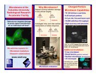
Why Microbeams? - raraf
Why Microbeams? - raraf
Why Microbeams? - raraf

Create successful ePaper yourself
Turn your PDF publications into a flip-book with our unique Google optimized e-Paper software.
<strong>Why</strong> <strong>Microbeams</strong><br />
<br />
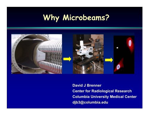
A microbeam can deposit ionizing radiation damage<br />
in microscopic or sub-microscopic regions of cells<br />
David J Brenner<br />
Center for Radiological Research<br />
Columbia University Medical Center<br />
djb3@columbia.edu
<strong>Why</strong> <strong>Microbeams</strong><br />
<br />
<br />
<br />
A microbeam can deposit ionizing radiation damage<br />
in microscopic or sub-microscopic regions of cells<br />
Allows investigation of single-particle effects<br />
Allows investigation of intra- and inter-cellular<br />
mechanisms of stress response
Single-particle effects: Radon
Single-particle effects: Radon<br />
1518 Woodcut:<br />
Doctor and<br />
nurse treating a<br />
sick miner at<br />
the Joachimsthal<br />
hospital
Radon-Related Rationale for Microbeam:<br />
Number of target cells exposed to a<br />
given number of alpha particle traversals<br />
# of<br />
traversals<br />
Colorado<br />
miners<br />
Domestic<br />
radon<br />
exposure<br />
“Conventional”<br />
in vitro<br />
experiments<br />
0 700,000 100,000,000 37,000<br />
1 3,500,000 20,000 37,000<br />
2 8,500,000 0 18,000<br />
3 14,000,000 0 6,000<br />
4 17,000,000 0 1,500<br />
5 17,000,000 0 300
Oncogenic transformation by exactly one alpha<br />
particle and a Poisson mean of one alpha particle<br />
Transformation Frequency<br />
(Miller et al 99)
<strong>Why</strong> <strong>Microbeams</strong><br />
<br />
<br />
<br />
A microbeam can deposit ionizing radiation damage<br />
in microscopic or sub-microscopic regions of cells<br />
Allows investigation of single-particle effects<br />
Allows investigation of intra- and inter-cellular<br />
mechanisms of stress response
Cytoplasmic irradiation
<strong>Why</strong> <strong>Microbeams</strong><br />
<br />
<br />
<br />
A microbeam can deposit ionizing radiation damage<br />
in microscopic or sub-microscopic regions of cells<br />
Allows investigation of single-particle effects<br />
Allows investigation of intra- and inter-cellular<br />
mechanisms of stress response
“Providing researchers with tools and<br />
capabilities to make new discoveries”<br />
Single-cell microbeams<br />
Intra-cellular studies<br />
Inter-cellular studies<br />
Single cells Cells Tissues Animals
A quantitative example of inter-cellular damage communication:<br />
Bystander Responses<br />
Damage is expressed in “bystander” cells,<br />
which are near to an irradiated cell, but have not<br />
themselves received any energy deposition
Bystander effects have been reported for a<br />
variety of endpoints using single-cell systems<br />
<br />
<br />
<br />
<br />
<br />
<br />
<br />
<br />
Sister-chromatid exchanges<br />
Cell killing (mitotic and apoptotic)<br />
Micronucleus induction<br />
Mutation induction<br />
In-vitro<br />
oncogenic transformation<br />
Changes in gene expression<br />
Altered cell growth<br />
And for almost every radiogenic endpoint<br />
that has been studied!
<strong>Why</strong> is there so much interest in<br />
bystander effects<br />
1. Basic interest in damage signal transduction<br />
2. Practical interest in low-dose risk estimation..
Low-dose risk estimation<br />
and the bystander effect<br />
Induced CD59 - Mutants per 10 5 Survivors<br />
140<br />
120<br />
100<br />
80<br />
60<br />
40<br />
20<br />
Measured mutations<br />
0<br />
0 20 40 60 80 100<br />
Percent of Cells Irradiated with One Alpha Particle<br />
Based on mutation data from the<br />
RARAF microbeam.<br />
Zhou et al PNAS 98, 14410-5 (2001)<br />
• Where bystander responses<br />
have been quantitated,<br />
they have shown saturation<br />
• In such cases,<br />
extrapolating linearly from<br />
low to very low doses<br />
could underestimate the<br />
risk at very low doses.
Various experimental approaches to<br />
bystander studies<br />
<br />
<br />
<br />
Irradiate with a broad beam of high-LET<br />
radiation at a very low dose, such that most<br />
cells not hit<br />
Intra-media signal transfer<br />
‣ Irradiate cells/medium, then transfer irradiated<br />
medium/cells onto fresh cells<br />
‣ Co-culturing dishes<br />
Microbeam studies:<br />
‣ Hit only specified cells in the field
Early Bystander Studies<br />
at the Columbia Microbeam<br />
Shoot α particles at the<br />
fibroblasts with blue-stained<br />
nuclei, but not at those with<br />
red-stained cytoplasm,<br />
then score micronuclei<br />
Frequency of micronuclei:<br />
• Controls 0.8±0.6%<br />
• Hit cells 30±4%<br />
• Non-hit cells 5±1%
We can hit a predetermined fraction of cells...
In-vitro oncogenic transformation with microbeam<br />
White: All cells hit by α particles<br />
Yellow: Only 1 in 10 cells hit<br />
Observed frequency / 10 4 surviving cells<br />
16<br />
14<br />
12<br />
10<br />
8<br />
6<br />
4<br />
2<br />
0<br />
Semi-confluent<br />
single-cell monolayer<br />
0 1 2 3 4 5 6 7 8<br />
# of α particles through hit cells<br />
Miller et al.<br />
PNAS
We can kill a defined fraction of cells on a dish,<br />
the remainder not being hit….<br />
20 α-particles<br />
X<br />
X<br />
20 α-particles through the nucleus kills the cells that are hit (SF< 1%)<br />
<br />
Mutations observed therefore come from unhit “bystander” cells.
Mutations in A L cells:<br />
20% of the cells each hit by 20 α particles<br />
Mutation Frequency<br />
T. K. Hei PNAS
Most bystander studies<br />
have been performed with<br />
single-cell systems<br />
In that bystander effects involve<br />
cell-to-cell communication,<br />
it is important to study these effects<br />
in normal three-dimensional human<br />
tissue
Microbeam-based bystander studies in<br />
human artificial 3-D skin
Microbeam-based bystander experiments<br />
in human 3-D tissue systems
Apoptosis<br />
Micronuclei<br />
Fraction of apoptotic cells<br />
0.06<br />
0.04<br />
0.02<br />
Bystanders<br />
Controls<br />
0.00<br />
100 300 500 700 900 1100<br />
Distance from irradiated cells (microns)<br />
Fraction of cells with micronuclei<br />
0.03<br />
0.02<br />
Unirradiated bystanders<br />
Controls<br />
Bystanders<br />
Controls<br />
0.01<br />
100 300 500 700 900 1100<br />
Distance from irradiated cells (microns)<br />
Belyakov et al PNAS 102, 14203-8 (2005)<br />
<br />
<br />
For both apoptosis and micronucleus induction,<br />
the range of the bystander effect in tissue is about<br />
1 mm, or 50 to 100 cells<br />
The average enhancement in effect, over this<br />
range of distances, is about 1.6 for micronuclei<br />
and 2.8 for apoptosis.
α-particle microbeam irradiation – 1 hour post-irradiation<br />
Phosphorylation of p53 at Ser 15<br />
0.35<br />
0.3<br />
Zone of Irradiation<br />
Intensity (AU)<br />
0.25<br />
0.2<br />
0.15<br />
0.1<br />
• Individual bystander / irradiated<br />
cells in partially irradiated tissue<br />
O Individual cells in control tissue<br />
Individual Cells in Control Tissue<br />
Individual Cells in Irradiated Tissue<br />
0.05<br />
0<br />
-10000 -500 10000 1500 1000 2000<br />
Distance from Distance irradiated (mm) cells (mm)
The mechanisms underlying<br />
bystander effects<br />
Due to unexpected circumstances<br />
this key slide cannot yet be shown
The growth of single-cell microbeams<br />
1<br />
Gray Laboratory, London<br />
2<br />
3<br />
4<br />
Dublin, Ireland**<br />
5<br />
Stresa, Italy<br />
6<br />
Oxford, England<br />
7<br />
International Workshops on Microbeam<br />
Probes of Cellular Radiation Response<br />
Pacific Northwest Labs, Washington<br />
Columbia University, New York**<br />
Columbia University, New York**<br />
Number of<br />
Year<br />
microbeams<br />
1993<br />
3<br />
1995<br />
3<br />
1997<br />
4<br />
1999<br />
7<br />
2001<br />
12<br />
2003<br />
17<br />
2006<br />
28<br />
8<br />
Chiba, Japan<br />
2008<br />
32<br />
10<br />
7<br />
9 Darmstadt, Germany 2010 36<br />
Number of single-cell microbeams<br />
30<br />
20<br />
10 Columbia University, New York** 2012<br />
40<br />
0<br />
1993 1995 1997 1999 2001 2003 2005 2007 2009
SINGLE-CELL MICROBEAMS, 2006<br />
LABORATORY LET ON LINE BIOLOGY <br />
Columbia University I, New York high Yes Yes<br />
Columbia University II, New York low to very high Yes Yes<br />
Gray Laboratory, London low, high Yes Yes<br />
Gray Laboratory, London soft X Yes Yes<br />
JAERI, Takasaki, Japan high Yes Yes<br />
PNL, Richland, Washington low Yes Yes<br />
GSI, Darmstadt, Germany high Yes Yes<br />
Texas A&M, College Station low Yes No<br />
PTB, Braunschweig, Germany low, high Yes Yes<br />
Bordeaux, France low, high Yes No<br />
Padua, Italy low, high Yes No<br />
Leipzig, Germany low, high Yes No<br />
MIT, Boston low, high Yes No<br />
Munich, Germany low, high Yes No<br />
L’Aquila, Italy soft X No No<br />
Padua, Italy high Yes No<br />
LBL, Berkeley very high No No<br />
University of Maryland low Yes Belfast, No N. Ireland<br />
Lund, Sweden low, high Yes Chiba, No Japan<br />
Tsukuba, Japan soft X Yes No<br />
Tsuruga, Japan<br />
Nagatani, Japan Low, high Yes Yes<br />
Seoul, South Korea Low Yes McMaster, Yes Canada<br />
..Helsinki, Finland High No Nagasaki, No Japan<br />
Chapel Hill, North Carolina Low No No<br />
Hefei, China<br />
Saclay, France Low, high Yes No<br />
Cracow, Poland Low Yes Shanghai, Yes China<br />
Gradignan, France High Yes Yes<br />
Lanzhou, China<br />
Surrey University, UK Low, High No No
The Single-Cell Microbeam at the Radiological<br />
Research Accelerator Facility - RARAF<br />
A biologist-friendly facility for probing<br />
intra-cellular and inter-cellular communication