LASER ION SOURCE DEVELOPMENT FOR THE - raraf
LASER ION SOURCE DEVELOPMENT FOR THE - raraf
LASER ION SOURCE DEVELOPMENT FOR THE - raraf
Create successful ePaper yourself
Turn your PDF publications into a flip-book with our unique Google optimized e-Paper software.
CENTER <strong>FOR</strong> RADIOLOGICAL RESEARCH • ANNUAL REPORT 2004<br />
RARAF − Table of Contents<br />
RARAF Staff and Picture..................................................................................................................................................66<br />
Research Using RARAF ...................................................................................................................................................67<br />
Development of Facilities .................................................................................................................................................70<br />
Accelerator Utilization and Operation ..............................................................................................................................73<br />
Training.............................................................................................................................................................................74<br />
Personnel...........................................................................................................................................................................74<br />
Recent Publications of Work Performed at RARAF (2003-2004)....................................................................................74<br />
RARAF PROFESS<strong>ION</strong>AL STAFF<br />
David J. Brenner, Ph.D., D.Sc. − RARAF Director<br />
Stephen A. Marino, M.S. − RARAF Manager<br />
Gerhard Randers-Pehrson, Ph.D. − RARAF Associate Director, Chief Physicist<br />
Charles R. Geard, Ph.D. − CRR Associate Director, Senior Biologist<br />
Alan Bigelow, Ph.D. − Associate Research Scientist<br />
Brian Ponnaiya, Ph.D. − Associate Research Scientist<br />
Guy Y. Garty, Ph.D. − Staff Associate<br />
Gregory Ross, Ph.D. − Programmer Analyst<br />
Stephen Mitchell, Ph.D. − Post-Doctoral Research Scientist<br />
Giuseppe Schettino, Ph.D. − Post-Doctoral Research Scientist<br />
Gloria Jenkins-Baker, B.A. − Biology Technician<br />
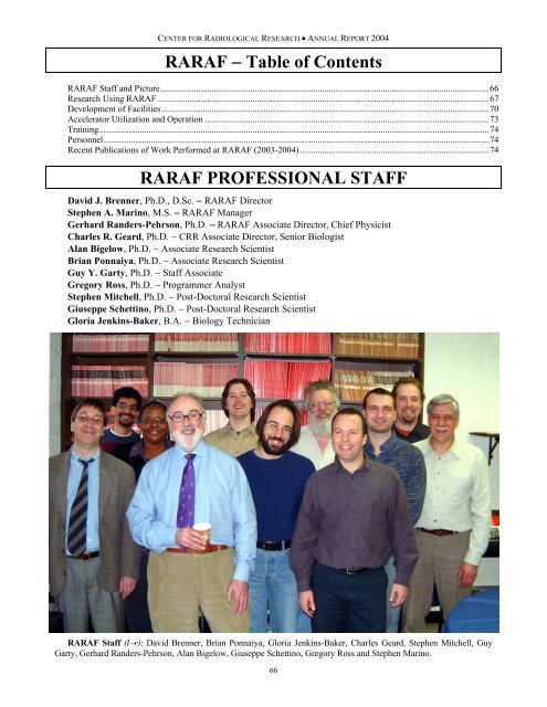
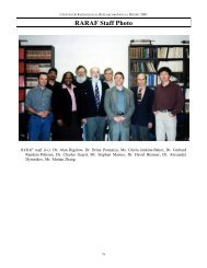
RARAF Staff (l−r): David Brenner, Brian Ponnaiya, Gloria Jenkins-Baker, Charles Geard, Stephen Mitchell, Guy<br />
Garty, Gerhard Randers-Pehrson, Alan Bigelow, Giuseppe Schettino, Gregory Ross and Stephen Marino.<br />
66
<strong>THE</strong> RADIOLOGICAL RESEARCH ACCELERATOR FACILITY<br />
The Radiological Research Accelerator Facility<br />
AN NIH-SUPPORTED RE<strong>SOURCE</strong> CENTER – WWW.RARAF.ORG<br />
Director: David J. Brenner, Ph.D., D.Sc.<br />
Manager: Stephen A. Marino, M.S.<br />
Chief Physicist: Gerhard Randers-Pehrson, Ph.D.<br />
This is a major year for RARAF. Our Van de Graaff accelerator,<br />
which is 55 years old and has provided us with<br />
charged particle beams for over 38 years, will be decommissioned<br />
this May. We will install a new Singletron from High<br />
Voltage Engineering (HVE) that should provide us with increased<br />
voltage, stability and beam current.<br />
Among the major accomplishments this year:<br />
• The first microbeam irradiations using the new microbeam<br />
facility (Microbeam II);<br />
• Construction of a successful quadrupole triplet, producing<br />
a helium ion beam spot 3.5 µm in diameter;<br />
• Construction of the stand-alone microbeam vacuum system<br />
and installation and testing of the quadrupole magnets.<br />
Research Using RARAF<br />
For several years, the focus of most of the biology experiments<br />
at RARAF has been the “bystander” effect, in<br />
which cells that are not irradiated show a response to radiation<br />
when in close contact with or even only in the presence<br />
of irradiated cells. Several experiments examining this effect<br />
were continued this year and new ones initiated, observing a<br />
variety of endpoints to determine the size of the effect and<br />
the mechanism(s) by which it is transmitted. Evidence has<br />
been obtained for both direct gap junction communication<br />
through cell membrane contact and indirect, long-range<br />
communication through media transfer. In some experiments,<br />
the unirradiated cells can be identified due to differential<br />
staining and scored directly, in other experiments unirradiated<br />
cells are physically separated from the irradiated<br />
cells during irradiation. Both the microbeam and the track<br />
segment facilities continue to be utilized in various investigations<br />
of this phenomenon. The single-particle microbeam<br />
facility provides precise control of the number and location<br />
of particles so that irradiated and bystander cells may be<br />
distinguished but is somewhat limited in the number of cells<br />
that can be irradiated. The track segment facility provides<br />
broad beam irradiation that provides a random pattern of<br />
charged particles but allows large numbers of cells to be<br />
irradiated and multiple users in a single day.<br />
In Table I are listed the experiments performed at<br />
RARAF from November 1, 2003 through October 31, 2004<br />
and the number of days each was run in this period. Use of<br />
the accelerator for experiments was 50% of the normal<br />
available time, 50% higher than last year and the highest we<br />
have attained at Nevis Labs. In addition, for the first time in<br />
over a decade outside users accounted for half the experiment<br />
time. Sixteen different experiments were run during<br />
this period, about the same as the average for 1997−2003.<br />
Seven experiments were undertaken by members of the<br />
CRR, supported by grants from the National Institutes of<br />
Health (NIH) and the Department of Energy (DOE). Nine<br />
experiments were performed by outside users, supported by<br />
grants and awards from the NIH, the National Aeronautics<br />
and Space Administration (NASA), the National Science<br />
Foundation (NSF), and the Ministry of Education, Science,<br />
Sports and Culture of Japan. Brief descriptions of these experiments<br />
follow.<br />
Eric Hall and Stephen Mitchell of the Center for Radiological<br />
Research (CRR) continued investigations involving<br />
the oncogenic neoplastic transformation of mouse C3H<br />
10T½ cells (Exp. 73). Using the microbeam facility, 10% of<br />
the cells were irradiated through the nucleus with 2 to 12<br />
helium ions. Cells were plated at densities of approximately<br />
200 and 2000 per dish to try to observe the relative contribution<br />
of cell-cell communication to the bystander effect. Cell<br />
killing and transformation were greater for the cells plated at<br />
the higher density relative to those plated at the lower density.<br />
The results imply that gap junction communication has<br />
a greater role in the bystander effect than media transfer. In<br />
another aspect of the study, a novel radiation apparatus<br />
where irradiated and non-irradiated cells were grown in<br />
close proximity was used to investigate the relationship between<br />
the bystander effect and adaptive response in C3H<br />
10T½ cells. Special “strip” track segment dishes were made<br />
by cutting the Mylar surface on the bottom of special cell<br />
dishes into many equal strips and removing alternate strips.<br />
The remaining Mylar strips are sufficiently thick to stop the<br />
incident ions, so that cells plated on these surfaces are not<br />
irradiated. These dishes were placed inside standard track<br />
segment dishes that have a complete Mylar surface 6 µm<br />
thick, through which the ions readily pass. Cells are plated<br />
on the combined surface. When the cells were left in situ for<br />
24 h for the non-hit cells to co-culture with cells irradiated<br />
with 5 Gy of α-particles using the track segment facility, a<br />
significant increase in both cell killing and oncogenic transformation<br />
frequency was observed. If these cells were<br />
treated with 2 cGy of x-rays 5 h prior to co-culture with irradiated<br />
cells, approximately 95% of the bystander effect was<br />
canceled out. A 2.5-fold decrease in the oncogenic transformation<br />
frequency was also observed. To investigate whether<br />
mouse embryo fibroblast cells haplo-insufficient for one or<br />
more of a number of genes of known importance, namely<br />
ATM, BRCA1 and RAD9, are radiosensitive to cell lethality<br />
and/or oncogenic transformation, cells that are heterozygous<br />
for these genes were irradiated on the track segment facility.<br />
To date no difference has been seen for survival, whereas<br />
cells haplo-insufficient for both ATM and RAD9 are signifi-<br />
67
Exp.<br />
No.<br />
Experimenter<br />
CENTER <strong>FOR</strong> RADIOLOGICAL RESEARCH • ANNUAL REPORT 2004<br />
Table I.<br />
Experiments Run at RARAF, Nov. 1, 2003–Oct. 31, 2004<br />
Institution<br />
Exp.<br />
Type<br />
Title of Experiment<br />
No.<br />
Days<br />
Run<br />
S. Mitchell,<br />
Neoplastic transformation of mouse cells<br />
73 CRR Biology<br />
E.J. Hall<br />
by α-particles<br />
6.9<br />
R. Eliassi,<br />
82<br />
(G. Garty)<br />
UCLA Physics 1. Detection of explosives 9.8<br />
R. H. Maurer, Johns Hopkins<br />
2. Calibration of a portable real-time<br />
89 Physics<br />
et al.<br />
University<br />
neutron spectrometry system<br />
0.6<br />
92 S. Amundson NIH<br />
Functional genomics of cellular response<br />
Biology<br />
to high-LET radiation<br />
5.0<br />
Single cell responses in hit and bystander<br />
B. Ponnaiya,<br />
94 CRR Biology cells: single-cell RT-PCR and protein<br />
C.R. Geard<br />
immunofluorescence<br />
8.3<br />
G. Jenkins,<br />
Damage induction and characterization<br />
103 CRR Biology<br />
C.R. Geard<br />
in known hit versus non-hit human cells<br />
9.6<br />
B. Ponnaiya,<br />
Track segment alpha-particles, cell cocultures<br />
and the bystander effect<br />
106 CRR Biology<br />
C.R. Geard<br />
2.3<br />
Identification of molecular signals of<br />
H. Zhou,<br />
110 CRR Biology alpha-particle-induced bystander<br />
T.K. Hei<br />
mutagenesis<br />
13.2<br />
113 A. Miller AFRRI<br />
Role of alpha-particle radiation in depleted<br />
uranium-induced cellular effects<br />
Biology 1.0<br />
R. Kolesnick, MSKCC; Mass. Characterization of the radiosensitive<br />
118 Biology<br />
G. Perez General Hosp.<br />
target for cell death in mouse oocytes<br />
3.5<br />
A. Zhu,<br />
The bystander effect in mouse embryo<br />
121 CRR Biology<br />
H. Lieberman<br />
stem cells with mutant Mrad9 gene<br />
14.6<br />
SSG Precision<br />
Strength of epoxy joints after neutron<br />
122 K. Kosakowski Physics<br />
Optics, Inc.<br />
irradiation<br />
1.3<br />
123 E. Aprile CU Astrophyics Physics Calibration of a liquid xenon detector 22.5<br />
Chromatid fragment induction detected<br />
Ntnl. Inst. of<br />
M. Suzuki (H.<br />
with the PCR technique by cytoplasmic<br />
125 Radiological Biology<br />
Zhou)<br />
irradiation in normal human bronchial<br />
Science, Japan<br />
cells<br />
16.8<br />
O. Sedelnikova<br />
γ-H2AX foci formation in directly irradiated<br />
and bystander cells<br />
126 NIH Biology<br />
(L. Smilenov)<br />
3.5<br />
129<br />
W. Morgan<br />
(S. Mitchell)<br />
University of<br />
Maryland<br />
Biology Investigation of the bystander effect 6.1<br />
Note: Names in parentheses are CRR members who collaborated with outside experimenters.<br />
68<br />
cantly more prone to transformation than wild-type cells.<br />
Development of a method to detect explosives in baggage<br />
(Exp. 82) was resumed this year. Ravash Eliassi, an<br />
undergraduate student at UCLA, in collaboration with Guy<br />
Garty and under the guidance of Gerhard Randers-Pehrson,<br />
both of the CRR, made measurements of neutron spectra and<br />
yield from the Be 9 (p,n) reaction using a very thin beryllium<br />
target. The detection system is based on resonant scattering<br />
of 0.43 MeV neutrons by nitrogen and oxygen. Measurements<br />
were made for several combinations of reaction angle<br />
and incident proton energy that produce 0.43 MeV neutrons<br />
to determine parameters producing the highest yield and the<br />
spectrum that contains the highest percentage of neutrons of<br />
the desired energy.<br />
Richard Maurer, David Roth and James Kinnison of<br />
Johns Hopkins University performed an irradiation (Exp. 89)<br />
with neutrons of a pair of charged-coupled devices (CCDs)<br />
to be used to send video signals from the NASA New Horizons<br />
probe that will travel past<br />
Pluto to the outer asteroid belt.<br />
Since the probe will spend<br />
much of its life too far away to<br />
use solar panels, there will be<br />
a radioisotope thermoelectric<br />
generator (RTG) on board that<br />
uses the radioactive decay of<br />
plutonium to produce electricity.<br />
The potential effect of<br />
neutrons emitted by the plutonium<br />
on the resolution of the<br />
CCDs was determined. A neutron<br />
energy of 2.1 MeV was<br />
used because it is near the<br />
average energy of the neutrons<br />
produced by the fission of<br />
plutonium.<br />
Sally Amundson, now a<br />
member of the CRR, is conducting<br />
two types of experiments<br />
concerning the radiation-induced<br />
gene expression<br />
profiles in human cell lines<br />
using cDNA microarray hybridization<br />
and other methods<br />
(Exp. 92). The first involves<br />
track segment irradiation for<br />
comparison of gene expression<br />
responses to direct and<br />
bystander irradiation. In these<br />
experiments, gene expression<br />
at 4 and 24 hours post treatment<br />
are compared. Early<br />
experiments have worked well<br />
and they are now being repeated<br />
to establish reproducibility<br />
and to obtain sufficient<br />
data to begin informatic<br />
analysis. The second type of<br />
irradiation involves use of the<br />
microbeam to irradiate either<br />
cell nuclei or cytoplasm. These experiments require optimization<br />
and validation of cDNA amplification techniques to<br />
produce sufficient material for microarray hybridization<br />
from the small number of cells irradiated. Initial results indicate<br />
the system is robust and accurate. RNA from single<br />
microbeam dishes has been isolated successfully and early<br />
amplification and hybridization results are highly encouraging.<br />
Gene expression profiles have been obtained from both<br />
nuclear and cytoplasmic irradiation at 4 and 24 hours after<br />
treatment. As in the case of the track-segment bystander<br />
studies, these experiments must still be repeated to obtain<br />
reproducible data that can be analyzed to reveal gene expression<br />
trends.<br />
Two studies investigating the bystander effect were continued<br />
by Brian Ponnaiya and Charles Geard of the CRR. In<br />
one study (Exp. 94), levels of p21 production were measured<br />
in individual normal human fibroblasts using immunofluo-
<strong>THE</strong> RADIOLOGICAL RESEARCH ACCELERATOR FACILITY<br />
rescent staining. This procedure permits observation of the<br />
variation in response of individual cells to radiation instead<br />
of just the average response of a large number of cells. From<br />
1 to 100% of the cell nuclei were irradiated with helium ions<br />
using the microbeam facility. The second investigation uses<br />
the track segment facility for broad-beam charged particle<br />
irradiations of human fibroblasts and epithelial cells immortalized<br />
with telomerase (Exp. 106). Special cell dishes are<br />
made from stainless steel rings with thin Mylar windows<br />
glued on both sides onto which cells are plated, eliminating<br />
any possibility of cell-cell contact between cells on opposing<br />
surfaces. The dish volume is filled with medium. Cells on<br />
one surface are irradiated with 4 He ions; cells on the opposite<br />
surface are unirradiated because the particles stop in the<br />
medium before reaching them. They have used this novel<br />
co-culturing protocol previously to demonstrate bystander<br />
responses observed by the induction of micronuclei and<br />
chromosomal aberrations in non-irradiated normal human<br />
fibroblasts following irradiation with helium ions using the<br />
track segment facility. These studies have since been expanded<br />
to include analyses of cellular signaling pathways in<br />
both irradiated and bystander cells at both the protein and<br />
mRNA levels. Cells were observed in situ after irradiation<br />
with doses from 0.05 to 1.6 Gy of 125 keV/µm 4 He ions or<br />
0.01 to 0.10 Gy of 12 keV/µm protons. The proteins examined<br />
by immunofluorescence techniques included<br />
p21/WAF1 and members of the MAP kinase signaling<br />
pathway, i.e., ERK1/2, p38 and pJNK, whose phosphorylation<br />
status has been shown to be altered in both irradiated<br />
and bystander cells. Levels of mRNA from early response<br />
genes, including c-fos, c-jun, junB and p21/WAF1 were also<br />
assayed using RT-PCR protocols.<br />
Charles Geard and Gloria Jenkins of the CRR continued<br />
their studies of the bystander effect in several cell lines using<br />
the microbeam facility (Exp. 103). Normal human fibroblasts<br />
and human mammary epithelial cells were irradiated<br />
with helium ions, targeting 1%, 10% and 100% of the cell<br />
nuclei. Endpoints for various experiments included micronucleus<br />
production in S phase, production of p21, p53 and<br />
H2AX in the fibroblasts and production of H2AX in the<br />
mammary cells. In some of the experiments the bystander<br />
cells were stained with a dye different than the irradiated<br />
cells so the two could be distinguished.<br />
Hongning Zhou and Tom Hei of the CRR continued to<br />
use the single-particle microbeam facility to try to identify<br />
the signaling transduction pathways involved in radiationinduced<br />
bystander mutagenesis (Exp. 110). Functional deficiency<br />
cell lines or cells treated with inhibitors are irradiated<br />
using the microbeam facility. A fraction of the cells is irradiated<br />
with alpha-particles. The cells are kept in situ for 2, 6,<br />
24 or 48 hours after irradiation, thereby increasing the number<br />
of cells and the time for interaction. In addition, some<br />
experiments have been performed using the track segment<br />
facility using the “strip” dishes described for Experiment 73.<br />
The mRNA extracted from the cells is analyzed using microarrays.<br />
Preliminary data show some changes in gene expression<br />
in the bystander cells.<br />
The Department of Defense is interested in the biological<br />
effects of depleted uranium (DU), especially after its significant<br />
relevance to the recent wars in the Middle East. The<br />
primary focus has been the chemical effects of DU on human<br />
cells. Alexandra Miller of the Armed Forces Radiobiological<br />
Research Institute (AFRRI) continued a study of<br />
neoplastic transformation of immortalized human osteoblast<br />
cells by helium ions (Exp. 113). Graded doses of helium<br />
ions were delivered using the track segment facility to try to<br />
determine the contribution to cell transformation of the alpha-particles<br />
emitted by the DU.<br />
Richard Kolesnick of the Memorial Sloan Kettering<br />
Cancer Center (MSKCC) and Gloria Perez of Massachusetts<br />
General Hospital continued their efforts to try to determine<br />
whether the radiosensitive target for mouse oocyte killing<br />
(Exp. 118) is the DNA, the plasma membrane or the cytoplasm.<br />
Understanding what the target is would help in the<br />
development of protective therapies to prevent the side effects<br />
of radiotherapy on female germ cells. For these experiments<br />
they have selected the mouse strain C57BL/6 because<br />
oocytes from these mice show low rates of spontaneous<br />
apoptosis. Mature and immature oocytes are irradiated in<br />
the nucleus, the cytoplasm or the cell membrane using the<br />
microbeam facility. Because the oocytes are spherical with a<br />
uniform diameter of about 80 µm, they are irradiated with<br />
protons because the range of the helium ions is insufficient<br />
to penetrate much more than half way through the cells.<br />
Howard Lieberman and Aiping Zhu of the CRR have<br />
completed experiments investigating the bystander effect in<br />
mouse embryonic stem cells with a mutation in the Mrad9<br />
gene (Exp. 121), which promotes radiation resistance and<br />
helps regulate the cell cycle and apoptosis. Cells plated on<br />
the special “strip” dishes were irradiated with 1 to 10 Gy of<br />
helium ions using the track segment facility and observed for<br />
cell survival, micronucleus production and apoptosis. Cells<br />
with the mutated gene show an enhanced bystander effect.<br />
The study was expanded to observe the survival of directly<br />
irradiated cells for LETs in the range 12 to 180 keV/µm. The<br />
cells with the mutated gene had significantly lower survival<br />
than normal cells for 12 keV/µm protons but there was little<br />
or no difference for 125 keV/µm helium ions, and these cohorts<br />
were growth phase dependent.<br />
Irradiations with 2.1 MeV neutrons were performed for<br />
Kris Kosakowski of SSG Precision Optics, Inc. (Exp. 122).<br />
His company is sending optical lenses on the New Horizons<br />
space probe mentioned above for Exp. 89. Part of the lens<br />
mounts consist of blocks of Invar epoxied to silicon carbide.<br />
The lenses had to be tested for degradation of their optical<br />
properties by neutron exposure from the RTG power supply<br />
and the lens mounts were tested for changes in the bonding<br />
strength of the epoxy.<br />
A group led by Elena Aprile of the Astrophysics section<br />
of Columbia University, in collaboration with researchers<br />
from Yale and Brown Universities, is calibrating a liquid<br />
xenon proportional counter (Exp. 123) to be used to detect<br />
WIMPs (weakly interacting massive particles). These are<br />
heavy neutral particles that only interact weakly with matter<br />
and may be the “dark matter” that will make up the “missing”<br />
mass required to “close” the universe, i.e. eventually<br />
cause the present expansion to reverse and implode. Neutrons<br />
scattered at a fixed angle by the xenon are detected in<br />
69
CENTER <strong>FOR</strong> RADIOLOGICAL RESEARCH • ANNUAL REPORT 2004<br />
coincidence with pulses produced by the neutrons in the<br />
xenon detector. Since the initial neutron energy is known,<br />
the energy imparted to the xenon nucleus can be calculated.<br />
This energy divided by the height of the xenon detector<br />
pulse provides the calibration of the detector.<br />
Masao Suzuki of the National Institute of Radiological<br />
Science, Japan, in collaboration with Hongning Zhou of the<br />
CRR, continued his efforts to determine whether alphaparticle<br />
irradiation can induce a bystander response in primary<br />
human bronchial epithelial (NHBE) cells (Exp. 125),<br />
extending the study to bystanders of cytoplasmic irradiation.<br />
Either all or ten percent of the cells were irradiated in the<br />
cytoplasm with helium ions using the microbeam facility.<br />
The cells were then accumulated in the G2 phase of the cell<br />
cycle and the process of premature chromosome condensation<br />
(G2PCC) was used to observe chromatin aberrations.<br />
Preliminary data show cytoplasmic alpha-particle irradiation<br />
can induce a bystander effect. However, more irradiated<br />
cells as well as non-irradiated bystander cells are needed to<br />
further confirm the results.<br />
The occurrence of non-targeted effects calls into question<br />
the use of simple linear extrapolations of cancer risk to low<br />
doses from data taken at higher doses. Olga Sedelnikova of<br />
the NIH, in collaboration with Lubomir Smilenov of the<br />
CRR, is investigating a model for bystander effects that<br />
would be potentially applicable to radiation risk estimation<br />
(Exp. 126). They are evaluating the lesions that are introduced<br />
into DNA by alpha-particles and the resulting nontargeted<br />
bystander effect. These lesions, and particularly the<br />
most dangerous – double strand breaks (DSB), can be revealed<br />
by phosphorylation of the histone H2AX. Primary<br />
WI38 human epithelial cells stained with Hoechst dye that is<br />
visible to the microbeam imaging system are mixed with<br />
others stained with cyto-orange (bystanders) that are not<br />
visible. The cells dyed with the Hoechst stain are irradiated<br />
with graded numbers of alpha-particles. The numbers of<br />
DSB in directly irradiated cells and in non-hit cells in close<br />
proximity to an irradiated cell are estimated by phosphorylation<br />
of the histone H2AX. The results from these experiments<br />
as well as from experiments from other labs will be<br />
used to make an overall best assessment of the public health<br />
significance of bystander-mediated responses.<br />
William Morgan of the University of Maryland, in collaboration<br />
with Stephen Mitchell of the CRR, performed an<br />
investigation of the bystander effect (Exp. 129) using the<br />
GM10115 cell line which does not show gap junction communication.<br />
Cells were irradiated with 2 to 12 helium ions<br />
using the microbeam facility or were plated on the “strip”<br />
dishes described in Exp. 73 and given a dose of 5 Gy of helium<br />
ions using the track segment facility. No difference was<br />
seen for cell killing, emphasizing the importance of gap<br />
junctions in mediating the bystander response.<br />
Development of Facilities<br />
Our development effort has somewhat decreased this<br />
year from last but is still very high. We have added another<br />
person to the development team: Giuseppe Schettino, who<br />
developed the x-ray microbeam for the Gray Cancer Institute.<br />
70<br />
Development continued or was initiated on the microbeam<br />
facilities and a number of extensions of their capabilities:<br />
• Development of focused accelerator microbeams<br />
• Source-based microbeam<br />
• Focused x-ray microbeam<br />
• Precision z-motion stage<br />
• Laser ion source<br />
• Secondary emission ion microscope (SEIM) for viewing<br />
focused beam spots<br />
• Non-scattering particle detector<br />
• Advanced imaging systems<br />
• New accelerator<br />
Development of focused accelerator microbeams<br />
A quadruplet lens with titanium-coated rods was<br />
mounted in the alignment tube for the double lens system<br />
and placed in the beam line for the new microbeam facility.<br />
It focused the beam to less than a 7 µm diameter. Measurements<br />
made of the voltages required to obtain various beam<br />
spot geometries when all and only some of the lens elements<br />
were used provided data for our consultant at the University<br />
of Louisiana, Alexander Dymnikov, to calculate parameters<br />
for the double quadrupole triplet lens assembly that will be<br />
used to focus the ion beam to a diameter of 0.5 µm.<br />
The first quadrupole triplet based on these calculations<br />
has been constructed, placed in a separate alignment tube<br />
and inserted in the beam line in place of the quadruplet lens.<br />
This triplet lens has produced a beam spot for helium ions<br />
3.5 µm in diameter. It required very little voltage conditioning,<br />
produced an acceptable beam in less than a week of<br />
adjustments and has been used for microbeam irradiations<br />
for several months. The second quadrupole triplet is under<br />
construction in our shop and will be inserted for testing in<br />
place of the present one, once it is completed. When the<br />
voltages on this second lens have been adjusted to produce<br />
the smallest beam spot attainable, the two lenses will be<br />
mounted in a single tube for testing of the compound lens<br />
system that will produce a sub-micron beam spot.<br />
Source-based microbeam<br />
A stand-alone microbeam (SAM) has been designed<br />
based on a small, relatively low activity radioactive alphaparticle<br />
emitter (5 mCi 210 Po) plated on the tip of a 1-mm<br />
diameter wire. Alpha-particles emitted from the source will<br />
be focused into a spot 10 µm in diameter using a compound<br />
quadrupole lens made from commercially available permanent<br />
magnets, since only a single type and energy of particle<br />
will be focused. The pair of quadrupole triplets is similar to<br />
the one designed for the sub-micron microbeam, the only<br />
difference being that it uses magnetic lenses, rather than<br />
electrostatic lenses. A small stepping motor rotating a disc<br />
with holes will be placed just above the source to chop the<br />
beam, enabling single particle irradiations. The end station<br />
for the original microbeam will be used to perform microbeam<br />
irradiations. The SAM will replace the acceleratorbased<br />
system in our original microbeam laboratory and can<br />
be used during the period when the Van de Graaff is being<br />
removed and the Singletron installed.
<strong>THE</strong> RADIOLOGICAL RESEARCH ACCELERATOR FACILITY<br />
Fig. 1 The new microbeam irradiation station (Microbeam<br />
II). On the right is a rack containing most of the electronics for<br />
the camera, moving the stage and data acquisition. The Mad<br />
City stage is in place on the microscope. The pivot for moving<br />
the microscope between the on-line and off-line positions is in<br />
the center of the picture.<br />
The magnets have been received and mounted in the<br />
support structure manufactured in our shop. To test the system<br />
and adjust the lenses, we are using a helium beam from<br />
the accelerator on a thin aluminum foil to produce an energy<br />
and energy spread that match those calculated for the polonium<br />
source. The end station for our original microbeam<br />
was moved to the floor above because additional room for<br />
the lens structure was required between the table and the<br />
bending magnet. The first lens was adjusted to provide a<br />
good line image on a CCD mounted at the focal point of the<br />
lens. By adjusting the second lens, the beam has been focused<br />
to a spot 30 µm in diameter using the compound lens<br />
system. Further adjustments of the lenses are expected to<br />
result in a beam spot 10 µm in diameter.<br />
A glove box has been purchased in which to plate the polonium<br />
on the wire to make the alpha source. This procedure<br />
is expected to be quite simple and exhaust all the polonium<br />
from the solution. A thin layer of gold will be plated over the<br />
source to contain the polonium. We are awaiting an amendment<br />
to our radioactive materials license so we can receive a<br />
small quantity of polonium as a test of the procedure.<br />
Focused x-ray microbeam<br />
We have investigated expanding the microbeam repertoire<br />
to include soft x-rays (Al k α , 1.49 keV). Microbeam<br />
studies with focused high-energy x-rays or gamma-rays are<br />
not feasible due to Compton scattering effects, so we are<br />
limited to x-ray energies where the predominant mode of<br />
interaction is photo-electron absorption. A proton beam will<br />
be focused onto an aluminum foil using the compound electrostatic<br />
lens. The characteristic x-rays produced in the foil<br />
then will be focused to a diameter of 1 µm using a zone plate<br />
with a focal length of 12.7 mm. Calculations performed indicate<br />
that a 1 nA proton beam should produce a dose rate of<br />
Fig. 2 The original microbeam irradiation station moved to<br />
the floor above the original microbeam room (normally storage)<br />
for use in testing the stand alone microbeam system. One<br />
of the permanent magnet quadrupole triplets is inside the white<br />
enclosure (to keep it clean) in the middle of the table on the<br />
left.<br />
0.1 Gy/sec of x-rays, adequate for the biological studies envisioned.<br />
An alternative system is being investigated in<br />
which a capillary tube with an inside diameter of a few microns<br />
would be used instead of a zone plate to collimate or<br />
focus the x-rays. The end of the microbeam line will be<br />
modified so that the target and focusing system can be rotated<br />
into or out of the beam path to change irradiation modalities<br />
quickly and without interrupting the vacuum system.<br />
Precision z-motion stage<br />
The high-precision stage from Mad City Labs in Wisconsin<br />
that also has a vertical motion is in routine use in the<br />
Microbeam II irradiation station. This stage has a range of<br />
motion of 200 µm in the x and y directions and 100 µm in<br />
the z direction, with nanometer positioning. Because of its<br />
limited range of motion in the horizontal plane, it has been<br />
mounted within a coarser stage in order to be able to access<br />
the entire area on which cells are plated. The vertical motion<br />
is required for the imaging techniques described below. The<br />
stage is also used to raise and lower the sample over the exit<br />
window during movement to minimize the separation from<br />
the window and thereby reduce beam spread due to scattering<br />
in the window.<br />
Laser ion source<br />
Development of the laser ion source continues to progress.<br />
The mounts for the mirrors and lenses to direct and<br />
focus the laser beam on the target have been constructed and<br />
installed and testing of the system has begun. The motor<br />
system advances the target surface along a spiral with each<br />
laser pulse in order to obtain a fresh surface and maintain<br />
yield. Methods are being examined to protect the focusing<br />
lens from material ejected by the target and to maintain the<br />
laser focus to prevent damage to the target.<br />
The terminal of the new Singletron has been designed to<br />
accommodate the ion source without any modification to our<br />
present design. We have decided not to install the source in<br />
71
CENTER <strong>FOR</strong> RADIOLOGICAL RESEARCH • ANNUAL REPORT 2004<br />
the Van de Graaff, which could take up to a month for modifications<br />
to the terminal and testing, because the accelerator<br />
will be decommissioned at the end of April.<br />
Secondary emission ion microscope<br />
As we improve the spatial characteristics of the microbeam<br />
system, it becomes increasingly important to be<br />
able to assess the beam quality in order to adjust the system<br />
to its optimum capabilities. A secondary electron ion microscope<br />
(SEIM) has been designed and is currently being constructed.<br />
This device will enable us to measure the beam<br />
profile and position in real time with sub-micron resolution<br />
and sensitivity to single ions (1−5 MeV protons, as well as<br />
heavier ions). The SEIM design was inspired by the technique<br />
of photoelectron microscopy (PEM) and we gratefully<br />
acknowledge the advice of a world expert in PEM, Dr.<br />
Gertrude Rempfer, in finalizing our design. The SEIM is<br />
based on secondary electron emission (SEE) by a film on<br />
which the ions in the beam are incident. The ejected electrons<br />
are focused to form a magnified image on an imageintensified<br />
CCD. In order to overcome the chromatic and<br />
spherical aberrations inherent in the electrostatic lens and<br />
provide a more compact instrument, the electrons are bent<br />
by a 45º angle, reflected by an electrostatic mirror and bent<br />
by an additional 45º before reaching the detector. This<br />
“folded” design of the SEIM is a novel one, developed at<br />
RARAF. Calculations indicate a magnification of ~500 can<br />
be achieved, yielding a resolution of 0.1−0.2 µm.<br />
We have built an “unfolded” SEIM, consisting of the<br />
electrostatic lens and the electron detector but without the<br />
magnet and mirror, in order to test the lens properties. For<br />
this version, simulations have shown that both the resolution<br />
and magnification are 10−20 times inferior to the folded<br />
SEIM. For testing and calibration purposes the SEE foil was<br />
replaced with a quartz window on which a micron scale pattern<br />
of aluminum was evaporated. The pattern was illuminated<br />
with low intensity UV light and the resulting photoelectrons<br />
were imaged, similar to a photoelectron microscope.<br />
In a sample image based on ~200,000 electrons the<br />
width of the spot edge allows us to estimate the SEIM resolution<br />
at 4.3 µm RMS, in good agreement with the prediction<br />
of 4−5µm made by simulations. The predicted magnification<br />
(16x) is also in good agreement with the measured<br />
value of 20x.<br />
The SAM would also be useful for groups that desire to<br />
perform microbeam experiments at their home institutions<br />
but lack an appropriate accelerator. It is estimated that a<br />
complete SAM system, including the microscope, could be<br />
built for ~$100k.<br />
Non-scattering particle detector<br />
To irradiate thick samples, such as model tissue systems<br />
or oocytes, or to use particles with very short ranges, such as<br />
the heavy ions from the laser ion source, a completely nonscattering<br />
upstream particle detector is necessary. A novel<br />
particle detector has been designed on the basis of a long<br />
series of inductive cells coupled together into a delay line.<br />
The Lumped Delay Line Detector (LD 2 ) will consist of 300<br />
silver cylinders 3 mm long with a 2.2 mm inside diameter<br />
72<br />
connected by inductors and capacitively coupled to ground.<br />
The cylinders are glued to a semi-cylindrical tube of dielectric<br />
material 1 m long for mechanical support. The dielectric<br />
has a semi-cylindrical metal tube around it that can be rotated<br />
about its axis to adjust the capacitance. If the individual<br />
segment delays are set (by adjustment of the capacitance)<br />
such that the propagation velocity of the pulse equals the<br />
projectile velocity, the pulses induced in all segments will<br />
add coherently, giving a fast electron pulse at one end of the<br />
delay line that is 150 times larger than the charge induced on<br />
a single cylinder. This easily detectable charge of at least<br />
150 electrons will be amplified to provide the detection<br />
pulse for the particle counter. The surface-mount inductors<br />
have been purchased. The silver cylinders originally purchased<br />
proved to be too eccentric and badly finished. Silver<br />
tubing has been purchased from which our shop will machine<br />
the cylinders. The rest of the detector parts have been<br />
designed and await machining. Testing of the detector will<br />
probably not begin until we have the new accelerator operational.<br />
It is anticipated that this detector will become the<br />
standard detector for all the irradiations on the new microbeam<br />
facility.<br />
Advanced imaging systems<br />
Development continued on new imaging techniques to<br />
view cells without using stain and to obtain threedimensional<br />
images of unstained cells. Two different techniques<br />
are being investigated: phase-shifted interference<br />
microscopy and quantitative non-interference phase microscopy<br />
(QPm).<br />
In phase-shifted interferometry images are obtained with<br />
an immersion Mirau interferometric objective in a sequence<br />
of three sub-wavelength path differences (phase shifts) between<br />
the sample and the lens. For this technique, it is important<br />
that the substrate for the cells be optically flat. The<br />
combined images can be used to produce a topographic image<br />
by solving for the phase shifts at each point. The essence<br />
of the algorithm for determining these phase shifts is to<br />
solve for three variables with an over-determined system of<br />
four equations. Results so far are encouraging. It has not yet<br />
been determined whether the cells will have to be on a reflective<br />
surface. The Mirau lens has been purchased and the<br />
immersion system has been designed, with assistance from<br />
Chun-Che Peng, one of the high school students.<br />
The other method being investigated is a relatively new<br />
technique that can generate phase images and phaseamplitude<br />
images using a standard microscope. To obtain a<br />
quantitative phase image, an in-focus image and very<br />
slightly positively and negatively defocused images are collected.<br />
The resulting data can be used to yield the phase distribution<br />
by Fourier-transform methods. Test images sent to<br />
the software manufacturer yielded surprisingly good resultant<br />
images. We are evaluating a trial copy of the Fourier<br />
transform-based software for generating phase images or<br />
phase-amplitude images from the three microscope images.<br />
Both of these techniques require rapid automated motion<br />
in the X-Y plane for locating the cells as well as in Z for<br />
changing the focal plane. In the case of immersion-based<br />
Mirau interferometry, the precision must be on the order of
<strong>THE</strong> RADIOLOGICAL RESEARCH ACCELERATOR FACILITY<br />
tens of nanometers. The Mad City stage will be able to provide<br />
the vertical motion required by both these methods to<br />
obtain the necessary images at different distances between<br />
the sample and the lens.<br />
A method of identifying the stage of the cell cycle using<br />
the microbeam image analysis system is being investigated.<br />
The Hoechst 33342 dye used to stain cell nuclei for<br />
identification in the microbeam irradiation system binds to<br />
the DNA in the nucleus. Cells in G2 have twice the DNA of<br />
cells in G0 or G1, with cells in S phase increasing from the<br />
level in G1 to that in G2. Consequently, it seems reasonable<br />
to believe that the amount of stain in a nucleus could be used<br />
to indicate which of these phases a cell is in. Initial measurements<br />
using cells synchronized in G1 by serum starvation<br />
have proven inconclusive. Additional measurements are<br />
continuing.<br />
New Accelerator<br />
The specifications for the Singletron from HVE in general<br />
exceed those for the Van de Graaff it is replacing. The<br />
maximum terminal voltage is 5 MV with 100 V or less ripple<br />
at 3 MV. The maximum voltage ever attained by the Van<br />
de Graaff was 4.4 MV and the ripple was never less than 1-2<br />
kV. The maximum beam currents are 200 µA of protons,<br />
100 µA of deuterons and 1 µA of helium ions, similar to that<br />
of the Van de Graaff.<br />
The Singletron is scheduled to be shipped by boat from<br />
the Netherlands about May 20 of this year and should arrive<br />
in port 1−2 weeks later. The Singletron will then be carried<br />
by truck to RARAF where it will be lifted by crane, placed<br />
on its rails and rolled into place. The Van de Graaff, of<br />
course, will have to be removed before then.<br />
We will begin the process of disconnecting the Van de<br />
Graaff wiring and plumbing on May 2. All the old control<br />
wires and the racks that supply power to the accelerator will<br />
be removed. At the console, all the voltage regulation electronics<br />
and controls for the ion source and charging system<br />
also will be removed. The Singletron will be controlled by a<br />
computer at the console through a fiber-optic link to the accelerator<br />
rack. The accelerator vacuum system will be disconnected<br />
from the rest of the beam line and the interior of<br />
the Van de Graaff dismantled. The accelerator pressure vessel,<br />
the interior components and especially the base plate<br />
will be assayed to determine whether the pieces can be disposed<br />
of as regular scrap or will have to be handled as radioactive<br />
material. The interior of the acceleration tube will be<br />
tested for tritium contamination that may have occurred<br />
from years of using tritium targets to make neutrons, and<br />
decontaminated before disposal, if necessary.<br />
Although the Van de Graaff was originally put in place<br />
using the overhead crane in the building, removal by the<br />
same method is no longer practical because of the shield<br />
blocks over the area and the labs that have been built on<br />
them, particularly the Microbeam II lab. Therefore, in order<br />
to remove the baseplate and pressure vessel, the rear wall of<br />
the building extension will be cut open and the accelerator<br />
pulled out onto a temporary platform where it will be lifted<br />
by crane onto a truck. Rails for the Singletron will be put in<br />
place on this platform temporarily so that the new accelerator<br />
can be rolled in through the same opening. We anticipate<br />
the entire process of removing the Van de Graaff and putting<br />
the Singletron into place will take about one month.<br />
After the building has been restored and the electric and<br />
fiber-optic cables are installed, a representative from HVE<br />
will supervise the assembly of the Singletron by the RARAF<br />
staff. Once the accelerator is assembled, performance tests<br />
will be made to certify that the accelerator meets (or exceeds)<br />
the specifications HVE stated in their bid. HVE estimates<br />
that this process will take 4−5 months, so that there<br />
will be a 5−6 month period when no accelerator will be<br />
available.<br />
Accelerator Utilization and Operation<br />
Accelerator usage is summarized in Table II. The accelerator<br />
now is started at 7:30 AM on most days and run into<br />
the evening on many nights for experiments, development<br />
and repair. In addition, Dr. Aprile’s Astrophysics group<br />
(Exp. 123) has run continuously over a few weekends. This<br />
has resulted in a total use (117%, including repairs) that considerably<br />
exceeds the nominal accelerator availability of one<br />
8-hour shift per weekday and is the highest we have had at<br />
Nevis Labs.<br />
Use of the accelerator for radiobiology and associated<br />
dosimetry increased almost 50% over 2002−2003 and was<br />
about the same as the average for 1999 to 2003. About half<br />
the accelerator use for all experiments was for microbeam<br />
irradiations. Because of the relatively low number of cells<br />
that can be irradiated in a day, microbeam experiments usually<br />
require considerably more beam time than broad beam<br />
irradiations to obtain sufficient biological material, especially<br />
for low probability events such as transformation and<br />
mutation, and therefore normally constitute a large fraction<br />
of the experimental use.<br />
Radiological physics utilization of the accelerator increased<br />
again this past year, primarily due to the calibration<br />
of the xenon detector (Exp. 123) that comprised about 18%<br />
of all the time used for experiments. On two weekends, this<br />
experiment ran continuously for 2½ days. As usual, there<br />
were no chemistry experiments this reporting period.<br />
Use of the accelerator for online development declined<br />
about 15% over last year but still comprised over 40% of all<br />
available time. For several months, many more than the<br />
usual number of extra shifts was worked in the evening, on<br />
weekends and holidays.<br />
Table II.<br />
Accelerator Use, Nov. 2003–Oct. 2004<br />
Percent Usage of Available Days<br />
Radiobiology and associated dosimetry 37<br />
Radiological physics and chemistry 14<br />
On-line facility development and testing 42<br />
Off-line facility development 17<br />
Safety system 2.5<br />
Accelerator-related repairs/maintenance 18<br />
Other repairs and maintenance 1<br />
73
CENTER <strong>FOR</strong> RADIOLOGICAL RESEARCH • ANNUAL REPORT 2004<br />
Accelerator maintenance and repair time increased by<br />
50% over last year, returning to the level of 2001−2002, and<br />
was also about 50% higher than the long-term average due<br />
to continued problems in the power supply in the terminal<br />
used to spray negative charge on the charging belt. Despite<br />
several modifications to the supply to reduce sparking, one<br />
of two strings of high voltage diodes in the supply would<br />
short out. We believe we now have located the cause of this<br />
problem and have repaired the power supply. The vacuum<br />
leak in one of the sections of the acceleration tube is a problem<br />
that has troubled us for several years, but at the moment<br />
is only an annoyance since a procedure has been developed<br />
to reseal the leak each time we open the accelerator for repair.<br />
No replacement of the section is planned because the<br />
accelerator will be dismantled in about 2 months to make<br />
room for the new one. No major repairs or modifications to<br />
the accelerator were performed. Once the new accelerator is<br />
installed, we anticipate much less accelerator maintenance,<br />
not only because the Singletron will be new, but also because<br />
it will be charged electronically (similar to a Cockroft-<br />
Walton) and will have few moving parts (no belt or chains).<br />
It has an RF ion source that also should require less maintenance<br />
than the Duoplasmatron source we are presently using.<br />
During the 5−6 months for the removal of the Van de<br />
Graaff and installation of the Singletron there will be no<br />
“on-line” development or accelerator-based experiments. All<br />
biology will be performed using the stand-alone microbeam.<br />
However, considerable development will continue since<br />
much of it is concerned with optical or other imaging issues<br />
and these don’t require an accelerator.<br />
Training<br />
This year we have had several students train at RARAF.<br />
During the summer of 2004, five students from Stuyvesant<br />
High School in Manhattan (Lusana Ahsan, Ross Kelly, Perry<br />
Leung, Deep Parikh, and Chun-Che Peng) spent at least two<br />
half days each week for 6 weeks working on projects in biology<br />
or physics that they selected. At the end of their projects,<br />
the students gave very professional presentations of<br />
their work. Their knowledge and commitment to their projects<br />
was impressive. This summer program for high school<br />
students now will be offered every year.<br />
Ravash Eliassi, an undergraduate student from UCLA,<br />
spent ten weeks during the summer measuring the yields and<br />
neutron spectra produced by protons on a very thin beryllium<br />
target (Exp. 82). This type of target might be used to<br />
produce neutrons for the detection of explosives by resonant<br />
neutron scattering.<br />
David Ross, an undergraduate student from the University<br />
of North Texas spent 4 weeks starting in December<br />
2004 studying whether the phase of the cell cycle could be<br />
determined by the microbeam image analysis system using<br />
quantitative analysis of the Hoechst stain.<br />
Personnel<br />
The Director of RARAF is Dr. David Brenner. The Van<br />
de Graaff accelerator facility is operated by Mr. Stephen<br />
Marino and Dr. Gerhard Randers-Pehrson. Our ranks have<br />
now swelled to a total of seven physicists, an increase of<br />
74<br />
two.<br />
Dr. Alan Bigelow, now an Associate Research Scientist,<br />
is continuing the development of the laser ion source and an<br />
optical system for 3-dimensional viewing of cells.<br />
Dr. Guy Garty, a Staff Associate, is working on the development<br />
of a stand alone microbeam, the secondary emission<br />
ion microscope (SEIM) and an inductive detector (LD 2 )<br />
for single ions.<br />
Mr. Greg Ross is a Programmer/Analyst, assisting with<br />
various programming tasks and working on the development<br />
of a stand alone microbeam and new methods of imaging<br />
cells.<br />
Dr. Giuseppe Schettino, a Post-Doctoral Fellow, arrived<br />
in November from the Gray Lab in England. He will work<br />
primarily on the development of the x-ray microbeam.<br />
Dr. Furu Zhan, a Post-Doctoral Fellow, returned to China<br />
in May, 2004.<br />
Biologists from the Center for Radiological Research are<br />
stationed at the facility in order to perform experiments:<br />
• Dr. Charles Geard, the Associate Director of the CRR,<br />
continues to spend most of each working day at RARAF.<br />
In addition to his own research, he is collaborates with<br />
some of the outside users on experiments using the single-particle<br />
microbeam facility.<br />
• Dr. Brian Ponnaiya is an Associate Research Scientist<br />
performing experiments using the track segment and microbeam<br />
irradiation facilities.<br />
• Ms. Gloria Jenkins, a Biology Technician, performs experiments<br />
on the microbeam facility for Dr. Geard.<br />
• Dr. Stephen Mitchell, a Post-Doctoral Fellow, continues<br />
to perform research involving neoplastic transformation<br />
of cells.<br />
Recent Publications of Work Performed at Raraf<br />
(2003−2004)<br />
1. Amundson SA, Do KT, Vinikoor L, Koch-Paiz CA,<br />
Bittner ML, Trent JM, Meltzer P and Fornace AJ Jr.<br />
Stress-specific signatures: Expression profiling of p53<br />
wild-type and null human cells. Oncogene Apr 11, 2005<br />
[Epub ahead of print].<br />
2. Balajee AS, Geard CR. Replication protein A and<br />
gamma-H2AX foci assembly is triggered by cellular response<br />
to DNA double-strand breaks. Exp Cell Res<br />
300:320-34, 2004.<br />
3. Balajee AS, Ponnaiya B, Baskar R and Geard CR. Induction<br />
of replication protein a in bystander cells. Radiat<br />
Res 162:677-86, 2004.<br />
4. Balajee AS and Geard CR. Replication protein A relocates<br />
into distinct nuclear foci and co-localizes with γ-<br />
H2AX in response to DNA damaging agents. (Submitted<br />
to Radiat Res, 2004.)<br />
5. Bigelow AW, Randers-Pehrson G, Kelly RP and Brenner<br />
DJ. Laser Ion Source for Columbia University’s Microbeam.<br />
Nucl Instr Meth B (in press 2005).<br />
6. Bigelow AW, Ross GJ, Randers-Pehrson G and Brenner<br />
DJ. The Columbia University Microbeam II endstation<br />
for cell imaging and irradiation. Nucl Instr Meth B (in<br />
press 2005).<br />
7. Garty G, Randers-Pehrson G and Brenner DJ. Develop-
<strong>THE</strong> RADIOLOGICAL RESEARCH ACCELERATOR FACILITY<br />
ment of a secondary-electron ion-microscope for microbeam<br />
diagnostics. Nucl Instrum Meth B (in press<br />
2005).<br />
8. Garty G, Ross GJ, Bigelow A, Randers-Pehrson G and<br />
Brenner DJ. A microbeam irradiator without an accelerator.<br />
Nucl Instrum Meth B (in press 2005).<br />
9. Geard CR and Ponnaiya B. Chromosomal changes and<br />
cell cycle checkpoints in Mammalian cells. Methods Mol<br />
Biol 241:315-28, 2004.<br />
10. Hei TK, Persaud R, Zhou H and Suzuki M. Genotoxicity<br />
in the eyes of bystander cells. Mutat Res 568:111-20,<br />
2004.<br />
11. Ponnaiya B, Jenkins-Baker G, Bigelow A, Marino S and<br />
Geard CR. Investigation of a radiation-induced bystander<br />
effect in human fibroblasts using co-culturing protocols.<br />
(Submitted to Int J Radiat Biol, 2004.)<br />
12. Ponnaiya B, Jenkins-Baker G, Bigelow A, Marino S and<br />
Geard CR. Investigation of the role of cell type specificity<br />
in the induction of a bystander effect. (Submitted to<br />
Int J Radiat Biol, 2004).<br />
13. Ponnaiya B, Jenkins-Baker G, Bigelow A, Marino S and<br />
Geard CR. Detection of chromosomal instability in α-<br />
irradiated and bystander human fibroblasts. Mutat Res<br />
568:41-8, 2004.<br />
14. Ponnaiya G, Jenkins-Baker G, Brenner DJ, Hall EJ,<br />
Randers-Pherson G and Geard CR. Biological responses<br />
in known bystander cells, relative to known microbeam<br />
irradiated cells. Radiat Res 162:426-32, 2004.<br />
15. Ross GJ, Garty G, Randers-Pehrson G and Brenner DJ.<br />
A single-particle/single-cell microbeam based on an isotopic<br />
alpha source. Nucl Instrum Meth B (in press 2005).<br />
16. Suzuki M, Zhou H, Geard CR and Hei TK. Effect of<br />
medium on chromatin damage in bystander mammalian<br />
cells. Radiat Res 162:264-269, 2004.<br />
17. Suzuki M, Zhou H, Hei TK, Tsuruoka C and Fujitaka K.<br />
Induction of a bystander chromosomal damage of He-ion<br />
microbeams in mammalian cells. Biol Sci Space 17:251-<br />
2, 2003. ■<br />
a)<br />
b)<br />
c)<br />
Biological target<br />
Voice coil stage<br />
Magnetic lens<br />
Aperture<br />
Magnetic lens<br />
d)<br />
Beam chopper<br />
Radioactive<br />
source<br />
The new RARAF stand-alone microbeam: a) Diagram of the stand-alone microbeam's principle elements. A small, high specificactivity<br />
α-emitter is used as a source. A compound magnetic lens, consisting of 24 permanent magnets arranged in two quadrupole<br />
triplets, focuses the emitted α-particles. The first triplet is placed 2 m above the source, with a second identical triplet placed 2 m above<br />
the focal plane of the first. The cells to be irradiated are placed at the image plane on a voice coil stage. The endstation consists of the<br />
voice coil stage and a microscope with a particle detector mounted on the objective lens. b) The microbeam endstation and immediately<br />
below it the upper magnetic quadrupole triplet. c) The source holder (near floor) and the lower quadrupole triplet. d) A system for electroplating<br />
polonium onto the end of a wire, to create the 1-mm diameter, α-particle source.<br />
75


