Hypertrophic Scar Following Burn Injury - American Burn Association
Hypertrophic Scar Following Burn Injury - American Burn Association
Hypertrophic Scar Following Burn Injury - American Burn Association

You also want an ePaper? Increase the reach of your titles
YUMPU automatically turns print PDFs into web optimized ePapers that Google loves.
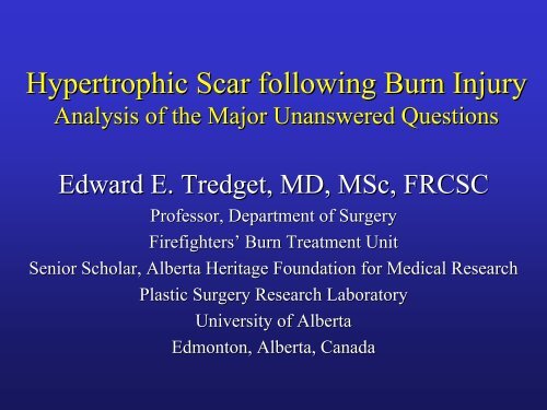
<strong>Hypertrophic</strong> <strong>Scar</strong> following <strong>Burn</strong> <strong>Injury</strong><br />
Analysis of the Major Unanswered Questions<br />
Edward E. Tredget, MD, MSc, FRCSC<br />
Professor, Department of Surgery<br />
Firefighters’ <strong>Burn</strong> Treatment Unit<br />
Senior Scholar, Alberta Heritage Foundation for Medical Research<br />
Plastic Surgery Research Laboratory<br />
University of Alberta<br />
Edmonton, Alberta, Canada
Unresolved Features of <strong>Hypertrophic</strong> <strong>Scar</strong><br />
following Thermal <strong>Injury</strong><br />
• definition of HTS vs normal skin, mature<br />
scar and keloid<br />
• diagnosis and scar severity<br />
• prevalence and socioeconomic impact<br />
• pathophysiology<br />
• treatment<br />
• future directions
How do you measure the severity of<br />
HTS and response to treatment<br />
• clinical observations<br />
• Vancouver <strong>Burn</strong> <strong>Scar</strong> Scale (VBSS)<br />
• scar volume<br />
• photography<br />
• scar color and vascularity<br />
• scar pliability and viscoelastic properties<br />
• ultrasound
• subjective ratings of<br />
pigmentation, vascularity,<br />
pliability, height<br />
• inter-rater rater reliability of 0.5-0.8<br />
0.8<br />
• correlated with subjective<br />
assessment at 1.5 years post-<br />
injury but not at 3 months<br />
Nedelec B et al. JBCR 2000
1. Baryza MJ et al. The Vancouver <strong>Scar</strong> Scale: An<br />
administration tool and its interrater reliably.Journal of<br />
<strong>Burn</strong> Care and Rehabilitation 1995, 16: 535-538.<br />
538.<br />
2. Sullivan T et al. Rating the burn scar. Journal of <strong>Burn</strong><br />
Care and Rehabilitation 1990, 11: 256-260.<br />
260.<br />
3. Nedelec et al. Rating the resolving bur hypertrophic scar:<br />
Comparison of the Vancouver <strong>Scar</strong> Scale and scar volume.<br />
Journal of <strong>Burn</strong> Care and Rehabilitation 2000, 21: 205-<br />
212.<br />
4. Draaijers LJ et al. The patient and observer scar<br />
assessment scale: A reliable and feasible tool for scar<br />
evaluation. Plastic and Reconstructive Surgery 2004, 113:<br />
1960-1965.<br />
1965.
Major Question regarding HTS<br />
What is the most objective, sensitive, specific<br />
and universally available measure to<br />
quantitate the severity of HTS and its<br />
response to treatment
Materials and Methods<br />
Outcome measurements included:<br />
– scar assessment using VBSS and rVBSS<br />
– skin pliability and elasticity using the<br />
Cutometer<br />
– skin vascularity and melanin level using the<br />
Mexameter<br />
– standardized photography
2<br />
4<br />
6<br />
8<br />
10<br />
A<br />
Mexameter<br />
color<br />
800<br />
700<br />
600<br />
500<br />
400<br />
300<br />
200<br />
100<br />
0<br />
Melanin<br />
*<br />
Erythema<br />
Normal<br />
Donor<br />
C<br />
Cutometer<br />
elasticity<br />
1.2<br />
1<br />
0.8<br />
0.6<br />
0.4<br />
*<br />
Normal<br />
Mature<br />
HSc<br />
0.2<br />
0<br />
*<br />
r0 r2 r3<br />
*<br />
B<br />
300<br />
250<br />
Serum TGF-ß (ng ng/ml)<br />
200<br />
150<br />
100<br />
50<br />
Figure<br />
0<br />
0<br />
Pre Rx Early Rx Late Rx Post Rx Control
1. Cheng W et al. Ultrasound assessment of scald scars in<br />
Asian children receiving pressure garment therapy.<br />
Journal of Pediatric Surgery 2001, 36: 466-469.<br />
469.<br />
2. Davey RB et al. Computerized color: A technique for the<br />
assessment of burn scar hypertrophy. <strong>Burn</strong>s 1999, 25:<br />
207-213.<br />
213.<br />
3. Li-Tsang CW et al. Validation of an objective scar<br />
pigmentation measurement by using a<br />
spectrocolorimeter. . <strong>Burn</strong>s 2003, 29:779-784.<br />
784.<br />
4. Martin D et al. Changes in subjective vs objective burn<br />
scar assessment: Does the patient agree with what we<br />
think Journal of <strong>Burn</strong> Care and Rehabilitation 2003, 24:<br />
239-244. 244. Discussion 238.
Is it possible to objectively assess the burn<br />
wound to determine if HTS will develop<br />
Riordon CL et al. JBCR 2003
Pathophysiology of HTS Formation<br />
• What are the factors locally within the burn<br />
wound that are important in the development<br />
of HTS<br />
• What role does the systemic response to<br />
injury play in the development of HTS
HEMOSTASIS<br />
WOUND<br />
Macrophages<br />
Neutrophils<br />
Fibrin<br />
Platelets<br />
PROLIFERATION<br />
Lymphocytes<br />
INFLAMMATION<br />
Fibroblasts<br />
Proteoglycans<br />
REMODELING<br />
Endothelium<br />
Epithelium<br />
Collagen Deposition<br />
<strong>Scar</strong> Maturation<br />
Collagen Fibril Crosslinking<br />
<strong>Hypertrophic</strong> <strong>Scar</strong><br />
Time after <strong>Injury</strong>
What are the important features of fibroblasts in hypertrophic scar s<br />
Collagenase activity<br />
Collagenase production<br />
Decorin<br />
Apoptosis<br />
Fibroblast density<br />
Fibronectin production<br />
Type I / III collagen production<br />
TGF-β<br />
Versican<br />
Biglycan<br />
Synthesis<br />
Degradation<br />
ECM<br />
Remodeling
Collagenase Expression in Fibroblasts from<br />
Different Layers of the Dermis
What are the important fibrogenic growth<br />
factors in HTS<br />
• TGF- β<br />
b1, b2, b3<br />
• CTGF<br />
• IL-4<br />
• IL-13<br />
• PDGF<br />
• others
What is the role of T cells in HTS<br />
and how do we control their effects<br />
1. Dunsmore SE et al. The bone marrow leaves its scar: New<br />
concepts in pulmonary fibrosis. The Journal of Clinical<br />
Investigations 2004, 113: 180-182.<br />
182.<br />
2. Tredget EE et al. Polarized T helper cells Th2 cytokine<br />
production in patients with hypertrophic scar following<br />
thermal injury. Journal of Interferon & Cytokine Research<br />
2006, 26:179-189.<br />
189.<br />
3. Yang L et al. Identification of fibrocytes in post-burn<br />
hypertrophic scar. Wound Repair and Regeneration 2005,<br />
13:398-404.
Native CD4+<br />
T cell<br />
IL-2<br />
IL-12<br />
IFN-γ<br />
IL-2; IL-4<br />
IL-4<br />
IL-10<br />
(TGF-β)<br />
T H 1<br />
T H 2<br />
IFN-γ<br />
IFN-γ<br />
IL-2<br />
IL-4<br />
IL-5<br />
IL-6<br />
IL-10<br />
pT H<br />
T H 3<br />
IL-4<br />
Anti-IL<br />
IL-12<br />
mAb<br />
IL-12<br />
IFN-γ<br />
<br />
TGF-β<br />
IL-4<br />
IL-10<br />
Letterio JL et al, Ann Rev Imm, , 1998
Percentage of Lymphocytes Producing<br />
IFN-γ and IL-4<br />
A<br />
IL-4 Percent positive cells(%)<br />
20<br />
15<br />
10<br />
5<br />
* *<br />
*<br />
0<br />
0.25 0.5 0.75 1 2 6 12 12+<br />
B<br />
IFN Percent positive cells (%)<br />
20<br />
15<br />
10<br />
5<br />
0<br />
*p
Tredget et al, J Interferon Cytokine Res, 2006<br />
Time Course of IL-10 and IL-12 from PBMC<br />
ex vivo in HSC and non-HSc Patients<br />
A<br />
IL-10 pg / ml<br />
16<br />
12<br />
8<br />
+<br />
*<br />
*<br />
HSc<br />
noHSc<br />
4<br />
B<br />
0<br />
16<br />
1 2 6 12 12+<br />
* p
CD4/TGF-β Staining in <strong>Hypertrophic</strong> <strong>Scar</strong><br />
→ CD4 +<br />
→ TGF-β +<br />
→ Double ++
Increased CD4+/TGF-β+ + T<br />
Lymphocytes in <strong>Burn</strong> Patients<br />
% of CD4+TGF-β T cells<br />
60.00%<br />
50.00%<br />
40.00%<br />
30.00%<br />
20.00%<br />
10.00%<br />
0.00%<br />
∗<br />
∗<br />
∗<br />
% of CD4+TGF-β T cells<br />
∗<br />
Normal
Increased CD4+/TGF-β+ + T Lymphocytes in<br />
<strong>Hypertrophic</strong> <strong>Scar</strong><br />
35<br />
∗<br />
∗<br />
p
1. Murphy TJ et al. CD4+CD25+ regulatory<br />
T cells control innate immune reactivity<br />
after injury. J Immunol 2005, 174: 2957-<br />
2963.<br />
2. Choileain NN et al. Enhanced regulatory<br />
T cell activity is an element of the host<br />
response to injury. The Journal of<br />
Immunology 2006, 176: 225-236<br />
236
Morphology of BLM-Induced<br />
Lung Fibrosis<br />
Hashimoto N et al, JCI, 2004
Dunsmore et al, JCI 2004<br />
What is the role of bone marrow hematopoietic stem<br />
cells and mesenchymal stem cells in HTS
Immunostaining for Type I Collagen<br />
A B C<br />
A. Fibroblasts B. Fibrocytes C. Fibrocytes with<br />
non-immune IgG
Day 0<br />
Femur<br />
Day 3<br />
Day 5
Chesney J et al, Proc Natl Acad Sci USA 1997<br />
Peripheral Blood Fibrocytes<br />
h have fibroblast-like like properties:<br />
– spindle shaped cells<br />
– produce types I and III collagen and fibronectin<br />
h display hemotopoietic cell features: CD34 +<br />
h have antigen-presenting ability<br />
h can rapidly enter subcutaneously implanted<br />
wound chambers in mice<br />
h present in scar tissue
Identification of LSP-1 1 in Fibrocytes by 2D<br />
SDS-PAGE of Cell Proteins
Fibrocytes in HSc from <strong>Burn</strong> Patients treated with IFN α-2b<br />
LSP-1<br />
Procollagen-1<br />
DAPI<br />
Merge<br />
Normal Skin<br />
HSc<br />
INFα-2b Treatment
Quantitation of Fibrocytes in HSc Tissue<br />
before and after IFN α−2b Treatment<br />
40<br />
35<br />
30<br />
HTS<br />
Normal Skin<br />
25<br />
20<br />
15<br />
10<br />
5<br />
0<br />
0 Months 2 Months 4 Months 6 Months 8 Months
What are the important inflammatory cytokines and receptors in HTS H<br />
<br />
Normal<br />
<strong>Burn</strong><br />
Gene Name Description <strong>Burn</strong>/Normal<br />
Ratio<br />
MCP-3 #77 Homo Sapiens mRNA for monocyte chemotactic protein-3 0.9/0.3 2.9<br />
IL-1R2 #37 Interleukin-1 receptor type II 0.9/0.3 2.7<br />
MPIF-2 #72 Small inducible cytokine subfamily A (Cy5-Cy5), member 24 1.0/0.4 2.7<br />
MCP-2 #78 Small inducible cytokine subfamily A (Cy5-Cy5), member 8 1.3/0.6 2.2<br />
(monocyte chemotactic protein 2)<br />
ENA-78 #82 Small inducible cytokine subfamily B (Cy50Cy5), member 5 0.0/0.7<br />
GCP-2 #83 Human chemokine alpha 3 (CKA-3) mRNA 0.0/0.2<br />
CCR1 #2 Chemokine (C-C motif) receptor 1 1.0/0.7 1.4<br />
IL-11Ra #19 Interleukin-11 receptor, alpha 0.1/0.1 1.5<br />
HCC-1 #61 Small inducible cytokine subfamily A (Cy5-Cy5), member 14 0.3/0.2 1.4<br />
MIP-1 delta #62 Small inducible cytokine subfamily A (Cy5-Cy5), member 15 0.6/0.5 1.3<br />
PARC#65 Small inducible cytokine subfamily A (Cys-Cy5), member 18 0.2/0.2 1.4<br />
Pulmonary and activation-regulated<br />
MCP-1(SCYA2) #67 Small inducible cytokine A2 (monocyte chemotactic protein 1) 10/0.8 1.3<br />
IFN-gamma #14 Interferon gamma 1.4/1.9 0.6<br />
CCR4 #5 Chemokine (C-C motif) receptor 4 1.1/1.7 0.6<br />
CXCR4 #13 Chemokine (C-X-C motif) receptor 4 1.0/1.6 0.6<br />
IL21 #40 Homo sapiens interleukin 21 1.2/1.8 0.6<br />
IL2Rβ #43 Interleukin2 receptor beta 0.5/0.9 0.6<br />
TNF-β/Lta #54 Lymphotoxin-alpha (TNF subfamily, member 1) 0.3/0.5 0.5<br />
LT-β #55 Lymphotoxin-beta 0.2/0.5 0.4<br />
LTbR #56 Homosapien lymphotoxin β receptor 0.2/0.6 0.3<br />
I309 #58 Small inducible cytokine A1 1.4/2.1 0.6<br />
SCYC2 #85 Small inducible cytokine subfamily C member 2 0.8/1.4 0.6<br />
Fractalkine #86 Small inducible cytokine subfamily D member 1 0.5/0.9 0.6<br />
SCYE1 #87 Small inducible cytokine subfamily E member 1 0.2/0.8 0.3
What is the role of other potential<br />
important cells in HTS<br />
• mast cells<br />
• mesenchymal stem cells<br />
• endothelial stem cells<br />
• transdifferentiating cells
What is the role of stem cells in injured tissue<br />
and circulating stem cells in HTS<br />
Korbling et al, NEJM 2003
Morphology of Early MSC<br />
Passage 1 murine BM-MSCs<br />
MSCs have large spindle-shaped<br />
shaped<br />
fibroblast-like like cells and small round cells.
sham<br />
ligation of<br />
coronary<br />
artery<br />
dilated and<br />
scarred<br />
ventricle<br />
MS cells<br />
mesenchy<br />
mal stems<br />
cells home<br />
to the site<br />
of injury<br />
regeneration<br />
of 75-80% of<br />
muscle<br />
function
BM-MSCs MSCs in the Wound<br />
Express Cytokeratins<br />
A<br />
B<br />
20<br />
50<br />
C<br />
D<br />
E<br />
F<br />
20
What is the optimal form of<br />
treatment for HTS<br />
1. Mustoe TA et al. International clinical recommendations on scar<br />
management.Plastic and Reconstructive Surgery 2002, 110: 560-571<br />
571<br />
2. Chang P et al. Prospective randomized study of the efficacy of<br />
pressure garment therapy in patients with burns. Journal of <strong>Burn</strong><br />
Care and Rehabilitation 1995, 16: 473-475.<br />
475.<br />
3. Kealy GP et al. Prospective randomized comparison of two types of<br />
pressure therapy garments. Journal of <strong>Burn</strong> Care and Rehabilitation<br />
ion<br />
1990, 11: 334-336.<br />
336.<br />
4. Patino O et al. Massage in hypertrophic scars. Journal of <strong>Burn</strong> Care<br />
and Rehabilitation 1999, 20:268-271. 271. Discussion 267.<br />
5. Costs AM et al. Mechanical forces induce scar remodeling: Study in<br />
non-pressure<br />
pressure-treated versus pressure-treated hypertrophic scars.<br />
<strong>American</strong> Journal of Pathology 1999, 155: 1671-1679.<br />
1679.
Sacket DL, Can J Physiol & Pharmacol (1986)<br />
Classification of Level of Evidence<br />
Level<br />
Description<br />
Strength<br />
Level I<br />
Level II<br />
Level III<br />
Level IV<br />
Level V<br />
Large randomized trials with clear cut results<br />
Good<br />
(and low risk of error)<br />
Small randomized trials with uncertain results Fair<br />
(and moderate to risk of error)<br />
Non-randomized, contemporaneous controls<br />
Fair<br />
Non-randomized, historical controls<br />
Fair<br />
No controls, case series only<br />
Poor
Pressure Garment Therapy for<br />
the Prevention of Abnormal<br />
<strong>Scar</strong>ring After <strong>Burn</strong> <strong>Injury</strong>: A<br />
Meta-Analysis<br />
A. Anzarut, MD, MSc (1,2,3) P. Singh, BSc (4) B. Rowe, MD, MSc, (2,5) E. Tredget, MD, MSc,<br />
FRCS(C) (1) E. Van den Kerckove (6) J. Olson, MD, FRCS(C) (1)<br />
From the Division of Plastic and Reconstructive Surgery (1), Department of Public Health Sciences (2),<br />
EPICORE Centre (3), Faculty of Medicine and Dentistry (4), and Department D<br />
of Emergency Medicine<br />
(5), all at the Faculty of Medicine and Dentistry, University of Alberta, Edmonton, Alberta; and the<br />
Department of Rehabilitation Sciences, Katholieke University, Leuven, Belgium (6).
Pressure Garment Therapy (PGT)<br />
Morbidity of PGT:<br />
– unattractive<br />
– over-heating<br />
– pruritis<br />
– wound breakdown<br />
– abnormal bone growth<br />
(Johnson, Journal of <strong>Burn</strong> Care and Rehab, , 1994)<br />
(Fricke, Journal of <strong>Burn</strong> Care and Rehab, , 1999)<br />
Costs of PGT:<br />
> $ 100,000 / year
Characteristics of included trials<br />
Study<br />
Study design<br />
Mean burn size<br />
(%TBSA)<br />
Co-interventions<br />
Control<br />
Groce 2000b<br />
Within<br />
patient<br />
48.3 (11-99)<br />
Not reported<br />
Low pressure<br />
garment<br />
Tredget<br />
Within<br />
patient<br />
10.4 (6.9) None<br />
None<br />
Moore 2000<br />
Within<br />
patient<br />
Not reported<br />
Not reported<br />
Low pressure<br />
garment<br />
Chang 1995<br />
Between<br />
patient<br />
21.1 (15.8) Not reported<br />
None<br />
Groce 2000a<br />
Between<br />
patient<br />
11.2 (1-30)<br />
Not reported<br />
None<br />
Van den<br />
Kerckhove<br />
2005<br />
Between<br />
patient<br />
8.5 (1-30)<br />
Not reported<br />
Low pressure<br />
garment
Results – Global scar score<br />
Pooled estimate: -0.46 (-1.07, 0.16)
What is the best animal models of<br />
HTS to assess new treatments<br />
• red duroc pig<br />
• transgenic mice<br />
– TGF-b, IL-4, IL-13,<br />
Smad 7 KO, others<br />
• animals with HTS grafts<br />
• bleomycin model in mice<br />
• Human controlled incisions (coma shaped<br />
wounds)
1. Gallant-Behm<br />
CL. et al. Cytokine and growth factor mRNA expression patterns<br />
associated with the hypercontracted, hyperpigmented healing phenotype of red duroc<br />
pigs: a model of abnormal human scar development Journal of Cutaneous Medicine<br />
& Surgery. 9:165-77, 2005.<br />
2. Lee JP. et al. Antifibrogenic effects of liposome-encapsulated encapsulated IFN-alpha2b cream on<br />
skin wounds in a fibrotic rabbit ear model. Journal of Interferon n & Cytokine<br />
Research. 25(10):627-31, 2005.<br />
3. Liang Z. Engrav LH. Muangman P. Muffley LA. Zhu KQ. Carrougher GJ.<br />
Underwood RA. Gibran NS. Nerve quantification in female red Duroc pig (FRDP)<br />
scar compared to human hypertrophic scar. <strong>Burn</strong>s. 30:57-64, 2004.<br />
4. Zhu KQ. et al. The female, red Duroc pig as an animal model of hypertrophic<br />
scarring and the potential role of the cones of skin. [Journal Article] A<br />
<strong>Burn</strong>s.<br />
29(7):649-64, 64, 2003<br />
5. Hillmer MP, et al. MacLeod SM. Experimental keloid scar models: a review of<br />
methodological issues. Journal of Cutaneous Medicine & Surgery. 6:354-9, 2002.<br />
6. Polo M, et al. An in vivo model of human proliferative scar. Journal of Surgical<br />
Research. 74:187-95, 1998.
What are useful antifibrogenic<br />
agents for HTS<br />
• antagonists of TGF- β<br />
• Interferon α, β, γ<br />
• Bleomycin<br />
• 5-fluorouracil<br />
• others
Acknowledgements<br />
Firefighter’s s <strong>Burn</strong> Trust Fund<br />
Canadian Institute for Health Research<br />
Alberta Heritage Foundation for Medical Research
systemic<br />
circulation<br />
bone marrow<br />
T T<br />
T<br />
T T<br />
T<br />
T<br />
cytokines<br />
PBMC / fibrocytes<br />
myofibroblasts<br />
_<br />
TH 2<br />
TH 1<br />
_<br />
IFN γ<br />
IL-10, IL-4<br />
TGF-β<br />
_<br />
+<br />
+<br />
wound<br />
wound contraction<br />
TH 3<br />
extracellular matrix<br />
synthesis / degradation<br />
Figure<br />
fibroblasts
Prevalence of HTS<br />
• What is the frequency of HTS following burn injury<br />
• How large is the socioeconomic impact of HTS<br />
• Who will is more likely to develop HTS given similar<br />
severity of initial injury<br />
• What is the role of the severity of injury in the<br />
development of HTS in terms of depth of burn and total<br />
body surface area involved<br />
• How does age, sex, racial background, and treatment of the<br />
acute injury affect the development of HTS<br />
• What is the psychological impact of HTS to the surviving<br />
burn patient
The most common<br />
complication for burn<br />
survivors is abnormal scarring<br />
(Bombaro, <strong>Burn</strong>s, , 2003)<br />
30-70% of burn patients<br />
develop abnormal scars<br />
(Bombaro, <strong>Burn</strong>s, , 2003)
1. Deitch EA et al. <strong>Hypertrophic</strong> burn scars: Analysis of<br />
variables. Journal of Trauma 1983, 23: 895-898.<br />
898.<br />
2. Dedovic Z et al. Time trends in incidence of hypertrophic<br />
scarring in children treated for burns. Acta chirurgica<br />
plastique 1999, 41: 87-90.<br />
3. McDonald WS et al. <strong>Hypertrophic</strong> skin grafts in burned<br />
patients: A prospective analysis of variables. Journal of<br />
Trauma 1987, 27: 147-150.<br />
150.<br />
4. Spurr ED et al. Incidence of hypertrophic scarring in burn-<br />
injured children. <strong>Burn</strong>s 1990, 16: 179-181.<br />
181.<br />
5. Bombaro KM et al. What is the prevalence of<br />
hypertrophic scarring following burns <strong>Burn</strong>s 2003, 29:<br />
299-302.