Decoding the Epigenetic Language of Neuronal Plasticity
Decoding the Epigenetic Language of Neuronal Plasticity
Decoding the Epigenetic Language of Neuronal Plasticity

Create successful ePaper yourself
Turn your PDF publications into a flip-book with our unique Google optimized e-Paper software.
Neuron<br />
Review<br />
<strong>Decoding</strong> <strong>the</strong> <strong>Epigenetic</strong> <strong>Language</strong><br />
<strong>of</strong> <strong>Neuronal</strong> <strong>Plasticity</strong><br />
Emiliana Borrelli, 1,5 Eric J. Nestler, 2 C. David Allis, 3 and Paolo Sassone-Corsi 4,5, *<br />
1<br />
Department <strong>of</strong> Microbiology and Molecular Genetics, University <strong>of</strong> California, Irvine, CA 92697, USA<br />
2<br />
Fishberg Department <strong>of</strong> Neuroscience, Mount Sinai School <strong>of</strong> Medicine, New York, NY 10029, USA<br />
3<br />
The Rockefeller University, New York, NY 10065, USA<br />
4<br />
Department <strong>of</strong> Pharmacology, University <strong>of</strong> California, Irvine, CA 92697, USA<br />
5<br />
Unite 904 Inserm ‘<strong>Epigenetic</strong>s and <strong>Neuronal</strong> <strong>Plasticity</strong>’, University <strong>of</strong> California, Irvine, CA 92697, USA<br />
*Correspondence: psc@uci.edu<br />
DOI 10.1016/j.neuron.2008.10.012<br />
Neurons are submitted to an exceptional variety <strong>of</strong> stimuli and are able to convert <strong>the</strong>se into high-order functions,<br />
such as storing memories, controlling behavior, and governing consciousness. These unique properties<br />
are based on <strong>the</strong> highly flexible nature <strong>of</strong> neurons, a characteristic that can be regulated by <strong>the</strong> complex<br />
molecular machinery that controls gene expression. <strong>Epigenetic</strong> control, which largely involves events <strong>of</strong><br />
chromatin remodeling, appears to be one way in which transcriptional regulation <strong>of</strong> gene expression can<br />
be modified in neurons. This review will focus on how epigenetic control in <strong>the</strong> mature nervous system<br />
may guide dynamic plasticity processes and long-lasting cellular neuronal responses. We outline <strong>the</strong> molecular<br />
pathways underlying chromatin transitions, propose <strong>the</strong> presence <strong>of</strong> an ‘‘epigenetic indexing code,’’ and<br />
discuss how central findings accumulating at an exponential pace in <strong>the</strong> field <strong>of</strong> epigenetics are conceptually<br />
changing our perspective <strong>of</strong> adult brain function.<br />
The ability to store information over long periods <strong>of</strong> time lies at<br />
<strong>the</strong> heart <strong>of</strong> cellular identity. This cellular ‘‘memory’’ is encoded<br />
in <strong>the</strong> specific pattern <strong>of</strong> expressed genes and allows a cell to<br />
ensure that it ‘‘remembers’’ who it is and how it should move<br />
along elaborate pathways during cellular development and<br />
differentiation. During development, germ cells or totipotent<br />
stem cells give rise to a diverse array <strong>of</strong> specialized cell types,<br />
including nerve cells, which become more hard-wired. These<br />
changes allow specialized cells to appropriately function in <strong>the</strong>ir<br />
specific niche—and, in <strong>the</strong> case <strong>of</strong> nerve cells, allows <strong>the</strong>m to<br />
properly control cognitive and behavioral functions. Once<br />
cellular differentiation processes are established, postmitotic<br />
nerve cells become committed to a variety <strong>of</strong> highly specialized<br />
functions that collectively determine our responses to external<br />
stimuli. Yet, insults, injury, and neurodegenerative diseases<br />
can dramatically affect nerve cells, calling into place a poorly<br />
understood ‘‘reprogramming process’’ that may be able to erase<br />
previously established cellular settings and, possibly, dedifferentiate<br />
or revert <strong>the</strong>se cells to a more primitive pluripotent state.<br />
Thus, it seems that developmental processes require ‘‘forward’’<br />
differentiation with a built-in memory component as well as<br />
a ‘‘reversible’’ reprogramming capability, allowing for plasticity<br />
at many levels (anatomical, electrical, synaptic, etc.). How could<br />
one relatively fixed genetic blueprint permit this flexibility to<br />
accommodate variability resulting from signals originated from<br />
environmental, dietary, and o<strong>the</strong>r influences<br />
How are cellular memories shaped by past experiences and<br />
environmental cues Does a molecular ‘‘sculpturing’’ process<br />
exist during development and adult life that takes adaptive<br />
cues from <strong>the</strong> environment (i.e., epigenetic mechanisms), or is<br />
this molding process purely stochastic in nature with selection<br />
doing <strong>the</strong> rest (i.e., genetic mechanisms) (Figure 1) The nervous<br />
system is characterized by a vast spectrum <strong>of</strong> cell types as well<br />
as a staggering number <strong>of</strong> reinforcing connections (synapses)<br />
that collectively shape and translate our daily experiences into<br />
complex thoughts and behaviors. Can 25,000 genes in our<br />
relatively fixed human genome explain who we are and how<br />
we act A wealth <strong>of</strong> accumulating evidence suggests that <strong>the</strong>re<br />
is much more to <strong>the</strong> genome than DNA sequence, permitting<br />
variability beyond <strong>the</strong> Watson-Crick DNA double helix. One<br />
way that such additional variability can be established is through<br />
epigenetic mechanisms (Figure 1). In this review, we explore <strong>the</strong><br />
evidence that suggests that <strong>the</strong>se mechanisms play a critical<br />
role in regulating neuronal function in <strong>the</strong> adult brain.<br />
<strong>Epigenetic</strong>s: An Old Word Takes on New Meaning<br />
and Renewed Interest<br />
A wealth <strong>of</strong> recent work from many laboratories has rekindled an<br />
interest in an old word: epigenetics. The general concept <strong>of</strong> an<br />
‘‘epigenetic landscape’’ was first articulated by developmental<br />
biologist Conrad Waddington, who used it to explain how identical<br />
genotypes could unfold a wide collection <strong>of</strong> phenotypes<br />
as development proceeded (Waddington, 1957). With time,<br />
Waddington’s concept <strong>of</strong> a phenotypic landscape took on additional<br />
meaning: ‘‘potentially heritable changes in gene expression<br />
that do not involve changes in DNA sequence’’ (Holliday<br />
and Pugh, 1975; Chambon, 1978; Jaenisch and Bird, 2003).<br />
While important questions remain, Waddington’s landscape<br />
has taken a clearer molecular form with <strong>the</strong> documentation <strong>of</strong><br />
a remarkable number <strong>of</strong> multisubunit complexes that act to<br />
remodel chromatin—to exchange specific histones (histone variants)<br />
in and out <strong>of</strong> assembled chromatin—or to enzymatically<br />
modify DNA and histones to bring about downstream events. It<br />
is not fully clear how <strong>the</strong>se dedicated machines are guided to<br />
Neuron 60, December 26, 2008 ª2008 Elsevier Inc. 961
Neuron<br />
Review<br />
Figure 1. Genetic versus <strong>Epigenetic</strong> Control<br />
Regulation <strong>of</strong> biological processes can be<br />
achieved via genetic and epigenetic programs.<br />
Variation in genetic information is obtained by<br />
mutagenesis <strong>of</strong> <strong>the</strong> DNA sequence that irreversibly<br />
changes <strong>the</strong> encoded message. <strong>Epigenetic</strong><br />
control operates ei<strong>the</strong>r on DNA, via DNA methylation,<br />
or on chromatin. Variation in <strong>the</strong> chromatin<br />
template can be brought about by posttranslational<br />
modifications (PTMs; colored beads) added<br />
to histones, exchange and replacement <strong>of</strong> major<br />
histones with specialized variants (colored<br />
wedges), or ATP-dependent nucleosome remodeling<br />
(not depicted), which alters histone:DNA<br />
contacts. All <strong>of</strong> <strong>the</strong>se mechanisms, along with<br />
DNA methylation and potential interactions with<br />
noncoding RNAs (not depicted), likely act toge<strong>the</strong>r<br />
to bring about <strong>the</strong> plasticity that helps to define<br />
epigenetic phenomena. PTMs <strong>of</strong> histones occur<br />
at highly conserved residues <strong>of</strong> <strong>the</strong> N-terminal tails<br />
<strong>of</strong> <strong>the</strong> core histones (see Figure 3) and include<br />
acetylation, methylation, phosphorylation, ubiquitination,<br />
etc. Examples <strong>of</strong> combined DNA methylation<br />
and histone modifications have been<br />
reported.<br />
<strong>the</strong>ir target sequences, but it is likely to involve constellations <strong>of</strong><br />
cis-acting regulatory proteins and noncoding RNAs that engage<br />
<strong>the</strong> DNA template directly (Bernstein and Allis, 2005). Some<br />
epigenetic marks, such as DNA methylation, appear to provide<br />
more stable, if not permanent, indexing marks that extend over<br />
long chromosomal domains, giving rise to ‘‘memorized’’ states<br />
<strong>of</strong> gene expression that may be inherited from one cell generation<br />
to <strong>the</strong> next (Figure 1). O<strong>the</strong>r modifications, such as histone<br />
acetylation, may be more labile and mediate regulation <strong>of</strong> gene<br />
expression over shorter-term periods. Considering <strong>the</strong> staggering<br />
complexity <strong>of</strong> neurogenesis, to what extent do changes<br />
in synaptic connections, guided by experiences, environment,<br />
diet, etc., influence <strong>the</strong> epigenomes <strong>of</strong> postmitotic nerve cells<br />
that underlie animal behavior, normal or abnormal Chromatin<br />
remodeling in <strong>the</strong> nervous system would not be directly heritable.<br />
Ra<strong>the</strong>r, heritability at this level could be through <strong>the</strong> reproducibility<br />
<strong>of</strong> behavioral patterns from a parent on its <strong>of</strong>fspring.<br />
In this review, we concentrate on emerging findings that tie<br />
epigenetic pathways to <strong>the</strong> special requirements exhibited by<br />
nerve cells, specifically <strong>of</strong> having seemingly opposing mechanisms<br />
allowing for cellular ‘‘memory’’ as well as cellular ‘‘plasticity.’’<br />
Somewhat a reflection <strong>of</strong> our interests in chromatin remodeling,<br />
we focus our remarks on specific examples <strong>of</strong> how<br />
histone posttranslational modifications (PTMs) are ‘‘written,’’<br />
‘‘erased,’’ and ‘‘interpreted’’ in ways that might contribute to<br />
both stable and plastic neuronal properties (Figure 2). Of course<br />
it is expected that many variations on this general <strong>the</strong>me exist<br />
and that a multitude <strong>of</strong> interconnected molecular signaling pathways<br />
dictates <strong>the</strong> elaborate gene expression networks that<br />
direct <strong>the</strong> complexities <strong>of</strong> nerve cell function.<br />
Several broad questions can be posed at this juncture. Are<br />
PTMs suited for on-<strong>of</strong>f behavior <strong>of</strong> short-lived, binary switches<br />
(Fischle et al., 2003) belonging to a different set from <strong>the</strong> PTMs<br />
compatible with more lasting, graded responses Are some<br />
PTMs, or <strong>the</strong> protein complexes that engage <strong>the</strong>m, more<br />
compatible with short-term versus long-term memory, and is<br />
<strong>the</strong>re enough combinatorial readout <strong>of</strong> PTMs to deal with <strong>the</strong><br />
diversity and plasticity <strong>of</strong> neuronal cells A wealth <strong>of</strong> histone<br />
PTMs has been uncovered in a variety <strong>of</strong> cell models. Our goal<br />
here was not to provide a compilation <strong>of</strong> <strong>the</strong>m but to illustrate<br />
general principles as <strong>the</strong>y might apply to <strong>the</strong> nervous system,<br />
by specifically highlighting distinct examples for fur<strong>the</strong>r discussion,<br />
including synaptic transmission, behavioral memory, drug<br />
addiction, circadian rhythms, and mental retardation-autism<br />
syndromes. Numerous reviews on chromatin biology (Berger,<br />
2007; Lee and Workman, 2007), DNA methylation (Miranda<br />
and Jones, 2007), and noncoding RNAs (Bernstein and Allis,<br />
2005) have appeared in <strong>the</strong> literature (see Allis et al., 2007a, for<br />
a textbook on epigenetics). Finally, a comprehensive nomenclature<br />
has been proposed for chromatin-modifying enzymes (Allis<br />
et al., 2007b).<br />
Chromatin Remodeling: Defining Lasting States <strong>of</strong> Gene<br />
Expression in <strong>the</strong> Nervous System<br />
Chromatin contributes in ensuring that <strong>the</strong> storage, organization,<br />
and readout <strong>of</strong> <strong>the</strong> genetic information occurs in a proper spatial<br />
and temporal sequence during cellular differentiation and organismal<br />
development. The fundamental repeating unit <strong>of</strong> chromatin,<br />
<strong>the</strong> nucleosome core particle, consists <strong>of</strong> 147 bp <strong>of</strong><br />
DNA organized in approximately two superhelical turns <strong>of</strong> DNA<br />
wrapped around an octamer <strong>of</strong> core histone proteins (two copies<br />
each <strong>of</strong> H2A, H2B, H3, and H4 or variants <strong>the</strong>re<strong>of</strong>). When associated<br />
with o<strong>the</strong>r components, higher-order nucleosomal structures<br />
are formed. Variations introduced into nucleosome array<br />
structures by a variety <strong>of</strong> mechanisms (see below) cause subtle,<br />
but meaningful, differences in chromatin compaction that correlate<br />
closely with more ‘‘open’’ versus ‘‘closed’’ states. These<br />
states loosely correlate with ‘‘euchromatin’’ versus ‘‘heterochromatin’’<br />
states that <strong>of</strong>ten, but not always, align with ‘‘active’’<br />
versus ‘‘inactive’’ states <strong>of</strong> gene expression, respectively<br />
(Berger, 2007). Covalent histone modifications, histone variants,<br />
or chromatin remodeling complexes work toge<strong>the</strong>r to alter <strong>the</strong><br />
chromatin fiber (Cheung et al., 2000a; Strahl and Allis, 2000).<br />
For example, histone acetylation, a charge-altering modification<br />
962 Neuron 60, December 26, 2008 ª2008 Elsevier Inc.
Neuron<br />
Review<br />
Table 1. Linking Histone Modifications to Metabolism<br />
Phosphorylation<br />
ATP/ADP<br />
Methylation SAM/SAH, FAD/FADH 2<br />
Acetylation<br />
Ubiquitylation/sumoylation<br />
Glycosylation<br />
Acetyl-CoA/CoA, NAD/NADH,<br />
Acetyl-ADP-ribose<br />
glucose<br />
UDP-GlcNAc/UDP<br />
Posttranslational modifications are elicited by specific enzymes whose<br />
activity depends on <strong>the</strong> intracellular levels <strong>of</strong> essential metabolites, thus<br />
sensing cellular metabolism, nutrients, and energy levels in <strong>the</strong> cell.<br />
PTMs target specific sites on histones, indicating that transient states<br />
<strong>of</strong> chromatin remodeling are under dynamic regulation <strong>of</strong> cellular<br />
physiology.<br />
Figure 2. Distinct Classes <strong>of</strong> Chromatin Remodeling Molecules<br />
Specific marks on <strong>the</strong> N-terminal tails <strong>of</strong> core histones are PTMs elicited by<br />
chromatin remodeling machineries that include a large variety <strong>of</strong> regulatory<br />
molecules, many <strong>of</strong> which interact physically and functionally. Conceptually,<br />
<strong>the</strong> regulators may be indicated as follows. (1) Writers. These are enzymes<br />
such as kinases, HATs, and HMTs that modify specific substrates adding<br />
phosphate, acetyl, or methyl groups. (2) Readers. These include a large variety<br />
<strong>of</strong> regulatory proteins that share unique domains implicated in recognizing<br />
acetyl or methyl groups. Some o<strong>the</strong>r domains, such as BRCT (Manke et al.,<br />
2003) and a specific region in 14-3-3 (Macdonald et al., 2005), can be considered<br />
as readers <strong>of</strong> phosphate. Often, ‘‘readers’’ recruit to chromatin additional<br />
epigenetic effectors. (3) Erasers. These enzymes include phosphatases,<br />
HDACs, and DMTs, which directly remove PTMs.<br />
that negates <strong>the</strong> positive charge on <strong>the</strong> 3-amino groups <strong>of</strong> lysine,<br />
has long been correlated with transcriptional activation (Cheung<br />
et al., 2000a), likely due to weakening <strong>of</strong> histone:DNA contacts.<br />
In contrast, histone hypoacetylation correlates closely with<br />
gene silencing (Lee and Workman, 2007).<br />
For <strong>the</strong> purposes <strong>of</strong> this review, several well-studied PTMs<br />
serve to illustrate paradigms emerging in present-day chromatin<br />
biology, with neurobiologists only beginning to consider how<br />
<strong>the</strong>se modifications contribute to neuronal functions (Crosio<br />
et al., 2003; Levenson and Sweatt, 2005; Tsankova et al.,<br />
2007). For example, acetylation and methylation result from <strong>the</strong><br />
addition or removal <strong>of</strong> acetyl and methyl or groups enzymatically<br />
donated from respective high-energy donors (acetyl coenzymeA<br />
[acetyl-CoA] and S-adenosyl methionine [SAM]; see Table 1).<br />
Histones are acetylated by histone acetyltransferases (HATs),<br />
which comprise a large family <strong>of</strong> enzymes, and deacetylated<br />
by histone deacetylases (HDACs), which are divided in different<br />
families. Class I HDACs are believed to provide <strong>the</strong> major HDAC<br />
catalytic activity present in brain, which is <strong>the</strong>n modified by<br />
class II HDACs through direct binding interactions. Class III<br />
HDACs represent a distinct subfamily, as discussed under circadian<br />
regulation below. Both histone H3 and H4 undergo polyacetylation<br />
at nearby lysine residues in <strong>the</strong> proteins’ N termini;<br />
however, still much more needs to be uncovered about <strong>the</strong> targeted<br />
recruiting and specificity <strong>of</strong> individual HATs and HDACs<br />
involved in <strong>the</strong>se reactions.<br />
Methylation affects DNA, RNA, and histone and nonhistone<br />
proteins, at least to varying degrees in different organisms. In<br />
some cases, intriguing links have emerged between histone<br />
modifications, and specifically methylation, and DNA methylation<br />
(Mutskov et al., 2002; Ooi et al., 2007). Second, within any<br />
potential histone or nonhistone protein that is methylated,<br />
multiple lysines or arginines can be modified, <strong>of</strong>ten existing<br />
toge<strong>the</strong>r in localized regions <strong>of</strong> <strong>the</strong> same or different histone<br />
domains. Thus, at least for lysine- and arginine-rich proteins<br />
like histones, a wealth <strong>of</strong> biological readouts may be possible.<br />
Third, and expanding on <strong>the</strong> complexity <strong>of</strong> methylation, individual<br />
lysine residues can be mono-, di-, or trimethylated; similarly,<br />
arginine residues can be mono- or dimethylated, and <strong>the</strong>se<br />
can be dimethylated in a symmetric or asymmetric fashion.<br />
These various methylation reactions are mediated by distinct<br />
subtypes <strong>of</strong> histone methyltransferases and demethylases, as<br />
discussed in <strong>the</strong> next section. Note that, unlike acetylation,<br />
methylation does not alter <strong>the</strong> positive charge <strong>of</strong> <strong>the</strong> targeted<br />
lysine or arginine, suggesting potential differences in regulatory<br />
outputs. Fourth, methylation <strong>of</strong> distinct lysine residues has opposite<br />
functional consequences on gene activation (see below).<br />
Fifth, all <strong>of</strong> <strong>the</strong> core histones (Shi and Whetstine, 2007) are methylated<br />
depending upon <strong>the</strong> physiological setting. Moreover, in<br />
keeping with studies on acetylation, methylation is not limited<br />
to histone proteins. Nonhistone proteins, such as <strong>the</strong> tumor<br />
suppressor p53 (Huang et al., 2007; Shi et al., 2007), are physiological<br />
targets <strong>of</strong> methylation reactions, a growing list likely to<br />
cross over into neuronal-specific proteins. Thus, methylation<br />
alone provides a remarkable number <strong>of</strong> regulatory options to<br />
<strong>the</strong> cell (Ru<strong>the</strong>nburg et al., 2007a). Combining methylation with<br />
o<strong>the</strong>r PTMs, such as acetylation, causes a remarkable array <strong>of</strong><br />
possibilities. As discussed in <strong>the</strong> next paragraph, we favor<br />
a scenario where <strong>the</strong> controlled addition and removal <strong>of</strong> specific<br />
PTMs results into unique combinations that constitute a sort <strong>of</strong><br />
‘‘epigenetic indexing code’’ that corresponds to distinct physiological<br />
states and genomic functions.<br />
<strong>Decoding</strong> <strong>the</strong> ‘‘<strong>Epigenetic</strong> Indexing Code’’: Writing,<br />
Reading, and Erasing Key Marks<br />
Remarkable progress has been made in characterizing what is<br />
<strong>of</strong>ten being referred to as ‘‘writers, readers, and erasers’’ <strong>of</strong><br />
PTMs in histone and nonhistone proteins (Figure 2). Thus,<br />
Neuron 60, December 26, 2008 ª2008 Elsevier Inc. 963
Neuron<br />
Review<br />
Synaptic<br />
activity<br />
Injury<br />
Stress<br />
Neurotransmitters<br />
Signaling<br />
PTMs<br />
Enzymatic Machinery<br />
Environmental<br />
stimuli<br />
Metabolism<br />
Figure 3. Multiple Posttranslational<br />
Modifications on Histone Tails<br />
The H3 N-terminal tail, here presented as a paradigm<br />
<strong>of</strong> all histone tails, can undergo numerous<br />
modifications. Here, only phosphorylation, acetylation,<br />
and methylation are indicated. Methylation<br />
can be mono-, di-, or trimethyl. The enzymatic<br />
machinery that elicits <strong>the</strong>se PTMs is believed to<br />
be under <strong>the</strong> physiological control <strong>of</strong> neuronal<br />
stimuli. Specific combinations <strong>of</strong> PTMs correspond<br />
to selective states <strong>of</strong> chromatin, ei<strong>the</strong>r<br />
permissive or not for transcription, responsive to<br />
damage and stress, or modulated by physiological<br />
changes in cellular metabolism.<br />
A RT<br />
KQTA RK S TGGK A PRKQ LATK A A R K SA PA TG<br />
20<br />
30 G VK K P…<br />
10<br />
considerable attention has been placed in documenting <strong>the</strong><br />
global distribution (or ‘‘patterns’’) <strong>of</strong> histone PTMs using a variety<br />
<strong>of</strong> high-resolution genomic pr<strong>of</strong>iling methods (Bernstein et al.,<br />
2005; Barski et al., 2007). It emerges from <strong>the</strong>se studies that ‘‘epigenomes’’<br />
are highly organized and strikingly nonrandom with<br />
respect to histone and DNA modifications (reviewed in Bernstein<br />
et al., 2007). Evidence discussed in this review indicates that <strong>the</strong><br />
enzymatic machinery that elicits <strong>the</strong>se PTMs operates under <strong>the</strong><br />
control <strong>of</strong> a variety <strong>of</strong> neuronal stimuli, which link physiological<br />
variations to modulated chromatin remodeling and <strong>the</strong>reby<br />
controlled gene expression (Figure 3). One important consideration<br />
relates to <strong>the</strong> intracellular pathways involved in <strong>the</strong> marking<br />
<strong>of</strong> <strong>the</strong>se PTMs. Interestingly, all use physiological metabolites,<br />
<strong>the</strong>reby indicating that <strong>the</strong> dynamic process <strong>of</strong> chromatin remodeling<br />
may ‘‘sense’’ cellular metabolism and changes in<br />
energy levels (Table 1), which are highly controlled and functionally<br />
essential in neuronal responses.<br />
Selected combinations <strong>of</strong> numerous PTMs occur at specific<br />
residues <strong>of</strong> <strong>the</strong> histone H3 N-terminal tail (Figure 3). For example,<br />
high levels <strong>of</strong> H3 and H4 acetylation and H3 lysine 4 methylation<br />
(H3K4me) are generally present in promoter regions <strong>of</strong> active<br />
genes (Ru<strong>the</strong>nburg et al., 2007b). In contrast, elevated levels <strong>of</strong><br />
H3 lysine 27 (H3K27me) correlate with gene repression mediated<br />
by <strong>the</strong> protein Polycomb (Trojer and Reinberg, 2006). Interestingly,<br />
<strong>the</strong> ‘‘degree’’ <strong>of</strong> histone lysine methylation matters, with<br />
significant differences in large-scale patterns <strong>of</strong> mono-, di-,<br />
and trimethylation at specific lysine residues, whe<strong>the</strong>r activating<br />
or repressing (Barski et al., 2007). These sorts <strong>of</strong> epigenetic<br />
indexing patterns vary in different cell types or during different<br />
stages <strong>of</strong> development (Marin-Husstege et al., 2002; Lessard<br />
et al., 2007; Putignano et al., 2007). For example, a ‘‘bivalent<br />
domain,’’ characterized by a configuration <strong>of</strong> marking genes<br />
H3<br />
with both ‘‘ON’’ (H3K4me) and ‘‘OFF’’<br />
(H3K27me), was noted in key developmental<br />
genes in embryonic stem cells<br />
(Bernstein et al., 2006). How bivalent<br />
domains are resolved during development<br />
is not known. As well, <strong>the</strong> extent<br />
to which neuronal cells use <strong>the</strong> strategies<br />
<strong>of</strong> histone PTMs as part <strong>of</strong> a mechanism<br />
for establishing ‘‘epigenomes’’ underlying<br />
neuronal diversity is not clear. How<br />
does DNA methylation and noncoding<br />
RNA enter into <strong>the</strong> above equation<br />
Recent findings suggest that all <strong>of</strong> <strong>the</strong>se components work<br />
toge<strong>the</strong>r to bring about a ‘‘language’’ that greatly exceeds that<br />
<strong>of</strong> DNA alone (Ooi and Henik<strong>of</strong>f, 2007).<br />
If histone methylation does not alter <strong>the</strong> charge <strong>of</strong> target<br />
lysines or arginines, how does histone methylation function<br />
Unlike acetylation, where charge-based mechanisms are likely<br />
to apply, histone:DNA contacts might not be affected by histone<br />
methylation in what can be referred to as ‘‘cis’’ (structural) effects<br />
on nucleosome structure. Likely, histone methylation is ‘‘read’’<br />
by effector proteins that bring about meaningful downstream<br />
events by ‘‘trans’’ mechanisms (Figure 2). Effector proteins<br />
contain ‘‘reader’’ modules that structural and functional<br />
evidence identify as relatively short (50–100 aa) binding motifs,<br />
such as chromodomains or PHD fingers, which read histone<br />
methyl-lysine marks with remarkable and elegant precision<br />
(Ru<strong>the</strong>nburg et al., 2007a; Lee and Workman, 2007). Similarly,<br />
as suggested in early articulations <strong>of</strong> <strong>the</strong> histone and epigenetic<br />
code hypo<strong>the</strong>ses (Cheung et al., 2000a; Strahl and Allis, 2000;<br />
Jenuwein and Allis, 2001; Turner, 2002), specific acetyl-lysine<br />
marks are also read in histone and nonhistone proteins by bromodomains.<br />
Also, interplays exist between adjacent or neighboring<br />
modifications that may serve to govern <strong>the</strong> binding and<br />
interactions <strong>of</strong> <strong>the</strong> effector proteins (Fischle et al., 2003). Understanding<br />
<strong>the</strong> extent to which such repeated modules read PTMs,<br />
ei<strong>the</strong>r on a single histone tail or on multiple tails or even distinct<br />
nucleosomes, remains a challenge for future studies.<br />
The general stability <strong>of</strong> DNA and histone methylation marks, as<br />
compared to rapid turnover kinetics PTMs such as acetylation<br />
and phosphorylation (Figure 4), prompted early speculation<br />
that methylation might be <strong>the</strong> ideal epigenetic indexing system<br />
(Jenuwein and Allis, 2001; Bannister and Kouzarides, 2005).<br />
While <strong>the</strong> situation with DNA demethylation remains unclear,<br />
964 Neuron 60, December 26, 2008 ª2008 Elsevier Inc.
Neuron<br />
Review<br />
A<br />
R P<br />
T KQ T ARKSTGGK AP RKQLATKAARK SAPATG GVKKP…<br />
DAPI<br />
H3S10-P<br />
NaCl<br />
Kainic<br />
Acid<br />
Figure 4. Dynamic Changes in Histone Modifications<br />
in <strong>the</strong> Hippocampus<br />
Chromatin remodeling occurs in hippocampal neurons <strong>of</strong> <strong>the</strong> dentate gyrus<br />
30 min after administering kainic acid (35 mg/kg) to a mouse. Kainate receptors<br />
are involved in epileptogenesis and synaptic plasticity. H3S10 phosphorylation<br />
in <strong>the</strong>se neurons is induced by stimulation <strong>of</strong> dopamine, acetylcholine,<br />
and glutamate receptors and is <strong>of</strong>ten associated with acetylation at H3K14<br />
(Crosio et al., 2003). These events are coupled to transcriptional activation in<br />
hippocampal neurons. An antibody that recognizes specifically H3 P-S10<br />
was used.<br />
with some reports <strong>of</strong> rapid DNA demethylation occurring in brain<br />
(Fan et al., 2001; Miller and Sweatt, 2007), progress has been<br />
made in <strong>the</strong> area <strong>of</strong> histone demethylation. Demethylase activities<br />
are being described for essentially all <strong>of</strong> <strong>the</strong> well-known<br />
lysine methyl sites in histones, not to mention those that are<br />
selective for mono-, di-, or trimethylation states (Trojer and Reinberg,<br />
2006), promising to carry this family <strong>of</strong> enzymes into <strong>the</strong><br />
‘‘celebrity status’’ <strong>of</strong> o<strong>the</strong>r chromatin modifiers, such as HATs<br />
and HDACs. Recent findings indicate that some <strong>of</strong> <strong>the</strong>se<br />
enzymes turn out to be critical in neuronal functions. For<br />
example, <strong>the</strong> histone H3K4 tridemethylase SMCX links repression<br />
<strong>of</strong> target genes (e.g., <strong>the</strong> sodium channel type 2A and synapsin<br />
1) to mental retardation and epilepsy (Tahiliani et al., 2007).<br />
These studies underscore several important points. First, <strong>the</strong>y<br />
point to a general model wherein failure to remove, or most<br />
certainly add, ‘‘ON’’ epigenetic marks, such as H3K4me3 (or<br />
likely ‘‘OFF’’ marks as well), impairs downstream neuronal<br />
gene regulation leading to disease states by yet undefined<br />
mechanisms. Second, disease phenotypes are not always associated<br />
with alterations in enzyme activity. For example,<br />
H3K4me3 demethylase activity was examined in a small series<br />
<strong>of</strong> SMCX mutations, which interestingly correlated roughly with<br />
<strong>the</strong> severity <strong>of</strong> mental retardation in human patients affected<br />
with <strong>the</strong>se mutations. In some cases, <strong>the</strong> mutations studied<br />
H3<br />
affected enzymatic activity, while in o<strong>the</strong>r cases <strong>the</strong> mutations<br />
affected <strong>the</strong> intracellular location <strong>of</strong> <strong>the</strong> protein or its association<br />
with o<strong>the</strong>r SMCX complex components. Third, SMCX contains<br />
multiple modules, including two PHD fingers, suggestive <strong>of</strong><br />
a yet unknown methylation ‘‘reading’’ function. Stabilization <strong>of</strong><br />
<strong>the</strong> protein in <strong>the</strong> remodeling complexes within <strong>the</strong> chromatin<br />
template may occur in part through this type <strong>of</strong> binding module<br />
(Wysocka et al., 2006; Taverna et al., 2006; Li et al., 2007). Interestingly,<br />
several <strong>of</strong> <strong>the</strong> mutations in <strong>the</strong> mental retardation-associated<br />
SMCX demethylase fall within some <strong>of</strong> its reading<br />
modules (Jensen et al., 2005). Similarly, <strong>the</strong> intriguing interaction<br />
between ATRX, a DNA helicase/ATPase mutated in <strong>the</strong> ATRX<br />
syndrome (a-thalassemia/mental retardation, X-linked) and<br />
MeCP2, a regulator <strong>of</strong> DNA methylation causally linked to Rett’s<br />
syndrome (see later), is disrupted by mutations responsible for<br />
<strong>the</strong> pathologies associated with <strong>the</strong>se two epigenetic regulators<br />
(Nan et al., 2007). As a footnote, we point out that many chromatin<br />
remodeling activities and histone-modifying enzymes<br />
contain one or more modules, such as bromodomains, chromodomains,<br />
and PHD fingers, linked in various combinations (Lee<br />
and Workman, 2007). Thus, while it is convenient to discuss<br />
‘‘writers’’ and ‘‘readers’’ <strong>of</strong> PTMs separately (Figure 1), many<br />
writers and erasers contain <strong>the</strong>ir own reading modules whose<br />
functions remain unclear (Figure 2). In mixed lineage leukemia<br />
(MLL), for example, <strong>the</strong> writer <strong>of</strong> H3K4 methyl marks (Ru<strong>the</strong>nburg<br />
et al., 2007a) contains a series <strong>of</strong> PHD fingers and an atypical<br />
bromodomain whose function is unknown. We predict that deciphering<br />
<strong>the</strong> ‘‘epigenetic indexing code’’ will help reveal <strong>the</strong> pathways<br />
responsible for <strong>the</strong> timing, intensity, and precision <strong>of</strong><br />
‘‘memorized’’ gene expression.<br />
This evolving knowledge <strong>of</strong> chromatin remodeling mechanisms<br />
has already begun to inform our understanding <strong>of</strong> <strong>the</strong><br />
regulation <strong>of</strong> brain function under normal conditions and certain<br />
pathophysiological states, as will be seen in <strong>the</strong> sections that<br />
follow.<br />
Does Chromatin Remodeling Influence<br />
Synaptic <strong>Plasticity</strong><br />
Repeated patterns <strong>of</strong> synaptic transmission lead to diverse<br />
forms <strong>of</strong> synaptic plasticity at excitatory and inhibitory synapses,<br />
e.g., long-term potentiation (LTP) or long-term depression (LTD),<br />
whereby <strong>the</strong> efficacy <strong>of</strong> synaptic transmission is up- or downregulated,<br />
respectively. Certain forms <strong>of</strong> LTP and LTD are long<br />
lived and thought to be dependent on lasting changes in gene<br />
expression. Based on <strong>the</strong> critical role that chromatin remodeling<br />
plays in dictating a transcription-permissive or silencing state <strong>of</strong><br />
<strong>the</strong> genome (Felsenfeld and Groudine, 2003), it is notable that<br />
growing evidence suggests that histone PTMs may be involved<br />
in <strong>the</strong>se processes. For example, H4 acetylation at specific<br />
promoters in Aplysia is altered after LTP and LTD (Guan et al.,<br />
2002), and HDAC inhibitors promote LTP in mammalian neurons<br />
(Levenson et al., 2004). Additionally, during synaptic transmission,<br />
neurotransmitters trigger responses in target neurons by<br />
activating two major families <strong>of</strong> receptors, ligand-gated ion<br />
channels and G protein-coupled receptors. Likewise, growth<br />
factors and cytokines are released from neurons in an activitydependent<br />
manner and act on target neurons through<br />
receptor-mediated signaling. Triggering signaling cascades in<br />
Neuron 60, December 26, 2008 ª2008 Elsevier Inc. 965
Neuron<br />
Review<br />
target neurons leads to more long-lasting effects, including<br />
changes in gene expression via control <strong>of</strong> transcription and<br />
<strong>the</strong>reby chromatin remodeling (Figures 3 and 4).<br />
One paradigmatic example involves <strong>the</strong> transcription factor<br />
CREB, which, once activated by signaling-induced phosphorylation,<br />
recruits CREB-binding protein (CBP), a coactivator with<br />
intrinsic HAT activity (Lonze and Ginty, 2002). In addition, an<br />
increase in cellular Ca 2+ levels in muscle activates Ca 2+ /calmodulin-dependent<br />
kinases, which phosphorylate class II HDACs.<br />
This phosphorylation provides a docking site for <strong>the</strong> ‘‘reader’’<br />
protein 14-3-3, which mediates <strong>the</strong> export <strong>of</strong> <strong>the</strong> phosphorylated<br />
HDACs from <strong>the</strong> nucleus (McKinsey et al., 2000). This pathway<br />
operates in hippocampal, striatal, and cerebellar granule<br />
neurons (Chawla et al., 2003; Renthal et al., 2007) and could<br />
represent a widespread mechanism <strong>of</strong> Ca 2+ -mediated histone<br />
acetylation and gene expression.<br />
Independent <strong>of</strong> histone deacetylation, class II HDACs can<br />
recruit cyclin-dependent kinase-5 (Cdk5) to phosphorylate myocyte<br />
enhancing factor 2 (MEF2), a protein that mediates activitydependent<br />
changes in synapse formation in neurons, and<br />
repress its transcriptional activity (Gong et al., 2003; Pulipparacharuvil<br />
et al., 2008). These studies reveal mechanisms by which<br />
synaptic activity may mediate long-lasting changes in brain,<br />
such as those associated with drug addiction. As discussed<br />
later, intriguing similarities indicate that some common pathways<br />
<strong>of</strong> epigenetic control may be relevant to memory and neurodegenerative<br />
disease.<br />
Synaptic activity is also reported to influence DNA methylation.<br />
This is unexpected, as it suggests that DNA methylation,<br />
classically thought as a very stable modification, undergoes<br />
rapid and dynamic regulation in <strong>the</strong> nervous system (Levenson<br />
et al., 2006). An important example is <strong>the</strong> activity-dependent<br />
control <strong>of</strong> Bdnf gene expression that has been correlated with<br />
reduced DNA methylation and release <strong>of</strong> a repressor complex<br />
comprising methyl-CpG binding protein (MeCP2; a protein that<br />
binds to and represses methylated DNA; Chen et al., 2003; Martinowich<br />
et al., 2003; Chang et al., 2006). It is proposed that<br />
neural activity, via increases in cellular Ca 2+ levels and activation<br />
<strong>of</strong> Ca 2+ /calmodulin kinases, leads to <strong>the</strong> phosphorylation <strong>of</strong><br />
MeCP2 and its release from <strong>the</strong> Bdnf promoter. This induces<br />
Bdnf expression and attendant dendritic outgrowth (Chen<br />
et al., 2003).<br />
In addition to direct mediators <strong>of</strong> <strong>the</strong>se synaptic changes,<br />
various immediate-early genes seem to be particular targets <strong>of</strong><br />
synaptic-plasticity-regulated epigenetic control. For example,<br />
<strong>the</strong> induction <strong>of</strong> c-fos is associated with dramatic increases in<br />
H4 acetylation and H3 phospho-acetylation (Figure 4; Crosio<br />
et al., 2003; Li et al., 2004; Brami-Cherrier et al., 2005; Kumar<br />
et al., 2005). Perturbation <strong>of</strong> dopamine signaling triggers <strong>the</strong><br />
dramatic induction <strong>of</strong> c-fos and o<strong>the</strong>r immediate-early genes in<br />
distinct subsets <strong>of</strong> striatal neurons in response to psychostimulant<br />
drugs <strong>of</strong> abuse (e.g., cocaine and amphetamine) and in<br />
response to antipsychotic drugs that block D2 dopamine receptors<br />
(Welter et al., 2007). The nuclear accumulation <strong>of</strong> DARPP32,<br />
a protein phosphatase 1 inhibitor, has been recently invoked in<br />
cocaine-induced H3 S-10 phosphorylation (Stipanovich et al.,<br />
2008). Ano<strong>the</strong>r study implicated mitogen and stress activated<br />
kinase 1 (MSK1) in cocaine stimulation <strong>of</strong> H3 P-S10 and c-fos<br />
induction (Brami-Cherrier et al., 2005), although o<strong>the</strong>r kinases<br />
may be also implicated in response to various stimuli (Sassone-Corsi<br />
et al., 1999; Nowak and Corces, 2004). Likewise,<br />
induction <strong>of</strong> c-fos by seizure activity or memory paradigms in<br />
o<strong>the</strong>r brain regions such as hippocampus is associated with<br />
H3 phosphoacetylation and ERKs (extracellular signal regulated<br />
kinases) activation (Crosio et al., 2003; Tsankova et al., 2004;<br />
Levenson and Sweatt, 2005; Chwang et al., 2006). Several o<strong>the</strong>r<br />
kinases can phosphorylate H3 S10 in nonneuronal cells, but <strong>the</strong>ir<br />
actions in brain are unexplored.<br />
Histone Modifications Affect Behavioral Memory<br />
Consistent with <strong>the</strong> coupling <strong>of</strong> histone modifications with<br />
synaptic plasticity and correlations between behavioral plasticity<br />
and epigenetic control <strong>of</strong> immediate-early gene transcription,<br />
<strong>the</strong>re are numerous reports <strong>of</strong> <strong>the</strong> importance <strong>of</strong> histone modifications<br />
in behavioral memory. Mice deficient in CBP exhibit<br />
memory deficits, and administration <strong>of</strong> an HDAC inhibitor can<br />
restore normal long-term memory formation in <strong>the</strong> mutants,<br />
and even enhance it in normal animals (Alarcon et al., 2004; Korzus<br />
et al., 2004; Levenson et al., 2004). Contextual fear conditioning,<br />
or activation <strong>of</strong> <strong>the</strong> ERK pathway that is thought to<br />
contribute to memory formation, increases levels <strong>of</strong> H3S10-<br />
K14 phospho-acetylation in <strong>the</strong> CA1 area <strong>of</strong> hippocampus,<br />
without affecting H4 acetylation (Levenson and Sweatt, 2005;<br />
Chwang et al., 2006). Interestingly, changes in histone acetylation<br />
and methylation in hippocampus have also been implicated<br />
in depression and antidepressant action, where HDAC inhibitors<br />
exert antidepressant-like effects (Tsankova et al., 2006;<br />
Schroeder et al., 2007).<br />
Recent work has implicated changes in DNA methylation in<br />
learning and memory as well. Contextual fear conditioning<br />
induces <strong>the</strong> expression <strong>of</strong> Dnmt3A and -3B in CA1 <strong>of</strong> hippocampus,<br />
and administration <strong>of</strong> <strong>the</strong> DNMT inhibitors, zebularine<br />
and 5-aza-2-deoxycytidine, blocks <strong>the</strong> induction <strong>of</strong> both contextual<br />
fear conditioning (Miller and Sweatt, 2007) and hippocampal<br />
LTP (Levenson et al., 2006). However, it remains unknown how<br />
<strong>the</strong>se drugs, which are thought to regulate DNA methylation in<br />
dividing cells only, affect gene expression in mature neurons.<br />
Fear conditioning causes rapid methylation and silencing <strong>of</strong> <strong>the</strong><br />
protein phosphatase 1 (Pp1) gene promoter (Miller and Sweatt,<br />
2007), a gene important for LTP and memory formation. Interestingly,<br />
fear conditioning also induces demethylation <strong>of</strong> <strong>the</strong> Reelin<br />
promoter (Dong et al., 2007), indicating that both DNA methylation<br />
and demethylation may be highly regulated in <strong>the</strong> adult brain.<br />
Chromatin Remodeling Mechanisms in Drug-Induced<br />
<strong>Plasticity</strong> and Addiction<br />
Drug addiction can be viewed as a form <strong>of</strong> drug-induced neural<br />
plasticity, whereby repeated exposure to drugs <strong>of</strong> abuse leads to<br />
long-lasting changes in <strong>the</strong> brain’s natural reward centers and<br />
associated memory circuits, which underlie <strong>the</strong> addiction phenotype<br />
(Hyman et al., 2006). One major site for <strong>the</strong>se lasting<br />
changes is <strong>the</strong> nucleus accumbens (NAc)—<strong>the</strong> ventral portion<br />
<strong>of</strong> <strong>the</strong> striatum, although many o<strong>the</strong>r regions are also involved.<br />
Drug-induced changes in gene expression, largely driven by<br />
dopaminergic signaling, occur in <strong>the</strong> NAc and o<strong>the</strong>r relevant<br />
regions (Tsankova et al., 2007). Some <strong>of</strong> <strong>the</strong>se changes persist<br />
966 Neuron 60, December 26, 2008 ª2008 Elsevier Inc.
Neuron<br />
Review<br />
even after months <strong>of</strong> abstinence (Grimm et al., 2003), consistent<br />
with <strong>the</strong> persisting behavioral abnormalities. These observations<br />
have driven research into chromatin remodeling as one molecular<br />
basis <strong>of</strong> sustained, even life-long, alterations in gene expression<br />
in brain reward regions that underlie an addicted state<br />
(Tsankova et al., 2007).<br />
As stated earlier, an acute dose <strong>of</strong> cocaine induces <strong>the</strong><br />
expression <strong>of</strong> fos family immediate-early genes in <strong>the</strong> NAc and<br />
dorsal striatum, an event associated with a rapid and transient<br />
increase in H4K5 acetylation (Brami-Cherrier et al., 2005; Kumar<br />
et al., 2005). CBP, with its intrinsic HAT activity, has been implicated<br />
in <strong>the</strong>se effects (Levine et al., 2005). Acute cocaine also<br />
induces Ser10/Lys14 H3 phospho-acetylation at <strong>the</strong> c-fos<br />
promoter (Crosio et al., 2003; Kumar et al., 2005), an event that<br />
is mediated by activation <strong>of</strong> ERKs and could require <strong>the</strong> protein<br />
kinase MSK1 (Lu et al., 2006; Brami-Cherrier et al., 2005). In<br />
contrast, chronic exposure to cocaine activates or represses<br />
many distinct genes compared to acute treatment. The c-fos<br />
gene, for instance, desensitizes in <strong>the</strong> NAc during a course <strong>of</strong><br />
chronic drug exposure, while <strong>the</strong> fosB gene continues to be<br />
induced, and <strong>the</strong>se changes are associated with decreased<br />
and increased histone acetylation at <strong>the</strong>se genes, respectively.<br />
In addition, chronic cocaine increases H3K9 methylation at <strong>the</strong><br />
c-fos gene, a modification tightly associated with gene repression.<br />
This effect could be mediated by <strong>the</strong> induction in <strong>the</strong> NAc<br />
<strong>of</strong> SUV39H1, a Lys9 histone methyltransferase (Renthal et al.,<br />
2008). Interestingly, several genes that are selectively induced<br />
in <strong>the</strong> chronic state, such as Cdk5 and Bdnf (Bibb et al., 2001;<br />
Grimm et al., 2003), are associated with increased H3 acetylation.<br />
Cocaine induction <strong>of</strong> H3 acetylation at <strong>the</strong> Bdnf promoter<br />
builds over a week <strong>of</strong> withdrawal (Kumar et al., 2005), and this<br />
change precedes <strong>the</strong> progressive increase in Bdnf expression<br />
in this brain region (Grimm et al., 2003). Little is known about<br />
<strong>the</strong> specific HATs and HDACs that may mediate <strong>the</strong>se changes<br />
in histone acetylation at cocaine-regulated genes. Yet <strong>the</strong>se<br />
results highlight <strong>the</strong> importance <strong>of</strong> exploring genome-wide chromatin<br />
alterations, by use <strong>of</strong> ChIP on chip (Lee et al., 2003) or<br />
SACO (Impey et al., 2004) or related techniques, to identify<br />
many o<strong>the</strong>r genes whose dysregulation contributes to cocaine<br />
addiction. Genome-wide epigenetic approaches have yielded<br />
exciting results in <strong>the</strong> fields <strong>of</strong> developmental (Lessard et al.,<br />
2007) and cancer biology (Lee et al., 2003).<br />
The transcription factor DFosB is implicated in <strong>the</strong> transition to<br />
an addicted state in chronic drug treatment (Nestler, 2008).<br />
DFosB is a truncated product <strong>of</strong> <strong>the</strong> fosB gene, which accumulates<br />
uniquely during chronic drug treatment due to its unique<br />
protein stability (Ulery et al., 2006). Gene expression array experiments<br />
indicate that DFosB accounts for >25% <strong>of</strong> all changes in<br />
steady-state mRNA levels induced in <strong>the</strong> NAc by chronic cocaine<br />
administration (McClung and Nestler, 2003). ChIP assays have<br />
shown that <strong>the</strong> induction by cocaine <strong>of</strong> one <strong>of</strong> <strong>the</strong>se mRNAs,<br />
Cdk5, represents a direct, activating effect <strong>of</strong> DFosB on <strong>the</strong><br />
Cdk5 gene (Kumar et al., 2005). By contrast, Bdnf is not a direct<br />
target <strong>of</strong> DFosB, consistent with a different mechanism being<br />
involved in its induction (McClung and Nestler, 2003). Induction<br />
<strong>of</strong> Cdk5, in turn, partly mediates <strong>the</strong> effects <strong>of</strong> chronic cocaine<br />
and <strong>of</strong> DFosB on dendritic remodeling in <strong>the</strong> NAc (Norrholm<br />
et al., 2003; Lee et al., 2006). DFosB is also responsible for<br />
c-fos repression after chronic cocaine, where it recruits<br />
HDAC1, leading to <strong>the</strong> deacetylation <strong>of</strong> nearby histones (Renthal<br />
et al., 2008). These findings support a model in which <strong>the</strong> accumulating<br />
DFosB interacts with distinct chromatin remodeling<br />
factors at specific promoters <strong>of</strong> genes that control reward<br />
neurons in this brain region.<br />
Cocaine-induced chromatin remodeling is mediated in part via<br />
regulation <strong>of</strong> <strong>the</strong> enzymatic machineries that control histone<br />
acetylation and methylation. For example, phosphorylation <strong>of</strong><br />
HDAC5, a class II HDAC enriched in <strong>the</strong> NAc, is induced by<br />
chronic cocaine exposure (Renthal et al., 2007), consistent<br />
with activation <strong>of</strong> Ca 2+ /calmodulin-dependent kinases in this<br />
brain region (Mattson et al., 2005). Ano<strong>the</strong>r example is <strong>the</strong> transcriptional<br />
regulator nucleus accumbens 1 (NAC-1), which is<br />
highly induced by cocaine in <strong>the</strong> NAc and interacts with<br />
HDAC3 and HDAC4 (Korutla et al., 2005). Chronic cocaine also<br />
induces <strong>the</strong> HMT SUV39H1 in <strong>the</strong> NAc (Renthal et al., 2008).<br />
Drug-induced chromatin remodeling is behaviorally relevant.<br />
Systemic or intra-NAc administration <strong>of</strong> HDAC inhibitors, or<br />
knockout <strong>of</strong> HDAC5, potentiates behavioral responses to<br />
cocaine, whereas viral-mediated overexpression <strong>of</strong> HDAC5<br />
specifically in <strong>the</strong> NAc has <strong>the</strong> opposite effect (Kumar et al.,<br />
2005; Renthal et al., 2007). Interestingly, overexpression <strong>of</strong><br />
HDAC4, but not HDAC9, has a similar effect, suggesting some<br />
specificity <strong>of</strong> HDAC action, <strong>the</strong> basis <strong>of</strong> which is still poorly<br />
understood. Moreover, CBP-deficient mice show reduced<br />
cocaine-induced locomotor activity (Levine et al., 2005).<br />
There are also several reports <strong>of</strong> chromatin changes after<br />
chronic ethanol administration (Mahadev and Vemuri, 1998;<br />
Bonsch et al., 2006; Bardag-Gorce et al., 2007). Interestingly,<br />
maternal ethanol consumption has been associated with lasting<br />
changes in DNA methylation in <strong>the</strong> fetal heart, which may<br />
contribute to increased risk <strong>of</strong> ischemic injury (Zhang et al.,<br />
2007). Identification <strong>of</strong> chromatin changes in blood and o<strong>the</strong>r<br />
peripheral tissues as a consequence <strong>of</strong> chronic ethanol administration<br />
raises <strong>the</strong> possibility <strong>of</strong> using such epigenetic markers to<br />
monitor drug use in vulnerable human populations.<br />
Intriguing connections have also been found between dopamine<br />
neurotransmission and <strong>the</strong> circadian clock machinery, via<br />
D2 receptors (Yujnovsky et al., 2006; Doi et al., 2006b; Yan<br />
et al., 2006). Dopamine is <strong>the</strong> major catecholamine in retinal<br />
neurons and plays a central role in neural adaptation to light (Iuvone<br />
et al., 1978), mostly through D2 receptors highly expressed<br />
in <strong>the</strong>se neurons (Doi et al., 2006b). Light stimulates <strong>the</strong><br />
syn<strong>the</strong>sis, turnover, and release <strong>of</strong> retinal dopamine, and dopaminergic<br />
activity is higher during <strong>the</strong> day than at night. Thereby,<br />
dopamine is a likely mediator <strong>of</strong> light signaling to <strong>the</strong> retinal circadian<br />
clock. Activation <strong>of</strong> D2 receptors stimulates CLOCK:BMAL1<br />
function through <strong>the</strong> MAPK signaling pathway. Since CLOCK is<br />
a HAT (see <strong>the</strong> next section; Doi et al., 2006a), dopamine may<br />
exert its function by signaling directly to <strong>the</strong> HAT potential <strong>of</strong><br />
CLOCK (Figure 5). Ano<strong>the</strong>r tempting speculation includes <strong>the</strong><br />
involvement <strong>of</strong> <strong>the</strong> AKT/GSK-3 transduction pathway, which is<br />
implicated in D2 receptor-dependent signaling (Beaulieu et al.,<br />
2007). GSK-3 kinase function is tightly regulated by lithium, an<br />
antimanic and antidepressant agent (Beaulieu et al., 2008), and<br />
shaggy, <strong>the</strong> GSK-3 Drosophila homolog, plays an essential role<br />
in circadian control (Martinek et al., 2001). Thus, this regulatory<br />
Neuron 60, December 26, 2008 ª2008 Elsevier Inc. 967
Neuron<br />
Review<br />
GSK-3<br />
P<br />
Rev-Erb<br />
BMAL1 P<br />
CLOCK<br />
Dopamine D2 Receptor<br />
-Arrestin<br />
AKT<br />
Ac<br />
P<br />
RAS/RAF-1<br />
pathway may link circadian rhythms, depression cycles, and<br />
chromatin remodeling.<br />
The Circadian Clock: How to ‘‘Time’’ Periodic<br />
Chromatin Remodeling<br />
Unlike memory and addiction behaviors, <strong>the</strong> neuronal dynamic<br />
response that underlies circadian regulation is peculiar, as it is<br />
characterized by an oscillatory and recurrent pattern <strong>of</strong> gene<br />
expression. About 15,000 neurons within <strong>the</strong> hypothalamic<br />
suprachiasmatic nucleus (SCN) constitute <strong>the</strong> mammalian<br />
master pacemaker, a highly organized structure that synchronizes<br />
peripheral oscillators to ensure temporally coordinated<br />
physiology (Saper et al., 2005). In SCN neurons, a number <strong>of</strong><br />
clock genes display <strong>the</strong> unique pattern <strong>of</strong> oscillatory gene<br />
expression that respects a rhythm <strong>of</strong> about 24 hr (Dunlap,<br />
1999). Similarly, almost all peripheral cells in <strong>the</strong> organism<br />
show circadian rhythmicity and oscillatory expression <strong>of</strong> clockcontrolled<br />
genes. Remarkably, at least 10% <strong>of</strong> all mammalian<br />
transcripts oscillate with circadian rhythmicity (Panda et al.,<br />
2002), revealing that a highly efficient molecular machinery<br />
must <strong>the</strong>reby operate to insure periodic chromatin remodeling.<br />
<br />
MEK<br />
ERKs<br />
MSK1<br />
RSK2<br />
P<br />
CBP<br />
Figure 5. Signaling Pathways Linking Dopamine to <strong>the</strong> Circadian<br />
Clock<br />
Dopamine controls many brain functions, including roles in behavior and<br />
cognition, motor activity, motivation, and reward. Signaling mediated by D2<br />
receptor is envisaged to control chromatin remodeling via modulation <strong>of</strong><br />
various enzymatic functions. By using ei<strong>the</strong>r <strong>the</strong> ERK transduction pathway<br />
or <strong>the</strong> alternative AKT/GSK-3 signaling system, dopamine could control<br />
kinases that phosphorylate H3, such as Rsk2 and MSK1, or <strong>the</strong> enzymatic<br />
function <strong>of</strong> HATs, such as CBP or CLOCK. The GSK-3 kinase is inhibited by<br />
lithium and <strong>the</strong>reby is implicated in <strong>the</strong> treatment <strong>of</strong> mood disorders. These<br />
regulatory pathways may integrate circadian control, dopamine signaling,<br />
and chromatin remodeling.<br />
Several studies have described that histone modifications occur<br />
at promoters <strong>of</strong> clock-controlled genes and that <strong>the</strong>se—specifically<br />
acetylation—show a rhythmic pattern (Etchegaray et al.,<br />
2003; Naruse et al., 2004; Ripperger and Schibler, 2006). These<br />
correlative analyses indicated that transcription-permissive<br />
chromatin states are dynamically established in a circadiantime-specific<br />
manner but did not demonstrate whe<strong>the</strong>r unique<br />
chromatin remodeling events are required for clock control,<br />
i.e., cause versus effect. Additional studies indicated that<br />
H3K27 methylation also oscillates at mammalian clock gene<br />
promoters (Etchegaray et al., 2006), whereas ATP-dependent<br />
chromatin remodeling operates within <strong>the</strong> circadian machinery,<br />
at least in Neurospora (Belden et al., 2007). This collection <strong>of</strong><br />
reports underscored <strong>the</strong> complexity <strong>of</strong> <strong>the</strong> circadian machinery,<br />
which is constituted in large part <strong>of</strong> a variety <strong>of</strong> transcription<br />
factors whose structural features have been analyzed during<br />
<strong>the</strong> past 10 years. The finding that a master clock regulator, <strong>the</strong><br />
protein CLOCK, is an enzyme with HAT activity provided <strong>the</strong> key<br />
to unlock <strong>the</strong> regulatory code that causally couples circadian<br />
chromatin plasticity and transcription (Doi et al., 2006a).<br />
CLOCK shares a number <strong>of</strong> structural features with ACTR,<br />
a HAT previously shown to function as a coactivator for some<br />
nuclear receptors (Doi et al., 2006a). In addition to <strong>the</strong> carboxylterminal<br />
glutamine-rich region—<strong>the</strong> domain where <strong>the</strong> HAT function<br />
resides—similarities include <strong>the</strong> highly conserved bHLH-<br />
PAS domain at <strong>the</strong> N13 termini, a nuclear receptor interaction<br />
domain (NRID), as well as serine-rich regions within <strong>the</strong> middle<br />
portion <strong>of</strong> both proteins. Yet, some features show that CLOCK<br />
is a unique HAT. Indeed, while <strong>the</strong> overall organization resembles<br />
<strong>the</strong> SRC-type <strong>of</strong> HAT, <strong>the</strong> acetyl-coenzyme A (CoA) binding motif<br />
is more similar to <strong>the</strong> MYST-family <strong>of</strong> HAT proteins, a combination<br />
not found in o<strong>the</strong>r HATs. In addition, when dimerized with its<br />
partner BMAL1, CLOCK binds DNA at E box promoter elements<br />
<strong>of</strong> clock-controlled genes, <strong>the</strong>reby functioning as a classical<br />
bHLH type <strong>of</strong> transcription factor.<br />
CLOCK acetylates histones H3 and H4, with specific preference<br />
for H3K14 (Doi et al., 2006a), an event associated with transcriptional<br />
activation. Acetylation at K14 is enhanced by prior<br />
phosphorylation at Ser10 in fibroblasts (Cheung et al., 2000b;<br />
Lo et al., 2000). Importantly, light-induces phosphorylation at<br />
H3S10 in SCN’s neurons (Crosio et al., 2000), indicating that<br />
light-mediated signaling influences <strong>the</strong> state <strong>of</strong> higher chromatin<br />
organization and suggesting that coordinated PTMs <strong>of</strong> histones<br />
may contribute to <strong>the</strong> integration <strong>of</strong> light signaling and clock<br />
function (Figure 2).<br />
As o<strong>the</strong>r HATs (Sterner and Berger, 2000), CLOCK also acetylates<br />
nonhistone proteins. CLOCK acetylates its own partner,<br />
BMAL1, at position Lys537, an event that is regulated in<br />
a rhythmic manner and that is critical for circadian control (Hirayama<br />
et al., 2007). This finding suggests that CLOCK may<br />
have several putative targets and that <strong>the</strong>ir identification is likely<br />
to provide significant clues about <strong>the</strong> neuronal pathways influenced<br />
by <strong>the</strong> circadian clock. In this respect, ano<strong>the</strong>r protein<br />
may play a relevant regulatory function: NPAS2. This is an alternative<br />
partner <strong>of</strong> BMAL1, whose structure is loosely similar to<br />
CLOCK (Reick et al., 2001). Interestingly, NPAS2 displays<br />
a neuronal-specific distribution, being abundant in <strong>the</strong> forebrain<br />
areas, including <strong>the</strong> cortex, hippocampus, striatum, amygdala,<br />
968 Neuron 60, December 26, 2008 ª2008 Elsevier Inc.
Neuron<br />
Review<br />
and thalamus (Garcia et al., 2000) and appears to have a role in<br />
sleep and behavioral adaptability (Dudley et al., 2003). While it is<br />
yet unclear whe<strong>the</strong>r NPAS2 may have acetyltransferase activity,<br />
its dimerization with BMAL1 confers to it a potential role in indirectly<br />
regulating CLOCK’s HAT activity.<br />
Interestingly, CLOCK expression is not rhythmic (Gekakis<br />
et al., 1998), whereas CLOCK’s enzymatic function is, indicating<br />
that its chromatin remodeling activity is critical for circadian<br />
physiology (Doi et al., 2006a). The finding <strong>of</strong> a circadian HAT<br />
opened <strong>the</strong> search for a counterbalancing HDAC. Recent reports<br />
indicate that SIRT1 may play this role (see Nakahata et al., 2008;<br />
and Figure 3). SIRT1 belongs to <strong>the</strong> family <strong>of</strong> sirtuins that constitutes<br />
<strong>the</strong> so-called class III <strong>of</strong> HDACs. These are <strong>the</strong> only HDACs<br />
whose enzymatic activity is NAD + dependent and that has been<br />
intimately linked to <strong>the</strong> control <strong>of</strong> metabolism and aging (Bishop<br />
and Guarente, 2007). SIRT1 directly associates with CLOCK and<br />
functions as a rheostat in modulating <strong>the</strong> acetylation state <strong>of</strong><br />
histone H3 and BMAL1 (Nakahata et al., 2008). These results<br />
are relevant in establishing a direct molecular coupling between<br />
circadian control and energy metabolism (Figure 6). The CLOCK-<br />
SIRT1 complex is in fact regulated by <strong>the</strong> NAD + /nicotinamide<br />
balance in <strong>the</strong> cell (Nakahata et al., 2008; Asher et al., 2008),<br />
providing a novel perspective to <strong>the</strong> links between circadian<br />
rhythms, metabolism, and cellular reduction-oxidation pathways<br />
(Wijnen and Young, 2006). Intriguingly, SIRT1 has been found to<br />
regulate aging and neurodegeneration (Gan and Mucke, 2008).<br />
For example, inhibitors <strong>of</strong> SIRT1 rescue a-synuclein-mediated<br />
toxicity in animal models <strong>of</strong> Parkinson’s disease (Outeiro et al.,<br />
2007). As <strong>the</strong> role <strong>of</strong> dopamine in neurotoxicity and neuroprotection<br />
is established (Bozzi and Borrelli, 2006), this may represent<br />
an intriguing link between dopamine signaling and SIRT1-mediated<br />
metabolic control in neurons. Indeed, SIRT1 has also been<br />
found to contribute to <strong>the</strong> redox-dependent fate <strong>of</strong> neural<br />
progenitors (Prozorovski et al., 2008).<br />
Getting ‘‘Nervous’’ about Chromatin Remodeling:<br />
Disease Links<br />
Given <strong>the</strong> involvement <strong>of</strong> epigenetic mechanisms in nervous<br />
system function, it is not surprising that a growing number <strong>of</strong><br />
disorders, in particular mental retardation and autism spectrum<br />
syndromes, have been linked to chromatin remodeling defects.<br />
This likely relates to <strong>the</strong> elegant control pathways that exist to<br />
regulate activation <strong>of</strong> neuronal gene programs as well as repression<br />
<strong>of</strong> nonneuronal gene sets in neuronal tissues (Feng et al.,<br />
2007). Well-studied, for example, is <strong>the</strong> REST/coREST repressor<br />
complex that serves to silence numerous neuronal genes in nonneuronal<br />
cells (Ballas and Mandel, 2005). Interestingly, REST/<br />
coREST contains HDACs and o<strong>the</strong>r chromatin regulators <strong>of</strong>ten<br />
associated with gene silencing. Thus, a neuronal differentiation<br />
program, from stem or progenitor cell to a well-connected postmitotic<br />
neuron, requires ‘‘undoing’’ <strong>the</strong> above-mentioned epigenetic<br />
silencing program. Taking clues from o<strong>the</strong>r well-studied<br />
‘‘ying-yang’’ chromatin relationships (Ru<strong>the</strong>nburg et al., 2007a),<br />
it seems likely that reversal <strong>of</strong> silencing includes hyperacetylation<br />
<strong>of</strong> <strong>the</strong> H3 and H4 N tails along with hypermethylation <strong>of</strong><br />
H3K4. Interestingly, <strong>the</strong> finding that HATs (such as MOF, for maless<br />
on <strong>the</strong> first) and HMTs (for histone methyltransferases such<br />
as MLL) coexist in large complexes suggests that this machinery<br />
Ac<br />
BMAL1<br />
Ac<br />
Signaling<br />
CLOCK<br />
Ac<br />
Active<br />
Ac<br />
Metabolism<br />
NAD +<br />
SIRT1<br />
Silenced<br />
Figure 6. CLOCK-Mediated Acetylation and Regulation by SIRT1,<br />
an NAD + -Dependent HDAC<br />
CLOCK acetylates H3 and its dimerization partner BMAL1 to regulate clockcontrolled<br />
genes. SIRT1 associates with CLOCK and, in response to metabolic<br />
changes in intracellular NAD + levels, modulates clock-controlled genes by<br />
virtue <strong>of</strong> its HDAC enzymatic activity. Thus, metabolic, nutritional, and environmental<br />
cues modulate <strong>the</strong> circadian machinery via chromatin remodeling.<br />
operates to bring about concerted gene-activating reactions<br />
(Dou et al., 2006; Taverna et al., 2006; Li et al., 2007). The<br />
H3K4 tridemethylase complex SMCX (Tahiliani et al., 2007), for<br />
example, contains <strong>the</strong> repressive HMT G9a, a writer <strong>of</strong> repressive<br />
H3K9me marks, and a H3K9me reader known as HP1ã,<br />
a heterochromatin-associated protein, which uses its chromodomain<br />
module to ‘‘dock’’ onto H3K9me marks. These findings<br />
are paralleled by previous observations on <strong>the</strong> concerted function<br />
<strong>of</strong> kinases and HATs (Lo et al., 2001; Merienne et al.,<br />
2001) and <strong>of</strong> interdependent trans-modifications on distinct<br />
histone tails (Sun and Allis, 2002).<br />
How might disruption <strong>of</strong> <strong>the</strong> concerted action <strong>of</strong> chromatin<br />
modifiers translate into pathological conditions Some important<br />
examples exist (Ausio et al., 2003; Levenson and Sweatt,<br />
2005; Tsankova et al., 2007). The most well-studied ‘‘epigenetic<br />
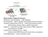
disease’’ associated with altered neurological function, is Rett’s<br />
syndrome, an X-linked postnatal autism spectrum disorder characterized<br />
by stereotypical motor, learning, and social abnormalities<br />
that generally worsen over time (Moretti and Zoghbi, 2006).<br />
Candidate gene analyses identified MeCP2 as <strong>the</strong> causative<br />
gene (Amir et al., 1999). MeCP2 was identified on <strong>the</strong> basis <strong>of</strong><br />
binding selectively to methylated CpG dinucleotides in heterochromatic<br />
regions and functioning in a methylation-dependent<br />
repressive fashion (Nan et al., 1997). Thus, MeCP2 can be<br />
described as a DNA ‘‘reader’’ in analogy to readers <strong>of</strong> histone<br />
methylation (Figure 2). In keeping with <strong>the</strong> general <strong>the</strong>me developed<br />
above for REST/coREST and SXMC repressive<br />
complexes, engagement with additional corepressors and<br />
HDACs provide <strong>the</strong> enzymatic ‘‘punch’’ to <strong>the</strong> silencing activity<br />
Ac<br />
Ac<br />
Neuron 60, December 26, 2008 ª2008 Elsevier Inc. 969
Neuron<br />
Review<br />
elicited by MeCP2. Interestingly, MeCP2 may switch partners<br />
and <strong>the</strong>reby its activity, for example by interacting with CREB<br />
and thus eliciting activation (Chahrour et al., 2008).<br />
In Rett’s syndrome, MeCP2 stands as <strong>the</strong> key DNA-binding<br />
‘‘hook’’ to bring <strong>the</strong> repressive chromatin remodeling machinery<br />
to target loci. In Rubinstein-Taybi syndrome (RSTS), characterized<br />
by mental retardation and developmental abnormalities,<br />
<strong>the</strong> DNA-binding hook is provided by CREB. Phosphorylation<br />
<strong>of</strong> CREB leads to CBP recruitment and activation <strong>of</strong> target<br />
promoters. Causative mutations in RSTS map to <strong>the</strong> CBP gene<br />
and may result in impairment <strong>of</strong> HAT activity (Murata et al.,<br />
2001). Mice haploinsufficient for CBP display impaired cognitive<br />
function, altered neuronal plasticity, and aberrant histone acetylation<br />
at target promoters (Alarcon et al., 2004; Korzus et al.,<br />
2004). Interestingly, <strong>the</strong> behavioral symptoms can be ameliorated<br />
by administration <strong>of</strong> HDAC inhibitors (Vo and Goodman,<br />
2001).<br />
Additional examples show how HAT activity may be modulated<br />
in pathological conditions because <strong>of</strong> unique interaction between<br />
epigenetic regulators. A polyglutamine-expanded protein, spinocerebellar<br />
ataxia-7, regulates several HAT complexes (McMahon<br />
et al., 2005; Palhan et al., 2005). The degree <strong>of</strong> polyglutamine<br />
expansion correlates with <strong>the</strong> impairment <strong>of</strong> HAT activity, which<br />
may, in turn, correlate with disease progression.<br />
Docking <strong>of</strong> effector proteins, especially proteins containing<br />
modules that bind to more stable methyl marks, is a rapidly<br />
expanding area <strong>of</strong> chromatin biology (Ru<strong>the</strong>nburg et al., 2007a).<br />
Equally exciting are ‘‘cross-talk’’ mechanisms wherein an adjacent<br />
or nearby modification can affect histone modifications on<br />
‘‘cis’’ or ‘‘trans’’ tails (Briggs et al., 2002; Fischle et al., 2003).<br />
For example, binding <strong>of</strong> HP1 to H3K9 methyl marks is regulated<br />
by phosphorylation at <strong>the</strong> adjacent H3S10 (Figure 3) in what has<br />
been referred to as ‘‘methyl/phos switching’’ (Fischle et al.,<br />
2003). Interestingly, H3S10 phosphorylation can be induced<br />
during mitosis or during immediate-early gene activation, elicited<br />
by distinct kinases (Nowak and Corces, 2004). One kinase that<br />
induces H3S10 phosphorylation during gene activation is<br />
Rsk2, a kinase causally linked to C<strong>of</strong>fin-Lowry syndrome,<br />
a type <strong>of</strong> human mental retardation (Sassone-Corsi et al.,<br />
1999). Chromatin changes, in part brought about by H3S10<br />
phosphorylation, have been directly demonstrated in hippocampal<br />
neurons (Crosio et al., 2003), leading to wider speculations<br />
that chromatin remodeling could contribute to learning<br />
and memory. Similar conclusions have been reached using<br />
HDAC inhibitors (Fischer et al., 2007). These studies cast new<br />
light on using ‘‘epigenetic <strong>the</strong>rapies’’ to develop strategies for<br />
neuronal dysfunctions, an approach that has proved effective<br />
in <strong>the</strong> treatment <strong>of</strong> cancer (Jones and Baylin, 2007).<br />
Summary, Conclusions, and Future Challenges<br />
Remarkable progress has been made in documenting marks,<br />
writers, readers, and erasers <strong>of</strong> a still incompletely defined<br />
epigenetic histone code. While many questions remain, several<br />
trends and conclusions can be reached. An elaborate series <strong>of</strong><br />
concerted enzymatic reactions remodel chromatin, leading to<br />
<strong>the</strong> generation and maintenance <strong>of</strong> quasi-stable epigenetic<br />
states. For example, HDAC recruitment to chromatin targets<br />
could be considered a late step in a silencing pathway wherein<br />
removal <strong>of</strong> acetyl groups from histone lysines, leading to <strong>the</strong><br />
re-establishment <strong>of</strong> positive charge, causes condensation <strong>of</strong><br />
<strong>the</strong> chromatin fiber in what is considered ‘‘cis mechanisms.’’<br />
However, HDACs might also act earlier in a silencing pathway,<br />
‘‘resetting’’ a particular lysine for subsequent methylation by<br />
HMTs that, in turn, serve as docking surfaces for methyl-binding<br />
effectors in what is being referred to as ‘‘trans mechanisms.’’<br />
O<strong>the</strong>r variations on this general <strong>the</strong>me may apply. For example,<br />
acetylation may be used to recruit downstream machinery (activating<br />
or repressive) through bromodomain recognition or o<strong>the</strong>r<br />
mechanisms. The general concept <strong>of</strong> multivalency in chromatin,<br />
<strong>the</strong> combinatorial association <strong>of</strong> multiple ‘‘Velcro’’ modules to<br />
bring about distinct effector:chromatin interactions, promises<br />
to be an exciting area <strong>of</strong> future work (Ru<strong>the</strong>nburg et al.,<br />
2007a). Early glimpses <strong>of</strong> this strategy at work have already<br />
been reported (Li et al., 2007).<br />
How do epigenetic signals translate into short-term versus<br />
long-term memory effects in <strong>the</strong> nervous system We favor<br />
a scenario where neuronal-specific transcription factors recruit<br />
chromatin remodeling activities that <strong>the</strong>n lock in positive or negative<br />
epigenetic states. We like to speculate that more stable, noncharge-altering<br />
modifications, such as methylation <strong>of</strong> DNA or<br />
histones, participate in ‘‘lock-down’’ activities by engaging chromatin-opening<br />
or chromatin-closing effectors. In this view, highly<br />
reversible modifications, such as acetylation and phosphorylation,<br />
may act as <strong>the</strong> dynamic ‘‘on-<strong>of</strong>f switches’’ in response to<br />
changing conditions and environmental cues (Figure 7).<br />
However, much <strong>of</strong> biology, particularly that displayed by multicellular<br />
organisms, requires less ‘‘black versus white’’ outputs and<br />
ra<strong>the</strong>r gradual, ‘‘gray’’ responses. Here, we propose that ‘‘polymodifications,’’<br />
like polyubiquitylation, etc., may be more in<br />
keeping with graded responses. While well-documented in pathways<br />
<strong>of</strong> protein degradation, polymodifications should receive<br />
more attention in chromatin biology (Verhey and Gaertig, 2007),<br />
not only because <strong>of</strong> <strong>the</strong>ir biological significance, but also since<br />
<strong>the</strong>y may identify novel avenues <strong>of</strong> pharmacological intervention.<br />
Considerable progress has been made in identifying chromatin-binding<br />
modules that function to read selective chromatin<br />
marks. Less clear are functions <strong>of</strong> adding (or removing) chromatin-binding<br />
modules to nonhistone proteins. We note that<br />
many chromatin-modifying enzymes (writers) also contain<br />
reading modules, suggesting that <strong>the</strong> writing and reading <strong>of</strong><br />
epigenetic marks may work toge<strong>the</strong>r in bringing about defined<br />
chromatin states. Along this line, multiple prion-like domains<br />
(glutamine- and asparagine-rich domains) have been noted in<br />
Polycomb, leading to provocative suggestions that ‘‘self-aggregation’’<br />
properties may exist in chromatin regulators to carry or<br />
to propagate long-term ‘‘memory formation and transcriptional<br />
memory’’ through altered chromatin conformational states<br />
(Shorter and Lindquist, 2005).<br />
Finally, is <strong>the</strong>re an epigenetic-based chromatin remodeling<br />
process that serves to ‘‘shape’’ or to ‘‘mold’’ <strong>the</strong> landscape <strong>of</strong><br />
our genome as it deals with environmental cues It may not be<br />
a coincidence that many well-studied chromatin-modifying<br />
enzymes are regulated by, or use directly as substrates, key<br />
intermediates from central metabolism. Acetyl-coenzyme A is<br />
required for most known HAT activities, while certain HDACs<br />
are regulated by nicotinamide. S-adenyosyl methonine is<br />
970 Neuron 60, December 26, 2008 ª2008 Elsevier Inc.
Neuron<br />
Review<br />
Signaling<br />
Poly-modifications<br />
miRNA<br />
Allis, C.D., Berger, S.L., Cote, J., Dent, S., Jenuwien, T., Kouzarides, T., Pillus,<br />
L., Reinberg, D., Shi, Y., Shiekhattar, R., et al. (2007b). New nomenclature for<br />
chromatin modifying enzymes. Cell 131, 633–636.<br />
Amir, R.E., Van den Veyver, I.B., Wan, M., Tran, C.Q., Francke, U., and Zoghbi,<br />
H.Y. (1999). Rett syndrome is caused by mutations in X-linked MECP2, encoding<br />
methyl-CpG-binding protein 2. Nat. Genet. 23, 185–188.<br />
Histone<br />
Phosphorylation<br />
Acetylation<br />
Histone<br />
Methylation<br />
DNA<br />
Methylation<br />
Asher, G., Garfield, D., Stratman, M., Reinke, H., Dibner, C., Kreppel, F.,<br />
Mostoslavsky, R., Alt, F.W., and Schibler, U. (2008). SIRT1 regulates circadian<br />
clock gene expression through Per2 deacetylation. Cell 134, 317–328.<br />
‘Unlocked’<br />
State<br />
Commitment<br />
Consolidation<br />
Information Storage<br />
‘Locked’<br />
State<br />
Ausio, J., Levin, D.B., De Amorim, G.V., Bakker, S., and Macleod, P.M. (2003).<br />
Syndromes <strong>of</strong> disordered chromatin remodeling. Clin. Genet. 64, 83–95.<br />
Ballas, N., and Mandel, G. (2005). The many faces <strong>of</strong> REST oversee epigenetic<br />
programming <strong>of</strong> neuronal genes. Curr. Opin. Neurobiol. 15, 500–506.<br />
Charged PTMs<br />
required for HMTs, and FAD is required for certain classes <strong>of</strong> <strong>the</strong><br />
histone demethylases. We predict that o<strong>the</strong>r ‘‘exotic’’ enzyme<br />
systems will be uncovered that function to regulate chromatin<br />
output following instructions from <strong>the</strong> environment. It follows<br />
that diet, vitamin intake (or depletion), calorie restriction, oxidative<br />
stress, aging, etc., will contribute to and impact epigenetic<br />
signatures (DNA and histone) in ways that have yet to be fully<br />
determined (Denu, 2007). Moreover, nonhistone proteins, in<br />
some cases, those <strong>of</strong> cytosolic origin, will be critical substrates<br />
that are only beginning to receive much-needed attention. Determining<br />
how this overall regulation impinges directly on neuronal<br />
function, survival, and regeneration remains a worthwhile challenge<br />
for future studies. Finding <strong>the</strong> ‘‘writers,’’ ‘‘erasers,’’<br />
‘‘readers,’’ and ‘‘interpreters’’ <strong>of</strong> this ‘‘epigenetic language’’ will<br />
keep neurobiologists, as well as many o<strong>the</strong>r biologists, busy<br />
for years to come.<br />
ACKNOWLEDGMENTS<br />
We would like to thank all <strong>the</strong> members <strong>of</strong> our laboratories for help and stimulating<br />
discussions. We also wish to apologize to all <strong>the</strong> colleagues whose<br />
work could not be cited because <strong>of</strong> space limitations.<br />
REFERENCES<br />
Non-Charged PTMs<br />
Figure 7. Consolidation <strong>of</strong> <strong>Epigenetic</strong> Information by Progressive<br />
Enzymatic Modifications<br />
Scheme <strong>of</strong> <strong>the</strong> proposed step-wise consolidation process <strong>of</strong> epigenetic information,<br />
in which successive and interconnected histone PTMs elicit transition<br />
from an ‘‘unlocked’’ chromatin state to a ‘‘locked,’’ fully committed state to<br />
ei<strong>the</strong>r gene activation or silencing. The unlocked state is characterized by<br />
dynamic, transient, and charged PTMs, such as phosphorylation and acetylation.<br />
The locked state is achieved through noncharged, stable modification <strong>of</strong><br />
both histones and DNA by methylation. Graded modulation <strong>of</strong> this process is<br />
under <strong>the</strong> control <strong>of</strong> intracellular signaling, polymodifications (polyubiquitylation,<br />
etc.), and microRNA pathways.<br />
Alarcon, J.M., Malleret, G., Touzani, K., Vronskaya, S., Ishii, S., Kandel, E.R.,<br />
and Barco, A. (2004). Chromatin acetylation, memory, and LTP are impaired<br />
in CBP+/ mice: a model for <strong>the</strong> cognitive deficit in Rubinstein-Taybi<br />
syndrome and its amelioration. Neuron 42, 947–959.<br />
Allis, C.D., Jenuwein, T., Reinberg, D., and Caparros, M.L. (2007a). <strong>Epigenetic</strong>s<br />
(New York: Cold Spring Harbor Laboratory Press).<br />
Bannister, A.J., and Kouzarides, T. (2005). Reversing histone methylation.<br />
Nature 436, 1103–1106.<br />
Bardag-Gorce, F., French, B.A., Joyce, M., Baires, M., Montgomery, R.O., Li,<br />
J., and French, S. (2007). Histone acetyltransferase p300 modulates gene<br />
expression in an epigenetic manner at high blood alcohol levels. Exp. Mol.<br />
Pathol. 82, 197–202.<br />
Barski, A., Cuddapah, S., Cui, K., Roh, T.Y., Schones, D.E., Wang, Z., Wei, G.,<br />
Chepelev, I., and Zhao, K. (2007). High-resolution pr<strong>of</strong>iling <strong>of</strong> histone methylations<br />
in <strong>the</strong> human genome. Cell 129, 823–837.<br />
Beaulieu, J.M., Tirotta, E., Sotnikova, T.D., Masri, B., Salahpour, A., Gainetdinov,<br />
R.R., Borrelli, E., and Caron, M.G. (2007). Regulation <strong>of</strong> Akt signaling by<br />
D2 and D3 dopamine receptors in vivo. J. Neurosci. 27, 881–885.<br />
Beaulieu, J.M., Marion, S., Rodriguiz, R.M., Medvedev, I.O., Sotnikova, T.D.,<br />
Ghisi, V., Wetsel, W.C., Lefkowitz, R.J., Gainetdinov, R.R., and Caron, M.G.<br />
(2008). A betaarrestin 2 signaling complex mediates lithium action on behavior.<br />
Cell 132, 125–136.<br />
Belden, W.J., Loros, J.J., and Dunlap, J.C. (2007). Execution <strong>of</strong> <strong>the</strong> circadian<br />
negative feedback loop in Neurospora requires <strong>the</strong> ATP-dependent chromatin-remodeling<br />
enzyme CLOCKSWITCH. Mol. Cell 25, 587–600.<br />
Berger, S.L. (2007). The complex language <strong>of</strong> chromatin regulation during transcription.<br />
Nature 447, 407–412.<br />
Bernstein, E., and Allis, C.D. (2005). RNA meets chromatin. Genes Dev. 19,<br />
1635–1655.<br />
Bernstein, B.E., Kamal, M., Lindblad-Toh, K., Bekiranov, S., Bailey, D.K.,<br />
Huebert, D.J., McMahon, S., Karlsson, E.K., Kulbokas, E.J., 3rd, Gingeras,<br />
T.R., et al. (2005). Genomic maps and comparative analysis <strong>of</strong> histone modifications<br />
in human and mouse. Cell 120, 169–181.<br />
Bernstein, E., Duncan, E.M., Masui, O., Gil, J., Heard, E., and Allis, C.D. (2006).<br />
Mouse polycomb proteins bind differently to methylated histone H3 and RNA<br />
and are enriched in facultative heterochromatin. Mol. Cell. Biol. 26, 2560–2569.<br />
Bernstein, B.E., Meissner, A., and Lander, E.S. (2007). The mammalian epigenome.<br />
Cell 128, 669–681.<br />
Bibb, J.A., Chen, J.S., Taylor, J.R., Svenningsson, P., Nishi, A., Snyder, G.L.,<br />
Yan, Z., Sagawa, Z.K., Ouimet, C.C., Nairn, A.C., et al. (2001). Effects <strong>of</strong><br />
chronic exposure to cocaine are regulated by <strong>the</strong> neuronal protein Cdk5.<br />
Nature 410, 376–380.<br />
Bishop, N.A., and Guarente, L. (2007). Genetic links between diet and lifespan:<br />
shared mechanisms from yeast to humans. Nat. Rev. Genet. 8, 835–844.<br />
Bonsch, D., Lenz, B., Fiszer, R., Frieling, H., Kornhuber, J., and Bleich, S.<br />
(2006). Lowered DNA methyltransferase (DNMT-3b) mRNA expression is<br />
associated with genomic DNA hypermethylation in patients with chronic alcoholism.<br />
J. Neural Transm. 113, 1299–1304.<br />
Bozzi, Y., and Borrelli, E. (2006). Dopamine in neurotoxicity and neuroprotection:<br />
what do D2 receptors have to do with it Trends Neurosci. 29, 167–174.<br />
Brami-Cherrier, K., Valjent, E., Hervé, D., Darragh, J., Corvol, J.C., Pages, C.,<br />
Arthur, S.J., Girault, J.A., and Caboche, J. (2005). Parsing molecular and<br />
behavioral effects <strong>of</strong> cocaine in mitogen- and stress-activated protein<br />
kinase-1-deficient mice. J. Neurosci. 25, 11444–11454.<br />
Neuron 60, December 26, 2008 ª2008 Elsevier Inc. 971
Neuron<br />
Review<br />
Briggs, S.D., Xiao, T., Sun, Z.W., Caldwell, J.A., Shabanowitz, J., Hunt, D.F.,<br />
Allis, C.D., and Strahl, B.D. (2002). Gene silencing: trans-histone regulatory<br />
pathway in chromatin. Nature 418, 498.<br />
Chahrour, M., Jung, S.Y., Shaw, C., Zhou, X., Wong, S.T., Qin, J., and Zoghbi,<br />
H.Y. (2008). MeCP2, a key contributor to neurological disease, activates and<br />
represses transcription. Science 320, 1224–1229.<br />
Chambon, P. (1978). The molecular biology <strong>of</strong> <strong>the</strong> eukaryotic genome is<br />
coming <strong>of</strong> age. Cold Spring Harb. Symp. Quant. Biol. 42, 1209–1234.<br />
Chang, Q., Khare, G., Dani, V., Nelson, S., and Jaenisch, R. (2006). The disease<br />
progression <strong>of</strong> Mecp2 mutant mice is affected by <strong>the</strong> level <strong>of</strong> BDNF expression.<br />
Neuron 49, 341–348.<br />
Chawla, S., Vanhoutte, P., Arnold, F., Huang, C., and Bading, H. (2003).<br />
<strong>Neuronal</strong> activity-dependent nucleocytoplasmic shuttling <strong>of</strong> HDAC4 and<br />
HDAC5. J. Neurochem. 85, 151–159.<br />
Chen, W.G., Chang, Q., Lin, Y., Meissner, A., West, A.E., Griffith, E.C.,<br />
Jaenisch, R., and Greenberg, M.E. (2003). Derepression <strong>of</strong> BDNF transcription<br />
involves calcium-dependent phosphorylation <strong>of</strong> MeCP2. Science 302,<br />
885–889.<br />
Cheung, P., Allis, C.D., and Sassone-Corsi, P. (2000a). Signaling to chromatin<br />
through histone modifications. Cell 103, 263–271.<br />
Cheung, P., Tanner, K.G., Cheung, W.L., Sassone-Corsi, P., Denu, J.M., and<br />
Allis, C.D. (2000b). Synergistic coupling <strong>of</strong> histone H3 phosphorylation and<br />
acetylation in response to epidermal growth factor stimulation. Mol. Cell 5,<br />
905–915.<br />
Chwang, W.B., O’Riordan, K.J., Levenson, J.M., and Sweatt, J.D. (2006). ERK/<br />
MAPK regulates hippocampal histone phosphorylation following contextual<br />
fear conditioning. Learn. Mem. 13, 322–328.<br />
Crosio, C., Cermakian, N., Allis, C.D., and Sassone-Corsi, P. (2000). Light<br />
induces chromatin modification in cells <strong>of</strong> <strong>the</strong> mammalian circadian clock.<br />
Nat. Neurosci. 3, 1241–1247.<br />
Crosio, C., Heitz, E., Allis, C.D., Borrelli, E., and Sassone-Corsi, P. (2003).<br />
Chromatin remodeling and neuronal response: multiple signaling pathways<br />
induce specific histone H3 modifications and early gene expression in hippocampal<br />
neurons. J. Cell Sci. 116, 4905–4914.<br />
Denu, J.M. (2007). Vitamins and aging: pathways to NAD+ syn<strong>the</strong>sis. Cell 129,<br />
453–454.<br />
Doi, M., Hirayama, J., and Sassone-Corsi, P. (2006a). Circadian regulator<br />
CLOCK is a histone acetyltransferase. Cell 125, 497–508.<br />
Doi, M., Yujnovsky, I., Hirayama, J., Malerba, M., Tirotta, E., Sassone-Corsi,<br />
P., and Borrelli, E. (2006b). Impaired light masking in dopamine D2 receptornull<br />
mice. Nat. Neurosci. 9, 732–734.<br />
Dong, E., Guidotti, A., Grayson, D.R., and Costa, E. (2007). Histone hyperacetylation<br />
induces demethylation <strong>of</strong> reelin and 67-kDa glutamic acid decarboxylase<br />
promoters. Proc. Natl. Acad. Sci. USA 104, 4676–4681.<br />
Dou, Y., Milne, T.A., Ru<strong>the</strong>nburg, A.J., Lee, S., Lee, J.W., Verdine, G.L., Allis,<br />
C.D., and Roeder, R.G. (2006). Regulation <strong>of</strong> MLL1 H3K4 methyltransferase<br />
activity by its core components. Nat. Struct. Mol. Biol. 13, 713–719.<br />
Dudley, C.A., Erbel-Sieler, C., Estill, S.J., Reick, M., Franken, P., Pitts, S., and<br />
McKnight, S.L. (2003). Altered patterns <strong>of</strong> sleep and behavioral adaptability in<br />
NPAS2- deficient mice. Science 301, 379–383.<br />
Dunlap, J.C. (1999). Molecular bases for circadian clocks. Cell 96, 271–290.<br />
Etchegaray, J.P., Lee, C., Wade, P.A., and Reppert, S.M. (2003). Rhythmic<br />
histone acetylation underlies transcription in <strong>the</strong> mammalian circadian clock.<br />
Nature 421, 177–182.<br />
Etchegaray, J.P., Yang, X., DeBruyne, J.P., Peters, A.H., Weaver, D.R.,<br />
Jenuwein, T., and Reppert, S.M. (2006). The polycomb group protein EZH2<br />
is required for mammalian circadian clock function. J. Biol. Chem. 281,<br />
21209–21215.<br />
Fan, G., Beard, C., Chen, R.Z., Csankovszki, G., Sun, Y., Siniaia, M., Biniszkiewicz,<br />
D., Bates, B., Lee, P.P., Kuhn, R., et al. (2001). DNA hypomethylation<br />
perturbs <strong>the</strong> function and survival <strong>of</strong> CNS neurons in postnatal animals. J. Neurosci.<br />
21, 788–797.<br />
Felsenfeld, G., and Groudine, M. (2003). Controlling <strong>the</strong> double helix. Nature<br />
421, 448–453.<br />
Feng, J., Fouse, S., and Fan, G. (2007). <strong>Epigenetic</strong> regulation <strong>of</strong> neural gene<br />
expression and neuronal function. Pediatr. Res. 61, 58R–63R.<br />
Fischer, A., Sananbenesi, F., Wang, X., Dobbin, M., and Tsai, L.H. (2007).<br />
Recovery <strong>of</strong> learning and memory is associated with chromatin remodelling.<br />
Nature 447, 178–182.<br />
Fischle, W., Wang, Y., and Allis, C.D. (2003). Binary switches and modification<br />
cassettes in histone biology and beyond. Nature 425, 475–479.<br />
Gan, L., and Mucke, L. (2008). Paths <strong>of</strong> convergence: Sirtuins in aging and neurodegeneration.<br />
Neuron 58, 10–14.<br />
Garcia, J.A., Zhang, D., Estill, S.J., Michn<strong>of</strong>f, C., Rutter, J., Reick, M., Scott, K.,<br />
Diaz-Arrastia, R., and McKnight, S.L. (2000). Impaired cued and contextual<br />
memory in NPAS2-deficient mice. Science 288, 2226–2230.<br />
Gekakis, N., Staknis, D., Nguyen, H.B., Davis, F.C., Wilsbacher, L.D., King,<br />
D.P., Takahashi, J.S., and Weitz, C.J. (1998). Role <strong>of</strong> <strong>the</strong> CLOCK protein in<br />
<strong>the</strong> mammalian circadian mechanism. Science 280, 1564–1569.<br />
Gong, X., Tang, X., Wiedmann, M., Wang, X., Peng, J., Zheng, D., Blair, L.A.,<br />
Marshall, J., and Mao, Z. (2003). Cdk5-mediated inhibition <strong>of</strong> <strong>the</strong> protective<br />
effects <strong>of</strong> transcription factor MEF2 in neurotoxicity-induced apoptosis.<br />
Neuron 38, 33–46.<br />
Grimm, J.W., Lu, L., Hayashi, T., Hope, B.T., Su, T.P., and Shaham, Y. (2003).<br />
Time dependent increases in brain-derived neurotrophic factor protein levels<br />
within <strong>the</strong> mesolimbic dopamine system after withdrawal from cocaine: implications<br />
for incubation <strong>of</strong> cocaine craving. J. Neurosci. 23, 742–747.<br />
Guan, Z., Giustetto, M., Lomvardas, S., Kim, J.H., Miniaci, M.C., Schwartz,<br />
J.H., Thanos, D., and Kandel, E.R. (2002). Integration <strong>of</strong> long-term-memoryrelated<br />
synaptic plasticity involves bidirectional regulation <strong>of</strong> gene expression<br />
and chromatin structure. Cell 111, 483–493.<br />
Hirayama, J., Sahar, S., Grimaldi, B., Tamaru, T., Takamatsu, K., Nakahata, Y.,<br />
and Sassone-Corsi, P. (2007). CLOCK-mediated acetylation <strong>of</strong> BMAL1<br />
controls circadian function. Nature 450, 1086–1090.<br />
Holliday, R., and Pugh, J.E. (1975). DNA modification mechanisms and gene<br />
activity during development. Science 187, 226–232.<br />
Huang, J., Sengupta, R., Espejo, A.B., Lee, M.G., Dorsey, J.A., Richter, M.,<br />
Opravil, S., Shiekhattar, R., Bedford, M.T., Jenuwein, T., and Berger, S.L.<br />
(2007). p53 is regulated by <strong>the</strong> lysine demethylase LSD1. Nature 449, 105–108.<br />
Hyman, S., Malenka, R., and Nestler, E. (2006). Neural mechanisms <strong>of</strong> addiction:<br />
<strong>the</strong> role <strong>of</strong> reward-related learning and memory. Annu. Rev. Neurosci. 26,<br />
565–598.<br />
Impey, S., McCorkle, S.R., Cha-Molstad, H., Dwyer, J.M., Yochum, G.S.,<br />
Boss, J.M., McWeeney, S., Dunn, J.J., Mandel, G., and Goodman, R.H.<br />
(2004). Defining <strong>the</strong> CREB regulon: a genome-wide analysis <strong>of</strong> transcription<br />
factor regulatory regions. Cell 119, 1041–1054.<br />
Iuvone, P.M., Galli, C.L., Garrison-Gund, C.K., and Neff, N.H. (1978). Light<br />
stimulates tyrosine hydroxylase activity and dopamine syn<strong>the</strong>sis in retinal<br />
amacrine neurons. Science 202, 901–902.<br />
Jaenisch, R., and Bird, A. (2003). <strong>Epigenetic</strong> regulation <strong>of</strong> gene expression:<br />
how <strong>the</strong> genome integrates intrinsic and environmental signals. Nat. Genet.<br />
33, 245–254.<br />
Jensen, L.R., Amende, M., Gurok, U., Moser, B., Gimmel, V., Tzschach, A.,<br />
Janecke, A.R., Tariverdian, G., Chelly, J., Fryns, J.P., et al. (2005). Mutations<br />
in <strong>the</strong> JARID1C gene which is involved in transcriptional regulation and chromatin<br />
remodeling cause X-linked mental retardation. Am. J. Hum. Genet. 76,<br />
227–236.<br />
Jenuwein, T., and Allis, C.D. (2001). Translating <strong>the</strong> histone code. Science 293,<br />
1074–1080.<br />
Jones, P.A., and Baylin, S.B. (2007). The epigenomics <strong>of</strong> cancer. Cell 128,<br />
683–692.<br />
Korutla, L., Wang, P.J., and Mackler, S.A. (2005). The POZ/BTB protein NAC1<br />
interacts with two different histone deacetylases in neuronal-like cultures. J.<br />
Neurochem. 94, 786–793.<br />
972 Neuron 60, December 26, 2008 ª2008 Elsevier Inc.
Neuron<br />
Review<br />
Korzus, E., Rosenfeld, M.G., and Mayford, M. (2004). CBP histone acetyltransferase<br />
activity is a critical component <strong>of</strong> memory consolidation. Neuron 42,<br />
961–972.<br />
Kumar, A., Choi, K.H., Renthal, W., Tsankova, N.M., Theobald, D.E., Truong,<br />
H.T., Russo, S.J., Laplant, Q., Sasaki, T.S., Whistler, K.N., et al. (2005). Chromatin<br />
remodeling is a key mechanism underlying cocaine-induced plasticity in<br />
striatum. Neuron 48, 303–314.<br />
Lee, K.K., and Workman, J.L. (2007). Histone acetyltransferase complexes:<br />
one size doesn’t fit all. Nat. Rev. Mol. Cell Biol. 8, 284–295.<br />
Lee, Y., Lee, E.K., Cho, Y.W., Matsui, T., Kang, I.C., Kim, T.S., and Han, M.H.<br />
(2003). ProteoChip: a highly sensitive protein microarray prepared by a novel<br />
method <strong>of</strong> protein immobilization for application <strong>of</strong> protein-protein interaction<br />
studies. Proteomics 3, 2289–2304.<br />
Lee, M.G., Wynder, C., Schmidt, D.M., McCafferty, D.G., and Shiekhattar, R.<br />
(2006). Histone H3 lysine 4 demethylation is a target <strong>of</strong> nonselective antidepressive<br />
medications. Chem. Biol. 13, 563–567.<br />
Lessard, J., Wu, J.I., Ranish, J.A., Wan, M., Winslow, M.M., Staahl, B.T., Wu,<br />
H., Aebersold, R., Graef, I.A., and Crabtree, G.R. (2007). An essential switch in<br />
subunit composition <strong>of</strong> a chromatin remodeling complex during neural development.<br />
Neuron 55, 201–215.<br />
Levenson, J.M., and Sweatt, J.D. (2005). <strong>Epigenetic</strong> mechanisms in memory<br />
formation. Nat. Rev. Neurosci. 6, 108–118.<br />
Levenson, J.M., O’Riordan, K.J., Brown, K.D., Trinh, M.A., Molfese, D.L., and<br />
Sweatt, J.D. (2004). Regulation <strong>of</strong> histone acetylation during memory formation<br />
in <strong>the</strong> hippocampus. J. Biol. Chem. 279, 40545–40559.<br />
Levenson, J.M., Roth, T.L., Lubin, F.D., Miller, C.A., Huang, I.C., Desai, P.,<br />
Malone, L.M., and Sweatt, J.D. (2006). Evidence that DNA (cytosine-5) methyltransferase<br />
regulates synaptic plasticity in <strong>the</strong> hippocampus. J. Biol. Chem.<br />
281, 15763–15773.<br />
Levine, A.A., Guan, Z., Barco, A., Xu, S., Kandel, E.R., and Schwartz, J.H.<br />
(2005). CREB-binding protein controls response to cocaine by acetylating<br />
histones at <strong>the</strong> fosB promoter in <strong>the</strong> mouse striatum. Proc. Natl. Acad. Sci.<br />
USA 102, 19186–19191.<br />
Li, J., Guo, Y., Schroeder, F.A., Youngs, R.M., Schmidt, T.W., Ferris, C.,<br />
Konradi, C., and Akbarian, S. (2004). Dopamine D2-like antagonists induce<br />
chromatin remodeling in striatal neurons through cyclic AMP-protein kinase<br />
A and NMDA receptor signaling. J. Neurochem. 90, 1117–1131.<br />
Li, B., Gogol, M., Carey, M., Lee, D., Seidel, C., and Workman, J.L. (2007).<br />
Combined action <strong>of</strong> PHD and chromo domains directs <strong>the</strong> Rpd3S HDAC to<br />
transcribed chromatin. Science 316, 1050–1054.<br />
Lo, W.S., Trievel, R.C., Rojas, J.R., Duggan, L., Hsu, J.Y., Allis, C.D., Marmorstein,<br />
R., and Berger, S.L. (2000). Phosphorylation <strong>of</strong> serine 10 in histone H3 is<br />
functionally linked in vitro and in vivo to Gcn5-mediated acetylation at lysine<br />
14. Mol. Cell 5, 917–926.<br />
Lo, W.S., Duggan, L., Emre, N.C., Belotserkovskya, R., Lane, W.S., Shiekhattar,<br />
R., and Berger, S.L. (2001). Snf1—a histone kinase that works in concert<br />
with <strong>the</strong> histone acetyltransferase Gcn5 to regulate transcription. Science<br />
293, 1142–1146.<br />
Lonze, B.E., and Ginty, D.D. (2002). Function and regulation <strong>of</strong> CREB family<br />
transcription factors in <strong>the</strong> nervous system. Neuron 35, 605–623.<br />
Lu, L., Koya, E., Zhai, H., Hope, B.T., and Shaham, Y. (2006). Role <strong>of</strong> ERK in<br />
cocaine addiction. Trends Neurosci. 29, 695–703.<br />
Macdonald, N., Welburn, J.P., Noble, M.E., Nguyen, A., Yaffe, M.B., Clynes,<br />
D., Moggs, J.G., Orphanides, G., Thomson, S., Edmunds, J.W., et al. (2005).<br />
Molecular basis for <strong>the</strong> recognition <strong>of</strong> phosphorylated and phosphoacetylated<br />
histone h3 by 14-3-3. Mol. Cell 20, 199–211.<br />
Mahadev, K., and Vemuri, M.C. (1998). Effect <strong>of</strong> ethanol on chromatin and<br />
nonhistone nuclear proteins in rat brain. Neurochem. Res. 23, 1179–1184.<br />
Manke, I.A., Lowery, D.M., Nguyen, A., and Yaffe, M.B. (2003). BRCT repeats<br />
as phosphopeptide binding modules involved in protein targeting. Science<br />
302, 636–639.<br />
Marin-Husstege, M., Muggironi, M., Liu, A., and Casaccia-Bonnefil, P. (2002).<br />
Histone deacetylase activity is necessary for oligodendrocyte lineage progression.<br />
J. Neurosci. 22, 10333–10345.<br />
Martinek, S., Inonog, S., Manoukian, A.S., and Young, M.W. (2001). A role for<br />
<strong>the</strong> segment polarity gene shaggy/GSK-3 in <strong>the</strong> Drosophila circadian clock.<br />
Cell 105, 769–779.<br />
Martinowich, K., Hattori, D., Wu, H., Fouse, S., He, F., Hu, Y., Fan, G., and Sun,<br />
Y.E. (2003). DNA methylation-related chromatin remodeling in activity-dependent<br />
BDNF gene regulation. Science 302, 890–893.<br />
Mattson, B., Bossert, J., Simmons, D., Nozaki, N., Nagarkar, D., Kreuter, J.,<br />
and Hope, B. (2005). Cocaine-induced CREB phosphorylation in nucleus accumbens<br />
<strong>of</strong> cocainesensitized rats is enabled by enhanced activation <strong>of</strong> extracellular<br />
signal-related kinase, but not protein kinase A. J. Neurochem. 95,<br />
1481–1494.<br />
McClung, C.A., and Nestler, E.J. (2003). Regulation <strong>of</strong> gene expression and<br />
cocaine reward by CREB and DFosB. Nat. Neurosci. 11, 1208–1215.<br />
McKinsey, T., Zhang, C., and Olson, E. (2000). Activation <strong>of</strong> <strong>the</strong> myocyte<br />
enhancer factor-2 transcription factor by calcium/calmodulin-dependent<br />
protein kinasestimulated binding <strong>of</strong> 14-3-3 to histone deacetylase 5. Proc.<br />
Natl. Acad. Sci. USA 97, 14400–14405.<br />
McMahon, S.J., Pray-Grant, M.G., Schieltz, D., Yates, J.R., and Grant, P.A.<br />
(2005). Polyglutamine-expanded spinocerebellar ataxia-7 protein disrupts<br />
normal SAGA and SLIK histone acetyltransferase activity. Proc. Natl. Acad.<br />
Sci. USA 102, 8478–8482.<br />
Merienne, K., Pannetier, S., Harel-Bellan, A., and Sassone-Corsi, P. (2001).<br />
Mitogen-regulated RSK2-CBP interaction controls <strong>the</strong>ir kinase and acetylase<br />
activities. Mol. Cell. Biol. 21, 7089–7096.<br />
Miller, C.A., and Sweatt, J.D. (2007). Covalent modification <strong>of</strong> DNA regulates<br />
memory formation. Neuron 53, 857–869.<br />
Miranda, T.B., and Jones, P.A. (2007). DNA methylation: <strong>the</strong> nuts and bolts <strong>of</strong><br />
repression. J. Cell. Physiol. 213, 384–390.<br />
Moretti, P., and Zoghbi, H.Y. (2006). MeCP2 dysfunction in Rett syndrome and<br />
related disorders. Curr. Opin. Genet. Dev. 16, 276–281.<br />
Murata, T., Kurokawa, R., Krones, A., Tatsumi, K., Ishii, M., Taki, T., Masuno,<br />
M., Ohashi, H., Yanagisawa, M., Rosenfeld, M.G., et al. (2001). Defect <strong>of</strong><br />
histone acetyltransferase activity <strong>of</strong> <strong>the</strong> nuclear transcriptional coactivator<br />
CBP in Rubinstein-Taybi syndrome. Hum. Mol. Genet. 10, 1071–1076.<br />
Mutskov, V.J., Farrell, C.M., Wade, P.M., Wolffe, A.P., and Felsenfeld, G.<br />
(2002). The barrier function <strong>of</strong> an insulator couples high histone acetylation<br />
levels with specific protection <strong>of</strong> promoter DNA from methylation. Genes<br />
Dev. 16, 1540–1554.<br />
Nakahata, Y., Kaluzova, M., Grimaldi, B., Sahar, S., Hirayama, J., Chen, D.,<br />
Guarente, L., and Sassone-Corsi, P. (2008). The NAD+ dependent deacetylase<br />
SIRT1 acts as a rheostat <strong>of</strong> CLOCK-mediated chromatin remodeling and<br />
circadian control. Cell 134, 329–340.<br />
Nan, X., Campoy, F.J., and Bird, A. (1997). MeCP2 is a transcriptional<br />
repressor with abundant binding sites in genomic chromatin. Cell 88, 471–481.<br />
Nan, X., Hou, J., Maclean, A., Nasir, J., Lafuente, M.J., Shu, X., Kriaucionis, S.,<br />
and Bird, A. (2007). Interaction between chromatin proteins MECP2 and ATRX<br />
is disrupted by mutations that cause inherited mental retardation. Proc. Natl.<br />
Acad. Sci. USA 104, 2709–2714.<br />
Naruse, Y., Oh-hashi, K., Iijima, N., Naruse, M., Yoshioka, H., and Tanaka, M.<br />
(2004). Circadian and light-induced transcription <strong>of</strong> clock gene Per1 depends<br />
on histone acetylation and deacetylation. Mol. Cell. Biol. 24, 6278–6287.<br />
Nestler, E.J. (2008). Transcriptional mechanisms <strong>of</strong> addiction: role <strong>of</strong> delta-<br />
FosB. Philos. Trans. R. Soc. Lond. B Biol. Sci. 363, 3245–3255.<br />
Norrholm, S.D., Bibb, J.A., Nestler, E.J., Ouimet, C.C., Taylor, J.R., and Greengard,<br />
P. (2003). Cocaine-induced proliferation <strong>of</strong> dendritic spines in nucleus<br />
accumbens is dependent on <strong>the</strong> activity <strong>of</strong> cyclin-dependent kinase-5. Neuroscience<br />
116, 19–22.<br />
Neuron 60, December 26, 2008 ª2008 Elsevier Inc. 973
Neuron<br />
Review<br />
Nowak, S.J., and Corces, V. (2004). Phosphorylation <strong>of</strong> histone H3: a balancing<br />
act between chromosome condensation and transcriptional activation. Trends<br />
Genet. 20, 214–220.<br />
Ooi, S.L., and Henik<strong>of</strong>f, S. (2007). Germline histone dynamics and epigenetics.<br />
Curr. Opin. Cell Biol. 19, 257–265.<br />
Ooi, S.K., Qiu, C., Bernstein, E., Li, K., Jia, D., Yang, Z., Erdjument-Bromage,<br />
H., Tempst, P., Lin, S.P., Allis, C.D., et al. (2007). DNMT3L connects unmethylated<br />
lysine 4 <strong>of</strong> histone H3 to de novo methylation <strong>of</strong> DNA. Nature 448, 714–<br />
717.<br />
Outeiro, T.F., Kontopoulos, E., Altmann, S.M., Kufareva, I., Stra<strong>the</strong>arn, K.E.,<br />
Amore, A.M., Volk, C.B., Maxwell, M., Rochet, J.C., McLean, P.J., et al.<br />
(2007). Sirtuin 2 inhibitors rescue alpha-synuclein-mediated toxicity in models<br />
<strong>of</strong> Parkinson’s disease. Science 317, 516–519.<br />
Palhan, V.B., Chen, S., Peng, G., Tjernberg, A., Gamper, A.M., Fan, Y., Chait,<br />
B.T., La Spada, A.R., and Roeder, R.G. (2005). Polyglutamine-expanded<br />
ataxin-7 inhibits STAGA histone acetyltransferase activity to produce retinal<br />
degeneration. Proc. Natl. Acad. Sci. USA 102, 8472–8477.<br />
Panda, S., Antoch, M.P., Miller, B., Su, A.I., Schook, A.B., Straume, M.,<br />
Schultz, P.G., Kay, S.A., Takahashi, J.S., and Hogenesch, J.B. (2002). Coordinated<br />
transcription <strong>of</strong> key pathways in <strong>the</strong> mouse by <strong>the</strong> circadian clock. Cell<br />
109, 307–320.<br />
Prozorovski, T., Schulze-Topph<strong>of</strong>f, U., Glumm, R., Baumgart, J., Schröter, F.,<br />
Ninnemann, O., Siegert, E., Bendix, I., Brüstle, O., Nitsch, R., et al. (2008). Sirt1<br />
contributes critically to <strong>the</strong> redox-dependent fate <strong>of</strong> neural progenitors. Nat.<br />
Cell Biol. 10, 385–394.<br />
Pulipparacharuvil, S., Renthal, W., Hale, C.F., Taniguchi, M., Xiao, G.H.,<br />
Kumar, A., Russo, S.J., Sikder, D., Dewey, C.M., Davis, M.M., et al. (2008).<br />
Cocaine regulates MEF2 to control synaptic and behavioral plasticity. Neuron<br />
59, 621–633.<br />
Putignano, E., Lonetti, G., Cancedda, L., Ratto, G., Costa, M., Maffei, L., and<br />
Pizzorusso, T. (2007). Developmental downregulation <strong>of</strong> histone posttranslational<br />
modifications regulates visual cortical plasticity. Neuron 53, 747–759.<br />
Reick, M., Garcia, J., Dudley, C., and McKnight, S.L. (2001). NPAS2: an analog<br />
<strong>of</strong> clock operative in <strong>the</strong> mammalian forebrain. Science 293, 506–509.<br />
Renthal, W., Maze, I., Krishnan, V., Covington, H.E., Xiao, G., Kumar, A.,<br />
Russo, S., Graham, A., Tsankova, N., Kippin, T.E., et al. (2007). Histone deacetylase<br />
5 epigenetically controls behavioral adaptations to chronic emotional<br />
stimuli. Neuron 56, 517–529.<br />
Renthal, W., Carle, T.L., Maze, I., Covington, H.E., III, Truong, H., Alibhai, I.,<br />
Kumar, A., Olson, E.N., and Nestler, E.J. (2008). DFosB mediates epigenetic<br />
desensitization <strong>of</strong> <strong>the</strong> c-fos gene after chronic amphetamine. J. Neurosci.<br />
28, 7344–7349.<br />
Ripperger, J.A., and Schibler, U. (2006). Rhythmic CLOCK-BMAL1 binding to<br />
multiple E-box motifs drives circadian Dbp transcription and chromatin transitions.<br />
Nat. Genet. 38, 369–374.<br />
Ru<strong>the</strong>nburg, A.J., Li, H., Patel, D.J., and Allis, C.D. (2007a). Multivalent<br />
engagement <strong>of</strong> chromatin modifications by linked binding modules. Nat.<br />
Rev. Mol. Cell Biol. 8, 983–994.<br />
Ru<strong>the</strong>nburg, A.J., Allis, C.D., and Wysocka, J. (2007b). Methylation <strong>of</strong> Lysine 4<br />
on Histone H3: Intricacy <strong>of</strong> writing and reading a single epigenetic mark. Mol.<br />
Cell 25, 15–30.<br />
Saper, C.B., Lu, J., Chou, T.C., and Gooley, J. (2005). The hypothalamic integrator<br />
for circadian rhythms. Trends Neurosci. 28, 152–157.<br />
Sassone-Corsi, P., Mizzen, C.A., Cheung, P., Crosio, C., Monaco, L., Jacquot,<br />
S., Hanauer, A., and Allis, C.D. (1999). Requirement <strong>of</strong> Rsk-2 for epidermal<br />
growth factor-activated phosphorylation <strong>of</strong> histone H3. Science 285, 886–891.<br />
Schroeder, F.A., Lin, C.L., Crusio, W.E., and Akbarian, S. (2007). Antidepressant-like<br />
effects <strong>of</strong> <strong>the</strong> histone deacetylase inhibitor, sodium butyrate, in <strong>the</strong><br />
mouse. Biol. Psychiatry 62, 55–64.<br />
Shi, Y., and Whetstine, J.R. (2007). Dynamic regulation <strong>of</strong> histone lysine methylation<br />
by demethylases. Mol. Cell 25, 1–14.<br />
Shi, X., Kachirskaia, I., Yamaguchi, H., West, L.E., Wen, H., Wang, E., Dutta, S.,<br />
Appella, E., and Gozani, O. (2007). Modulation <strong>of</strong> p53 function by SET8 mediated<br />
methylation at lysine 382. Mol. Cell 27, 636–646.<br />
Shorter, J., and Lindquist, S. (2005). Prions as adaptive conduits <strong>of</strong> memory<br />
and inheritance. Nat. Rev. Genet. 6, 435–450.<br />
Sterner, D.E., and Berger, S.L. (2000). Acetylation <strong>of</strong> histones and transcription-related<br />
factors. Microbiol. Mol. Biol. Rev. 64, 435–459.<br />
Stipanovich, A., Valjent, E., Matamales, M., Nishi, A., Ahn, J., Maroteaux, M.,<br />
Bertran-Gonzalez, J., Brami-Cherrier, K., Enslen, H., Corbillé, A.G., et al.<br />
(2008). A phosphatase cascade by which rewarding stimuli control nucleosomal<br />
response. Nature 453, 879–884.<br />
Strahl, B.D., and Allis, C.D. (2000). The language <strong>of</strong> covalent histone modifications.<br />
Nature 403, 41–45.<br />
Sun, Z.W., and Allis, C.D. (2002). Ubiquitination <strong>of</strong> histone H2B regulates H3<br />
methylation and gene silencing in yeast. Nature 418, 104–108.<br />
Tahiliani, M., Mei, P., Fang, R., Leonor, T., Rutenberg, M., Shimizu, F., Li, J.,<br />
Rao, A., and Shi, Y. (2007). The histone H3K4 demethylase SMCX links<br />
REST target genes to Xlinked mental retardation. Nature 447, 601–605.<br />
Taverna, S.D., Ilin, S., Rogers, R.S., Tanny, J.C., Lavender, H., Li, H., Baker, L.,<br />
Boyle, J., Blair, L.P., Chait, B., et al. (2006). Yng1 PHD finger binding to H3 trimethylated<br />
at K4 promotes NuA3 HAT activity at K14 <strong>of</strong> H3 and transcription at<br />
a subset <strong>of</strong> targeted ORFs. Mol. Cell 24, 785–796.<br />
Trojer, P., and Reinberg, D. (2006). Histone lysine demethylases and <strong>the</strong>ir<br />
impact on epigenetics. Cell 125, 213–217.<br />
Tsankova, N.M., Kumar, A., and Nestler, E.J. (2004). Histone modifications at<br />
gene promoter regions in rat hippocampus after acute and chronic electroconvulsive<br />
seizures. J. Neurosci. 24, 5603–5610.<br />
Tsankova, N., Berton, O., Renthal, W., Kumar, A., Neve, R., and Nestler, E.J.<br />
(2006). Sustained hippocampal chromatin regulation in a mouse model <strong>of</strong><br />
depression and antidepressant action. Nat. Neurosci. 9, 519–525.<br />
Tsankova, N., Renthal, W., Kumar, A., and Nestler, E.J. (2007). <strong>Epigenetic</strong><br />
regulation in psychiatric disorders. Nat. Rev. Neurosci. 8, 355–367.<br />
Turner, B. (2002). Cellular memory and <strong>the</strong> histone code. Cell 111, 285–291.<br />
Ulery, P.G., Rudenko, G., and Nestler, E.J. (2006). Regulation <strong>of</strong> DeltaFosB<br />
stability by phosphorylation. J. Neurosci. 26, 5131–5142.<br />
Verhey, K.J., and Gaertig, J. (2007). The tubulin code. Cell Cycle 6, 2152–2160.<br />
Vo, N., and Goodman, R.H. (2001). CREB-binding protein and p300 in transcriptional<br />
regulation. J. Biol. Chem. 276, 13505–13508.<br />
Waddington, C.H. (1957). The Strategy <strong>of</strong> <strong>the</strong> Genes; a Discussion <strong>of</strong> Some<br />
Aspects <strong>of</strong> Theoretical Biology (London: Allen & Unwin).<br />
Welter, M., Vallone, D., Samad, T.A., Meziane, H., Usiello, A., and Borrelli, E.<br />
(2007). Absence <strong>of</strong> dopamine D2 receptors unmasks an inhibitory control<br />
over <strong>the</strong> brain circuitries activated by cocaine. Proc. Natl. Acad. Sci. USA<br />
104, 6840–6845.<br />
Wijnen, H., and Young, M.W. (2006). Interplay <strong>of</strong> circadian clocks and metabolic<br />
rhythms. Annu. Rev. Genet. 40, 409–448.<br />
Wysocka, J., Swigut, T., Xiao, H., Milne, T.A., Kwon, S.Y., Landry, J., Kauer,<br />
M., Tackett, A., Chait, B.T., Badenhorst, P., et al. (2006). A PHD finger <strong>of</strong><br />
NURF couples histone H3 lysine 4 trimethylation with chromatin remodelling.<br />
Nature 442, 86–90.<br />
Yan, L., Bobula, J.M., Svenningsson, P., Greengard, P., and Silver, R. (2006).<br />
DARPP-32 involvement in <strong>the</strong> photic pathway <strong>of</strong> <strong>the</strong> circadian system. J. Neurosci.<br />
26, 9434–9438.<br />
Yujnovsky, I., Hirayama, J., Doi, M., Borrelli, E., and Sassone-Corsi, P. (2006).<br />
Signaling mediated by <strong>the</strong> dopamine D2 receptor potentiates circadian regulation<br />
by CLOCK:BMAL1. Proc. Natl. Acad. Sci. USA 103, 6386–6391.<br />
Zhang, H., Darwanto, A., Linkhart, T.A., Sowers, L., and Zhang, L. (2007).<br />
Maternal cocaine administration causes an epigenetic modification <strong>of</strong> protein<br />
kinase Cå gene expression in fetal rat heart. Mol. Pharmacol. 71, 1319–1328.<br />
974 Neuron 60, December 26, 2008 ª2008 Elsevier Inc.