Inhibition of lactate dehydrogenase A induces oxidative stress and ...
Inhibition of lactate dehydrogenase A induces oxidative stress and ...
Inhibition of lactate dehydrogenase A induces oxidative stress and ...
- No tags were found...

You also want an ePaper? Increase the reach of your titles
YUMPU automatically turns print PDFs into web optimized ePapers that Google loves.
<strong>Inhibition</strong> <strong>of</strong> <strong>lactate</strong> <strong>dehydrogenase</strong> A <strong>induces</strong><strong>oxidative</strong> <strong>stress</strong> <strong>and</strong> inhibits tumor progressionAnne Le a , Charles R. Cooper a , Arvin M. Gouw b , Ramani Dinavahi a , Anirban Maitra b,c , Lorraine M. Deck d , Robert E. Royer e ,David L. V<strong>and</strong>er Jagt e , Gregg L. Semenza c,f,g,h,1 <strong>and</strong> Chi V. Dang a,b,c,i,j,k,2a Division <strong>of</strong> Hematology, Department <strong>of</strong> Medicine, Departments <strong>of</strong> b Pathology, c Oncology, h Pediatrics, i Molecular Biology, j Genetics, <strong>and</strong> k Cell Biology,f Institute for Cell Engineering, <strong>and</strong> g McKusick–Nathans Institute <strong>of</strong> Genetic Medicine, The Johns Hopkins University School <strong>of</strong> Medicine, Baltimore, MD 21205;d Department <strong>of</strong> Chemistry, University <strong>of</strong> New Mexico, Albuquerque, NM 87131; <strong>and</strong> e Department <strong>of</strong> Biochemistry <strong>and</strong> Molecular Biology, University <strong>of</strong> NewMexico School <strong>of</strong> Medicine, Albuquerque, NM 87131Contributed by Gregg L. Semenza, December 14, 2009 (sent for review October 30, 2009)As the result <strong>of</strong> genetic alterations <strong>and</strong> tumor hypoxia, manycancer cells avidly take up glucose <strong>and</strong> generate <strong>lactate</strong> through<strong>lactate</strong> <strong>dehydrogenase</strong> A (LDHA), which is encoded by a targetgene <strong>of</strong> c-Myc <strong>and</strong> hypoxia-inducible factor (HIF-1). Previousstudies with reduction <strong>of</strong> LDHA expression indicate that LDHA isinvolved in tumor initiation, but its role in tumor maintenance <strong>and</strong>progression has not been established. Furthermore, how reduction<strong>of</strong> LDHA expression by interference or antisense RNA inhibitstumorigenesis is not well understood. Here, we report that reduction<strong>of</strong> LDHA by siRNA or its inhibition by a small-molecule inhibitor (FX11[3-dihydroxy-6-methyl-7-(phenylmethyl)-4-propylnaphthalene-1-carboxylic acid]) reduced ATP levels <strong>and</strong> induced significant <strong>oxidative</strong><strong>stress</strong> <strong>and</strong> cell death that could be partially reversed by theantioxidant N-acetylcysteine. Furthermore, we document that FX11inhibited the progression <strong>of</strong> sizable human lymphoma <strong>and</strong> pancreaticcancer xenografts. When used in combination with the NAD +synthesis inhibitor FK866, FX11 induced lymphoma regression.Hence, inhibition <strong>of</strong> LDHA with FX11 is an achievable <strong>and</strong> tolerabletreatment for LDHA-dependent tumors. Our studies document atherapeutical approach to the Warburg effect <strong>and</strong> demonstratethat <strong>oxidative</strong> <strong>stress</strong> <strong>and</strong> metabolic phenotyping <strong>of</strong> cancers arecritical aspects <strong>of</strong> cancer biology to consider for the therapeuticaltargeting <strong>of</strong> cancer energy metabolism.glycolysis | lymphoma | pancreatic cancer | redox <strong>stress</strong> | xenograft modelsThe propensity <strong>of</strong> cancer cells to take up glucose avidly <strong>and</strong>convert it to <strong>lactate</strong>, even under experimental conditions withadequate oxygen, has been called the Warburg effect or aerobicglycolysis (1, 2). The Warburg effect has been directly linked to theactivation <strong>of</strong> oncogenes, such as MYC, Ras, <strong>and</strong> Akt, <strong>and</strong> loss <strong>of</strong>tumor suppressor genes, which result in the deregulated conversion<strong>of</strong> glucose to <strong>lactate</strong> (3–9). As a result <strong>of</strong> the pervasivepresence <strong>of</strong> hypoxia in tumors, hypoxia-inducible factor (HIF-1) iscommonly increased. HIF-1 levels can also be increased downstream<strong>of</strong> signal transduction pathways <strong>and</strong> by loss <strong>of</strong> the vonHippel-Lindau (VHL) tumor suppressor (10). HIF-1 is a criticaltranscription factor for hypoxic adaptation with its activation <strong>of</strong>glycolytic enzyme genes, including <strong>lactate</strong> <strong>dehydrogenase</strong> A(LDHA), which converts pyruvate to <strong>lactate</strong> coupled with therecycling <strong>of</strong> NAD + (11).Lactate <strong>dehydrogenase</strong> is a tetrameric enzyme comprising twomajor subunits A <strong>and</strong>/or B, resulting in five isozymes (A4, A3B1,A2B2, A1B3, <strong>and</strong> B4) that can catalyze the forward <strong>and</strong> backwardconversion <strong>of</strong> pyruvate to <strong>lactate</strong>. LDHA (LDH-5, M-LDH, or A4), which is the predominant form in skeletal muscle,kinetically favors the conversion <strong>of</strong> pyruvate to <strong>lactate</strong>. LDHB(LDH-1, H-LDH, or B4), which is found in heart muscle, converts<strong>lactate</strong> to pyruvate that is further oxidized. It has long beenknown that many human cancers have higher LDHA levels thannormal tissues (12), but the link between oncogenes <strong>and</strong> glycolysiswas poorly understood. LDHA was identified as a directtarget gene <strong>of</strong> the c-Myc oncogenic transcription factor (13, 14).Because HIF also activates LDHA (11, 15), we reasoned thatreduction <strong>of</strong> LDHA expression would inhibit cellular transformation<strong>and</strong> in vivo tumorigenesis, because tumor tissues arehypoxic <strong>and</strong> normal tissues are neither pr<strong>of</strong>oundly hypoxic norhave activated MYC (14). Studies now document that reduction<strong>of</strong> LDHA reduced cellular transformation <strong>and</strong> markedly delayedtumor formation, indicating that LDHA is important for tumorinitiation (16, 17). These studies provide pro<strong>of</strong>-<strong>of</strong>-concept thatLDHA is a tractable therapeutical target but do not establishwhether LDHA is required for tumor progression. Furthermore,the mechanisms by which LDHA inhibition leads to inhibition <strong>of</strong>tumorigenesis are poorly understood.Here, we report that reduction <strong>of</strong> LDHA causes bioenergetic <strong>and</strong><strong>oxidative</strong> <strong>stress</strong> leading to cell death. Furthermore, using a drug-likesmall molecule (FX11 [3-dihydroxy-6-methyl-7-(phenylmethyl)-4-propylnaphthalene-1-carboxylic acid]), we document that LDHAis required not only for tumor initiation but for tumor maintenance<strong>and</strong> progression. When combined with another metabolic inhibitor,FK866 (APO866), that inhibits NAD + synthesis throughdirect inhibition <strong>of</strong> nicotinamide phosphoribosyltransferase(NAMPT) (18, 19), FX11 is able to induce lymphoma regression.These studies indicate that targeting cancer metabolism throughsmall drug-like molecules is achievable, <strong>and</strong> hence paves the wayfor the development <strong>of</strong> unique classes <strong>of</strong> anticancer drugs.Results <strong>and</strong> DiscussionReduction <strong>of</strong> LDHA Induces Oxidative Stress <strong>and</strong> Cell Death. Wesought to underst<strong>and</strong> the mechanisms <strong>of</strong> cell death followingLDHA reduction by short interfering RNA (siLDHA), which hasbeen shown to inhibit tumorigenesis. First, we determined theeffect <strong>of</strong> reduced LDHA expression on oxygen consumption byhuman Panc (P) 493 B-lymphoid cells, because reduction <strong>of</strong>LDHA would favor the entry <strong>of</strong> pyruvate into mitochondria for<strong>oxidative</strong> phosphorylation, thereby enhancing oxygen consumption.Reduction <strong>of</strong> LDHA expression by siRNA in P493 cellsresulted in an increase in oxygen consumption (Fig. 1 A <strong>and</strong> B).Author contributions: A.L., A.M., D.L.V.J., G.L.S., <strong>and</strong> C.V.D. designed research; A.L., C.R.C.,A.M.G., <strong>and</strong> R.D. performed research; C.R.C., R.D., A.M., L.M.D., R.E.R., <strong>and</strong> D.L.V.J. contributednew reagents/analytic tools; A.L., C.R.C., A.M.G., R.D., A.M., D.L.V.J., G.L.S., <strong>and</strong>C.V.D. analyzed data; <strong>and</strong> A.L., C.R.C., G.L.S., <strong>and</strong> C.V.D. wrote the paper.The authors declare no conflict <strong>of</strong> interest. C.V.D. is member <strong>of</strong> the Scientific AdvisoryBoard <strong>of</strong> Agios Pharmaceuticals; there is no sponsored research or technology licensingactivities involving the company. An invention relating to this work was reported to theJohns Hopkins Technology Transfer <strong>of</strong>fice.Freely available online through the PNAS open access option.1 To whom correspondence may be addressed at: The Johns Hopkins University School <strong>of</strong>Medicine, BRB 671, 733 N. Broadway Ave., Baltimore, MD, 21205. E-mail: gsemenza@jhmi.edu.2 To whom correspondence may be addressed at: The Johns Hopkins University School <strong>of</strong>Medicine, Ross Building, Room 1032, 720 Rutl<strong>and</strong> Avenue, Baltimore, MD 21205. E-mail:cvdang@jhmi.edu.This article contains supporting information online at www.pnas.org/cgi/content/full/0914433107/DCSupplemental.CELL BIOLOGYwww.pnas.org/cgi/doi/10.1073/pnas.0914433107 PNAS | February 2, 2010 | vol. 107 | no. 5 | 2037–2042
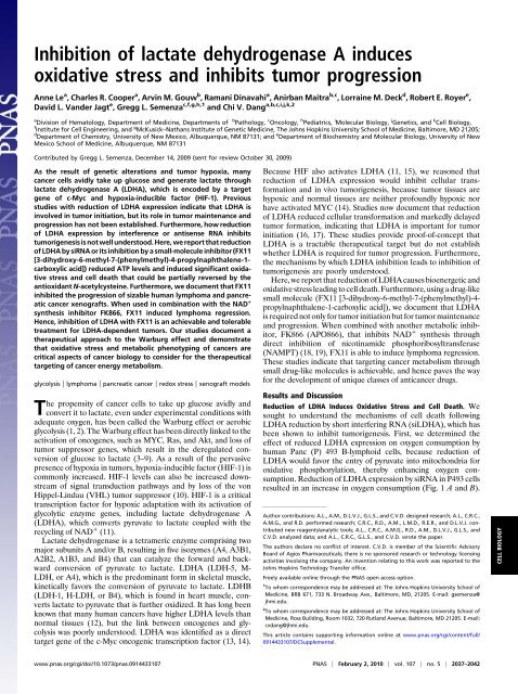
Fig. 2. <strong>Inhibition</strong> <strong>of</strong> LDHA by FX11 resulted in increased oxygen consumption,ROS production, <strong>and</strong> cell death. (A) Oxygen consumption <strong>of</strong> P493cells was determined by a Clark-type oxygen electrode in the presence (slope =−2.4) <strong>and</strong> absence (slope = −1.7) <strong>of</strong> FX11. Data are representative <strong>of</strong> duplicateexperiments. (B) ROS levels were determined by DCFDA fluorescence in P493cells treated with FX11 or FK866. Data are representative <strong>of</strong> triplicate samples<strong>of</strong> two separate experiments. (C) Cell death was determined by flow cytometry<strong>of</strong> Annexin V- <strong>and</strong> 7-AAD-stained cells after 24 h <strong>of</strong> FX11 treatment as comparedwith control. The number in each figure represents the average percentage<strong>of</strong> dead cells. The FX11-treated cells compared with the control grouphave a P value <strong>of</strong> 8.92e-06. (D <strong>and</strong> E) Cell population growth <strong>of</strong> control cellscompared with cells treated with FX11 or FK866 in the presence or absence <strong>of</strong>20 mM NAC. All cells were grown at 1 × 10 5 cells/mL. Cell counts were performedin triplicate <strong>and</strong> shown as mean ± SD, <strong>and</strong> the entire experiment wasreplicated, with similar results.GAPDH is another pivotal glycolytic enzyme that converts NAD +to NADH, we also sought to determine whether FX11 can inhibitits NAD + -dependent conversion <strong>of</strong> glyceraldehyde-3-phosphateto bis-phosphoglycerate. We found that even at 74 μM FX11,GAPDH activity was not inhibited. Through formal Michaelis–Menten kinetics, the estimated K i is >>300 μM for GAPDH,indicating that FX11 was selective for LDHA among glycolyticenzymes that use the c<strong>of</strong>actor NAD.As observed with siRNA-mediated reduction <strong>of</strong> LDHA,inhibition <strong>of</strong> LDHA by FX11 also resulted in increased oxygenconsumption, ROS production, <strong>and</strong> cell death (Fig. 2 A–C). Wefound that the NAMPT inhibitor <strong>of</strong> NAD + synthesis, FK866,also increased ROS production (Fig. 2B). Because the increasein ROS levels might result from lowered NADPH productionattributable to the possible inhibition <strong>of</strong> glucose transport (<strong>and</strong>hence diminished hexose monophosphate shunt activity, whichproduces NADPH) by an indirect effect <strong>of</strong> FX11 <strong>and</strong> FK866, wemeasured <strong>and</strong> found that glucose uptake, as measured by NBDglucose[2-(N-(7-nitrobenz-2-oxa-1,3-diazol-4-yl)amino)-2-deoxyglucose],was not significantly diminished <strong>and</strong> that NADPHlevels were virtually unaltered by treatment with FX11 or FK866(Fig. S3). NAC could partially rescue the diminished proliferation<strong>of</strong> P493 cells treated with either FX11 or FK866 (Fig. 2D <strong>and</strong> E), indicating that <strong>oxidative</strong> <strong>stress</strong> contributed partially tothe inhibition <strong>of</strong> cell proliferation. Given the significant effect <strong>of</strong>FX11 on the proliferation <strong>of</strong> P493 cells, which are dependent onMyc, we ruled out the trivial possibility that FX11 could inhibitMyc expression itself (Fig. S1C). In aggregate, these studiesdocument that reduction <strong>of</strong> LDHA levels or activity triggers<strong>oxidative</strong> <strong>stress</strong> <strong>and</strong> cell death.FX11 Inhibits Glycolysis <strong>and</strong> Alters Cellular Energy Metabolism. Inaddition to <strong>oxidative</strong> <strong>stress</strong> induced by inhibition <strong>of</strong> LDHA, wesought to determine how FX11 affects cellular bioenergetics.First, we observed that both FX11 <strong>and</strong> FK866 decreased mitochondrialmembrane potential <strong>and</strong> that the combination accentuatedthe abnormality (Fig. 3A). In this regard, the combination<strong>of</strong> FX11 <strong>and</strong> FK866 was more toxic to P493 cells than either onealone, causing a more pr<strong>of</strong>ound inhibition <strong>of</strong> cell proliferation(Fig. 3B). A dose–response study using a combination <strong>of</strong> FX11<strong>and</strong> FK866 doses revealed a combination index (CI-50) <strong>of</strong> 0.78,suggesting a slightly synergistic effect <strong>of</strong> the combination on cellproliferation (Fig. S4A). After 20 h <strong>of</strong> exposure to FX11 orFK866, a decrease in ATP levels was accompanied by activation<strong>of</strong> AMP kinase <strong>and</strong> phosphorylation <strong>of</strong> its substrate acetyl-CoAcarboxylase (Fig. 3 C <strong>and</strong> D <strong>and</strong> Fig. S1D), suggesting that, inaddition to induction <strong>of</strong> <strong>oxidative</strong> <strong>stress</strong>, these agents depletedcellular energy levels. Decreased ATP levels, despite an increasein cellular oxygen consumption, suggests that FX11 treatmentincreased nonproductive mitochondrial respiration as reportedwith shRNA-mediated knock-down <strong>of</strong> LDHA (16). Becauseinhibition <strong>of</strong> LDHA decreases NAD + recycling, treatment <strong>of</strong>P493 cells with FX11 was associated with increases in theNADH/NAD + ratio (Fig. 3E) <strong>and</strong> cellular aut<strong>of</strong>luorescence,reflective <strong>of</strong> elevated cellular NADH levels (Fig. S4 B <strong>and</strong> C). Incontrast to FK866, FX11 significantly diminished but did notcompletely inhibit cellular production <strong>of</strong> <strong>lactate</strong> (Fig. 3F). Theseobservations suggest that FX11 targets LDHA, inhibits glycolysis,<strong>and</strong> shunts pyruvate into the mitochondrion.FX11 Inhibits Cells that Are Dependent on Glycolysis. It st<strong>and</strong>s toreason that if FX11 targets LDHA, cells that depend on LDHAfor glycolysis would be more susceptible to FX11 inhibition thanthose that primarily use <strong>oxidative</strong> phosphorylation. In thisregard, we sought to determine whether metabolic phenotypescould affect the sensitivity <strong>of</strong> cancer cells to FX11. We used thehuman RCC4 renal cell carcinoma cell line <strong>and</strong> the RCC4 cellline reconstituted with VHL (RCC4-VHL). Loss <strong>of</strong> VHL inRCC4 rendered these cells constitutively glycolytic because <strong>of</strong>the stabilization <strong>and</strong> expression <strong>of</strong> HIF-1 <strong>and</strong> HIF-2. Reconstitutionwith VHL resulted in degradation <strong>of</strong> HIF-1α <strong>and</strong> HIF-2α <strong>and</strong> increased mitochondrial biogenesis <strong>and</strong> oxygen consumption(23). Given the metabolic differences in these isogeniccell lines, we expect a more significant influence <strong>of</strong> FX11 on theRCC4 cells as compared with the RCC4-VHL cells. Indeed, adose–response study revealed that RCC4 is more sensitive toFX11 compared with RCC4-VHL (Fig. S5 A <strong>and</strong> B).To corroborate these findings further, we studied the glycolyticMCF-7 <strong>and</strong> the <strong>oxidative</strong> MDA-MB-453 breast carcinoma celllines (24). We confirmed that MCF-7 was more dependent onglucose, whereas MDA-MB-453 was more dependent on glutamineoxidation (Fig. S6 A <strong>and</strong> B), such that deprivation <strong>of</strong> glucosehas a more pr<strong>of</strong>ound growth inhibitory effect on MCF-7. Adose-dependent study further revealed that MCF-7 is moresensitive to FX11 (Fig. S5 C <strong>and</strong> D). Although there are manyother differences between these cell lines, the correlation <strong>of</strong>FX11 sensitivity <strong>and</strong> glucose dependency <strong>of</strong> MCF-7 supports thenotion that glycolysis predisposes cancer cells to growth inhibitionby FX11.CELL BIOLOGYLe et al. PNAS | February 2, 2010 | vol. 107 | no. 5 | 2039
further slowed by FX11 (Fig. S5F). Although we noted thatsiControl cells had a modestly diminished proliferation rate ascompared with untreated cells, the siLDHA treatment remarkablydisabled cell proliferation contemporaneous with reducedLDHA expression (Fig. S6 C <strong>and</strong> D). These observations collectivelyindicate that the growth inhibitory effect <strong>of</strong> FX11 isconsistent with its ability to inhibit LDHA.We surmised that the human P493 B-lymphoma cells would besensitive to FX11 because they express LDHA (25) but that thesensitivity would be heightened under hypoxia when glycolysis isfavored. This is particularly important, because P493 cellsdepend on both glucose <strong>and</strong> glutamine metabolism when culturedat 20% (vol/vol) O 2 (26). When P493 cells were subjectedto a dose–response study with FX11, we found that growthinhibition by 9 μM FX11 was increased when the cells werecultured at 1% O 2 (Fig. S7 A <strong>and</strong> B). Hypoxia also sensitized thehuman P198 pancreatic cancer cell line to inhibition by FX11(Fig. S7 C <strong>and</strong> D), suggesting that reliance on LDHA for hypoxicmetabolism caused cancer cells to be susceptible to the growthinhibitoryeffects <strong>of</strong> LDHA inhibition by FX11.Fig. 3. (A) FK866 enhances FX11-induced loss <strong>of</strong> mitochondrial membranepotential. P493 cells treated with control vehicle, FK866, FX11, or bothinhibitors were stained with JC-1 <strong>and</strong> subjected to flow cytometric analysis,with FL2 representing red fluorescence intensity <strong>and</strong> FL1 representing greenfluorescence intensity, which is reflective <strong>of</strong> cells with decreased mitochondrialmembrane. The average percentage (±SEM) <strong>of</strong> cells with decreasedmembrane potential is indicated in each panel. The P values were 0.03,0.002, <strong>and</strong> 0.0008 when comparing FX11-treated cells, FK866-treated cells, orcells treated with both, respectively, with the control group. (B) Effect <strong>of</strong>FK866 <strong>and</strong> FX11 on P493-6 cell proliferation. Live cells were counted usingtrypan blue dye exclusion. Data are shown as the mean ± SD <strong>of</strong> triplicatesamples. (C) Effect <strong>of</strong> FX11 or FK866 on ATP levels. P493 cells were treatedwith 9 μM FX11 or 0.5 nM FK866 for 20 h <strong>and</strong> counted. ATP levels (mean ±SEM, n = 5 experiments) were determined by luciferin–luciferase-based assayon aliquots containing an equal number <strong>of</strong> live cells. *P = 0.008; **P = 0.003.(D) Immunoblot <strong>of</strong> phosphor-AMP kinase (PAMPK) in lysates <strong>of</strong> cell treatedwith FX11 or FK866. Tubulin serves as a loading control. AICAR, an AMPanalogue that activates AMPK, was used to treat the cells as a positivecontrol. (E) FX11 increases the NADH/NAD + ratio. NADH/NAD + ratio in P493cells treated with 9 μM FX11 for 24 h as compared with vehicle control.*P = 0.028. (F) FX11 inhibits <strong>lactate</strong> production. Lactate levels in the media <strong>of</strong>P493 human B cells treated with 9 μM FX11 or 0.5 nM FK866 for 24 h ascompared with control. Control RPMI contained 10.7 mmol/L glucose <strong>and</strong> nodetectable <strong>lactate</strong>. *P = 6.9E-06.We further tested whether the inhibition <strong>of</strong> human P493 Bcells by FX11 depended on glucose or on LDHA. The growth <strong>of</strong>P493 was inhibited by about 60% when depleted <strong>of</strong> glucose ascompared with growth in normal medium (Fig. S5E). Addition <strong>of</strong>FX11 could not inhibit P493 cells further in the absence <strong>of</strong>glucose, suggesting that the effect <strong>of</strong> FX11 on cell proliferationwas glucose-dependent. Furthermore, knock-down <strong>of</strong> LDHAexpression by two sequential electroporations with siRNAcaused a markedly diminished proliferative rate that was notFX11 Inhibits Tumorigenesis in Vivo. Although the renewed interestin the Warburg effect is accompanied by a greater underst<strong>and</strong>ing<strong>of</strong> its molecular underpinnings, targeting it for therapeuticalpurposes remains a major challenge (27). By characterizing asmall-molecule inhibitor <strong>of</strong> LDHA, we found that it is effectivein inhibiting cellular growth <strong>and</strong> triggering cell death by bothinducing ROS production <strong>and</strong> depleting ATP. We observed thathypoxia further sensitized human P493 lymphoma cells toLDHA inhibition by FX11. In this regard, the pervasive hypoxictumor microenvironment, as compared with normal tissues,ought to force a further dependency <strong>of</strong> these lymphoma cells onglycolysis, <strong>and</strong> particularly on LDHA (Fig. 4A <strong>and</strong> Fig. S8A). Ofnote, primary human lymphomas have been documented to haveelevated LDHA expression particularly in hypoxic regions (28).Hence, we sought to determine whether in vivo efficacy could bedemonstrated with FX11 as an inhibitor <strong>of</strong> LDHA. We calculateda desired FX11 dose <strong>of</strong> 42 μg for daily i.p. injection. Weexpected an initial serum level <strong>of</strong> ∼100 μM, assuming a uniform<strong>and</strong> immediate distribution in the vascular system withoutaccounting for the drug half-life or drug metabolism. It should benoted that solubility was a significant dose-limiting factor,because we could only double the dose further before reachingthe limited solubility <strong>of</strong> FX11 in aqueous solution.First, we sought to determine whether FX11 could inhibitP493 tumor initiation after a palpable tumor developed. Ascontrols, we injected animals with vehicle (2% (vol/vol) DMSO)or with 0.8 mg <strong>of</strong> doxycycline to inhibit MYC expression in thesetransformed human B cells. As expected, doxycycline pr<strong>of</strong>oundlyinhibited palpable tumor xenograft growth as compared withvehicle-injected control animals (Fig. 4B). Intriguingly, daily i.p.injection <strong>of</strong> 42 μg <strong>of</strong> FX11 also resulted in a remarkable inhibition<strong>of</strong> tumor growth. It is notable that we have ruled out thetrivial possibility that FX11 might directly inhibit Myc expressionto mediate this pr<strong>of</strong>ound effect (Fig. S1C). These observationsindicate that LDHA is necessary for tumor initiation.To test whether LDHA is required for tumor maintenance orprogression, we challenged the ability <strong>of</strong> FX11 to inhibit tumorxenograft growth by treating P493 lymphomas or human P198pancreatic tumors that reached the size <strong>of</strong> 200 mm 3 beforetreatment commenced. For comparison, we treated animals withvehicle control or a compound related to FX11, called E, thatlacks the benzyl group <strong>and</strong> has a K i for LDHA <strong>of</strong> >90 μM, ormore than 10-fold higher than that <strong>of</strong> FX11. We found that Ehad no antitumor activity as compared with vehicle. At thissolubility-limiting dose, FX11 displayed a static but significanteffect over 10 days (Fig. 4C). Intriguingly, we observed that thehypoxic regions associated with untreated control tumors were2040 | www.pnas.org/cgi/doi/10.1073/pnas.0914433107 Le et al.
Fig. 4. In vivo efficacy <strong>of</strong> FX11 as an antitumor agent. (A) Immunohistochemicalstaining to detect hypoxic regions (dark brown) <strong>of</strong> spleen, liver,<strong>and</strong> P493 lymphoma by pimonidazole labeling. (B) Effect <strong>of</strong> FX11 on growth<strong>of</strong> palpable human P493 B-cell xenografts. Control animals were treatedwith daily i.p. injection <strong>of</strong> vehicle (2% (vol/vol) DMSO), <strong>and</strong> doxycycline(0.8 mg/day) was used as a positive control because it inhibits Myc expression<strong>and</strong> tumorigenesis in P493 cells. (C) Effect <strong>of</strong> FX11 <strong>and</strong>/or FK866 dailytreatment as compared with control or compound E (a weak LDHA inhibitor)on established human lymphoma xenografts. (Inset) Photographs <strong>of</strong> representativeanimals treated with control vehicle or FX11. (D) FX11 inhibitedP198 human pancreatic cancer xenografts as compared with compound E.For experiments in all panels, 2.0 × 10 7 P493 cells or 5 × 10 6 P198 cells wereinjected s.c. into SCID mice or athymic nude mice, respectively. When thetumor volume reached 200 mm 3 ,42μg <strong>of</strong> FX11 <strong>and</strong>/or 100 μg <strong>of</strong> FK866 wasinjected i.p. daily <strong>and</strong> observed for 10–14 days. The tumor volumes weremeasured using digital calipers every 4 days <strong>and</strong> calculated using the followingformula: [length (mm) × width (mm) × width (mm) × 0.52]. Theresults represent the average ± SEM.relatively diminished in the FX11-treated tumors (Fig. S8B). Wespeculated that hypoxic tumor cells are more dependent onglycolysis, <strong>and</strong> hence are diminished by FX11 relative to thenonhypoxic tumor cells. We also observed a significant response<strong>of</strong> human P198 tumor xenografts to FX11 as compared with E(Fig. 4D). Although a more aggressive human pancreatic tumorxenograft LZ10.7 grew significantly faster than P198, it was alsosensitive to FX11 as a single agent (29) (Fig. S8C). The structure<strong>and</strong> activity relation <strong>of</strong> FX11 <strong>and</strong> E in vivo correlated with theinhibition <strong>and</strong> binding <strong>of</strong> LDHA by these compounds in vitro,further supporting the notion that FX11 targets LDHA. Theseresults collectively indicate that LDHA plays a role in tumorprogression <strong>and</strong> maintenance.On the basis <strong>of</strong> our studies <strong>of</strong> cultured cells (Fig. 3B), wehypothesized that FX11 might accentuate the effect <strong>of</strong> FK866 inthe treatment <strong>of</strong> the P493 human lymphomas. Hence, weselected a dose <strong>of</strong> 100 μg <strong>of</strong> FK866, which gave a static outcome,<strong>and</strong>, indeed, we found remarkable tumor regression when animalswere treated with both FX11 <strong>and</strong> FK866 (Fig. 4C). Thesefindings underscore the fact that targeting cancer metabolism isfeasible <strong>and</strong> that LDHA is a significant c<strong>and</strong>idate target forfurther development.Given the significant effects <strong>of</strong> FX11 as an LDHA inhibitor invivo, we studied a group <strong>of</strong> treated animals to begin to underst<strong>and</strong>the potential side effects <strong>of</strong> FX11. It is notable thathumans lacking LDHA develop normally but have been shownto display exertional myopathy (30). In this regard, although wedid not formally exercise the animals to examine exertional tolerance,we did not note lethargy or the inability to eat <strong>and</strong> drink.In fact, animals treated with FX11 did not lose weight. In initialstudies <strong>of</strong> the hematology <strong>and</strong> blood chemistry, we did not seecytopenia in animals treated with FX11 alone; however, two (<strong>of</strong>five studied) animals treated with FK866 did show mild thrombocytopenia(Fig. S9). The average leukocyte count in the controlgroup was skewed upward by two animals that hadleukocytosis with >15 K/μL (normal range: 1.8–to 10.7 K/μL).The blood chemistries did not reveal any evidence <strong>of</strong> kidney[blood urea nitrogen (BUN) or creatinine] or liver (aspartateaminotransferase, alanine aminotransferase, <strong>and</strong> alkaline phosphatase)toxicity in animals treated with FX11 or FK866 alone atdoses that affected tumor growth in vivo (Fig. S9). However, thecombination <strong>of</strong> FX11 <strong>and</strong> FK866, as compared with control,increased BUN.ConclusionsTargeting cancer energy metabolism has been partly elusivebecause <strong>of</strong> our poor underst<strong>and</strong>ing <strong>of</strong> metabolic phenotypes <strong>of</strong>different cancers, compounded by the lack <strong>of</strong> underst<strong>and</strong>ing <strong>of</strong>cellular responses to inhibition <strong>of</strong> specific enzymes involved inenergy metabolism. Here, we document that LDHA inhibition,which was previously shown through genetically engineered cellsto reduce tumor initiation (16, 17), not only resulted indecreased ATP levels <strong>and</strong> reduced mitochondrial membranepotential but in a remarkable increase in <strong>oxidative</strong> <strong>stress</strong> that islinked to cell death. Furthermore, inhibition <strong>of</strong> LDHA by FX11inhibited tumor xenograft progression, confirming that LDHA isrequired for tumor maintenance.We found that similar to siRNA reduction <strong>of</strong> LDHA expression,FX11 could increase cellular oxygen consumption, increase ROSproduction, <strong>and</strong> induce cell death that could be partially rescuedby the antioxidant NAC. Seemingly paradoxical to the antiapoptoticeffect <strong>of</strong> NAC in vitro observed in the current work, wehave documented previously that NAC has an antitumorigeniceffect in vivo because <strong>of</strong> its inhibition <strong>of</strong> HIF-1 in tumors (31). Theillusion <strong>of</strong> a conundrum with NAC treatment arises only whenresults <strong>of</strong> experiments performed in vitro under normoxic conditionsare compared with the results <strong>of</strong> NAC effects on in vivotumorigenesis when HIF-1 is induced within the hypoxic tumormicroenvironment. Our current observations are corroborated bystudies in Saccharomyces cerevisiae with enhanced or defectiverespiration (32). Genetic knock-down or glucose repression <strong>of</strong>respiration in yeast reduced apoptosis <strong>and</strong> enhanced clonogenicsurvival, whereas forced enhancement <strong>of</strong> respiration increasedROS production <strong>and</strong> reduced colony growth that could be partiallyrescued by the antioxidant glutathione. In this regard, a recentperspective on cancer energy metabolism emphasizes the importance<strong>of</strong> redox homeostasis in cancer cell survival (6). FX11 alsoreduced ATP levels, suggesting that inhibition <strong>of</strong> LDHA causedbioenergetic <strong>and</strong> <strong>oxidative</strong> <strong>stress</strong>, which, together, inhibits tumorxenograft maintenance <strong>and</strong> progression.Because FX11 has a catechol moiety, it could hypotheticallybe converted in vivo to a dihydroquinone that is reactive <strong>and</strong>could cause effects other than inhibition <strong>of</strong> LDHA. Although theCELL BIOLOGYLe et al. PNAS | February 2, 2010 | vol. 107 | no. 5 | 2041
Supporting InformationLe et al. 10.1073/pnas.0914433107SI Experimental ProceduresCell Lines <strong>and</strong> Hypoxic Exposure. P493 human lymphoma B cells weremaintained in RPMI 1640 with 10% (vol/vol) FBS <strong>and</strong> 1% (wt/vol)penicillin–streptomycin. P198 human pancreatic cancer cells,RCC4 <strong>and</strong> RCC4-VHL human renal carcinoma cells, <strong>and</strong> MCF-7<strong>and</strong> MDA-MB-453 breast cancer cells were maintained in highglucose(4.5 mg/mL) DMEM with 10% (vol/vol) FBS <strong>and</strong> 1% (wt/vol) penicillin–streptomycin. Nonhypoxic cells (20% (vol/vol) O 2 )were maintained at 37 °C in a 5% (vol/vol) CO 2 <strong>and</strong> 95% (vol/vol)air incubator. Hypoxic cells (1% O 2 ) were maintained in a controlledatmosphere chamber (PLAS-LABS) with a gas mixturecontaining 1% O 2 , 5% (vol/vol) CO 2 , <strong>and</strong> 94% (vol/vol) N 2 at37 °C for the indicated time. Bright live cells were counted dailyin a hemacytometer using trypan blue dye to exclude dead cells. Allcells were grown at a concentration <strong>of</strong> 10 5 cells /mL. All drug treatmentsbegan at day 0. FX11 was added daily.RNA Interference Experiments. siRNAs targeting human LDHA(ON-TARGETplus SMARTpool) were purchased from DharmaconResearch, Inc. Targeting sequences for LDHA were a pool<strong>of</strong> the following four target sequences: sequence 1, GGAGAA-AGCCGUCUUAAUU; sequence 2, GGCAAAGACUAUA-AUGUAA; sequence 3, UAAGGGUCUUUACGGAAUA; <strong>and</strong>sequence 4, AAAGUCUUCUGAUGUCAUA. For P493 humanlymphoma B cells, transfection <strong>of</strong> siRNAs was performed using anAmaxa Nucleotransfection device according to the manufacturer’sinstructions. Briefly, 2 μg <strong>of</strong> siLDHA ON-TARGETplus SMARTpoolor ON-TARGETplus Nontargeting Pool (Dharmacon Research,Inc.) was transfected into 2 × 10 6 cells at 0 h. At 24 h, 10 5 cellswere treated with 0.1% DMSO or FX11 for 48 more hours. Theremaining cells were harvested for immunoblot analysis. For P198human pancreatic cancer cells, transfection <strong>of</strong> siRNAs was performedusing X-tremeGENE siRNA Transfection Reagent (Roche)according to the manufacturer’s instructions.Western Blot Analysis. Cell pellets were harvested after washingwith PBS. Protein concentration was determined by BCA assay(Pierce) <strong>and</strong> 30 μg <strong>of</strong> protein per well was separated by SDS/PAGE <strong>and</strong> transferred by iBlot gel transfer stacks (Invitrogen).Rabbit monoclonal anti-LDHA (Epitomics) was used to detecthuman LDHA; phosphor-AMP kinase α <strong>and</strong> phospho-acetyl-CoA carboxylase antibodies were purchased from Cell Signaling.Membranes were reprobed with anti-α-tubulin as a loadingcontrol. The enhanced chemiluminescence reagent ECL (GEHealthcare) was used for detection.Oxygen Consumption. Oxygen consumption was measured using aClark-type oxygen electrode (Oxytherm System; HansatechInstruments Ltd). Then, 5 × 10 6 cells in 1 mL <strong>of</strong> medium wereplaced in the chamber above a membrane that is permeable tooxygen. Oxygen diffuses through the membrane <strong>and</strong> is reduced atthe cathode surface so that a current flows through the circuit,which is completed by a thin layer <strong>of</strong> KCl solution. The current thatis generated bears a direct stoichiometric relation to the oxygenreduced <strong>and</strong> is converted to a digital signal. Determinations weredone in triplicate, <strong>and</strong> the entire experiment was done twice.ROS Measurement by Flow Cytometry. Intracellular ROS productionwas measured by staining cells with 5-(<strong>and</strong>-6)-carboxy-2′,7′-dichlorodihydr<strong>of</strong>luorescein diacetate (carboxy-H 2 DCFDA;Molecular Probes) according to the manufacturer’s instructions.Next, 10 5 cells/mL were treated with 9 μM FX11 or 0.5 nMFK866 for 24 h. Stained cells were analyzed in FACScan flowcytometers (BD Bioscience).Annexin V Assay. After 24 h <strong>of</strong> FX11 treatment, cells were harvested<strong>and</strong> washed twice with cold PBS <strong>and</strong> the assay was performedusing the Annexin V–7-AAD apoptosis detection Kit I(BD Biosciences Pharmingen) according to the manufacturer’sinstructions.Determination <strong>of</strong> K i <strong>and</strong> Enzyme Kinetics. The reaction velocity <strong>of</strong>purified human LDHA or GAPDH was determined by a decreaseor increase, respectively, in absorbance at 340 nm <strong>of</strong> NADH. TheLDHA activity was assessed using the protocol described in theWorthington Enzyme Manual, with varying concentrations <strong>of</strong>NADH. The GADPH activity was assessed using the protocoldescribed in the Worthington Enzyme Manual with varying concentrations<strong>of</strong> NAD + . K i values were determined from doublereciprocalplots by linear regression analysis using SigmaPlotEnzyme Kinetic s<strong>of</strong>tware.CarboxyLink Immobilization <strong>and</strong> Affinity Columns. FX11 <strong>and</strong> Emolecules that contain carboxyl groups were coupled to immobilizeddiaminodipropylamine resins according to the manufacturer’sinstructions (Pierce). About 1.9 mg (95% couplingefficiency) <strong>of</strong> FX11 or E was coupled to 2 mL <strong>of</strong> resin as estimatedby the amount <strong>of</strong> either molecule recovered after conjugation.An equal amount <strong>of</strong> cell lysate was loaded onto thesebeads <strong>and</strong> eluted with 1 mM NADH after washing with six columnvolumes <strong>of</strong> high salt (1 M NaCl). LDHA activity was performedfrom the eluates to assess the binding affinity <strong>of</strong> themolecules with LDHA.Mitochondrial Membrane Potential Measurement by Flow Cytometry.The lipophilic cation dye [JC-1(5,5′,6,6′-tetrachloro-1,1′,3,3′-tetraethylbenzimidazolcarbocyanine iodide); Invitrogen) wasused to detect the loss <strong>of</strong> the mitochondrial membrane potential.The negative charge established by the intact mitochondrialmembrane potential lets the lipophilic dye stain the mitochondriabright red, which emits in channel 2 (FL2). When the mitochondrialmembrane potential collapses, JC-1 remains in thecytoplasm in a green fluorescent monomeric emission in channel1 (FL1). JC-1 reversibly changes its color from green to orangeas membrane potentials increase (over values <strong>of</strong> 80–100 mV).Then, 10 5 cells/mL were treated with 9 μM FX11 <strong>and</strong>/or FK866for 24 h. Stained cells were analyzed in FACScan flow cytometers(BD Bioscience).Measurement <strong>of</strong> ATP. P493 cells were treated with 9 μM FX11 or0.5 nM FK866 for 20 h <strong>and</strong> counted. ATP levels were determinedby a luciferin–luciferase-based assay (Promega) onaliquots containing equal number <strong>of</strong> cells according to a st<strong>and</strong>ardprotocol.Determination <strong>of</strong> NADH/NAD + <strong>and</strong> NADPH/NADP + Ratios. The NADH/NAD + <strong>and</strong> NADPH /NADP + ratios were assessed using theprotocol described by the manufacturer. The assay is based on anenzyme-catalyzed kinetic reaction in which a tetrazolium dye 3-[4,5-dimethylthiazol-2-yl]-2,5-diphenyltetrazolium bromide is reducedby NAD(P)H in the presence <strong>of</strong> phenazine methosulfate.The intensity <strong>of</strong> the reduced product color, measured at 565 nm, isproportionate to the NAD(P)H /NAD(P) + concentration in thesamples. The NAD(P)H or NAD(P) + content <strong>of</strong> the cells washarvested separately using a corresponding extraction buffer.Le et al. www.pnas.org/cgi/content/short/09144331071<strong>of</strong>11
Fig. S1. (A) Effect <strong>of</strong> reduced LDHA expression by siRNA on oxygen consumption in P198 human pancreatic cancer cells. Oxygen consumption was determinedby the use <strong>of</strong> a Clark-type oxygen electrode at 72 h posttransfection with siLDHA (slope = −2.1) or siControl (slope = −1.1). (B) Immunoblotting was performedon whole-cell lysates, probed with rabbit monoclonal anti-LDHA antibody, <strong>and</strong> reprobed with anti-α-tubulin as a loading control. (C) FX11 does not affect c-Myc levels in P493 cells. Tetracycline, known to repress c-Myc expression, was used as a control. Equivalent amounts <strong>of</strong> proteins were immunoblotted with antic-Mycantibody, <strong>and</strong> α-tubulin served as a loading control. (D) Activation <strong>of</strong> phospho-acetyl-CoA carboxylase (P-ACC) in lysates <strong>of</strong> cells treated with FX11 orFK866. Western blot analysis was performed after 24 h <strong>of</strong> treatment. Equivalent amounts <strong>of</strong> proteins were immunoblotted with anti-P-ACC, <strong>and</strong> α-tubulinserved as a loading control.Le et al. www.pnas.org/cgi/content/short/09144331073<strong>of</strong>11
Fig. S2. (A <strong>and</strong> B) FX11 <strong>and</strong> its derivative E are competitive inhibitors <strong>of</strong> LDHA with NADH as substrate. Lineweaver–Burk plots were determined from triplicateexperiments using averages <strong>of</strong> activities. K i determination was performed with 13.5 μM FX11 or 27 μME.(C)Affinity chromatography <strong>of</strong> LDHA using sepharoseimmobilizedFX11 or E. Equal volumes <strong>of</strong> P493 human B cell lysates were chromatographed with six column volumes <strong>of</strong> high salt (1 M NaCl) wash, followed byelution with 1 mM NADH. LDHA activity was determined for each fraction, <strong>and</strong> the experiment was replicated with a representative experiment shown.Le et al. www.pnas.org/cgi/content/short/09144331074<strong>of</strong>11
Fig. S3. (A) FX11 <strong>and</strong>/or FK866 effects on NADPH/NADP + ratio. P493 cells were treated with 10 μM FX11 <strong>and</strong>/or 0.1 nM FK866 for 24 h before the NADPH/NADP ratio was assayed. FX11 treatment compared with the control (P = 0.8) <strong>and</strong> FK866 treatment compared with the control (P = 0.9). (B) Effects <strong>of</strong> FX11treatment on glucose uptake by P493 cells. A fluorescent analogue <strong>of</strong> 2-deoxyglucose (2-NBDG) was used to assay the glucose uptake <strong>of</strong> P493 cells, followed byflow cytometry. Viable cells taking up glucose were determined by negative 7-AAD <strong>and</strong> positive 2-NBDG. The number in each panel indicates the geometricalmean <strong>of</strong> green fluorescence intensity (520 nm) <strong>of</strong> the cell population.Le et al. www.pnas.org/cgi/content/short/09144331075<strong>of</strong>11
Fig. S4. (A) Effect <strong>of</strong> FK866 <strong>and</strong> FX11 on P493-6 cell proliferation. Live cells were counted using trypan blue dye exclusion. Experiments were done in duplicate,<strong>and</strong> the entire experiment was repeated two times with similar results. The bars represent the averages <strong>of</strong> duplicate samples from one experiment. Thecombination <strong>of</strong> FX11 <strong>and</strong> FK866 doses revealed a CI-50 = 0.78 calculated using R 2.9.1 s<strong>of</strong>tware. (B) FX11 treatment increases cellular NADH fluorescence <strong>of</strong>P493-6 cells compared with control, compound E, or FK866 treatment. The number in the P2 box represents the average percentage <strong>of</strong> cellular NADH aut<strong>of</strong>luorescence(n = 3). FX11 treatment compared with control (P = 0.005), FK866 compared with control (P = 0.144), <strong>and</strong> E treatment compared with control(P = 0.38). (C) Fluorescence emission spectrum <strong>of</strong> NADH (green line), FX11 (red line), or E (blue line). Note that the emission maxima <strong>of</strong> FX11 or E (390 nm) donot coincide with that <strong>of</strong> NADH (460 nm), excluding the direct contribution <strong>of</strong> FX11 to the aut<strong>of</strong>luorescence <strong>of</strong> FX11-treated cells at 460 nm.Le et al. www.pnas.org/cgi/content/short/09144331076<strong>of</strong>11
Fig. S5. (A–D) RCC4 cells <strong>and</strong> MCF-7 cells are more sensitive to FX11 than RCC4-VHL <strong>and</strong> MDA-MB-453 cells. Cell population growth <strong>of</strong> human renal carcinomaRCC4 <strong>and</strong> RCC4-VHL cells or human breast cancer MCF-7 <strong>and</strong> MDA-453 cells in response to different doses <strong>of</strong> FX11. (E) Effect <strong>of</strong> FX11 on the proliferation <strong>of</strong>P493 cells is glucose-dependent. Cell population growth <strong>of</strong> P493 cells in the absence <strong>of</strong> glucose <strong>and</strong> in the presence or absence <strong>of</strong> 9 μM FX11. (F) LDHAdependenteffect <strong>of</strong> FX11. Cell population growth <strong>of</strong> P493 cells with siRNA-mediated reduced LDHA expression in the presence or absence <strong>of</strong> 9 μM FX11. Cellnumbers are shown as mean ± SD.Le et al. www.pnas.org/cgi/content/short/09144331077<strong>of</strong>11
Fig. S6. (A <strong>and</strong> B) Characterization <strong>of</strong> glucose, glutamine, <strong>and</strong> pyruvate dependency <strong>of</strong> different human breast cancer cell lines, MCF-7 <strong>and</strong> MDA-MB-453.Cells were cultured in media with different availability <strong>of</strong> glucose, glutamine, <strong>and</strong> pyruvate. All media were supplemented with 10% (vol/vol) bovine fetalserum <strong>and</strong> 1% penicillin–streptomycin. Averages <strong>of</strong> cell numbers from triplicate experiments are shown ± SD. (C) Growth curves for control, electroporated,<strong>and</strong> siControl cells compared with cells treated with siLDHA. ON-TARGETplus pool was used as siRNAs targeting human LDHA, <strong>and</strong> Nontargeting Pool was usedas siControl. The siRNAs were transfected into 2 × 10 6 cells at 0 <strong>and</strong> 24 h by electroporation. (D) Cells were harvested for immunoblot analysis. Rabbitmonoclonal anti-LDHA was used to detect LDHA, <strong>and</strong> the membrane was reprobed with anti-α-tubulin as a loading control.Le et al. www.pnas.org/cgi/content/short/09144331078<strong>of</strong>11
Fig. S7. Hypoxia accentuates the sensitivity <strong>of</strong> human P493 B cells (A <strong>and</strong> B) <strong>and</strong> human P198 pancreatic cancer (C <strong>and</strong> D) cells to FX11 inhibition <strong>of</strong> growth. Cellpopulation growth <strong>of</strong> P493 B cells or pancreatic cancer P198 cells in normoxia or hypoxia with different doses <strong>of</strong> FX11. Normoxic cells were grown at 37 °C in a5% (vol/vol) CO 2 <strong>and</strong> 95% (vol/vol) air incubator. Hypoxic cells (1% O 2 ) were maintained for the indicated time in a controlled atmosphere chamber with a gasmixture containing 1% O 2 , 5% (vol/vol) CO 2 , <strong>and</strong> 94% (vol/vol) N 2 at 37 °C. There is no significant difference in cell numbers between the 0% <strong>and</strong> 0.1% DMSOgroups.Le et al. www.pnas.org/cgi/content/short/09144331079<strong>of</strong>11
Fig. S8. (A) Pimonidazole-labeling <strong>of</strong> hypoxic regions (dark brown) <strong>of</strong> spleen, liver, <strong>and</strong> P493-6 lymphoma. (B) FX11 treatment diminishes hypoxic regions <strong>of</strong>P493 lymphoma. Rabbit anti-Hypoxyprobe antibody was used as the primary antibody. Texas-red anti-rabbit <strong>and</strong> DakoCytomation EnVision+ System-HRP anti-Rabbit antibody were used as secondary antibodies for immun<strong>of</strong>luorescence <strong>and</strong> immunohistochemistry, respectively. Samples were analyzed under anAxiovert 200 (Zeiss) fluorescence microscope at ×0 magnification. Ten r<strong>and</strong>om fields from an untreated tumor <strong>and</strong> a treated tumor were photographed. (C)Effect <strong>of</strong> FX11 treatment vs. vehicle control on LZ10.7 human pancreatic cancer xenografts (P = 0.03). A total <strong>of</strong> 5 × 10 6 LZ10.7 cells were injected s.c. intoathymic nude mice. When the tumor volume reaches 150 mm 3 ,42μg <strong>of</strong> FX11 or DMSO, which is used as vehicle control, was injected i.p. daily <strong>and</strong> observed for10 days. The tumor volumes were measured using digital calipers <strong>and</strong> calculated using the following formula: [length (mm) × width (mm) × width (mm) × 0.52].The results represent the average ± SEM.Le et al. www.pnas.org/cgi/content/short/0914433107 10 <strong>of</strong> 11
Fig. S9. Effect <strong>of</strong> FX11 treatment on blood chemistries <strong>and</strong> hematology. Average values ± SD are shown for five animals in each group. ALB, albumin; ALP,alkaline phosphatase; ALT, alanine aminotransferase; AMYL, amylase; AST, aspartate aminotransferase; BA, basophils; CAL, calcium; Chol, cholesterol; CL,chloride; CREAT, creatinine; EO, eosinophils; GLU, glucose; HB, hemoglobin; HCT, hematocrit; K, potassium; LY, lymphocytes; MCH, mean corpuscular hemoglobin;MCHC, mean corpuscular hemoglobin concentration; MCV, mean corpuscular volume; MO, monocytes; NA, sodium; NE, neutrophils; RBC, red bloodcells; RDW, red cell distribution width; TBILI, total bilirubin; TPROT, total protein; Tri, triglyceride; UA, uric acid; WBC, white blood cells.Le et al. www.pnas.org/cgi/content/short/0914433107 11 <strong>of</strong> 11