TGen Tissue Microarray Center | - Translational Genomics Research ...
TGen Tissue Microarray Center | - Translational Genomics Research ...
TGen Tissue Microarray Center | - Translational Genomics Research ...

You also want an ePaper? Increase the reach of your titles
YUMPU automatically turns print PDFs into web optimized ePapers that Google loves.
<strong>TGen</strong> <strong>Tissue</strong> <strong>Microarray</strong> <strong>Center</strong> |<br />
Clean, high quality TMAs optimized<br />
for the detection of your specifi c targets<br />
The <strong>TGen</strong> TMA <strong>Center</strong> plays a<br />
critical role in the translational<br />
research process through standardized<br />
collection and organization<br />
of clinical tissues. The TMA format<br />
offers high throughput capacity for<br />
molecular analyses and provides<br />
inherent assay standardization.<br />
Active collaborations with imaging<br />
experts and computer programmers<br />
are an integral component<br />
of the TMA <strong>Center</strong>’s efforts to<br />
continually extend the applications<br />
of TMA technology. Most notable<br />
of these was a two-year collaborative<br />
development of an automated<br />
fluorescence in situ hybridization<br />
(FISH) spot counting program. This<br />
highly interactive program was<br />
designed to enumerate probe<br />
signals after hybridization of<br />
specific DNA sequences. In situ<br />
hybridization (ISH) applications<br />
using DNA or RNA probes remains<br />
an active part of Dr. Hostetter’s<br />
research.<br />
About the <strong>Center</strong> Director<br />
Dr. Galen Hostetter is the<br />
Director of the <strong>Tissue</strong> Microarry<br />
(TMA) <strong>Center</strong> at <strong>TGen</strong> and an<br />
Associate Investigator at <strong>TGen</strong>.<br />
Dr. Hostetter is a board-certified<br />
pathologist and has significant<br />
expertise and experience in<br />
constructing tissue microarrays<br />
and the molecular assays of IHC<br />
and FISH on TMA slides.<br />
Board Certifications:<br />
• Diplomate of the American Board<br />
of Pathology, Certified in Clinical<br />
Pathology, September 2001.<br />
• Board Eligible, Clinical Genetics<br />
and Molecular Genetics, American<br />
Board of Medical Genetics, June<br />
2002.<br />
http://www.tgen.org/<br />
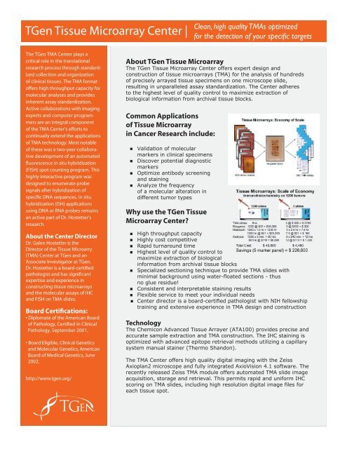
About <strong>TGen</strong> <strong>Tissue</strong> <strong>Microarray</strong><br />
The <strong>TGen</strong> <strong>Tissue</strong> <strong>Microarray</strong> <strong>Center</strong> offers expert design and<br />
construction of tissue microarrays (TMA) for the analysis of hundreds<br />
of precisely arrayed tissue specimens on one microscope slide,<br />
resulting in unparalleled assay standardization. The <strong>Center</strong> adheres<br />
to the highest level of quality control to maximize extraction of<br />
biological information from archival tissue blocks.<br />
Common Applications<br />
of <strong>Tissue</strong> <strong>Microarray</strong><br />
in Cancer <strong>Research</strong> include:<br />
• Validation of molecular<br />
markers in clinical specimens<br />
• Discover potential diagnostic<br />
markers<br />
• Optimize antibody screening<br />
and staining<br />
• Analyze the frequency<br />
of a molecular alteration in<br />
different tumor types<br />
Why use the <strong>TGen</strong> <strong>Tissue</strong><br />
<strong>Microarray</strong> <strong>Center</strong><br />
• High throughput capacity<br />
• Highly cost competitive<br />
• Rapid turnaround time<br />
• Highest level of quality control to<br />
maximize extraction of biological<br />
information from archival tissue blocks<br />
• Specialized sectioning technique to provide TMA slides with<br />
minimal background using water-floated sections - thus<br />
no glue residue!<br />
• Consistent and interpretable staining results<br />
• Flexible service to meet your individual needs<br />
• <strong>Center</strong> director is a board-certified pathologist with NIH fellowship<br />
training and extensive experience in TMA design and construction<br />
Technology<br />
The Chemicon Advanced <strong>Tissue</strong> Arrayer (ATA100) provides precise and<br />
accurate sample extraction and TMA construction. The IHC staining is<br />
optimized with advanced epitope retrieval methods utilizing a capillary<br />
system manual stainer (Thermo Shandon).<br />
The TMA <strong>Center</strong> offers high quality digital imaging with the Zeiss<br />
Axioplan2 microscope and fully integrated AxioVision 4.1 software. The<br />
recently released Zeiss TMA module offers automated TMA slide image<br />
acquisition, storage and retrieval. This permits rapid and uniform IHC<br />
scoring on TMA slides, including high resolution digital image files for<br />
each tissue spot.<br />
®
<strong>TGen</strong> <strong>Tissue</strong> <strong>Microarray</strong> <strong>Center</strong> |<br />
Clean, high quality TMAs optimized<br />
for the detection of your specifi c targets<br />
Services<br />
<strong>TGen</strong> offers the following TMA services:<br />
• Design and construction of tissue microarrays (with expert<br />
sectioning on standard glass slides) for investigators with<br />
parrafin tissue block collections<br />
• Access to high quality “tester” TMA slides for optimization of<br />
molecular assay protocols<br />
• Tumor specific TMA slides are under development for IRB approved<br />
protocols<br />
• Immunohistochemistry (IHC) and fluorescent in situ hybridization<br />
(FISH) services adapted to paraffin tissue sections<br />
* Available in October 2004 – in situ hybridization assays!<br />
• High-throughput analysis of protein and DNA targets in formalin<br />
fixed paraffin embedded tissue section<br />
• Specialized antibody optimization services for immunostaining on<br />
formalin fixed paraffin embedded tissue sections<br />
Our service can include any combination of the following:<br />
1. literature search for application guidelines<br />
2. choice goat, rabbit, mouse kits<br />
3. chromagen choice: Dako DAB, Vector Red<br />
4. expert consultation with <strong>TGen</strong> investigators<br />
Fees<br />
Please contact us directly for a complete fee schedule of all of our<br />
services. The following pricing is for not-for-profit institutions and is to<br />
be used as a guideline only as each investigator’s project is unique.<br />
Array Construction and Design<br />
0.6 mm Core - $5,600 ($28 TMA slide)<br />
1.0 mm core - $4,100 ($20 TMA slide)<br />
Antibody tester slide - $90 each<br />
IHC staining only:<br />
• 1-6 slides: $465.00<br />
• 7-12 slides: $645.00<br />
• Greater than 12 slides: contact us for pricing<br />
Questions<br />
For antibody optimization,<br />
immunohistochemistry services<br />
or more information, please<br />
contact Galen Hostetter, MD,<br />
Director, <strong>TGen</strong> <strong>Tissue</strong> <strong>Microarray</strong><br />
<strong>Center</strong>.<br />
Phone: (602) 343-8810<br />
E-mail: ghostetter@tgen.org<br />
<strong>TGen</strong> <strong>Tissue</strong> Micorarray<br />
1255 W. Washington Street<br />
Tempe, AZ 85281<br />
Antibody optimization: (includes 6 slides post)<br />
• low complexity: $680.00<br />
• high complexity: $1,800.00<br />
Note: <strong>Research</strong>er provides IHC antibody to the <strong>TGen</strong> TMA <strong>Center</strong>.<br />
Results<br />
<strong>Tissue</strong> <strong>Microarray</strong> data is collected using an imaging station comprised<br />
of a Zeiss microscope and Spot Insight digital camera for high<br />
resolution imaging in both brightfield and florescent applications. We<br />
can supply you with a report that includes digital files of your images.<br />
For more information, please visit: www.tgen.org