Pancreatic Tuberculosis: A Case Report - IAGH
Pancreatic Tuberculosis: A Case Report - IAGH
Pancreatic Tuberculosis: A Case Report - IAGH

Create successful ePaper yourself
Turn your PDF publications into a flip-book with our unique Google optimized e-Paper software.
Middle East Journal of Digestive Diseases<br />
<strong>Case</strong> <strong>Report</strong><br />
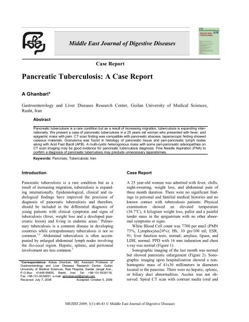
<strong>Pancreatic</strong> <strong>Tuberculosis</strong>: A <strong>Case</strong> <strong>Report</strong><br />
A Ghanbari*<br />
Gastroenterology and Liver Diseases Research Center, Guilan University of Medical Sciences,<br />
Rasht, Iran<br />
Abstract<br />
<strong>Pancreatic</strong> tuberculosis is a rare condition but as a result of increasing migration, tuberculosis is expanding internationally.<br />
We present a case of pancreatic tuberculosis in a 25 years old woman who presented with fever, and<br />
epigastric mass with pain. CT scan finding was compatible with pancreatic abscess, laparoscopic finding showed<br />
caseous materials. Granuloma was found in histology of pancreatic tissue and peri-pancreatic lymph nodes<br />
along with Acid Fast Bacill (AFB). A multi-cystic heterogonous mass with some peri-pancreatic adenopathies on<br />
CT scan imaging may be good evidence for pancreatic tuberculosis diagnosis. Fine Needle Aspiration (FNA) to<br />
confirm a diagnosis of pancreatic tuberculosis may preclude unnecessary laparatomies.<br />
Keywords: Pancreas; <strong>Tuberculosis</strong>; Iran<br />
Introduction<br />
<strong>Pancreatic</strong> tuberculosis is a rare condition but as a<br />
result of increasing migration, tuberculosis is expanding<br />
internationally. Epidemiological, clinical and radiological<br />
findings have improved the precision of<br />
diagnosis of pancreatic tuberculosis and therefore,<br />
should be included in the differential diagnosis of<br />
young patients with clinical symptoms and signs of<br />
tuberculosis (fever, weight loss and a developed pancreatic<br />
lesion) and living in endemic areas. 1 Pulmonary<br />
tuberculosis is a common disease in developing<br />
countries while extrapulmonary tuberculosis is not so<br />
common. 1,2 Abdominal tuberculosis is often accompanied<br />
by enlarged abdominal lymph nodes involving<br />
the ilio-cecal region. Hepatic, splenic, and peritoneal<br />
involvement are less common. 3<br />
*Correspondence: Abbas Ghanbari, MD, Assistant Professor of<br />
Gastroenterology and Liver Diseases Research Center, Guilan<br />
University of Medical Sciences, Razi Hospital, Sardar Jangal Ave.,<br />
P.O.Box: 41448-95655, Rasht, Iran. Tel: +98-131-5535116,<br />
Fax: +98-131-5534951, e-mail: abmotamed@gmail.com<br />
Received: July 7, 2008 Accepted: October 5, 2008<br />
<strong>Case</strong> <strong>Report</strong><br />
A 25 year-old woman was admitted with fever, chills,<br />
night-sweating, weight loss, and abdominal pain of<br />
three month duration. There were no significant findings<br />
in personal and familial medical histories and no<br />
known contact with tuberculosis patients. Physical<br />
examination showed an elevated temperature<br />
(38.7 o C), 6 kilogram weight loss, pallor and a painful<br />
tender mass in the epigastrium with no other abnormal<br />
symptoms or signs.<br />
White Blood Cell count was 7700 per mm3 (PMN<br />
73%, Lymphocytes24%); Hb, 10 grs/100 ml; ESR,<br />
91; liver function tests, normal; amylase, lipase, and<br />
LDH, normal; PPD with 16 mm induration and chest<br />
x-ray was normal (Figure 1).<br />
Sonographic imaging of the last month was normal<br />
but showed pancreatic enlargement (Figure 2). Sonographic<br />
imaging upon hospitalization showed a nonhomogenic<br />
mass of 41x36 millimeters in diameters<br />
located in the pancreas. There were no hepatic, splenic,<br />
or biliary duct abnormalities. Ascites was not observed.<br />
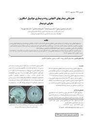
Spiral CT scan with contrast media (oral and<br />
MEJDD 2009; 1(1):40-43 © Middle East Journal of Digestive Diseases
<strong>Pancreatic</strong> tuberculosis<br />
Fig 1: Chest X ray of the patient. Fig 2: Sonography of the patient. Fig 3: CT scan before treatment.<br />
injected) of hepatic and biliary ducts revealed no abnormalities.<br />
There was inflammation in the pancreatic<br />
corpus and the head parenchyma, with some nonhomogenous,<br />
fluid-filled, and air-filled foci.<br />
There was a high density of peri-pancreatic fat tissue<br />
which expanded to the small intestine. Additionally,<br />
there was compression of the gastric greater curvature<br />
and duodenal C-loop and a small amount of fluid in subhepatic<br />
space. Findings also showed necrotic tissue and<br />
abscess formation (Figure 3). The pancreatic abscess<br />
was drained by laparatomy (Figure 4) which revealed<br />
adenopathies in the pancreatic and para-aortic areas.<br />
Lymph node biopsy for pathologic investigation was<br />
performed and the pus was sent for Acid Fast Bacile<br />
(AFB) stain (Figure 5). Pathology revealed granulomatous<br />
inflammation in lymphatic nodes and pancreatic<br />
tissue and some AFB by Kinyoun staining (Figure 6).<br />
The patient was treated with three medications<br />
(isoniazid, rifampicine, and pyrazinamid). She had<br />
no fever in three weeks and was doing well thereafter.<br />
Abdominal CT-scan six months later showed<br />
normal pancreatic size and parenchyma (Figure 7).<br />
Fig 5: Acid Fast Bacile (AFB) stain.<br />
Fig 6: Granulomatous inflammation in lymphatic<br />
nodes and pancreatic tissue.<br />
Discussion<br />
Fig 4: <strong>Pancreatic</strong> abscess in laparotomy.<br />
Although pulmonary tuberculosis is increasing in endemic<br />
areas, 3 pancreatic tuberculosis involving adjacent<br />
lymphatic nodes is very rare. In a report of 300 tuberculosis<br />
cases by Bhansali et al. covering a 12 year period,<br />
there were no pancreatic tuberculosis cases. 4<br />
mejdd.sums.ac.ir Vol 1 January 2009 41
Ghanbari et al.<br />
Fig 7: CT scan after treatment.<br />
In two autopsy reports, pancreatic tuberculosis<br />
was reported in only 2.1% to 4.7% of cases. 3,5,6 <strong>Pancreatic</strong><br />
tuberculosis prevalence is similar in males and<br />
females, with the mean age of fo rty years and it is<br />
more prevalent in North-East Asian countries. 7,8<br />
More pancreatic tuberculosis cases have been reported<br />
in recent years. 7 The most complete report of<br />
pancreatic tuberculosis includes 42 cases from 1991<br />
to 2001. 8 Up to year 2004, 75 cases of pancreatic tuberculosis<br />
in patients without immunodeficiency were<br />
reported in literature. 9 Till now, 80 cases of pancreatic<br />
tuberculosis were reported. 1 The increase in pancreatic<br />
tuberculosis cases is attributed to immigration<br />
from tuberculosis endemic regions to non endemic<br />
countries. 7 Additionally, there is an association<br />
between tuberculosis prevalence and HIV infection. It<br />
is recommended that all pancreatic tuberculosis patients<br />
be tested for HIV infection. 8,10<br />
Signs and symptoms of pancreatic tuberculosis are as<br />
follows: abdominal pain in 81% of cases, weight loss<br />
in 55%, fever in 36%, vomiting in 19%, jaundice in<br />
17%, and pancreatic mass. Often patients have a high<br />
erythrocyte sedimentation rate (ESR) and 70% of patients<br />
have positive PPD. 7,8,11 <strong>Pancreatic</strong> mass is the<br />
most common form of pancreatic tuberculosis (more<br />
than 50% of cases). 3,5,6,7 <strong>Pancreatic</strong> tuberculosis may<br />
presents as acute or chronic pancreatitis, cholestatic<br />
jaundice or may cause portal vein thrombosis,<br />
splenomegaly, thrombocytopenia and ascitis. 2-15<br />
Radiographic features are non-specific. Sonographic<br />
imaging shows hypoecho or heterogenic<br />
masses, and hypodense image on CT-scan. There may<br />
be multicystic lesions on imaging. Presence of a thick<br />
margin around the pancreatic lesion is indicative of<br />
pancreatic tuberculosis. 9,12,16 Peri-pancreatic hypodense<br />
lymphatic nodes in adjacent mesentery is evidences<br />
of pancreatic tuberculosis. 3 Conclusive diagnosis<br />
may be by histological investigation, performed<br />
by fine needle aspiration (FNA) or laparatomic biopsy<br />
and looking for AFB. 7 Success rate with FNA<br />
examination is estimated at 50%. 9 AFB can be seen<br />
only in 20-40% of cases. 3-7 Interestingly, AFB was<br />
found in histologic slides in our case, too.<br />
Conflict of interest: None declared.<br />
References<br />
1 Uma K, Reddy JJm, Kumar A,<br />
Prayag A. <strong>Tuberculosis</strong> of pancreas:<br />
a case report. Ind J Radiol Imag<br />
2005;15(1):29-30.<br />
2 Pombo F, Diaz Candamio MJ,<br />
Rodringuez E, Pombo S. <strong>Pancreatic</strong><br />
tuberculosis: CT findings. Abdom<br />
Imaging 1998;23(4):394-7.<br />
3 Franco-Paredes C, Leonard M, Jurado<br />
R, Blumberg HM, Smith RM. <strong>Tuberculosis</strong><br />
of the pancreas: report of<br />
two cases and review of the literature.<br />
Am J Med Sci 2002;323(1):54-8.<br />
4 Bhansali SK. Abdominal tuberculosis.<br />
Experiences with 300 cases. Am J<br />
Gastroenterol 1977;67(4):324-37.<br />
5 Demir K, Kaymakoglu S, Besisik F,<br />
Durakoglu Z, Ozdil S, Kaplan Y,<br />
Boztas G, Cakaloglu Y, Okten A.<br />
Solitary pancreatic tuberculosis in<br />
immunocompetent patients mimicking<br />
pancreatic carcinoma. J Gastroenterol<br />
Hepatol. 2001; 16(9):1071-4.<br />
6 Turan M, Sen M, Koyuncu A, Aydin<br />
C, Elaldi N, Arici S. <strong>Pancreatic</strong><br />
pseudotumor due to peripancreatic<br />
tuberculous lymphadenitis. Pancreatology<br />
2002;2(6):561-4.<br />
7 Ladas SD, Vaidakis E, Lariou C,<br />
Anastasiou K, Chalevelakis G,<br />
Kintzonidis D, Raptis SA. <strong>Pancreatic</strong><br />
tuberculosis in nonimmunocompromised<br />
patients: reports<br />
of two cases, and a literature<br />
review. Eur J Gastroenterol Hepatol<br />
1998;10(11):973-6.<br />
8 Schneider A, von Birgelen C,<br />
Duhrsen U, Gerken G, Runzi M.<br />
Two cases of pancreatic tuberculosis<br />
in non immunocompromised<br />
patients. A diagnostic challenge<br />
and a rare cause of portal hypertension.<br />
Pancrtatology 2002;2(1):<br />
69-73.<br />
9 Woodfield JC, Windsor JA, Godfrey<br />
CC, Orr DA, Officer NM. Diagnosis<br />
and management of isolated pancreatic<br />
tuberculosis: recent experience<br />
and literature review. ANZ J<br />
Surg 2004;74(5):368-71.<br />
10 Corbett EL, Watt CJ, Walker N,<br />
Maher D, Williams BG, Raviglione<br />
MC, Dye C. The growing burden of tuberculosis:<br />
global trends and interactions<br />
with the HIV epidemic. Arch Intern<br />
Med 2003;163(9):1009-21.<br />
11 Haddad FS, Ghossain A, Sawaya E,<br />
Nelson AR. Abdominal tuberculosis.<br />
DIS Colon Rectum 1987;30(9):724-35.<br />
12 Rezeig MA, Fashir BM, At-<br />
Suhaibani H, Al-Fadda M, Amin T,<br />
Eisa H. <strong>Pancreatic</strong> tuberculosis<br />
mimicking pancreatic carcinoma:<br />
four case reports and review of the<br />
literature. Dig Dis Sci 1998;<br />
43(2):329-31.<br />
13 Takhtani D, Gupta S, Suman K,<br />
Kakkar N, Challa S, Wig JD, Suri<br />
S. Radiology of pancreatic tuber-<br />
42<br />
mejdd.sums.ac.ir Vol 1 January 2009
<strong>Pancreatic</strong> tuberculosis<br />
culosis: a report of three cases.<br />
Am J Gastroenterol 1996;91(9):<br />
1832-4.<br />
14 Ruttenberg D, Graham S, Burns D,<br />
Solomon D, Bornman P. Abdominal<br />
tuberculosis- a cause of portal vein<br />
thrombosis and portal hypertension.<br />
Dig Dis Sci 1991;36(1):112-5.<br />
15 Caroli-Bosc FX, Conio M, Maes B,<br />
Chevallier P, Hastier P, Delmont JP.<br />
Abdominal tuberculosis involving<br />
hepatic hilar lymph nodes. A cause<br />
of portal vein thrombosis and portal<br />
hypertension. J Clin Gastroenterol<br />
1997;25(3):541-3.<br />
16 Sunderam G, McDonald RJ, Maniatis<br />
T, Oleske J, Kapila R,<br />
Reichman LB. <strong>Tuberculosis</strong> as a<br />
manifestation of the acquired immunodeficiency<br />
syndrome (AIDS).<br />
JAMA 1986;256(3):362-6.<br />
mejdd.sums.ac.ir Vol 1 January 2009 43