peqGOLD Protein-Marker X Rabbit - Peqlab
peqGOLD Protein-Marker X Rabbit - Peqlab
peqGOLD Protein-Marker X Rabbit - Peqlab

Create successful ePaper yourself
Turn your PDF publications into a flip-book with our unique Google optimized e-Paper software.
Data sheet<br />
<strong>peqGOLD</strong> <strong>Protein</strong> <strong>Marker</strong> X <strong>Rabbit</strong><br />
Lot No.<br />
________<br />
Usage Standard gel application 5 – 10 μl/lane<br />
Cat. No. 27-2710 1 x 250 μl<br />
27-2711 5 x 250 μl<br />
Description<br />
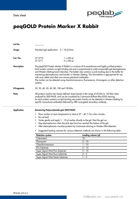
The <strong>peqGOLD</strong> <strong>Protein</strong> <strong>Marker</strong> X <strong>Rabbit</strong> is a mixture of 8 recombinant and highly purified proteins.<br />
Each protein contains an IgG-binding site and is proportioned to yield comparable gel electrophoresis<br />
and Western blotting band intensities. The ladder also contains a pink tracking dye in the buffer for<br />
monitoring electrophoresis and transfer in Western blotting. This formulation is appropriate for use<br />
with most rabbit and other non-mouse polyclonal antibodies.<br />
The marker can be detected using chemiluminescence, fluorescence, chromogenic or other detection<br />
systems.<br />
8 fragments 20, 30, 40, 50, 60, 80, 100 and 150 kDa<br />
Note<br />
All proteins resolve into clearly defined, sharp bands in the range of 20 kDa to 150 kDa when<br />
analysed by SDS-PAGE, and can be visualized by Coomassie Brilliant Blue R250 staining.<br />
As each protein contains an IgG-binding site protein bands can be detected in Western blotting by<br />
specific monoclonal antibodies followed by HRP-conjugated secondary antibody.<br />
Application<br />
Denaturing Polyacrylamide gels (SDS-PAGE)<br />
• Thaw marker at room temperature or heat at 37 – 40 °C for a few minutes.<br />
• Do not boil!<br />
• Vortex gently and apply 1 – 10 μl marker directly to the gel. Start the gel run.<br />
• Stop electrophoresis when the pink dye front has reached the bottom of the gel.<br />
• After electrophoresis visualize proteins by Coomassie staining or Western Blot detection.<br />
• Suggested loading volumes for various detection methods are shown in the following table:<br />
Detection system<br />
Loading volume (μl)<br />
Colorimetric 10<br />
Fluorescent 5 – 10<br />
Chemiluminescence 1 – 10<br />
ECL Substrate 5 – 10<br />
Super Signal West Pico Substrate 5 – 10<br />
Super Signal West Dura Substrate 2 – 5<br />
Super Signal West Femto Substrate 1 – 5<br />
PEQLAB_v0314_E<br />
info@peqlab.com · www.peqlab.com
<strong>peqGOLD</strong> <strong>Protein</strong> <strong>Marker</strong> X <strong>Rabbit</strong><br />
Note<br />
The marker was optimized for staining using Coomassie<br />
Brilliant Blue R-250, but can also be visualized by other<br />
staining methods (e.g. silver staining). As this method is<br />
10 to 100fold more sensitive than Coomassie staining, the<br />
amount of applied marker should be reduced accordingly.<br />
<strong>peqGOLD</strong> <strong>Protein</strong> <strong>Marker</strong> X contains SDS and is therefore<br />
not recommended to be used in native polyacrylamide gels<br />
for determing native molecular weights of proteins.<br />
If the marker shall be diluted use reducing loading buffer.<br />
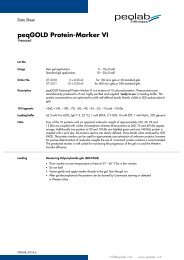
Chemiluminescent detection of <strong>Protein</strong> <strong>Marker</strong> X <strong>Rabbit</strong>:<br />
The marker was separated by electrophoresis and transferred<br />
to a nitrocellulose membrane. <strong>Protein</strong>s were detected by<br />
hybridization with monoclonal rabbit antibodies followed by<br />
HRP-conjugated donkey-anti-rabbit secondary IgG, using ECL<br />
Western Blotting substrate.<br />
• Shipment: Blue ice<br />
• Storage: –20 °C<br />
• Stability: __________<br />
PEQLAB_v0314_E<br />
info@peqlab.com · www.peqlab.com