visual field defects in optic chiasm lesions - Acta Medica Transilvanica
visual field defects in optic chiasm lesions - Acta Medica Transilvanica
visual field defects in optic chiasm lesions - Acta Medica Transilvanica

Create successful ePaper yourself
Turn your PDF publications into a flip-book with our unique Google optimized e-Paper software.
CLINICAL ASPECTS<br />
VISUAL FIELD DEFECTS IN OPTIC CHIASM LESIONS<br />
MARIETA DUMITRACHE 1 , RODICA LASCU 2<br />
1 Ophthalmological Emergency Cl<strong>in</strong>ical Hospital, Bucureşti, 2 Emergency Cl<strong>in</strong>ical County Hospital, Sibiu<br />
Keywords: bitemporal<br />
defect-quadrantic or<br />
hemianopic,<br />
Wilbrand’knee,<br />
junctional scotoma,<br />
bitemporal hemianopic<br />
scotomas, arcuate defect<br />
Cuv<strong>in</strong>te cheie: defect<br />
bitemporal-cvadrantic<br />
sau hemianopic,<br />
genunchiul lui Wilbrand,<br />
scotom joncţional,<br />
scotoame hemianopice<br />
bitemporale, defect<br />
arcuat<br />
Abstract: Temporal <strong>visual</strong> <strong>field</strong> <strong>defects</strong> result from the compression of the <strong>optic</strong> <strong>chiasm</strong> medially<br />
<strong>in</strong>volv<strong>in</strong>g the cross<strong>in</strong>g of nasal ret<strong>in</strong>al fibres, while nasal <strong>visual</strong> <strong>field</strong> <strong>defects</strong> occur with lateral <strong>optic</strong><br />
<strong>chiasm</strong> compression <strong>in</strong>volv<strong>in</strong>g the temporal ret<strong>in</strong>al fibres. The <strong>visual</strong> <strong>field</strong> <strong>defects</strong> reported with <strong>optic</strong><br />
<strong>chiasm</strong> compression <strong>in</strong>clude nasal <strong>visual</strong> <strong>field</strong> loss, arcuate <strong>visual</strong> <strong>field</strong> <strong>defects</strong>, scotomatous <strong>visual</strong> <strong>field</strong><br />
<strong>defects</strong> and homonymous <strong>visual</strong> <strong>field</strong> loss, <strong>in</strong> addition to the typical temporal <strong>visual</strong> <strong>field</strong> loss.<br />
Rezumat: Defectele câmpului vizual temporal rezultă de la compresiunea <strong>chiasm</strong>ei <strong>optic</strong>e implicând<br />
medial încrucişarea fibrelor ret<strong>in</strong>iene nazale, în timp ce defectele câmpului vizual nazal apar cu<br />
compresia <strong>chiasm</strong>ei <strong>optic</strong>e laterale implicând fibrele ret<strong>in</strong>iene temporale. Defectele câmpului vizual<br />
raportate cu compresia <strong>chiasm</strong>ei <strong>optic</strong>e <strong>in</strong>clud pierderea câmpului vizual nazal, defecte ale câmpului<br />
vizual arcuat, defecte câmp vizual scotoame şi pierderea câmpului vizual omonim, pe lângă pierderea<br />
câmpului vizual temporal tipic.<br />
„Rules of the road” for the <strong>optic</strong> <strong>chiasm</strong>:<br />
Three rules describe the course of major fibres bundles<br />
<strong>in</strong> the <strong>chiasm</strong>:<br />
• The nasal ret<strong>in</strong>al fibres (<strong>in</strong>clud<strong>in</strong>g the nasal half of the<br />
macula) of each eye cross <strong>in</strong> the <strong>chiasm</strong> to the contralateral<br />
<strong>optic</strong> tract. Temporal fibres rema<strong>in</strong> uncrossed. Thus, a<br />
<strong>chiasm</strong>al lesion will cause a bitemporal hemianopia due to<br />
<strong>in</strong>terruption of decussat<strong>in</strong>g nasal fibres.<br />
• Lower ret<strong>in</strong>al fibres project through the <strong>optic</strong> nerve and<br />
<strong>chiasm</strong> to lie laterally <strong>in</strong> the tracts; upper ret<strong>in</strong>al fibres will<br />
lie medially (there is a 90-degree rotation of fibres from the<br />
nerves through the <strong>chiasm</strong> <strong>in</strong>to the tracts)<br />
• Inferonasal ret<strong>in</strong>al fibres cross <strong>in</strong>to the <strong>chiasm</strong> and cross<br />
anteriorly approximately 4 mm <strong>in</strong> the contralateral <strong>optic</strong><br />
nerve (Wilbrand ś knee) then redress back <strong>in</strong> to opposite<br />
<strong>optic</strong> tract. The existence of Wilbrand ś knee is<br />
controversial.<br />
• „Macular” cross<strong>in</strong>g fibres are distributed throughout the<br />
<strong>chiasm</strong> and if primarily affected, cause a „central”<br />
bitemporal hemianopia.<br />
• Cl<strong>in</strong>ical “pearl”: if a patient comes <strong>in</strong> with poor vision <strong>in</strong><br />
the left eye, the important eye for <strong>visual</strong> exam<strong>in</strong>ation is the<br />
right due to the <strong>in</strong>volvement of Wilbrand‘s knee. The<br />
lesion is now <strong>in</strong>tracranial at the junction of the left <strong>optic</strong><br />
nerve and <strong>chiasm</strong>. The <strong>field</strong> <strong>defects</strong> constitute a junctional<br />
scotoma.<br />
Although there are many variations <strong>in</strong> the <strong>visual</strong> <strong>field</strong><br />
<strong>defects</strong> caused by the damage to the <strong>optic</strong> <strong>chiasm</strong>, the essential<br />
feature is some type of bitemporal defect, the hallmark of<br />
damage to fibres that cross with<strong>in</strong> the <strong>chiasm</strong>. The bitemporal<br />
<strong>defects</strong> may be superior, <strong>in</strong>ferior, or complete, and they may be<br />
peripheral, central, or both. Many <strong>lesions</strong> that arise <strong>in</strong> the region<br />
of the <strong>chiasm</strong> affect not only the entire <strong>chiasm</strong> but also the<br />
<strong>in</strong>tracranial <strong>optic</strong> nerves. Most <strong>visual</strong> <strong>field</strong> <strong>defects</strong> produced by<br />
<strong>lesions</strong> that damage the <strong>optic</strong> <strong>chiasm</strong> seem to result from the<br />
damage at one of three locations: (a) the anterior angle of the<br />
<strong>chiasm</strong>, (b) the body of the <strong>chiasm</strong>, or (c) the posterior angle of<br />
the <strong>chiasm</strong>.<br />
Lesions that damage the body of the <strong>optic</strong> <strong>chiasm</strong><br />
The <strong>lesions</strong> that damage the body of the <strong>optic</strong> <strong>chiasm</strong><br />
produce a bitemporal defect that may be quadrantic or<br />
hemianopic and that may be peripheral, central, or both, with or<br />
without the so-called „splitt<strong>in</strong>g of the macula”. In most cases,<br />
<strong>visual</strong> acuity is normal. In some patients, however, <strong>visual</strong> acuity<br />
is dim<strong>in</strong>ished and a bitemporal hemianopia is present. When the<br />
lesion compresses the <strong>chiasm</strong> from below, such as occurs with a<br />
pituitary adenoma, the <strong>field</strong> <strong>defects</strong> are typical. When the<br />
peripheral fibres are pr<strong>in</strong>cipally affected, the <strong>field</strong> <strong>defects</strong><br />
usually commence <strong>in</strong> the outer upper quadrants of both eyes. In<br />
the <strong>field</strong> of the right eye, the defect usually progresses <strong>in</strong> a<br />
clockwise direction and <strong>in</strong> the left eye <strong>in</strong> a counterclockwise<br />
direction. Alternatively, suprasellar, supra<strong>chiasm</strong>al compressive<br />
<strong>lesions</strong>-such as tuberculum sellae men<strong>in</strong>giomas,<br />
craniopharyngiomas, aneurysms, and dolichoectatic anterior<br />
cerebral arteries-may damage the superior fibers of the <strong>optic</strong><br />
<strong>chiasm</strong>, as may <strong>in</strong>filtrat<strong>in</strong>g <strong>lesions</strong> such as benign and malignant<br />
gliomas, and cavernous angiomas. The <strong>defects</strong> <strong>in</strong> the <strong>visual</strong><br />
<strong>field</strong>s <strong>in</strong> such cases are still bitemporal, but are located <strong>in</strong> the<br />
<strong>in</strong>ferior rather than the superior <strong>field</strong>s of both eyes. Papilledema,<br />
which is quite unusual <strong>in</strong> patients with suprasellar, <strong>in</strong>fra<strong>chiasm</strong>al<br />
<strong>lesions</strong>, is somewhat more common <strong>in</strong> supra<strong>chiasm</strong>al <strong>lesions</strong><br />
because such <strong>lesions</strong> can extend <strong>in</strong>to the 3rd ventricle.<br />
Lesions that damage the posterior angle of the<br />
<strong>optic</strong> <strong>chiasm</strong><br />
1 Correspond<strong>in</strong>g author: Rodica Lascu, Str. Aleea Infanteriştilor, Bloc I, Scara B, Etaj III, Ap.25, Sibiu, România; e-mail: lascughrodica@yahoo.com;<br />
tel +40-720547341<br />
Article received on 26.01.2012 and accepted for publication on 23.03.2012<br />
ACTA MEDICA TRANSILVANICA June 2012;2(2):185-187<br />
AMT, v. II, no. 2, 2012, p. 185
Lesions that damage the posterior aspect of the <strong>optic</strong><br />
<strong>chiasm</strong> produce characteristic <strong>defects</strong> <strong>in</strong> the <strong>visual</strong> <strong>field</strong>s:<br />
bitemporal hemianopic scotomas. Such <strong>defects</strong> may be<br />
cecocentral scotomas and attributed to a toxic, metabolic, or<br />
even hereditary process rather than to a tumour. True bitemporal<br />
hemianopic scotomas are almost always associated with normal<br />
<strong>visual</strong> acuity and colour perception, whereas cecocentral<br />
scotomas are <strong>in</strong>variably associated with reduced <strong>visual</strong> acuity<br />
and dyschromatopsia. Lesions that damage the posterior aspect<br />
of the <strong>optic</strong> <strong>chiasm</strong> may also damage one of the <strong>optic</strong> tracts, thus<br />
produc<strong>in</strong>g an homonymous <strong>field</strong> defect that is comb<strong>in</strong>ed with<br />
whatever <strong>field</strong> defect has occurred from damage to the <strong>optic</strong><br />
<strong>chiasm</strong>. Bitemporal homonymous scotomas are important <strong>in</strong><br />
localiz<strong>in</strong>g a lesion.<br />
Visual Field Defects Caused by Lesions That Damage the<br />
Optic Chiasm after Initially Damag<strong>in</strong>g the Optic Nerve or<br />
Optic Tract<br />
If there is extension of a lesion from the <strong>optic</strong> nerve or<br />
the <strong>optic</strong> tract to the <strong>optic</strong> <strong>chiasm</strong>, the bl<strong>in</strong>d eye usually is on the<br />
side of the lesion. When there is extension of a lesion from an<br />
<strong>optic</strong> nerve or <strong>optic</strong> tract to the <strong>optic</strong> <strong>chiasm</strong>, the bl<strong>in</strong>d (or nearbl<strong>in</strong>d)<br />
eye is always on the side of the orig<strong>in</strong>al lesion, and when<br />
there is extension of a lesion from the <strong>optic</strong> <strong>chiasm</strong> to the <strong>optic</strong><br />
nerve or to the <strong>optic</strong> tract, the bl<strong>in</strong>d (or near-bl<strong>in</strong>d) eye is always<br />
on the side of the extension of the lesion.<br />
The degree of <strong>visual</strong> <strong>field</strong> loss is usually asymmetrical.<br />
Optic atrophy is only present <strong>in</strong> 50% of cases with <strong>visual</strong> <strong>field</strong><br />
<strong>defects</strong>. For this reason, it is extremely important to perform<br />
careful exam<strong>in</strong>ation of the <strong>visual</strong> <strong>field</strong>s <strong>in</strong> all patients with<br />
unexpla<strong>in</strong>ed <strong>visual</strong> loss.<br />
Compression of the <strong>optic</strong> <strong>chiasm</strong> may be symmetrical<br />
or asymmetrical relat<strong>in</strong>g to the size of lesion and its degree of<br />
<strong>in</strong>volvement of the <strong>optic</strong> <strong>chiasm</strong>, <strong>optic</strong> nerve and <strong>optic</strong> tract.<br />
Symmetrical or asymmetrical compression is reflected by the<br />
presence of bilateral or unilateral <strong>visual</strong> <strong>field</strong> <strong>defects</strong>.<br />
At the junction of the <strong>optic</strong> nerve and <strong>optic</strong> <strong>chiasm</strong>, the<br />
crossed and uncrossed ret<strong>in</strong>al nerve fibres are separated;<br />
consequently, a small lesion of the <strong>optic</strong> nerve at this level<br />
affect<strong>in</strong>g either the crossed or the uncrossed fibres may give rise<br />
to a unilateral hemianopic defect. The <strong>in</strong>volvement of the<br />
ipsilateral <strong>optic</strong> nerve close enough to the <strong>optic</strong> <strong>chiasm</strong> (to<br />
impair selectively conduction <strong>in</strong> cross<strong>in</strong>g nasal ret<strong>in</strong>al fibres<br />
from the ipsilateral eye, but too anterior to affect the cross<strong>in</strong>g<br />
nasal ret<strong>in</strong>al fibres from the contralateral eye) produces<br />
monocular temporal hemi<strong>field</strong> loss. Junctional scotoma:<br />
temporal hemianopsic central scotoma associated with<br />
deficiency <strong>in</strong> opposite superior temporal quadrant, situation<br />
described by Traquir. Junctional scotoma presents a lesion<br />
placed <strong>in</strong> <strong>in</strong>side angle, anterior of <strong>chiasm</strong>. The junction scotoma<br />
is met particularly <strong>in</strong> tumours of the anterior angle of <strong>chiasm</strong> at<br />
the junction between the <strong>optic</strong> nerve and the <strong>chiasm</strong>. The<br />
suffer<strong>in</strong>g of these fibres at the junction between the <strong>optic</strong> nerve<br />
and <strong>chiasm</strong>, that is the suffer<strong>in</strong>g of the „anterior knee”, produces<br />
a superior temporal peripherial deficiency, to the opposite eye.<br />
The fibres from the nasal <strong>in</strong>ferior quadrant of the ret<strong>in</strong>a go<br />
through the anterior part of the <strong>chiasm</strong>, those com<strong>in</strong>g from the<br />
superior nasal quadrants go through the posterior side of the<br />
<strong>chiasm</strong>. That is why pituitar tumours produce <strong>in</strong>itially<br />
deficiencies <strong>in</strong> the superior temporal quadrant, and the<br />
craniopharyngiomas produce <strong>in</strong>itially deficiencies <strong>in</strong> the <strong>in</strong>ferior<br />
temporal <strong>field</strong>.<br />
Arcuate <strong>visual</strong> <strong>field</strong> <strong>defects</strong> have been proposed to<br />
result from vascular changes <strong>in</strong> the <strong>optic</strong> nerve rather than at the<br />
<strong>optic</strong> <strong>chiasm</strong>. Compression of the <strong>optic</strong> nerve at the anterior<br />
<strong>optic</strong> <strong>chiasm</strong> level might also expla<strong>in</strong> the presence of an arcuate<br />
defect. Trobe (1974) described hemianopic temporal arcuate<br />
CLINICAL ASPECTS<br />
AMT, v. II, no. 2, 2012, p. 186<br />
<strong>visual</strong> <strong>field</strong> <strong>defects</strong> due to a lesion <strong>in</strong> the anterocentral <strong>optic</strong><br />
<strong>chiasm</strong>, where cross<strong>in</strong>g and non-cross<strong>in</strong>g portions of the nerve<br />
fibre bundles separate; the lesion would selectively impair<br />
cross<strong>in</strong>g fibres.<br />
Bitemporal hemianopic central scotomas<br />
(heteronimous) are caused by a lesion strictly placed at <strong>chiasm</strong>,<br />
particularly on its posterior edge. The unilateral central scotoma<br />
sometimes expresses a compression lesion: tumour of sellar<br />
area, men<strong>in</strong>gioma of olfactory ditch. The bilateral central<br />
scotoma is mentioned among different types of <strong>visual</strong> troubles<br />
dur<strong>in</strong>g the opto<strong>chiasm</strong>a arachnoid. Hemianopia is a bilateral<br />
deficiency of <strong>visual</strong> <strong>field</strong>, represent<strong>in</strong>g an alteration of the<br />
pathways from the <strong>optic</strong> <strong>chiasm</strong> up to occipital cortex. With<br />
reference to the place of lesion, hemianopic deficiencies present<br />
different aspects which have a very important significance if<br />
neuropathological diagnosis. Bitemporal hemianopia is met <strong>in</strong><br />
sagittal lesion of <strong>chiasm</strong>, <strong>in</strong> pituitary adenoma,<br />
craniopharyngiomas, gliomas of <strong>optic</strong> <strong>chiasm</strong>, opto<strong>chiasm</strong>al<br />
arahnoid, tubersellar men<strong>in</strong>giomas, obstructive hydrocephalus<br />
with <strong>chiasm</strong>atic compression by the bottom of the third<br />
ventricle, carotid aneurysms situated <strong>in</strong> sellar, anterior cerebral<br />
aneurysms, craniocerebral trauma with <strong>chiasm</strong> <strong>in</strong>volvement.<br />
When we come across a temporal hemianopic<br />
deficiency to an eye and bl<strong>in</strong>dness to the other, it is difficult to<br />
state the <strong>chiasm</strong>atical orig<strong>in</strong> of the lesion. It is possible that, by<br />
means of a big <strong>in</strong>dex or by us<strong>in</strong>g a candle <strong>in</strong> an obscure room,<br />
we can observe the presence of temporal ret<strong>in</strong>al sensitivity of<br />
the atrophy of the eye and the absence of it <strong>in</strong> the nasal ret<strong>in</strong>al<br />
area. B<strong>in</strong>asal heteronymous hemianopia is the loss of both nasal<br />
half <strong>field</strong>s, correspond<strong>in</strong>g to the lesion of the fibres of temporal<br />
half ret<strong>in</strong>as. The lesion focuses on the temporal uncrossed direct<br />
bundle at the <strong>chiasm</strong> level (<strong>in</strong> lateral lesion of the <strong>chiasm</strong>).<br />
CRANIOPHARYNGIOMA<br />
Sometimes hemianopia appears as a result of the<br />
compression of the <strong>optic</strong> pathway by tumour of sellar area<br />
(craniopharyngiom).Visual <strong>field</strong> <strong>defects</strong> with<br />
craniopharyngiomas are frequently asymmetric bitemporal<br />
hemianopias or a homonymous pattern with reduced acuity. The<br />
craniopharyngiomas will not only cause <strong>in</strong>ferotemporal <strong>field</strong><br />
<strong>defects</strong> but also bitemporal hemianopic scotomas. Situations<br />
start<strong>in</strong>g with homonim hemianopic central scotomas and<br />
paradoxal situations of homonim hemianopic with spar<strong>in</strong>g of the<br />
macula are quoted.<br />
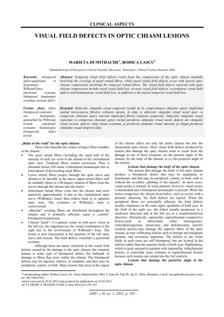
Figure no. 1. Humphrey perimeter <strong>visual</strong> <strong>field</strong> assessment:<br />
craniopharyngioma. Bitemporal hemianopia is present with<br />
some superior nasal <strong>visual</strong> <strong>field</strong> impairment also present<br />
bilaterally.
CLINICAL ASPECTS<br />
GLIOMA AND MENINGIOMA<br />
Men<strong>in</strong>giomas compress<strong>in</strong>g the junction of the <strong>optic</strong><br />
<strong>chiasm</strong> and <strong>optic</strong> nerve will <strong>in</strong>terfere with the anterior knee of<br />
Wilbrand. A lesion at this site will therefore give rise to an<br />
ipsilateral central scotoma and a contralateral upper temporal<br />
<strong>field</strong> defect (junctional scotoma). For this reason, it is very<br />
important to test the <strong>visual</strong> <strong>field</strong> of the opposite eye <strong>in</strong> all<br />
patients who present unexpla<strong>in</strong>ed unilateral <strong>visual</strong> impairment,<br />
particularly <strong>in</strong>clud<strong>in</strong>g a central <strong>visual</strong> <strong>field</strong> defect.<br />
Visual deficits due to men<strong>in</strong>giomas and gliomas<br />
usually take the form of slowly progressive monocular loss of<br />
vision. When both <strong>field</strong>s are <strong>in</strong>volved, there is a dist<strong>in</strong>ct<br />
tendency toward marked asymmetry.<br />
HYDROCEPHALUS<br />
The <strong>optic</strong> <strong>chiasm</strong> is situated <strong>in</strong> the antero<strong>in</strong>ferior region of<br />
the third ventricle. An enlarged third ventricle due to raised<br />
<strong>in</strong>tracranial pressure may press on the superior aspect of the <strong>chiasm</strong><br />
result<strong>in</strong>g <strong>in</strong> a <strong>visual</strong> deficit of the <strong>in</strong>ferior quadrants <strong>in</strong>itially.<br />
Figure no. 2. Humphrey perimeter <strong>visual</strong> <strong>field</strong> assessment:<br />
hydrocephalus. There is severe impairment of <strong>visual</strong> <strong>field</strong>,<br />
but particularly loss <strong>in</strong> the temporal <strong>visual</strong> <strong>field</strong> as evident<br />
on the pattern deviation plot and corrected pattern standard<br />
deviation plot. Note the decibel values for the bitemporal<br />
areas of the <strong>visual</strong> <strong>field</strong>s.<br />
directly above the sella. As the tumour grows upwards it splays<br />
the anterior <strong>chiasm</strong>al notch and compresses the cross<strong>in</strong>g<br />
<strong>in</strong>feronasal fibres, caus<strong>in</strong>g a defect <strong>in</strong> the upper <strong>visual</strong> <strong>field</strong>.<br />
With further tumour extension, the defect progresses <strong>in</strong> an<br />
anticlockwise direction <strong>in</strong> the left eye and clockwise <strong>in</strong> the right<br />
eye to <strong>in</strong>volve the lower <strong>visual</strong> <strong>field</strong>.<br />
Figure no. 3. Humphrey perimeter <strong>visual</strong> <strong>field</strong> assessment:<br />
pituitary adenoma. There is superior temporal <strong>visual</strong> <strong>field</strong><br />
impairment (left greater than right) due to <strong>in</strong>volvement of<br />
the <strong>in</strong>ferior ret<strong>in</strong>al nerve fibres first. Note the decibel values<br />
and probability plots.<br />
VASCULAR ABNORMALITIES<br />
Nasal <strong>visual</strong> <strong>field</strong> <strong>defects</strong> may also be caused by<br />
arterial aneurysms. A dilatation of an <strong>in</strong>ternal carotid aneurysm<br />
may cause lateral compression of the <strong>optic</strong> <strong>chiasm</strong>. The <strong>field</strong><br />
defect is usually unilateral, but may be bilateral with large<br />
aneurysms or bilateral carotid aneurysms.<br />
BIBLIOGRAPHY<br />
1. Arseni C, David M, Chiliman M, et al. Neurooftalmologie,<br />
Editura Didactică şi Pedagogică. Bucureşti; 1981. p.61-66, p.<br />
154-157, p. 174-177.<br />
2. Biousse V, Newman JN. Neuro-Ophthalmology Illustrated.<br />
Thieme. New York; 2009. p. 60-75.<br />
3. Kidd PD, Newman JN, Biousse V. Neuro-Ophthalmology,<br />
MULTIPLE SCLEROSIS<br />
Butterworth-He<strong>in</strong>emann. Oxford; 2008. p. 7-8, p. 243-249.<br />
This may commence with a central scotoma which 4. Kl<strong>in</strong>e BL, Bajandas JF. Neuro-Ophthalmology. SLACK<br />
progresses to hemianopic scotoma.<br />
Incorporated, Torofare, NJ, USA; 2008. p. 10-11.<br />
PITUITARY ADENOMA<br />
5. Miller NR, Newman NJ, Biousse V et al. Walsh and<br />
Bitemporal <strong>visual</strong> <strong>field</strong> <strong>defects</strong> are classically Hoyt’s ̉Cl<strong>in</strong>ical Neuro-Ophthalmology: The Essentials.<br />
associated with <strong>optic</strong> <strong>chiasm</strong> compression. The <strong>in</strong>feronasal Lipp<strong>in</strong>cott Williams & Wilk<strong>in</strong>s, Philadelphia; 2008. p. 224-<br />
ret<strong>in</strong>al nerve fibres cross low and anteriorly, and therefore are 227.<br />
most vulnerable to damage from expand<strong>in</strong>g sellar <strong>lesions</strong>, 6. Rowe F. Visual <strong>field</strong>s via the <strong>visual</strong> pathway. Blackwell<br />
typically pituitary adenomas.<br />
Publish<strong>in</strong>g, Oxford; 2006. p. 160-177.<br />
Compression of the <strong>optic</strong> <strong>chiasm</strong> may be<br />
asymmetrical, thus caus<strong>in</strong>g an asymmetrical <strong>visual</strong> <strong>field</strong> defect<br />
such that temporal <strong>visual</strong> <strong>field</strong> loss is present <strong>in</strong> one eye but the<br />
other eye can have very little <strong>in</strong>volvement. With extensive<br />
compression of the <strong>optic</strong> <strong>chiasm</strong>, there can be substantial loss of<br />
<strong>visual</strong> <strong>field</strong>.<br />
In approximately 10% of normal subjects, the <strong>optic</strong><br />
<strong>chiasm</strong> is situated more anteriorly over the tuberculum sellaeprefixed<br />
(Walsh and Hoyt 1982). In this situation, pituitary<br />
tumours may compress the <strong>optic</strong> tract first, result<strong>in</strong>g <strong>in</strong><br />
homonymous <strong>defects</strong> (Trobe1974). Elk<strong>in</strong>gton (1968) reported<br />
that homonymous hemianopia is uncommon, but is particularly<br />
associated with large and extensive tumours.<br />
In about 80% of normal subjects, the <strong>optic</strong> <strong>chiasm</strong> lies<br />
AMT, v. II, no. 2, 2012, p. 187