Diagnostic Hysteroscopy - A Retrospective Study of 1545 Cases
Diagnostic Hysteroscopy - A Retrospective Study of 1545 Cases
Diagnostic Hysteroscopy - A Retrospective Study of 1545 Cases

You also want an ePaper? Increase the reach of your titles
YUMPU automatically turns print PDFs into web optimized ePapers that Google loves.
VITAMIN D RECEPTOR FOKI (C/T) AND BSMI (A/G) & POLYCYSTIC OVARY SYNDROME<br />
Pathology Number %<br />
Submucous mioma 127 41<br />
Endometrial polyp 32 10<br />
Proximal tubal disease 80 26<br />
Uterine sinechiae and cervico-istmic sinechiae 70 23<br />
TABLE 3. Pathology suspected via another imagistic method.<br />
<strong>Diagnostic</strong> Preoperative Postoperative<br />
False<br />
positive<br />
results<br />
Submucous mioma 127 90 (70.86%) 29.10%<br />
Endometrial polyp 32 20 (62.50%) 37.50%<br />
Proximal tubal disease 80 48 (60.00%) 40.00%<br />
Uterine sinechiae and<br />
cervico-istmic sinechiae<br />
70 15 (21.42%) 79.50%<br />
TABLE 4. Concordance between the preoperative and postoperative<br />
diagnostic.<br />
Uterine malformations Number %<br />
Uterine septum 21 35<br />
Unicorn uterus 6 10<br />
arcuate uterus 28 45<br />
Other malformations 6 10<br />
TABLE 5. Distribution <strong>of</strong> uterine malformation in the study.<br />
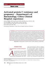
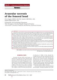
FIGURE 4. Distribution <strong>of</strong> different types <strong>of</strong> pathology for abnormal<br />
uterine bleeding.<br />
HYSBIOPSY: hysteroscopic biopsy; IUD : intrauterine devices.<br />
3. The uterine malformations for which a<br />
diagnostic hysteroscopy was performed<br />
showed the following distribution: uterine septum<br />
35% <strong>of</strong> cases, 10% <strong>of</strong> cases unicorn uterus,<br />
arcuate uterus 45% <strong>of</strong> cases and other uterine<br />
malformations in 10% <strong>of</strong> cases (Table 5).<br />
Concordance between HSG and diagnostic<br />
hysteroscopy was 100% for septate uterus and<br />
the unicorn, decreased to 66.6% for arcuate<br />
uterus and 50% for other uterine malformations.<br />
In conclusion, the highest accuracy <strong>of</strong> HSG<br />
was noted for uterine malformation and minimal<br />
accuracy was observed for intrauterine adhesions.<br />
Trans-vaginal ultrasonography had a<br />
better accuracy in sub mucosal miomas than in<br />
polyps.<br />
4. In our study, in 9% from the 1.545<br />
hysteros copies were performed for abnormal<br />
uterine bleeding. Distribution <strong>of</strong> different<br />
types <strong>of</strong> pathology diagnosed by hysteroscopy<br />
is illustrated in Figure 4.<br />
Figure 4 shows following aspects: no endouterine<br />
pathology observed in 21% <strong>of</strong> cases; in<br />
18% <strong>of</strong> cases hysteroscopy findings interested<br />
entire endometrial cavity requiring curettage<br />
bio psy for endometrial hyperplasia, in 24% <strong>of</strong><br />
cases focal pathology was detected (focal hyperplasia<br />
<strong>of</strong> the endometrium, small polyps)<br />
which was followed by hysteroscopy biopsy<br />
(HYSBIOPSY); a percentage <strong>of</strong> 19% was represented<br />
by the intracavitary foreign bodies (70%<br />
suture material remaining post caesarian) and<br />
imprisoned IUD 8% <strong>of</strong> endometrial polyps over<br />
1 cm resected with resectoscope; 4% sub mucosal<br />
miomas; 5% chronic endometritis which<br />
underwent hysteroscopy biopsy. <br />
DISCUSSION<br />
Since 1999, the specialists in infertility <strong>of</strong> the<br />
University <strong>of</strong> Jerusalem from the Department<br />
<strong>of</strong> Obstetrics and Gynecology started a<br />
debate about the opportunity <strong>of</strong> including hysteroscopy<br />
in the basic/common investigations<br />
<strong>of</strong> infertility (13). The specialists’ conclusions,<br />
ba sed in the studies performed throughout the<br />
years, lead to hysteroscopy being currently<br />
con sidered as absolutely necessary in the<br />
infertili ty investigations (14).<br />
However, the World Health Organization<br />
(WHO) recommends hysterosapingography<br />
(HSG) alone for management <strong>of</strong> infertile women<br />
(1). The explanation for this discrepancy is<br />
that HSG provides information on tuba patency<br />
or blockage. Office hysteroscopy is only<br />
recom mended by the WHO when clinical or<br />
complementary exams (ultrasound, HSG) suggest<br />
intrauterine abnormality (15) or after in vitro<br />
fertilization (IVF) failure (16). Nevertheless,<br />
many specialists feel that hysteroscopy is a<br />
more accurate tool because <strong>of</strong> the high falsepositive<br />
and false-negative rates <strong>of</strong> intra uterine<br />
abnormality with HSG (17-19). This explains<br />
why many specialists use hysteroscopy as a<br />
first-line routine exam for infertility patients regardless<br />
<strong>of</strong> guidelines (2).<br />
Our experience in the Department <strong>of</strong> Assisted<br />
Human Reproduction at the „Pr<strong>of</strong>.dr.Panait<br />
Sîrbu” Clinical Hospital <strong>of</strong> Obstetrics Gynecology<br />
with exploratory hysteroscopy used<br />
312 Maedica A Journal <strong>of</strong> Clinical Medicine, Volume 7 No.4 2012