Diagnostic Hysteroscopy - A Retrospective Study of 1545 Cases
Diagnostic Hysteroscopy - A Retrospective Study of 1545 Cases
Diagnostic Hysteroscopy - A Retrospective Study of 1545 Cases

You also want an ePaper? Increase the reach of your titles
YUMPU automatically turns print PDFs into web optimized ePapers that Google loves.
Mædica - a Journal <strong>of</strong> Clinical Medicine<br />
ORIGINAL PAPERS<br />
<strong>Diagnostic</strong> <strong>Hysteroscopy</strong> -<br />
A <strong>Retrospective</strong> <strong>Study</strong> <strong>of</strong><br />
<strong>1545</strong> <strong>Cases</strong><br />
Andreea STEFANESCU, MD a ; Bogdan MARINESCU, MD, PhD b<br />
a<br />
Elias University Hospital, Department <strong>of</strong> Obstetrics and Gynecology<br />
b<br />
„Pr<strong>of</strong>. Dr. Panait Sirbu“ Hospital <strong>of</strong> Obstetrics and Gynecology<br />
ABSTRACT<br />
Introduction: The development <strong>of</strong> hysteroscopy has provided a minimally invasive approach to common<br />
gynecologic problems, such as abnormal uterine bleeding. <strong>Diagnostic</strong> hysteroscopy is considered<br />
now „the gold standard” by the Association <strong>of</strong> Pr<strong>of</strong>essors <strong>of</strong> Gynecology and Obstetrics (2002) in investigation<br />
<strong>of</strong> abnormal uterine bleeding (AUB) in order to rule out organic endouterine causes <strong>of</strong> AUB.<br />
Although the World Health Organization (WHO) recommends hysterosapingography (HSG) alone for<br />
management <strong>of</strong> infertile women many specialists use hysteroscopy as a first-line routine exam for infertility<br />
patients regardless <strong>of</strong> guidelines.<br />
Material and method: This paper is a retrospective study <strong>of</strong> <strong>1545</strong> diagnostic hysteroscopies performed<br />
in the “Pr<strong>of</strong>. Dr. Panait Sirbu” Obstetrics and Gynecology Hospital between January 1, 2008<br />
and June 30, 2011. The following parameters were studied: diagnostic hysteroscopy indications, type <strong>of</strong><br />
anesthesia used, correlation between pre-and postoperative diagnoses.<br />
Outcomes: Of <strong>1545</strong> diagnostic hysteroscopies, 78% <strong>of</strong> cases were performed without anesthesia; <strong>of</strong><br />
the total <strong>of</strong> 299 cases <strong>of</strong> primary infertility diagnostic hysteroscopy showed in 34% <strong>of</strong> cases tubal obstruction<br />
and endouterine pathology; <strong>of</strong> the total 396 cases <strong>of</strong> secondary infertility under investigation,<br />
diagnostic hysteroscopy showed in 40% <strong>of</strong> cases tubal obstruction and endouterine pathology; the<br />
highest accuracy <strong>of</strong> HSG was noted for uterine malformation and minimal accuracy was observed for<br />
intrauterine adhesions.<br />
Conclusions: Our experience supports the opinion that diagnostic hysteroscopy should be a first-line<br />
routine exam in infertility.<br />
Because <strong>of</strong> the high rate <strong>of</strong> false positive results for HSG in our study and considering the other studies<br />
in specialty literature, we always perform a diagnostic hysteroscopy before Assisted Human Reproduction<br />
procedures regardless <strong>of</strong> the HSG aspect.<br />
Keywords: diagnostic hysteroscopy, abnormal uterine bleeding, infertility<br />
Address for correspondence:<br />
Andreea Stefanescu, Department <strong>of</strong> Obstetrics and Gynecology, Elias University Hospital, Marasti 17 Blvd, District 1, Bucharest.<br />
Tel./fax: 0040213161640, Mobile: 0040723878326.<br />
E-mail: andreea_pindaru@yahoo.com<br />
Article received on the 2 nd <strong>of</strong> February 2012. Article accepted on the 5 th <strong>of</strong> November 2012.<br />
Maedica A Journal <strong>of</strong> Clinical Medicine, Volume 7 No.4 2012<br />
309
VITAMIN D RECEPTOR FOKI (C/T) AND BSMI (A/G) & POLYCYSTIC OVARY SYNDROME<br />
INTRODUCTION<br />
<strong>Hysteroscopy</strong> is performed for<br />
eva luation or treatment <strong>of</strong> different<br />
pathologies <strong>of</strong> the endometrial<br />
cavity, tubal ostia, or endocervical<br />
canal for diagnostic<br />
alo ne or for diagnostic and treatment in the<br />
same operative time (1,2).<br />
A review published on Up To Date in 2011<br />
synthesizes the indications <strong>of</strong> diagnostic and<br />
operative hysteroscopy as follows: abnormal<br />
pre menopausal or postmenopausal uterine ble -<br />
e ding, endometrial polyps, sub-mucosal, and<br />
some intramural, fibroids, intrauterine ad hesions,<br />
Müllerian anomalies (eg, uterine septum),<br />
retained intrauterine devices (retained<br />
IUD ) or other foreign bodies, retained products<br />
<strong>of</strong> conception, desire for sterilization, endocervical<br />
polips (3).<br />
Contraindications to hysteroscopy are: viable<br />
intrauterine pregnancy, active pelvic infection<br />
(including genital herpes infection) (4),<br />
kno wn cervical or uterine cancer.<br />
The possibility to perform hysteroscopy<br />
u sing no anesthetic or local anesthesia allows<br />
use <strong>of</strong> outpatient settings and speeds recovery.<br />
The vaginoscopic, or “no touch,” technique is<br />
performed without a speculum or tenaculum<br />
and without anesthesia (5). Bettocchi introduced<br />
the ‘no-touch’ trans-vaginal approach,<br />
where no instruments expose or grasp the cervix<br />
(6,7). Most diagnostic and brief or minor<br />
operative pro cedures can be performed without<br />
anesthe tic or with a local anesthetic. Regional<br />
or gene ral anesthesia is reserved for patients<br />
who cannot tolerate a procedure under<br />
local anesthesia, extensive operative procedures,<br />
or patients with comorbidities that necessitate<br />
intensive monitoring (7).<br />
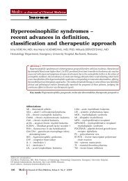
FIGURE 1. The type <strong>of</strong> anesthesia used for hysteroscopy.<br />
One factor in deciding whether to use a<br />
para-cervical block versus no anesthetic is the<br />
pain <strong>of</strong> the injection; some women find the injection<br />
<strong>of</strong> the anesthetic agent more painful<br />
than the procedure itself (8,9). Some surgeons<br />
advocate using no anesthetic (10,11). In our<br />
study, about 20% <strong>of</strong> the diagnostic hysteroscopies<br />
were performed using the „no-touch”<br />
technique with very good tolerance. This technique<br />
is used by a couple <strong>of</strong> surgeons who<br />
were trained by pr<strong>of</strong>essor Bettocchi at “Pr<strong>of</strong>.<br />
Dr. Panait Sirbu” Obstetrics and Gynecology<br />
Hospital in 2005. <br />
MATERIAL AND METHOD<br />
This paper is a retrospective study <strong>of</strong> <strong>1545</strong><br />
diagnostic hysteroscopies performed in the<br />
“Pr<strong>of</strong>. Dr. Panait Sirbu” Obstetrics and Gynecology<br />
Hospital between January 1, 2008 and<br />
June 30, 2011. Total number <strong>of</strong> hysteroscopies<br />
performed in this period was 3220. Those patients<br />
who underwent hysteroscopy for patholo<br />
gy suspected via another imagistic method<br />
were initially investigated using trans-vaginal ultrasonography<br />
or hysterosapingography (HSG).<br />
Before hysteroscopy, the standard investigations<br />
were represented by: PAP smear, vaginal<br />
bacteriologic tests, hemograms.<br />
Antibiotics are not routinely administered<br />
during hysteroscopy for prevention <strong>of</strong> surgical<br />
site infection or endocarditis since post<br />
hysteros copy infection occurs in less than 1<br />
percent <strong>of</strong> women (12).<br />
The following parameters were studied: diagnostic<br />
hysteroscopy indications, type <strong>of</strong> anesthesia<br />
used, correlation between pre-and po stoperative<br />
diagnoses. <br />
OUTCOMES<br />
During the period 1 st January 2008 till 30<br />
June 2011, in the „Pr<strong>of</strong>.dr.Panait Sîrbu”<br />
Clinical Hospital <strong>of</strong> Obstetrics Gynecology, in<br />
Bucharest, there was a total number <strong>of</strong> 3220<br />
hysteroscopies. The distribution <strong>of</strong> pathologies<br />
is expressed in Table 1. Of these 3220 hysteroscopies,<br />
a number <strong>of</strong> <strong>1545</strong> were diagnostic hysteroscopies.<br />
Anesthesia. The type <strong>of</strong> anesthesia used for<br />
the 3220 hysteroscopy shows the prevalence <strong>of</strong><br />
interventions performed without any type <strong>of</strong><br />
anesthesia (61%). General anesthesia with orotracheal<br />
intubation was used mainly for com-<br />
310 Maedica A Journal <strong>of</strong> Clinical Medicine, Volume 7 No.4 2012
VITAMIN D RECEPTOR FOKI (C/T) AND BSMI (A/G) & POLYCYSTIC OVARY SYNDROME<br />
bined interventions (laparoscopic and hysteroscopic)<br />
or hysteroscopic major surgeries like<br />
myo mectomy or metroplasty with a descending<br />
trend between 2008 and 2011 from 12%<br />
to 7%. We observe a slight ascending trend for<br />
ge neral anesthesia with sedation between<br />
2008 and 2011 from 21% to 25% (Figure 1).<br />
<strong>Diagnostic</strong> hysteroscopies were performed<br />
without anesthesia in 78% <strong>of</strong> cases.<br />
<strong>Diagnostic</strong> hysteroscopies performed during<br />
this period had different indications (Table<br />
2).<br />
1. Primary and secondary infertility<br />
Primary and secondary infertility cases included<br />
a total number <strong>of</strong> 695 patients, <strong>of</strong> which<br />
299 (43%) were <strong>of</strong> primary infertility and 396<br />
(57%) <strong>of</strong> secondary infertility.<br />
In the „Pr<strong>of</strong>. Dr. Panait Sîrbu” Clinical Hospital<br />
<strong>of</strong> Obstetrics Gynecology in the Department<br />
<strong>of</strong> Assisted Human Reproduction,<br />
diagnos tic hysteroscopy is used as a first line<br />
diagnostic method for the patients with infertility<br />
in order to detect endocervical, uterine and<br />
proximal tubal factor <strong>of</strong> infertility.<br />
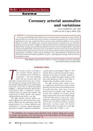
Of the total 299 cases <strong>of</strong> primary infertility,<br />
diagnostic hysteroscopy showed 197 cases<br />
(66%) with a normal hysteroscopy aspect; 60<br />
cases (20%) indicated a proximal tubal obstruction<br />
uni- or bilateral (cornual adhesions obstructing<br />
the tubal ostia, small polyps, endometrial<br />
hyperplasia) and in other 42 cases (14%)<br />
there were findings <strong>of</strong> endouterine pathology:<br />
polyps, miomas, chronic endometritis (Figure<br />
2).<br />
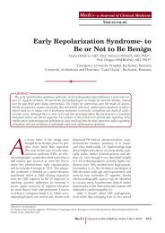
Of the total 396 cases <strong>of</strong> secondary infertility<br />
under investigation, diagnostic hysteroscopy<br />
showed a normal hysteroscopy aspect in 238<br />
cases (60%); in 138 cases (35%) there was proximal<br />
tubal uni or bilateral obstruction and in<br />
other 20 cases (5%) there was endouterine pathology:<br />
polyps, miomas, chronic endometritis<br />
(Figure 3).<br />
2. There were 309 cases which required diagnostic<br />
hysteroscopy in order to confirm the<br />
pathology suspected as a result <strong>of</strong> hysterosapingography<br />
or trans-vaginal sonography. The<br />
type <strong>of</strong> pathology under investigation is illustrated<br />
in Table 3.<br />
The imagistic methods used before hysteroscopy<br />
were HSG in 55% <strong>of</strong> cases and transvaginal<br />
sonography in 45% <strong>of</strong> cases.<br />
In Table 4 there is the concordance between<br />
the preoperative and postoperative<br />
diag nostic and the rate <strong>of</strong> false positive results<br />
Intervention Number %<br />
<strong>Diagnostic</strong> hysteroscopies <strong>1545</strong> 49<br />
Hysteroscopic adhesiolis <strong>of</strong> uterine synechiae<br />
and cervico-istmic synechiae<br />
653 20<br />
Hysteroscopic polipectomy 436 14<br />
Hysteroscopic myomectomy 206 6<br />
Hysteroscopic metroplasty 123 4<br />
Hysteroscopic endometrial biopsy, 92 3<br />
Others: hysteroscopic endometrial ablation,<br />
foreign bodies extraction, hysteroscopic tubar<br />
cannulation<br />
165 5<br />
TABLE 1. The distribution <strong>of</strong> pathologies in 3220 hysteroscopies.<br />
Indication Number %<br />
Primary or secondary infertility 695 45<br />
Pathology suspected by HSG or TVS 309 20<br />
Chronic endometritis 93 6<br />
Abnormal uterine bleeding 139 9<br />
Uterine malformations 62 4<br />
Postoperative control 139 9<br />
IVF 77 5<br />
Others 31 2<br />
TABLE 2. Indications for diagnostic hysteroscopies.<br />
HSG - histerosalpingography; TVS - transvaginal sonography; IVF - in vitro<br />
fertilization.<br />
FIGURE 2. Primary infertility -hysteroscopic results.<br />
FIGURE 3. Secondary infertility -hysteroscopic results.<br />
Looking at Table 4, we can conclude that<br />
the best diagnostic accuracy was for trans-vaginal<br />
sonography in cases <strong>of</strong> sub mucosal miomas<br />
(70.86%) and the lowest rate <strong>of</strong> detection<br />
was for HSG in cases with uterine and cervicoistmic<br />
sinechiae (21.42%).<br />
Maedica A Journal <strong>of</strong> Clinical Medicine, Volume 7 No.4 2012<br />
311
VITAMIN D RECEPTOR FOKI (C/T) AND BSMI (A/G) & POLYCYSTIC OVARY SYNDROME<br />
Pathology Number %<br />
Submucous mioma 127 41<br />
Endometrial polyp 32 10<br />
Proximal tubal disease 80 26<br />
Uterine sinechiae and cervico-istmic sinechiae 70 23<br />
TABLE 3. Pathology suspected via another imagistic method.<br />
<strong>Diagnostic</strong> Preoperative Postoperative<br />
False<br />
positive<br />
results<br />
Submucous mioma 127 90 (70.86%) 29.10%<br />
Endometrial polyp 32 20 (62.50%) 37.50%<br />
Proximal tubal disease 80 48 (60.00%) 40.00%<br />
Uterine sinechiae and<br />
cervico-istmic sinechiae<br />
70 15 (21.42%) 79.50%<br />
TABLE 4. Concordance between the preoperative and postoperative<br />
diagnostic.<br />
Uterine malformations Number %<br />
Uterine septum 21 35<br />
Unicorn uterus 6 10<br />
arcuate uterus 28 45<br />
Other malformations 6 10<br />
TABLE 5. Distribution <strong>of</strong> uterine malformation in the study.<br />
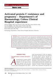
FIGURE 4. Distribution <strong>of</strong> different types <strong>of</strong> pathology for abnormal<br />
uterine bleeding.<br />
HYSBIOPSY: hysteroscopic biopsy; IUD : intrauterine devices.<br />
3. The uterine malformations for which a<br />
diagnostic hysteroscopy was performed<br />
showed the following distribution: uterine septum<br />
35% <strong>of</strong> cases, 10% <strong>of</strong> cases unicorn uterus,<br />
arcuate uterus 45% <strong>of</strong> cases and other uterine<br />
malformations in 10% <strong>of</strong> cases (Table 5).<br />
Concordance between HSG and diagnostic<br />
hysteroscopy was 100% for septate uterus and<br />
the unicorn, decreased to 66.6% for arcuate<br />
uterus and 50% for other uterine malformations.<br />
In conclusion, the highest accuracy <strong>of</strong> HSG<br />
was noted for uterine malformation and minimal<br />
accuracy was observed for intrauterine adhesions.<br />
Trans-vaginal ultrasonography had a<br />
better accuracy in sub mucosal miomas than in<br />
polyps.<br />
4. In our study, in 9% from the 1.545<br />
hysteros copies were performed for abnormal<br />
uterine bleeding. Distribution <strong>of</strong> different<br />
types <strong>of</strong> pathology diagnosed by hysteroscopy<br />
is illustrated in Figure 4.<br />
Figure 4 shows following aspects: no endouterine<br />
pathology observed in 21% <strong>of</strong> cases; in<br />
18% <strong>of</strong> cases hysteroscopy findings interested<br />
entire endometrial cavity requiring curettage<br />
bio psy for endometrial hyperplasia, in 24% <strong>of</strong><br />
cases focal pathology was detected (focal hyperplasia<br />
<strong>of</strong> the endometrium, small polyps)<br />
which was followed by hysteroscopy biopsy<br />
(HYSBIOPSY); a percentage <strong>of</strong> 19% was represented<br />
by the intracavitary foreign bodies (70%<br />
suture material remaining post caesarian) and<br />
imprisoned IUD 8% <strong>of</strong> endometrial polyps over<br />
1 cm resected with resectoscope; 4% sub mucosal<br />
miomas; 5% chronic endometritis which<br />
underwent hysteroscopy biopsy. <br />
DISCUSSION<br />
Since 1999, the specialists in infertility <strong>of</strong> the<br />
University <strong>of</strong> Jerusalem from the Department<br />
<strong>of</strong> Obstetrics and Gynecology started a<br />
debate about the opportunity <strong>of</strong> including hysteroscopy<br />
in the basic/common investigations<br />
<strong>of</strong> infertility (13). The specialists’ conclusions,<br />
ba sed in the studies performed throughout the<br />
years, lead to hysteroscopy being currently<br />
con sidered as absolutely necessary in the<br />
infertili ty investigations (14).<br />
However, the World Health Organization<br />
(WHO) recommends hysterosapingography<br />
(HSG) alone for management <strong>of</strong> infertile women<br />
(1). The explanation for this discrepancy is<br />
that HSG provides information on tuba patency<br />
or blockage. Office hysteroscopy is only<br />
recom mended by the WHO when clinical or<br />
complementary exams (ultrasound, HSG) suggest<br />
intrauterine abnormality (15) or after in vitro<br />
fertilization (IVF) failure (16). Nevertheless,<br />
many specialists feel that hysteroscopy is a<br />
more accurate tool because <strong>of</strong> the high falsepositive<br />
and false-negative rates <strong>of</strong> intra uterine<br />
abnormality with HSG (17-19). This explains<br />
why many specialists use hysteroscopy as a<br />
first-line routine exam for infertility patients regardless<br />
<strong>of</strong> guidelines (2).<br />
Our experience in the Department <strong>of</strong> Assisted<br />
Human Reproduction at the „Pr<strong>of</strong>.dr.Panait<br />
Sîrbu” Clinical Hospital <strong>of</strong> Obstetrics Gynecology<br />
with exploratory hysteroscopy used<br />
312 Maedica A Journal <strong>of</strong> Clinical Medicine, Volume 7 No.4 2012
VITAMIN D RECEPTOR FOKI (C/T) AND BSMI (A/G) & POLYCYSTIC OVARY SYNDROME<br />
as a first line diagnostic method for the patients<br />
with infertility (in order to detect endocervical,<br />
endouterine or proximal tubal factor <strong>of</strong> infertility)<br />
supports the opinion that it should be a firstline<br />
routine exam for infertility.<br />
The importance <strong>of</strong> diagnostic hysteroscopy<br />
in elucidating pathological aspects suspected<br />
by other diagnostic methods is obvious if we<br />
look at the high rate <strong>of</strong> false positive results given<br />
by trans-vaginal ultrasound and HSG. Note<br />
that high false positive rates are dependent on<br />
the performance <strong>of</strong> equipment used on the<br />
one hand and the experience <strong>of</strong> medical staff<br />
on the other hand. The group <strong>of</strong> 309 patients<br />
who have undergone diagnostic hysteroscopy<br />
to elucidate the diagnosis is extremely heterogeneous<br />
in terms <strong>of</strong> equipment used and the<br />
practitioner who performed the initial investigation.<br />
This could explain the differences between<br />
false positive rates existing in our study<br />
compared with other published studies in the<br />
literature: in the 1996 study run by Wang et al,<br />
which compared the diagnostic value <strong>of</strong><br />
hysteros copy and HSG, it was demonstrated<br />
that out <strong>of</strong> 135 patients with abnormal HSG,<br />
the hysteroscopy aspect was normal in 21 cases,<br />
which means a false positive rate <strong>of</strong> 15.6%.<br />
In the same study, the sensibility <strong>of</strong> HSG to diagnose<br />
the intrauterine abnormalities was<br />
80.3% and the specificity was 70.1% (20).<br />
There are many independent studies (17,21) in<br />
specialty literature with similar results which<br />
show that in about one third <strong>of</strong> cases interpreted<br />
with HSG as normal, there may be a false<br />
positive result. These false positive results may<br />
lead to a wrong diagnostic and therapeutic decision<br />
in these patients (22).<br />
<strong>Diagnostic</strong> hysteroscopy for patients with<br />
abnormal uterine bleeding in our study used to<br />
investigate organic endouterine causes <strong>of</strong> abnormal<br />
bleeding showed only in 21% <strong>of</strong> cases<br />
no endouterine pathology. The group <strong>of</strong> 83 cases<br />
(60%) benefited from a diagnosis <strong>of</strong> certainty<br />
with this investigation and the possibility <strong>of</strong><br />
treating in the same operative time the pathology<br />
detected by diagnostic hysteroscopy. <br />
CONCLUSIONS<br />
Our experience supports the opinion that<br />
diagnostic hysteroscopy should be a first<br />
–line routine exam in infertility.<br />
Because <strong>of</strong> the high rate <strong>of</strong> false positive results<br />
for HSG in our study and considering the<br />
other studies in specialty literature, we always<br />
perform a diagnostic hysteroscopy before Assisted<br />
Human Reproduction procedures<br />
regard less <strong>of</strong> the HSG aspect.<br />
REFERENCES<br />
1. Rowe PC, Hargreave T, Mellows H<br />
– WHO Manual for the Standardized<br />
Investigation and Diagnosis <strong>of</strong> the<br />
Infertile Couple, The Press Syndicate <strong>of</strong><br />
the University <strong>of</strong> Cambridge, Cambridge,<br />
UK, 1993<br />
2. Koskas M, Mergui JL, Yazbeck C, et al.<br />
– Office hysteroscopy for infertility: a<br />
series <strong>of</strong> 557 consecutive cases. Obstet<br />
Gynecol Int 2010:168096<br />
3. Bradley LD – Overview <strong>of</strong> hysteroscopy,<br />
UpToDate Last literature review<br />
version 2010;18:3<br />
4. Price TM, Harris JB – Fulminant<br />
hepatic failure due to herpes simplex<br />
after hysteroscopy. Obstet Gynecol 2001;<br />
98:954<br />
5. Garbin O, Kutnahorsky R, Göllner JL,<br />
et al – Vaginoscopic versus conventional<br />
approaches to outpatient<br />
diagnostic hysteroscopy: a two-centre<br />
randomized prospective study. Hum<br />
Reprod 2006; 21:2996-3000<br />
6. Bettocchi S, Selvaggi L – A vaginoscopic<br />
approach to reduce the pain <strong>of</strong><br />
<strong>of</strong>fice hysteroscopy. J Am Assoc Gynecol<br />
Laparosc 1997; 4:255-258<br />
7. Bettocchi S, Ceci O, Di Venere R, et al.<br />
– Advanced operative <strong>of</strong>fice hysteroscopy<br />
without anesthesia: analysis <strong>of</strong> 501<br />
cases treated with a 5 Fr bipolar<br />
electrode. Hum Reprod 2002; 17:2435-<br />
2438<br />
8. Giorda G, Scarabelli C, Franceschi S,<br />
et al. – Feasibility and pain control in<br />
outpatient hysteroscopy in postmenopausal<br />
women: a randomized trial. Acta<br />
Obstet Gynecol Scand 2000; 79:593<br />
9. Broadbent JA, Hill NC, Molnár BG, et<br />
al. – Randomized placebo controlled<br />
trial to assess the role <strong>of</strong> intracervical<br />
lignocaine in outpatient hysteroscopy.<br />
Br J Obstet Gynaecol 1992; 99:777<br />
10. De Iaco P, Marabini A, Stefanetti M, et<br />
al. – Acceptability and pain <strong>of</strong><br />
outpatient hysteroscopy. J Am Assoc<br />
Gynecol Laparosc 2000; 7:71<br />
11. Kremer C, Barik S, Duffy S – Flexible<br />
outpatient hysteroscopy without<br />
anaesthesia: a safe, successful and well<br />
tolerated procedure. Br J Obstet<br />
Gynaecol 1998; 105:672<br />
12. ACOG Committee on Practice Bulletins.<br />
ACOG Practice Bulletin No. 74.<br />
Antibiotic prophylaxis for gynecologic<br />
procedures. Obstet Gynecol 2006; 108:225<br />
13. Shushan A, Rojansky N – Should<br />
hysteroscopy be a part <strong>of</strong> the basic<br />
infertility workup Hum Reprod 1999;<br />
14:1923-1924<br />
14. Campo R, Van Belle Y, Rombauts L, et<br />
al. – Office mini-hysteroscopy. Hum<br />
Reprod Update 1999; 5:73-81<br />
15. De Sa Rosa e de Silva AC, Rosa e Silva<br />
JC, Candido dos Reis FJ, et al.<br />
– Routine <strong>of</strong>fice hysteroscopy in the<br />
investigation <strong>of</strong> infertile couples before<br />
assisted reproduction. J Reprod Med<br />
2005; 50:501-506.<br />
16. Balmaceda JP, Ciuffardi I – <strong>Hysteroscopy</strong><br />
and assisted reproductive<br />
technology. Obstet Gynecol Clin North<br />
Am 1995;22:507-518<br />
17. Golan A, Eilat E, Ron-El R, et al.<br />
– <strong>Hysteroscopy</strong> is superior to hystero-<br />
Maedica A Journal <strong>of</strong> Clinical Medicine, Volume 7 No.4 2012<br />
313
VITAMIN D RECEPTOR FOKI (C/T) AND BSMI (A/G) & POLYCYSTIC OVARY SYNDROME<br />
salpingography in infertility investigation.<br />
Acta Obstet Gynecol Scand 1996;<br />
75:654-656<br />
18. Valle RF – <strong>Hysteroscopy</strong> in the<br />
evaluation <strong>of</strong> female infertility. Am J<br />
Obstet Gynecol 1980; 137:425-431<br />
19. Prevedourakis C, Loutradis D,<br />
Kalianidis C, et al. – Hysterosalpingogra<br />
phy and hysteroscopy in female<br />
infertility. Hum Reprod 1994; 9:2353-<br />
2355<br />
20. Wang CW, Lee CL, Lay YM, et al.<br />
– Comparison <strong>of</strong> hysterosalpingography<br />
and hysteroscopy in female infertility.<br />
J Am Assoc Gynecol Laparosc 1996;<br />
3:581-4<br />
21. Prevedourakis C, Loutradis D,<br />
Kalianidis C, et al. – Hysterosalpingography<br />
and hysteroscopy in female<br />
infertility. Hum Reprod 1994; 9:2353-5<br />
22. Kumar S, Awasthi RT, Gokhale N<br />
– Assessment <strong>of</strong> Uterine Factor in<br />
Infertile Women: Hysterosalpingography<br />
vs <strong>Hysteroscopy</strong>. MJAFI 2003;<br />
60:39-41.<br />
314 Maedica A Journal <strong>of</strong> Clinical Medicine, Volume 7 No.4 2012