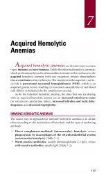
Hemolytic Anemia
Hemolytic Anemia
Hemolytic Anemia
- No tags were found...

Create successful ePaper yourself
Turn your PDF publications into a flip-book with our unique Google optimized e-Paper software.
The direct Coombs’ test (the direct antiglobulin<br />
test) is the hallmark of autoimmune hemolysis.<br />
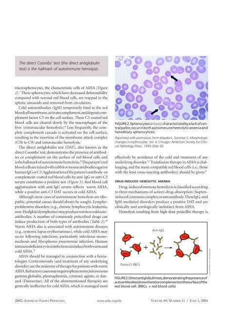
microspherocytes, the characteristic cells of AIHA (Figure<br />
2). 7 These spherocytes, which have decreased deformability<br />
compared with normal red blood cells, are trapped in the<br />
splenic sinusoids and removed from circulation.<br />
Cold autoantibodies (IgM) temporarily bind to the red<br />
blood cell membrane, activate complement, and deposit complement<br />
factor C3 on the cell surface. These C3-coated red<br />
blood cells are cleared slowly by the macrophages of the<br />
liver (extravascular hemolysis). 8 Less frequently, the complete<br />
complement cascade is activated on the cell surface,<br />
resulting in the insertion of the membrane attack complex<br />
(C5b to C9) and intravascular hemolysis.<br />
The direct antiglobulin test (DAT), also known as the<br />
direct Coombs’ test, demonstrates the presence of antibodies<br />
or complement on the surface of red blood cells and<br />
is the hallmark of autoimmune hemolysis. 9 The patient’s red<br />
blood cells are mixed with rabbit or mouse antibodies against<br />
human IgG or C3. Agglutination of the patient’s antibody- or<br />
complement-coated red blood cells by anti-IgG or anti-C3<br />
serum constitutes a positive test (Figure 3). Red blood cell<br />
agglutination with anti-IgG serum reflects warm AIHA,<br />
while a positive anti-C3 DAT occurs in cold AIHA.<br />
Although most cases of autoimmune hemolysis are idiopathic,<br />
potential causes should always be sought. Lymphoproliferative<br />
disorders (e.g., chronic lymphocytic leukemia,<br />
non-Hodgkin’s lymphoma) may produce warm or cold autoantibodies.<br />
A number of commonly prescribed drugs can<br />
induce production of both types of antibodies (Table 2). 10<br />
Warm AIHA also is associated with autoimmune diseases<br />
(e.g., systemic lupus erythematosus), while cold AIHA may<br />
occur following infections, particularly infectious mononucleosis<br />
and Mycoplasma pneumoniae infection. Human<br />
immunodeficiency virus infection can induce both warm and<br />
cold AIHA. 11<br />
AIHA should be managed in conjunction with a hematologist.<br />
Corticosteroids (and treatment of any underlying<br />
disorder) are the mainstay of therapy for patients with warm<br />
AIHA. Refractory cases may require splenectomy, intravenous<br />
gamma globulin, plasmapheresis, cytotoxic agents, or danazol<br />
(Danocrine). All of the aforementioned therapies are<br />
generally ineffective for cold AIHA, which is managed most<br />
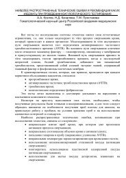
FIGURE 2. Spherocytes (arrows), characterized by a lack of central<br />
pallor, occur in both autoimmune hemolytic anemia and<br />
hereditary spherocytosis.<br />
Reprinted with permission from Maedel L, Sommer S. Morphologic<br />
changes in erythrocytes. Vol. 4. Chicago: American Society for Clinical<br />
Pathology Press, 1993:Slide 50.<br />
effectively by avoidance of the cold and treatment of any<br />
underlying disorder. 12 Transfusion therapy in AIHA is challenging,<br />
and the most compatible red blood cells (i.e., those<br />
with the least cross-reacting antibodies) should be given. 9<br />
DRUG-INDUCED HEMOLYTIC ANEMIA<br />
Drug-induced immune hemolysis is classified according<br />
to three mechanisms of action: drug-absorption (hapteninduced),<br />
immune complex, or autoantibody. These IgG- and<br />
IgM-mediated disorders produce a positive DAT and are<br />
clinically and serologically indistinct from AIHA.<br />
Hemolysis resulting from high-dose penicillin therapy is<br />
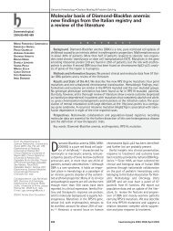
Patient’s RBCs<br />
Anti-IgG<br />
FIGURE 3. Direct antiglobulin test, demonstrating the presence of<br />
autoantibodies (shown here) or complement on the surface of the<br />
red blood cell. (RBCs = red blood cells)<br />
ILLUSTRATION BY DAVID KLEMM<br />
2602-AMERICAN FAMILY PHYSICIAN www.aafp.org/afp VOLUME 69, NUMBER 11 / JUNE 1, 2004