Development of Simplified, Inexpensive Flow Cytometry for CD4+ ...
Development of Simplified, Inexpensive Flow Cytometry for CD4+ ...
Development of Simplified, Inexpensive Flow Cytometry for CD4+ ...

Create successful ePaper yourself
Turn your PDF publications into a flip-book with our unique Google optimized e-Paper software.
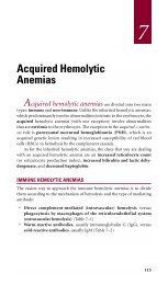
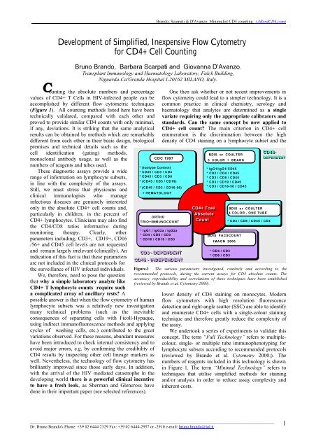
Brando, Scarpati & D'Avanzo: Minimalist CD4 counting _(Af<strong>for</strong>dCD4.com)C<strong>Development</strong> <strong>of</strong> <strong>Simplified</strong>, <strong>Inexpensive</strong> <strong>Flow</strong> <strong>Cytometry</strong><strong>for</strong> <strong>CD4+</strong> Cell CountingBruno Brando, Barbara Scarpati and Giovanna D’Avanzo.Transplant Immunology and Haematology Laboratory, Falck Building,Niguarda-Ca'Granda Hospital I-20162 MILANO, Italy.ounting the absolute numbers and percentagevalues <strong>of</strong> <strong>CD4+</strong> T Cells in HIV-infected people can beaccomplished by different flow cytometric techniques(Figure 1). All counting methods listed here have beentechnically validated, compared with each other andproved to provide similar CD4 counts with only minimal,if any, deviations. It is striking that the same analyticalresults can be obtained by methods which are remarkablydifferent from each other in their basic design, biologicalpremises and technical details such as thecell identification (gating) methods,monoclonal antibody usage, as well as thenumbers <strong>of</strong> reagents and tubes used.These diagnostic assays provide a widerange <strong>of</strong> in<strong>for</strong>mation on lymphocyte subsets,in line with the complexity <strong>of</strong> the assays.Still, we must stress that physicians andclinical immunologists who manageinfectious diseases are genuinely interestedonly in the absolute <strong>CD4+</strong> cell counts and,particularly in children, in the percent <strong>of</strong><strong>CD4+</strong> lymphocytes. Clinicians may also findthe CD4/CD8 ratios in<strong>for</strong>mative duringmonitoring therapy. Clearly, otherparameters including, CD3+, CD19+, CD16/56+ and CD45 cell levels are not requestedand remain largely irrelevant (clinically). Anindication <strong>of</strong> this fact is that these parametersare not included in the clinical protocols <strong>for</strong>the surveillance <strong>of</strong> HIV infected individuals.We, there<strong>for</strong>e, need to pose the questionthat why a simple laboratory analyte like<strong>CD4+</strong> T lymphocyte counts require sucha complicated array <strong>of</strong> ancillary tests? Apossible answer is that when the flow cytometry <strong>of</strong> humanlymphocyte subsets was a relatively new investigationmany technical problems (such as the inevitableconsequences <strong>of</strong> separating cells with Ficoll-Hypaque,using indirect immun<strong>of</strong>luorescence methods and applyingcycles <strong>of</strong> washing cells, etc.) contributed to the greatvariations observed. For these reasons, abundant measureshave been introduced to check internal consistency and toavoid major errors, e.g. by confirming the credibility <strong>of</strong>CD4 results by inspecting other cell lineage markers aswell. Nevertheless, the technology <strong>of</strong> flow cytometry hasbrilliantly improved since those early days. In addition,with the arrival <strong>of</strong> the HIV mediated catastrophe in thedeveloping world there is a powerful clinical incentiveto have a fresh look, as Sherman and Glencross havedone in their important paper (see selected references).One then ask whether or not recent improvements inflow cytometry could lead to a simpler technology. It is acommon practice in clinical chemistry, serology andhaematology that analytes are determined as a singlevariate requiring only the appropriate calibrators andstandards. Can the same concept be now applied to<strong>CD4+</strong> cell count? The main criterion in <strong>CD4+</strong> cellenumeration is the discrimination between the highdensity <strong>of</strong> CD4 staining on a lymphocyte subset and theCDC 1997* (Isotype Control)* CD45 / CD3 / CD4* CD45 / CD3 / CD8* (CD45 / CD3 / CD19)* (CD45 / CD3 / CD16-56)+ HEMATOLOGYORTHOTRIO+IMMUNOCOUNT* IgG1 / IgG2a / IgG2a* CD4 / CD8 / CD3* CD16 / CD19 / CD3CD3 - DEPENDENTCD45 - INDEPENDENT<strong>CD4+</strong> TcellAbsoluteCountBDIS or COULTER3 COLOR + BEADS* IgG1/IgG1/CD45* CD3 / CD4 / CD45* CD3 / CD8 / CD45* CD3 / CD19 / CD45* CD3 / CD16-56 / CD45BDIS FACSCOUNTIMAGN 2000* CD4 / CD3* CD8 / CD3BDIS or COULTER4 COLOR - ONE TUBE* CD3 / CD8 / CD45 / CD4CD45-DEPENDENTFigure.1 The various parameters investigated, routinely and according to therecommended protocols, during the current assays <strong>for</strong> CD4 absolute counts. Theaccuracy, reproducibility and correlations <strong>of</strong> these techniques have been established(reviewed by Brando et al. <strong>Cytometry</strong> 2000).lower density <strong>of</strong> CD4 staining on monocytes. Modernflow cytometers with high resolution fluorescencedetection and right-angle scatter (SSC) are able to identifyand enumerate <strong>CD4+</strong> cells with a single-colour stainingtechnique and there<strong>for</strong>e greatly reduce the complexity <strong>of</strong>the assay.We undertook a series <strong>of</strong> experiments to validate thisconcept. The term “Full Technology” refers to multiplecolour,single- or multiple tube immunophenotyping <strong>for</strong>lymphocyte subsets according to recommended protocols(reviewed by Brando et al. <strong>Cytometry</strong> 2000;). Thenumbers <strong>of</strong> reagents included in this technology is shownin Figure 1. The term “Minimal Technology” refers totechniques that utilise simplified methods <strong>for</strong> stainingand/or analysis in order to reduce assay complexity andinherent costs.___________________________________________________________________________________________________________________Dr. Bruno Brando's Phone: +39 02 6444 2329 Fax: +39 02 6444-2957 or -2910 e-mail: bruno.brando@iol.it1
Brando, Scarpati & D'Avanzo: Minimalist CD4 counting _(Af<strong>for</strong>dCD4.com)Comparison Between CD4 Assays per<strong>for</strong>medusing Full- and Minimal TechnologyIn our study blood samples from 125 HIV+ adultsubjects were analysed on single-plat<strong>for</strong>m. AbsoluteCD4 mean values ranged 11-3600 cells/µL. Three- andfour-colour Full Technology counting procedures weresimultaneously used on Ortho-Cytoron, BDFACSCalibur, Coulter EPICS XL and Dako Galaxy.On the Dako Galaxy volumetric counting wasper<strong>for</strong>med using two different staining protocols: (i)the same Trio staining as per<strong>for</strong>med on the OrthoCytoron and (ii) additional staining in two tubes withCD3/CD4/CD45 and CD3/CD8/CD45.From each analysis file the CD4 single-colourdataset was isolated and reanalysed according to theconcept <strong>of</strong> Minimal Technology. Each Absolute CD4count obtained by Full Technology methods wascompared, first, with its own Minimal Technology, andthen cross-compared with all Minimal Technology dataobtained on the other systems. The Bland-Altmanstatistics was used throughout.When each instrument data were comparedinternally as Full vs. Minimal Technology counts, themean difference (bias) ranged from –2 to +13 with avery narrow standard deviation <strong>of</strong> the mean difference.These results indicate an excellent agreement betweenthe two approaches as shown in Table 1. Theseexcellent agreements between the two differentapproaches in the various flow systems indicate thatthe additional markers and internal checks <strong>for</strong>consistency do not introduce any particular extraadvantage during CD4 analysis per<strong>for</strong>med with thesemodern flow cytometric systems.The agreement between the two methods shownin Table 1 may be criticised because data are partlyself-referential. Nevertheless, this criticism is no longervalid when each Full Technology approach is crosscomparedwith all the other Minimal Technology assays(Table 2). Obviously, however, during this comparisons theextra complications <strong>of</strong> using flow methods based ondifferent principles (see Footnote to Table 2) increase theinherent variations - although by a surprisingly low degree.Here the values <strong>of</strong> mean differences fall into two groups. Inthe first six rows the bias was
Brando, Scarpati & D'Avanzo: Minimalist CD4 counting _(Af<strong>for</strong>dCD4.com)Table 2 Absolute <strong>CD4+</strong> cell counting methods using Full Technology compared with all theother Minimal Technology counting systemsLimits <strong>of</strong> AgreementAbsolute <strong>CD4+</strong> Counting Method Mean Diff 2 x SD LOWER UPPER MEAN RangeFULL Tech vs MINIMALORTHO vs TRUCount 9 65 -56 74 20 - 3394ORTHO vs <strong>Flow</strong>Count -5 84.5 -89.5 79.5 22 - 3383TRUCount vs <strong>Flow</strong>Count -12 52 -64 40 24 - 3231TRUCount vs ORTHO -12 63.5 -75.5 51.5 23 - 3449<strong>Flow</strong>Count vs ORTHO 7 141.5 -134.5 148.5 24 - 3014<strong>Flow</strong>Count vs TRUCount 16 45.5 -29.5 61.5 23 - 2808GALAXY vs ORTHO -47 140 -187 93 18 - 3240GALAXY vs TRUCount -34 115 -149 81 17 - 3043GALAXY vs <strong>Flow</strong>Count -45 116 -161 71 19 - 3031The methods using Full Technology are compared to CD4 results obtained with the other, different, Minimal Technologies (shown here initalics). In these assays, similar bead-based principles are used with the TRUCount and <strong>Flow</strong>Count. ORTHO and GALAXY both representvolumetric measurements but based on different principles (reviewed by Brando et al. <strong>Cytometry</strong> 2000: in press). The lowest SDs are recordedbetween TRUCount and <strong>Flow</strong>Count, in either comparison. Markedly imbalanced mean difference and high SD is evident when DAKO Galaxy isinvolved, due to a bias <strong>of</strong> underestimation (between -34 and -47).The Issue <strong>of</strong> Clinically Significant Error inAbsolute CD4 CountingIn monitoring the CD4 absolute counts we mustnow take into account a hitherto poorly defined issue,namely the magnitude <strong>of</strong> the measurement variabilitythat can influence clinical decisions. A preliminaryattempt to define the critical error boundaries <strong>for</strong>absolute T <strong>CD4+</strong> cell count has been per<strong>for</strong>med at theS. Martino Hospital/University <strong>of</strong> Genova, Italy, by Dr.Annalisa Kunkl (akunkl@smartino.ge.it). The datacame from comparative analyses <strong>of</strong> 24 HIV+ andnormal blood samples by 18 laboratories participatingto a regional QC program. Each sample was analysedon double plat<strong>for</strong>m equipments (and there<strong>for</strong>e theprocedure may need to be repeated on single plat<strong>for</strong>msystems if the clinicians finds the reported variationsuncom<strong>for</strong>table high). True <strong>CD4+</strong> cell values weredefined by the consensus mean; the 99.9% confidenceinterval <strong>of</strong> the mean was calculated on the basis <strong>of</strong> atotal <strong>of</strong> some 300 valid data after the elimination <strong>of</strong>outliers and data without internal consistency.Regression <strong>of</strong> the upper and lower confidence limits <strong>of</strong>the 24 samples were used to extrapolate confidenceranges <strong>for</strong> any theoretical <strong>CD4+</strong> value (Upper Limitregression Line: Y=1.126 AbsCD4 +7.64; LowerLimit Regression Line:Y=0.874 AbsCD4 -7.64).Confidence intervals <strong>of</strong> true <strong>CD4+</strong> valueshave been taken as a measure <strong>of</strong> the error due to thevariation <strong>of</strong> <strong>CD4+</strong> counts. As an example, with thismethod, the 99.9% confidence intervals in the case <strong>of</strong>measured <strong>CD4+</strong> counts <strong>of</strong> 100 and 300 <strong>CD4+</strong> T cells/µLwere (80/µL, 120/µL) and (254/µL, 346/µL) respectively.From Kunkl’s studies it is also apparent that an observedvalue <strong>of</strong> 200 <strong>CD4+</strong> T Cells/µL can mean any real CD4 countfrom 167 to 216/µL, representing 24% variation underoptimal analytical conditions. These issues must also beviewed in the light <strong>of</strong> physiological, diurnal and circannualvariability <strong>of</strong> <strong>CD4+</strong> T cell counts .The estimation <strong>of</strong> test variability is a prerequisite <strong>of</strong> anyclinical study including threshold values as decisionalboundaries. Strangely enough, in almost any instance fixedthreshold values, including the 200 <strong>CD4+</strong> T cells/µL <strong>for</strong> anAIDS related boundary value, have been first established bysome powerful international committee without pointing outthe levels, and the importance, <strong>of</strong> laboratory variations. Theworld-wide accepted CDC guidelines on AIDS-definingconditions state that below 200 <strong>CD4+</strong> T Cells/µL a HIVinfectedsubject is classified as having AIDS by laboratorycriteria. But the variations <strong>of</strong> this threshold has not been atthat time or ever since <strong>of</strong>ficially assessed despite thecompelling results shown above following the use <strong>of</strong> doubleplat<strong>for</strong>m equipments. At this point the assay variability <strong>of</strong>the CD4 tests per<strong>for</strong>med on single plat<strong>for</strong>ms around thisthreshold value is still unknown.Consequently, when the analytical variability can still beso wide one must doubt the wisdom <strong>of</strong> investing time,money and technology into the propagation <strong>of</strong> apparentlyover-complicated assay procedures <strong>for</strong> <strong>CD4+</strong> cell counting.Furthermore, this analytical variability is just one <strong>of</strong> themany components that contributers to the total assayvariability. The additive factors include ‘center effect’,___________________________________________________________________________________________________________________Dr. Bruno Brando's Phone: +39 02 6444 2329 Fax: +39 02 6444-2957 or -2910 e-mail: bruno.brando@iol.it3
Brando, Scarpati & D'Avanzo: Minimalist CD4 counting _(Af<strong>for</strong>dCD4.com)patient status, drug treatment, concomitant disease anddiurnal as well as circannual variations.In conclusion, <strong>for</strong> the full clinical use <strong>of</strong> CD4 testsfour factors are important:(i) Af<strong>for</strong>dable, simple CD4 assays,(ii) an understanding <strong>of</strong> clinical and biologicalfactors during the interpretation <strong>of</strong> results,(iii) to appreciate assay variability at theboundaries <strong>of</strong> CD4 values that lead toclinically important decisions, and(iv) quality assurance programs <strong>for</strong> decreasinglaboratory variations and securing training aswell as the uni<strong>for</strong>mity <strong>of</strong> data handling.Selected References:Brando B, Barnett D, Janossy G, Mandy F, Autran B, Rothe G,Scarpati B, D’Avanzo G, D’Hautcourt JL, Lenkei R, Schmitz G,Kunkl A, Chianese R, Papa S, Gratama JW. Cyt<strong>of</strong>luorometricmethods <strong>for</strong> assessing absolute numbers <strong>of</strong> cell subsets in blood.<strong>Cytometry</strong> (Communications in Clinical <strong>Cytometry</strong>), 2000; 42:327-346.Brando B, Sommaruga E, Mazzola E, Sbressa C. <strong>CD4+</strong> T Cellabsolute count. A crossed comparison among three flowcytometric methods and plate ELISA test in 325 HIV+ patients.<strong>Cytometry</strong> 1996; Suppl. 8: Abstract IH32, pg 77.Brando B. <strong>Development</strong> <strong>of</strong> a ‘minimal technology’ procedure <strong>for</strong> absolutecounting <strong>of</strong> <strong>CD4+</strong> cells in HIV+ patients. <strong>Cytometry</strong> 2000; Suppl. 10:138 (Abstract 6390).Janossy G, Jani I, Göhde W. Af<strong>for</strong>dable <strong>CD4+</strong> T cell counts on ‘singleplat<strong>for</strong>m’flow cytometers. I. Primary CD4 gating. Br J Haematol,2000; 111: 1198-1208.Sherman GG, Galpin JS, Patel JM, Mendelow BV, Glencross DK. <strong>CD4+</strong>T cell enumeration in HIV infection with limited resources. J ImmunolMethods 1999; 222: 209-217.Kunkl A, Terranova MP, Mortara L, Bottone. Measurement error <strong>of</strong>absolute <strong>CD4+</strong> T lymphocyte count double plat<strong>for</strong>m evaluations <strong>for</strong> theassessment <strong>of</strong> laboratory per<strong>for</strong>mance in lymphocyteimmunophenotyping quality control. <strong>Cytometry</strong> Suppl. 10, 2000: 45,Abstract 6415.Hoover DR, Graham NMH, Chen B, Taylor JMG, Phair J, Zhou SYJ,Munoz A. Effect <strong>of</strong> <strong>CD4+</strong> cell count measurement variability onstaging HIV-1 infection. J Acquir Immune Defic Syndr Hum Retrovirol1992; 5: 794-802.Paglieroni TG, Holland PV. Circannual variation in lymphocyte subsets,revisited. Transfusion 1994; 34: 512-516.Hughes MD, Stein DS, Gundacker HM, Valentine FT, Phair JP, VolberdingPA. Within-subject variation in CD4 lymphocyte count inasymptomatic human immunodeficiency virus infection: implications<strong>for</strong> patient monitoring. J Infect Dis 1994; 169: 28-36.Raboud JM, Haley L, Montaner JS, Murphy C, Januszewska M, SchechterMT. Quantification <strong>of</strong> the variation due to laboratory and physiologicsources in CD4 lymphocyte counts <strong>of</strong> clinically stable HIV-infectedindividuals. J Acquir Immune Defic Syndr Hum Retrovirol 1995; 10Suppl 2: S67-S73.Jones AR, Twedt D, Swaim W, Gottfried E. Diurnal change <strong>of</strong> blood countanalytes in normal subjects. Am J Clin Pathol 1996; 106: 723-727.___________________________________________________________________________________________________________________Dr. Bruno Brando's Phone: +39 02 6444 2329 Fax: +39 02 6444-2957 or -2910 e-mail: bruno.brando@iol.it4