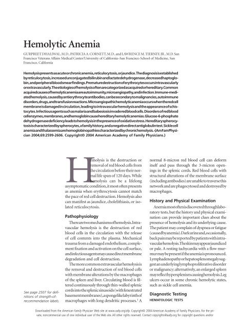
Hemolytic Anemia
Hemolytic Anemia
Hemolytic Anemia
- No tags were found...
You also want an ePaper? Increase the reach of your titles
YUMPU automatically turns print PDFs into web optimized ePapers that Google loves.
<strong>Hemolytic</strong> <strong>Anemia</strong><br />
GURPREET DHALIWAL, M.D., PATRICIA A. CORNETT, M.D., and LAWRENCE M. TIERNEY, JR., M.D. San<br />
Francisco Veterans Affairs Medical Center/University of California–San Francisco School of Medicine, San<br />
Francisco, California<br />
Hemolysis presents as acute or chronic anemia, reticulocytosis, or jaundice. The diagnosis is established<br />
by reticulocytosis, increased unconjugated bilirubin and lactate dehydrogenase, decreased haptoglobin,<br />
and peripheral blood smear findings. Premature destruction of erythrocytes occurs intravascularly<br />
or extravascularly. The etiologies of hemolysis often are categorized as acquired or hereditary. Common<br />
acquired causes of hemolytic anemia are autoimmunity, microangiopathy, and infection. Immune-mediated<br />
hemolysis, caused by antierythrocyte antibodies, can be secondary to malignancies, autoimmune<br />
disorders, drugs, and transfusion reactions. Microangiopathic hemolytic anemia occurs when the red cell<br />
membrane is damaged in circulation, leading to intravascular hemolysis and the appearance of schistocytes.<br />
Infectious agents such as malaria and babesiosis invade red blood cells. Disorders of red blood<br />
cell enzymes, membranes, and hemoglobin cause hereditary hemolytic anemias. Glucose-6-phosphate<br />
dehydrogenase deficiency leads to hemolysis in the presence of oxidative stress. Hereditary spherocytosis<br />
is characterized by spherocytes, a family history, and a negative direct antiglobulin test. Sickle cell<br />
anemia and thalassemia are hemoglobinopathies characterized by chronic hemolysis.-(Am Fam Physician<br />
2004;69:2599-2606. Copyright© 2004 American Academy of Family Physicians.)<br />
See page 2507 for definitions<br />
of strength-ofrecommendation<br />
labels.<br />
Hemolysis is the destruction or<br />
removal of red blood cells from<br />
the circulation before their normal<br />
life span of 120 days. While<br />
hemolysis can be a lifelong<br />
asymptomatic condition, it most often presents<br />
as anemia when erythrocytosis cannot match<br />
the pace of red cell destruction. Hemolysis also<br />
can manifest as jaundice, cholelithiasis, or isolated<br />
reticulocytosis.<br />
Pathophysiology<br />
There are two mechanisms of hemolysis. Intravascular<br />
hemolysis is the destruction of red<br />
blood cells in the circulation with the release<br />
of cell contents into the plasma. Mechanical<br />
trauma from a damaged endothelium, complement<br />
fixation and activation on the cell surface,<br />
and infectious agents may cause direct membrane<br />
degradation and cell destruction.<br />
The more common extravascular hemolysis is<br />
the removal and destruction of red blood cells<br />
with membrane alterations by the macrophages<br />
of the spleen and liver. Circulating blood is filtered<br />
continuously through thin-walled splenic<br />
cords into the splenic sinusoids (with fenestrated<br />
basement membranes), a spongelike labyrinth of<br />
macrophages with long dendritic processes. 1 A<br />
normal 8-micron red blood cell can deform<br />
itself and pass through the 3-micron openings<br />
in the splenic cords. Red blood cells with<br />
structural alterations of the membrane surface<br />
(including antibodies) are unable to traverse this<br />
network and are phagocytosed and destroyed by<br />
macrophages.<br />
History and Physical Examination<br />
<strong>Anemia</strong> most often is discovered through laboratory<br />
tests, but the history and physical examination<br />
can provide important clues about the<br />
presence of hemolysis and its underlying cause.<br />
The patient may complain of dyspnea or fatigue<br />
(caused by anemia). Dark urine and, occasionally,<br />
back pain may be reported by patients with intravascular<br />
hemolysis. The skin may appear jaundiced<br />
or pale. A resting tachycardia with a flow murmur<br />
may be present if the anemia is pronounced.<br />
Lymphadenopathy or hepatosplenomegaly suggest<br />
an underlying lymphoproliferative disorder<br />
or malignancy; alternatively, an enlarged spleen<br />
may reflect hypersplenism causing hemolysis. Leg<br />
ulcers occur in some chronic hemolytic states,<br />
such as sickle cell anemia.<br />
Diagnostic Testing<br />
HEMATOLOGIC TESTS<br />
Downloaded from the American Family Physician Web site at www.aafp.org/afp. Copyright© 2004 American Academy of Family Physicians. For the private,<br />
noncommercial use of one individual user of the Web site. All other rights reserved. Contact copyrights@aafp.org for copyright questions and/or
Along with anemia, a characteristic laboratory feature<br />
of hemolysis is reticulocytosis, the normal response of the<br />
bone marrow to the peripheral loss of red blood cells. In the<br />
absence of concomitant bone marrow disease, a brisk reticulocytosis<br />
should be observed within three to five days after a<br />
decline in hemoglobin. In a minority of patients, the bone marrow<br />
is able to chronically compensate, leading to a normal<br />
and stable hemoglobin concentration. The anemia of hemolysis<br />
usually is normocytic, although a marked reticulocytosis<br />
can lead to an elevated measurement of mean corpuscular<br />
volume, because the average mean corpuscular volume of a<br />
reticulocyte is 150 fL. 2<br />
Review of the peripheral blood smear is a critical step in<br />
the evaluation of any anemia. Along with an assessment<br />
for pathognomonic red blood cell morphologies, such as<br />
spherocytes or schistocytes, examination of the white blood<br />
cells and platelets for coexisting hematologic or malignant<br />
disorders is essential.<br />
CHEMISTRY TESTS<br />
The destruction of red blood cells is characterized by<br />
increased unconjugated bilirubin, increased lactate dehydrogenase,<br />
and decreased haptoglobin levels. Lactate dehydrogenase<br />
and hemoglobin are released into the circulation<br />
when red blood cells are destroyed. Liberated hemoglobin is<br />
converted into unconjugated bilirubin in the spleen or may be<br />
bound in the plasma by haptoglobin. The hemoglobin-hapto-<br />
<strong>Hemolytic</strong> <strong>Anemia</strong><br />
Indirect hyperbilirubinemia<br />
<strong>Anemia</strong><br />
Reticulocy-<br />
Evaluate for hemolysis:<br />
CBC, reticulocyte count, LDH,<br />
indirect bilirubin, haptoglobin,<br />
peripheral blood smear<br />
Positive<br />
Negative<br />
Consider alternative diagnoses,<br />
including other causes of<br />
normocytic anemia (e.g.,<br />
hemorrhage, chronic disease,<br />
chronic kidney disease)<br />
Spherocytes,<br />
positive DAT<br />
Immune hemolysis:<br />
lymphoproliferative<br />
disorder/cancer,<br />
autoimmune diseases,<br />
drugs, infections,<br />
transfusion<br />
Spherocytes,<br />
negative DAT,<br />
family history<br />
Hereditary<br />
spherocytosis<br />
Schistocytes<br />
Microangiopathic<br />
hemolytic anemia<br />
PT/PTT, renal and<br />
liver function,<br />
blood pressure<br />
Hypochromic,<br />
microcytic<br />
anemia<br />
Thalassemia<br />
Sickle cells<br />
Sickle cell<br />
anemia<br />
Hemoglobin<br />
electrophoresis<br />
Infection/<br />
drug expo-<br />
G6PD activity<br />
Fever/<br />
recent<br />
Thick and thin blood<br />
smears, Babesia<br />
serologies, bacterial<br />
cultures<br />
TTP, HUS, DIC, eclampsia,<br />
preeclampsia, malignant<br />
hypertension, prosthetic valve<br />
FIGURE 1. Algorithm for the evaluation of hemolytic anemia. (CBC = complete blood count; LDH = lactate dehydrogenase; DAT = direct<br />
antiglobulin test; G6PD = glucose-6-phosphate dehydrogenase; PT/PTT = prothrombin time/partial thromboplastin time; TTP = thrombotic<br />
thrombocytopenic purpura; HUS = hemolytic uremic syndrome; DIC = disseminated intravascular coagulation)<br />
2600-AMERICAN FAMILY PHYSICIAN www.aafp.org/afp VOLUME 69, NUMBER 11 / JUNE 1, 2004
TABLE 1<br />
Overview of the <strong>Hemolytic</strong> <strong>Anemia</strong>s<br />
Type Etiology Associations Diagnosis Treatment<br />
Acquired*<br />
Immune-mediated Antibodies to red Idiopathic, malignancy, Spherocytes and Treatment of underlying disorder;<br />
blood cell surface drugs, autoimmune positive DAT removal of offending drug;<br />
antigens disorders, infections, steroids, splenectomy, IV gamma<br />
transfusions<br />
globulin, plasmapheresis,<br />
cytotoxic agents, or danazol<br />
(Danocrine); avoidance of cold<br />
Microangiopathic Mechanical disruption TTP, HUS, DIC, pre- Schistocytes Treatment of underlying disorder<br />
of red blood cell eclampsia, eclampsia,<br />
in circulation<br />
malignant hypertension,<br />
prosthetic valves<br />
Infection Malaria, babesiosis, Cultures, thick and Antibiotics<br />
Clostridium<br />
thin blood smears,<br />
infections<br />
serologies<br />
Hereditary†<br />
Enzymopathies G6PD deficiency Infections, drugs, Low G6PD activity Withdrawal of offending drug,<br />
ingestion of fava beans measurement treatment of infection<br />
Membranopathies Hereditary Spherocytes, family Splenectomy in some moderate<br />
spherocytosis history, negative DAT and most severe cases<br />
Hemoglobinopathies Thalassemia and Hemoglobin Folate, transfusions<br />
sickle cell disease<br />
electrophoresis,<br />
genetic studies<br />
DAT = direct antiglobulin test; IV = intravenous; TTP = thrombotic thrombocytopenic purpura; HUS = hemolytic uremic syndrome; DIC = disseminated<br />
intravascular coagulation; G6PD = glucose-6-phosphate dehydrogenase.<br />
*—Other select causes of acquired hemolysis (not discussed in this article) include splenomegaly, end-stage liver disease/spur cell (acanthocyte)<br />
hemolytic anemia, paroxysmal cold hemoglobinuria, paroxysmal nocturnal hemoglobinuria, insect stings, and spider bites.<br />
†—Other select causes of inherited hemolysis (not discussed in this article) include Wilson’s disease and less common forms of membranopathy<br />
(hereditary elliptocytosis), enzymopathy (pyruvate kinase deficiency), and hemoglobinopathy (unstable hemoglobin variants).<br />
globin complex is cleared quickly by the liver, leading to low<br />
or undetectable haptoglobin levels. 3<br />
URINARY TESTS<br />
In cases of severe intravascular hemolysis, the binding<br />
capacity of haptoglobin is exceeded rapidly, and free hemoglobin<br />
is filtered by the glomeruli. The renal tubule cells may<br />
absorb the hemoglobin and store the iron as hemosiderin;<br />
hemosiderinuria is detected by Prussian blue staining of<br />
sloughed tubular cells in the urinary sediment approximately<br />
one week after the onset of hemolysis. 4 Hemoglobinuria,<br />
which causes red-brown urine, is indicated by a positive urine<br />
dipstick reaction for heme in the absence of red blood cells.<br />
Acquired Disorders<br />
Once the diagnosis of hemolysis is made on the basis of<br />
laboratory and peripheral smear findings (Figure 1), it is<br />
necessary to determine the etiology. While most forms of<br />
hemolysis are classified as predominantly intravascular or<br />
extravascular, the age of onset, accompanying clinical presentation,<br />
and co-existing medical problems usually guide<br />
the clinician to consider either an acquired or a hereditary<br />
cause 5,6 (Table 1).<br />
IMMUNE HEMOLYTIC ANEMIA<br />
Immune hemolytic anemias are mediated by antibodies<br />
directed against antigens on the red blood cell surface.<br />
Microspherocytes on a peripheral smear and a positive direct<br />
antiglobulin test are the characteristic findings. Immune<br />
hemolytic anemia is classified as autoimmune, alloimmune, or<br />
drug-induced, based on the antigen that stimulates antibodyor<br />
complement-mediated destruction of red blood cells.<br />
AUTOIMMUNE HEMOLYTIC ANEMIA<br />
Autoimmune hemolytic anemia (AIHA) is mediated by autoantibodies<br />
and further subdivided according to their maximal<br />
binding temperature. Warm hemolysis refers to IgG autoantibodies,<br />
which maximally bind red blood cells at body temperature<br />
(37°C [98.6°F]). In cold hemolysis, IgM autoantibodies<br />
(cold agglutinins) bind red blood cells at lower temperatures<br />
(0° to 4°C [32° to 39.2°F]).<br />
When warm autoantibodies attach to red blood cell<br />
surface antigens, these IgG-coated red blood cells are<br />
partially ingested by the macrophages of the spleen, leaving<br />
JUNE 1, 2004 / VOLUME 69, NUMBER 11 www.aafp.org/afp AMERICAN FAMILY PHYSICIAN-2601
The direct Coombs’ test (the direct antiglobulin<br />
test) is the hallmark of autoimmune hemolysis.<br />
microspherocytes, the characteristic cells of AIHA (Figure<br />
2). 7 These spherocytes, which have decreased deformability<br />
compared with normal red blood cells, are trapped in the<br />
splenic sinusoids and removed from circulation.<br />
Cold autoantibodies (IgM) temporarily bind to the red<br />
blood cell membrane, activate complement, and deposit complement<br />
factor C3 on the cell surface. These C3-coated red<br />
blood cells are cleared slowly by the macrophages of the<br />
liver (extravascular hemolysis). 8 Less frequently, the complete<br />
complement cascade is activated on the cell surface,<br />
resulting in the insertion of the membrane attack complex<br />
(C5b to C9) and intravascular hemolysis.<br />
The direct antiglobulin test (DAT), also known as the<br />
direct Coombs’ test, demonstrates the presence of antibodies<br />
or complement on the surface of red blood cells and<br />
is the hallmark of autoimmune hemolysis. 9 The patient’s red<br />
blood cells are mixed with rabbit or mouse antibodies against<br />
human IgG or C3. Agglutination of the patient’s antibody- or<br />
complement-coated red blood cells by anti-IgG or anti-C3<br />
serum constitutes a positive test (Figure 3). Red blood cell<br />
agglutination with anti-IgG serum reflects warm AIHA,<br />
while a positive anti-C3 DAT occurs in cold AIHA.<br />
Although most cases of autoimmune hemolysis are idiopathic,<br />
potential causes should always be sought. Lymphoproliferative<br />
disorders (e.g., chronic lymphocytic leukemia,<br />
non-Hodgkin’s lymphoma) may produce warm or cold autoantibodies.<br />
A number of commonly prescribed drugs can<br />
induce production of both types of antibodies (Table 2). 10<br />
Warm AIHA also is associated with autoimmune diseases<br />
(e.g., systemic lupus erythematosus), while cold AIHA may<br />
occur following infections, particularly infectious mononucleosis<br />
and Mycoplasma pneumoniae infection. Human<br />
immunodeficiency virus infection can induce both warm and<br />
cold AIHA. 11<br />
AIHA should be managed in conjunction with a hematologist.<br />
Corticosteroids (and treatment of any underlying<br />
disorder) are the mainstay of therapy for patients with warm<br />
AIHA. Refractory cases may require splenectomy, intravenous<br />
gamma globulin, plasmapheresis, cytotoxic agents, or danazol<br />
(Danocrine). All of the aforementioned therapies are<br />
generally ineffective for cold AIHA, which is managed most<br />
FIGURE 2. Spherocytes (arrows), characterized by a lack of central<br />
pallor, occur in both autoimmune hemolytic anemia and<br />
hereditary spherocytosis.<br />
Reprinted with permission from Maedel L, Sommer S. Morphologic<br />
changes in erythrocytes. Vol. 4. Chicago: American Society for Clinical<br />
Pathology Press, 1993:Slide 50.<br />
effectively by avoidance of the cold and treatment of any<br />
underlying disorder. 12 Transfusion therapy in AIHA is challenging,<br />
and the most compatible red blood cells (i.e., those<br />
with the least cross-reacting antibodies) should be given. 9<br />
DRUG-INDUCED HEMOLYTIC ANEMIA<br />
Drug-induced immune hemolysis is classified according<br />
to three mechanisms of action: drug-absorption (hapteninduced),<br />
immune complex, or autoantibody. These IgG- and<br />
IgM-mediated disorders produce a positive DAT and are<br />
clinically and serologically indistinct from AIHA.<br />
Hemolysis resulting from high-dose penicillin therapy is<br />
Patient’s RBCs<br />
Anti-IgG<br />
FIGURE 3. Direct antiglobulin test, demonstrating the presence of<br />
autoantibodies (shown here) or complement on the surface of the<br />
red blood cell. (RBCs = red blood cells)<br />
ILLUSTRATION BY DAVID KLEMM<br />
2602-AMERICAN FAMILY PHYSICIAN www.aafp.org/afp VOLUME 69, NUMBER 11 / JUNE 1, 2004
<strong>Hemolytic</strong> <strong>Anemia</strong><br />
TABLE 2<br />
Selected Drugs that Cause Immune-Mediated Hemolysis<br />
Drug absorption<br />
Mechanism (hapten) Immune complex Autoantibody<br />
DAT Positive anti-IgG Positive anti-C3 Positive anti-IgG<br />
Site of hemolysis Extravascular Intravascular Extravascular<br />
Medications Penicillin Quinidine Alpha-methyldopa<br />
Ampicillin Phenacetin Mefenamic acid<br />
Methicillin Hydrochlorothiazide (Ponstel)<br />
Carbenicillin Rifampin (Rifadin) L-dopa<br />
Cephalothin Sulfonamides Procainamide<br />
(Keflin)* Isoniazid Ibuprofen<br />
Cephaloridine Quinine Diclofenac<br />
(Loridine)* Insulin (Voltaren)<br />
Tetracycline<br />
Interferon alfa<br />
Melphalan (Alkeran)<br />
Acetaminophen<br />
Hydralazine (Apresoline)<br />
Probenecid<br />
Chlorpromazine (Thorazine)<br />
Streptomycin<br />
Fluorouracil (Adrucil)<br />
Sulindac (Clinoril)<br />
DAT = direct antiglobulin test.<br />
*—Not available in the United States.<br />
Adapted with permission from Schwartz RS, Berkman EM, Silberstein LE. Autoimmune hemolytic<br />
anemias. In: Hoffman R, Benz EJ Jr, Shattil SJ, Furie B, Cohen HJ, Silberstein LE, et al., eds. Hematology:<br />
basic principles and practice. 3d ed. Philadelphia: Churchill Livingstone, 2000:624.<br />
membrane protein and rendering it<br />
antigenic 13 ) induces the production<br />
of antierythrocyte IgG antibodies<br />
and causes an extravascular hemolysis.<br />
ALLOIMMUNE (TRANSFUSION) HEMOLYTIC<br />
ANEMIA<br />
The most severe alloimmune hemolysis<br />
is an acute transfusion reaction<br />
caused by ABO-incompatible red<br />
blood cells. For example, transfusion<br />
of A red cells into an O recipient (who<br />
has circulating anti-A IgM antibodies)<br />
leads to complement fixation and a<br />
brisk intravascular hemolysis. Within<br />
minutes, the patient may develop fever,<br />
chills, dyspnea, hypotension, and<br />
shock.<br />
Delayed hemolytic transfusion<br />
reactions occur three to 10 days after<br />
a transfusion and usually are caused<br />
by low titer antibodies to minor red<br />
blood cell antigens. On exposure to<br />
antigenic blood cells, these antibodies<br />
are generated rapidly and cause<br />
an extravascular hemolysis. Compared<br />
with the acute transfusion reaction,<br />
the onset and progression are more<br />
gradual. 14<br />
an example of the drug-absorption mechanism, in which a<br />
medication attached to the red blood membrane stimulates IgG<br />
antibody production. When large amounts of drug coat the<br />
cell surface, the antibody binds the cell membrane and causes<br />
extravascular hemolysis.<br />
Quinine-induced hemolysis is the prototype of the immune<br />
complex mechanism, in which the drug induces IgM antibody<br />
production. The drug-antibody complex binds to the red blood<br />
cell membrane and initiates complement activation, resulting in<br />
intravascular hemolysis.<br />
Alpha-methyldopa is the classic example of antierythrocyte<br />
antibody induction. Although the exact mechanism is<br />
unknown, the drug (perhaps by altering a red blood cell<br />
MICROANGIOPATHIC HEMOLYTIC ANEMIA<br />
Microangiopathic hemolytic anemia<br />
(MAHA), or fragmentation hemolysis,<br />
is caused by a mechanical disruption of the red blood cell<br />
membrane in circulation, leading to intravascular hemolysis<br />
and the appearance of schistocytes, the defining peripheral<br />
smear finding of MAHA (Figure 4). 7<br />
When red blood cells traverse an injured vascular endothelium—with<br />
associated fibrin deposition and platelet<br />
aggregation—they are damaged and shredded. This fragmentation<br />
occurs in a diverse group of disorders, including<br />
thrombotic thrombocytopenic purpura, hemolytic uremic syndrome,<br />
disseminated intravascular coagulation, preeclampsia,<br />
eclampsia, malignant hypertension, and scleroderma renal<br />
crisis. In addition, intravascular devices, such as prosthetic<br />
cardiac valves and transjugular intrahepatic portosystemic<br />
JUNE 1, 2004 / VOLUME 69, NUMBER 11 www.aafp.org/afp AMERICAN FAMILY PHYSICIAN-2603
shunts, can induce MAHA. 15<br />
INFECTION<br />
Numerous mechanisms link infection and hemolysis. 16<br />
Autoantibody induction (e.g., by M. pneumoniae), glucose-<br />
6-phosphate dehydrogenase (G6PD) deficiency, and antimicrobial<br />
drugs (e.g., penicillin) are discussed elsewhere in this<br />
article. In addition, certain infectious agents are directly<br />
toxic to red blood cells.<br />
Malaria is the classic example of direct red blood cell<br />
parasitization. Plasmodium species, introduced by the Anopheles<br />
mosquito, invade red blood cells and initiate a cycle of<br />
cell lysis and further parasitization. Both the cellular invasion<br />
and the metabolic activity of the parasite alter the cell<br />
membrane, leading to splenic sequestration. 16,17 Red cell lysis<br />
also contributes to the anemia and can be dramatic in the<br />
case of “blackwater fever,” named for the brisk intravascular<br />
hemolysis and hemoglobinuria that accompany overwhelming<br />
Plasmodium falciparum infection. The diagnosis is made<br />
by the observation of intracellular asexual forms of the<br />
parasite on thick and thin blood smears.<br />
Similarly, Babesia microti and Babesia divergens, tick-borne<br />
protozoa, and Bartonella bacilliformis, a gram-negative bacillus<br />
transmitted by the sandfly, cause extravascular hemolysis by<br />
direct red blood cell invasion and membrane alteration.<br />
Septicemia caused by Clostridium perfringens, which occurs<br />
in intra-abdominal infections and septic abortions, causes<br />
The Authors<br />
GURPREET DHALIWAL, M.D., is assistant clinical professor of medicine<br />
at the University of California–San Francisco School of Medicine. He<br />
graduated from Northwestern University Medical School, Chicago, and<br />
completed a residency in internal medicine at the University of California–San<br />
Francisco School of Medicine, where he was chief resident.<br />
PATRICIA A. CORNETT, M.D., is clinical professor of medicine at the University<br />
of California–San Francisco School of Medicine. She graduated<br />
from the Medical College of Pennsylvania, Philadelphia, and completed a<br />
residency in internal medicine and a fellowship in hematology/oncology<br />
at Letterman Army Medical Center, San Francisco.<br />
LAWRENCE M. TIERNEY, JR., M.D., is professor of medicine at the University<br />
of California–San Francisco School of Medicine. He graduated from<br />
the University of Maryland School of Medicine, Baltimore, and completed<br />
an internal medicine residency at the University of California–San<br />
Francisco School of Medicine, where he was chief resident.<br />
Address correspondence to Gurpreet Dhaliwal, M.D., San Francisco Veterans<br />
Affairs Medical Center, 4150 Clement St. (111), San Francisco, CA<br />
94121 (e-mail: gurpreet.dhaliwal@med.va.gov). Reprints are not available<br />
from the authors.<br />
FIGURE 4. Schistocytes (arrows).<br />
Reprinted with permission from Maedel L, Sommer S. Morphologic<br />
changes in erythrocytes. Vol. 4. Chicago: American Society for Clinical<br />
Pathology Press, 1993:Slide 52.<br />
hemolysis when the bacterium releases alpha toxin, a phospholipase<br />
that degrades the red blood cell membrane.<br />
Hereditary Disorders<br />
The mature red blood cell, while biochemically complex,<br />
is a relatively simple cell that has extruded its nucleus,<br />
organelles, and protein-synthesizing machinery. Defects in<br />
any of the remaining components—enzymes, membrane, and<br />
hemoglobin—can lead to hemolysis.<br />
ENZYMOPATHIES<br />
The most common enzymopathy causing hemolysis is<br />
G6PD deficiency. G6PD is a critical enzyme in the production<br />
of glutathione, which defends red cell proteins<br />
(particularly hemoglobin) against oxidative damage. This<br />
X-linked disorder predominantly affects men. More than<br />
300 G6PD variants exist worldwide, but only a minority<br />
cause hemolysis. 18<br />
Most patients have no clinical or laboratory evidence of<br />
ongoing hemolysis until an event—infection, drug reaction<br />
(Table 3), 19 or ingestion of fava beans—causes oxidative damage<br />
to hemoglobin. The oxidized and denatured hemoglobin<br />
cross-links and precipitates intracellularly, forming inclusions<br />
that are identified as Heinz bodies on the supravital stain<br />
of the peripheral smear. Heinz bodies are removed in the spleen,<br />
leaving erythrocytes with a missing section of cytoplasm;<br />
these “bite cells” can be seen on the routine blood smear. The<br />
altered erythrocytes undergo both intravascular and extravascular<br />
destruction. Older red blood cells are most suscep-<br />
2604-AMERICAN FAMILY PHYSICIAN www.aafp.org/afp VOLUME 69, NUMBER 11 / JUNE 1, 2004
<strong>Hemolytic</strong> <strong>Anemia</strong><br />
tible, because they have an intrinsic G6PD deficiency coupled<br />
with the normal age-related decline in G6PD levels.<br />
Hemolysis occurs two to four days following exposure<br />
and varies from an asymptomatic decline in hemoglobin to a<br />
marked intravascular hemolysis. Even with ongoing exposure,<br />
the hemolysis usually is self-limited, as the older G6PD-deficient<br />
cells are destroyed. There is no specific therapy other<br />
than treatment of the underlying infection and avoidance of<br />
implicated medications. In cases of severe hemolysis, which can<br />
occur with the Mediterranean-variant enzyme, transfusion<br />
may be required. 19<br />
G6PD activity levels may be measured as normal during<br />
an acute episode, because only nonhemolyzed, younger cells<br />
are assayed. If G6PD deficiency is suspected after a normal<br />
activity-level measurement, the assay should be repeated<br />
in two to three months, when cells of all ages are again<br />
present. 20<br />
TABLE 3<br />
Agents that Precipitate Hemolysis<br />
in Patients with G6PD Deficiency<br />
Acetanilid*<br />
Furazolidone (Furoxone)<br />
Isobutyl nitrite<br />
Methylene blue<br />
Nalidixic acid (NegGram)<br />
Naphthalene<br />
Niridazole*<br />
Nitrofurantoin (Furadantin,<br />
Macrobid, Macrodantin)<br />
Phenazopyridine (Pyridium)<br />
Phenylhydrazine<br />
Primaquine<br />
Sulfacetamide<br />
Sulfamethoxazole (Gantanol)<br />
Sulfapyridine<br />
Thiazolesulfone<br />
Toluidine blue<br />
Trinitrotoluene (TNT)<br />
Urate oxidase<br />
*—Not available in the United States.<br />
Adapted with permission from Beutler E. G6PD deficiency. Blood<br />
1994;84:3614.<br />
Inherited red blood cell disorders that can result in<br />
hemolysis are enzymopathies, membranopathies,<br />
and hemoglobinopathies.<br />
MEMBRANOPATHIES<br />
Hereditary spherocytosis is an autosomal dominant disorder<br />
caused by mutations in the red blood cell membrane<br />
skeleton protein genes. With a weakened protein backbone<br />
anchoring its lipid bilayer, the membrane undergoes a progressive<br />
deterioration in structure, resulting in a spherocyte, the<br />
characteristic abnormality seen on peripheral smear. As with<br />
AIHA, the spherocytes are unable to pass through the splenic<br />
cords and are degraded and ingested by the monocyte-macrophage<br />
system.<br />
Although there is marked variability in phenotype, hereditary<br />
spherocytosis is typically a chronically compensated,<br />
mild to moderate hemolytic anemia. The diagnosis is based on<br />
the combination of spherocytosis noted on peripheral smear,<br />
a family history (in 75 percent of cases), and a negative DAT.<br />
The mean corpuscular hemoglobin concentration frequently<br />
is elevated. 2,21<br />
Splenectomy effectively arrests the extravascular hemolysis<br />
and prevents its long-term complications, such as cholelithiasis<br />
and aplastic crises. Because of the inherent risk<br />
of infections and sepsis, however, splenectomy generally is<br />
reserved for use in patients older than five years with moderate<br />
to severe disease, characterized by hemoglobin concentrations<br />
of less than 11 g per dL (110 g per L) and jaundice. 21-23<br />
Partial splenectomy has been demonstrated to be effective in<br />
decreasing hemolysis while maintaining the phagocytic function<br />
of the spleen. 21,24,25 [Reference 25—strength of recommendation<br />
level C, case series]<br />
HEMOGLOBINOPATHIES<br />
Chronic hemolysis can be a characteristic of disorders of<br />
hemoglobin synthesis, including sickle cell anemia and thalassemias.<br />
The thalassemias are a heterogeneous group of inherited<br />
multifactorial anemias characterized by defects in the synthesis<br />
of the alpha or beta subunit of the hemoglobin tetramer<br />
( 2 2 ). The deficiency in one globin chain leads to an overall<br />
decrease in hemoglobin and the intracellular precipitation<br />
of the excess chain, which damages the membrane and leads<br />
to clinically evident hemolysis in the severe forms of alpha<br />
thalassemia (hemoglobin H disease) and beta thalassemia<br />
(intermedia and major). Beta thalassemia can be diagnosed by<br />
hemoglobin electrophoresis, which shows elevated levels of<br />
hemoglobins A 2 and F, while diagnosis of alpha thalassemia<br />
requires genetic studies. Thalassemias are characterized by<br />
hypochromia and microcytosis; target cells frequently are<br />
seen on the peripheral smear (Figure 5). 7<br />
Sickle cell anemia is an inherited disorder caused by a point<br />
mutation leading to a substitution of valine for glutamic acid<br />
JUNE 1, 2004 / VOLUME 69, NUMBER 11 www.aafp.org/afp AMERICAN FAMILY PHYSICIAN-2605
<strong>Hemolytic</strong> <strong>Anemia</strong><br />
FIGURE 5. Target cells (arrows).<br />
Reprinted with permission from Maedel L, Sommer S. Morphologic<br />
changes in erythrocytes. Vol. 4. Chicago: American Society for Clinical<br />
Pathology Press, 1993:Slide 66.<br />
in the sixth position of the chain of hemoglobin. Membrane<br />
abnormalities from sickling and oxidative damage caused by<br />
hemoglobin S, along with impaired deformability of sickle<br />
cells, leads to splenic trapping and removal of cells. Some<br />
degree of intravascular hemolysis occurs as well. Hemoglobin<br />
electrophoresis reveals a predominance of hemoglobin S.<br />
Sickle cells are observed on the peripheral smear.<br />
The authors indicate that they do not have any conflicts of interest.<br />
Sources of funding: none reported.<br />
REFERENCES<br />
1. Chadburn A. The spleen: anatomy and anatomical function. Semin<br />
Hematol 2000;37(1 suppl 1):13-21.<br />
2. Walters MC, Abelson HT. Interpretation of the complete blood<br />
count. Pediatr Clin North Am 1996;43:599-622.<br />
3. Marchand A, Galen S, Van Lente F. The predictive value of serum<br />
haptoglobin in hemolytic disease. JAMA 1980;243:1909-11.<br />
4. Tabbara IA. <strong>Hemolytic</strong> anemias. Diagnosis and management. Med<br />
Clin North Am 1992;76:649-68.<br />
5. Gutman JD, Kotton CN, Kratz A. Case records of the Massachusetts<br />
General Hospital. Weekly clinicopathological exercises. Case 29-<br />
2003. A 60-year-old man with fever, rigors, and sweats. N Engl J Med<br />
2003;349:1168-75.<br />
6. Ucar K. Clinical presentation and management of hemolytic anemias.<br />
Oncology [Huntingt] 2002;16(9 suppl 10):163-70.<br />
7. Maedel L, Sommer S. Morphologic changes in erythrocytes. Vol. 4.<br />
Chicago: American Society for Clinical Pathology Press, 1993: Slides<br />
50,52,66.<br />
8. Engelfriet CP, Overbeeke MA, von dem Borne AE. Autoimmune<br />
hemolytic anemia. Semin Hematol 1992;29:3-12.<br />
9. Jefferies LC. Transfusion therapy in autoimmune hemolytic anemia.<br />
Hematol Oncol Clin North Am 1994;8:1087-104.<br />
10. Schwartz RS, Berkman EM, Silberstein LE. Autoimmune hemolytic<br />
anemias. In: Hoffman R, Benz EJ Jr, Shattil SJ, Furie B, Cohen HJ,<br />
Silberstein LE, et al., eds. Hematology: basic principles and practice.<br />
3d ed. Philadelphia: Churchill Livingstone, 2000:624.<br />
11. Saif MW. HIV-associated autoimmune hemolytic anemia: an update.<br />
AIDS Patient Care STDS 2001;15:217-24.<br />
12. Gehrs BC, Friedberg RC. Autoimmune hemolytic anemia. Am J<br />
Hematol 2002;69:258-71.<br />
13. Petz LD. Drug-induced autoimmune hemolytic anemia. Transfus<br />
Med Rev 1993;7:242-54.<br />
14. Perrotta PL, Snyder EL. Non-infectious complications of transfusion<br />
therapy. Blood Rev 2001;15:69-83.<br />
15. Schrier SL. Extrinsic nonimmune hemolytic anemias. In: Hoffman<br />
R, Benz EJ Jr, Shattil SJ, Furie B, Cohen HJ, Silberstein LE, et al., eds.<br />
Hematology: basic principles and practice. 3d ed. Philadelphia:<br />
Churchill Livingstone, 2000:630-8.<br />
16. Berkowitz FE. Hemolysis and infection: categories and mechanisms<br />
of their interrelationship. Rev Infect Dis 1991;13:1151-62.<br />
17. Barrett-Connor E. <strong>Anemia</strong> and infection. Am J Med 1972;52:242-53.<br />
18. Beutler E, Luzzatto L. <strong>Hemolytic</strong> anemia. Semin Hematol 1999;36(4<br />
suppl 7):38-47.<br />
19. Beutler E. G6PD deficiency. Blood 1994;84:3613-36.<br />
20. Steensma DP, Hoyer JD, Fairbanks VF. Hereditary red blood cell disorders<br />
in middle eastern patients. Mayo Clinic Proc 2001;76:285-93.<br />
21. Bolton-Maggs PH. The diagnosis and management of hereditary<br />
spherocytosis. Baillieres Best Pract Res Clin Haematol 2000; 13:327-<br />
42.<br />
22. Marchetti M, Quaglini S, Barosi G. Prophylactic splenectomy and cholecystectomy<br />
in mild hereditary spherocytosis: analyzing the decision<br />
in different clinical scenarios. J Intern Med 1998;244:217-26.<br />
23. Bader-Meunier B, Gauthier F, Archambaud F, Cynober T, Mielot F,<br />
Dommergues JP, et al. Long-term evaluation of the beneficial effect<br />
of subtotal splenectomy for management of hereditary spherocytosis.<br />
Blood 2001;97:399-403.<br />
24. Tchernia G, Bader-Meunier B, Berterottiere P, Eber S, Dommergues<br />
JP, Gauthier F. Effectiveness of partial splenectomy in hereditary<br />
spherocytosis. Curr Opin Hematol 1997;4:136-41.<br />
25. De Buys Roessingh AS, de Lagausie P, Rohrlich P, Berrebi D, Aigrain Y.<br />
Follow-up of partial splenectomy in children with hereditary spherocytosis.<br />
J Pediatr Surg 2002;37:1459-63.<br />
2606-AMERICAN FAMILY PHYSICIAN www.aafp.org/afp VOLUME 69, NUMBER 11 / JUNE 1, 2004


