Microcystins-ADDA ELISA (Microtiter Plate) - Abraxis
Microcystins-ADDA ELISA (Microtiter Plate) - Abraxis
Microcystins-ADDA ELISA (Microtiter Plate) - Abraxis
- No tags were found...

You also want an ePaper? Increase the reach of your titles
YUMPU automatically turns print PDFs into web optimized ePapers that Google loves.
Importance of <strong>Microcystins</strong>/Nodularins Determination<br />
Most of the world’s population relies on surface freshwaters as its primary source for drinking water. The drinking water<br />
industry is constantly challenged with surface water contaminants that must be removed to protect human health. Toxic<br />
cyanobacterial blooms are an emerging issue worldwide due to increased source water nutrient pollution caused by<br />
eutrophication. <strong>Microcystins</strong> and Nodularins are cyclic toxin peptides. <strong>Microcystins</strong> (of which there are many structural<br />
variants, or congeners) have been found in fresh water throughout the world. To date, approximately 80 variants of<br />
Microcystin have been isolated. The most common variant is Microcystin-LR. Other common Microcystin variants include<br />
YR, RR, and LW. These toxins are produced by many types of cyanobacteria (blue-green algae), including Microcystis,<br />
Anabaena, Oscillatoria, Nostoc, Anabaenopsis, and terrestrial Hapalosiphon. Nodularins are produced by the genus<br />
Nodularia and are found in marine and brackish water.<br />
Acute poisoning of humans and animals constitutes the most obvious problem from toxic cyanobacterial blooms, and in<br />
several cases has lead to death. Human and animal exposure to these toxins occurs most frequently through ingestion of<br />
water, through drinking or during recreational activities in which water is swallowed. These toxins mediate their toxicity by<br />
inhibiting liver function and are potent inhibitors of the serine/threonine protein phosphatases, and therefore may act as<br />
tumor promoters.<br />
To protect consumers from adverse health effects caused by these toxins, the World Health Organization (WHO) has<br />
proposed a provisional upper limit for Microcystin-LR of 1.0 ppb (μg/L) in drinking water.<br />
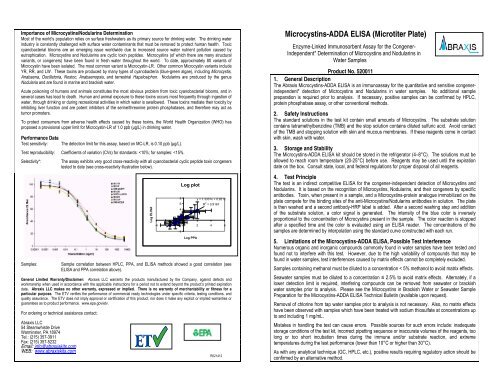
Performance Data<br />
Test sensitivity:<br />
The detection limit for this assay, based on MC-LR, is 0.10 ppb (μg/L).<br />
Test reproducibility: Coefficients of variation (CVs) for standards:
A. Materials Provided<br />
1. <strong>Microtiter</strong> plate (12 X 8 strips) coated with an analog of <strong>Microcystins</strong> conjugated to a protein<br />
2. Standards (6) and Control (1): 0, 0.15, 0.40, 1.0, 2.0, 5.0 ppb; Control at 0.75 ± 0.185 ppb<br />
3. Sample Diluent (for dilution of samples above the range of the curve)<br />
4. Antibody Solution<br />
5. Anti-Sheep-HRP Conjugate Solution<br />
6. Wash Solution (5X) Concentrate, must be diluted prior to use, see Test Preparation (Section D)<br />
7. Substrate (Color) Solution (TMB)<br />
8. Stop Solution<br />
B. Additional Materials (not delivered with the test kit)<br />
1. Micro-pipettes with disposable plastic tips (20-200 µL)<br />
2. Multi-channel pipette (50-300 µL) or stepper pipette with plastic tips (50-300 µL)<br />
3. Deionized or distilled water<br />
4. Paper towels or equivalent absorbent material<br />
5. Timer<br />
6. Tape or parafilm<br />
7. <strong>Microtiter</strong> plate reader (wavelength 450 nm)<br />
8. <strong>Microtiter</strong> plate washer (optional)<br />
C. Sample Collection and Handling<br />
Collect water samples in glass containers and test within 24 hours. If samples must be held for longer<br />
periods (up to 5 days), samples should be stored refrigerated. For storage periods greater than 5 days,<br />
samples should be stored frozen.<br />
If total <strong>Microcystins</strong> concentration (free and cell bound) is required, an appropriate cell lysing procedure<br />
(freeze and thaw, sonication, QuickLyse, etc.) must be performed prior to analysis.<br />
D. Test Preparation<br />
Micro-pipetting equipment and pipette tips for pipetting the standards and the samples are necessary. A<br />
multi-channel pipette or a stepping pipette is recommended for the addition of the antibody, enzyme<br />
conjugate, substrate, and stop solutions in order to equalize the incubation periods on the entire microtiter<br />
plate. Please use only the reagents and standards from one kit lot in one test, as they have been adjusted<br />
in combination.<br />
1. Allow the reagents and samples to reach ambient temperature before use.<br />
2. Remove the number of microtiter plate strips required from the resealable pouch. The remaining strips<br />
are stored in the pouch with the desiccant (tightly sealed).<br />
3. The standards, control, sample diluent, antibody, enzyme conjugate, substrate, and stop solutions are<br />
ready to use and do not require any further dilutions.<br />
4. Dilute the Wash Solution (5X) Concentrate at a ratio of 1:5 with deionized or distilled water. If using<br />
the entire bottle (100 mL), add to 400 mL of deionized or distilled water and mix thoroughly.<br />
5. The stop solution must be handled with care as it contains diluted H2SO4.<br />
E. Working Scheme<br />
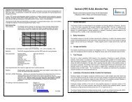
The microtiter plate consists of 12 strips of 8 wells, which can be used individually for the test. The<br />
standards must be run with each test. Never use the values of standards which have been determined in a<br />
test performed previously.<br />
Std 0-Std5: Standards<br />
Contr.: Control<br />
Samp1, Samp2, etc: Samples<br />
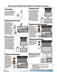
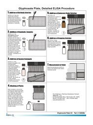
F. Assay Procedure<br />
1. Add 50 µL of the standard solutions, control, or samples into the wells of the test strips<br />
according to the working scheme given. Analysis in duplicate or triplicate is recommended.<br />
2. Add 50 µL of the antibody solution to the individual wells successively using a multi-channel<br />
pipette or a stepping pipette. Cover the wells with parafilm or tape and mix the contents by moving<br />
the strip holder in a circular motion on the benchtop for 30 seconds. Be careful not to spill the<br />
contents. Incubate the strips for 90 minutes at room temperature.<br />
3. Remove the covering and decant the contents of the wells into a sink. Wash the strips three<br />
times using the 1X wash buffer solution. Please use at least a volume of 250 µL of wash buffer<br />
for each well and each washing step. Remaining buffer in the wells should be removed by patting<br />
the plate dry on a stack of paper towels.<br />
4. Add 100 µL of the enzyme conjugate solution to the individual wells successively using a multichannel<br />
pipette or a stepping pipette. Cover the wells with parafilm or tape and mix the contents<br />
by moving the strip holder in a circular motion on the benchtop for 30 seconds. Be careful not to<br />
spill the contents. Incubate the strips for 30 minutes at room temperature.<br />
5. Remove the covering and decant the contents of the wells into a sink. Wash the strips three<br />
times using the 1X wash buffer solution. Please use at least a volume of 250 µL of wash buffer<br />
for each well and each washing step. Remaining buffer in the wells should be removed by patting<br />
the plate dry on a stack of paper towels.<br />
6. Add 100 µL of substrate (color) solution to the individual wells successively using a multichannel<br />
pipette or a stepping pipette. Cover the wells with parafilm or tape and mix the contents<br />
by moving the strip holder in a circular motion on the benchtop for 30 seconds. Be careful not to<br />
spill the contents. Incubate the strips for 20-30 minutes at room temperature. Protect the strips<br />
from sunlight.<br />
7. Add 50 µL of stop solution to the wells in the same sequence as for the substrate (color) solution<br />
using a multi-channel pipette or a stepping pipette.<br />
8. Read the absorbance at 450 nm using a microplate <strong>ELISA</strong> photometer within 15 minutes after the<br />
addition of the stopping solution.<br />
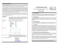
G. Evaluation<br />
The evaluation of the <strong>ELISA</strong> can be performed using commercial <strong>ELISA</strong> evaluation programs such as<br />
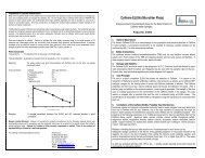
4-Parameter (preferred) or Logit/Log. For a manual evaluation, calculate the mean absorbance value<br />
for each of the standards. Calculate the %B/B0 for each standard by dividing the mean absorbance<br />
value for each standard by the Zero Standard (Standard 0) mean absorbance. Construct a standard<br />
curve by plotting the %B/B0 for each standard on the vertical linear (y) axis versus the corresponding<br />
<strong>Microcystins</strong> concentration on the horizontal logarithmic (x) axis on graph paper. %B/B0 for the control<br />
and samples will then yield levels in ppb of <strong>Microcystins</strong> by interpolation using the standard curve.<br />
Results can also be determined using a spreadsheet macro available from <strong>Abraxis</strong> upon request.<br />
The concentrations of the samples are determined using the standard curve run with each test.<br />
Samples showing a lower concentration of <strong>Microcystins</strong> than standard 1 (0.15 ppb) should be reported<br />
as containing < 0.15 ppb of <strong>Microcystins</strong>. Samples showing a higher concentration than standard 5 (5.0<br />
ppb) must be diluted to obtain accurate results. The concentration of the positive control provided<br />
should be 0.75 ± 0.185 ppb.<br />
Semi-quantitative results can be derived by simple comparison of the sample absorbances to the<br />
absorbances of the calibrators. Samples with lower absorbances than a calibrator will have<br />
concentrations of <strong>Microcystins</strong> greater than that calibrator. Samples which have higher absorbances<br />
than a calibrator will have concentrations of <strong>Microcystins</strong> less than that calibrator.<br />
H. References<br />
(1) W. J. Fischer, I. Garthwaite, C.O. Miles, K.M. Ross, J.B. Aggen, A.R. Chamberlin, N.A. Towers, and D.R.<br />
Dietrich, Congener-Independent Immunoassay for <strong>Microcystins</strong> and Nodularins. Environ. Sci. Technol. 35,<br />
2001, 4849-4858.<br />
(2) Worldwide Patenting PCT WO 01/18059 A2.<br />
(3) U.S. Patent Number 6,967,240.