β-N-methylamino-L-alanine (BMAA) ELISA* (Microtiter Plate) - Abraxis
β-N-methylamino-L-alanine (BMAA) ELISA* (Microtiter Plate) - Abraxis
β-N-methylamino-L-alanine (BMAA) ELISA* (Microtiter Plate) - Abraxis

You also want an ePaper? Increase the reach of your titles
YUMPU automatically turns print PDFs into web optimized ePapers that Google loves.
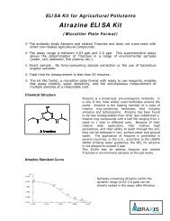
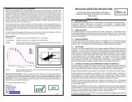
Importance of β-N-<strong>methylamino</strong>-L-<strong>alanine</strong> (<strong>BMAA</strong>) Determinationβ-N-<strong>methylamino</strong>-L-<strong>alanine</strong> (<strong>BMAA</strong>) is a non-protein amino acid produced by several types of cyanobacteria found infreshwaters, marine waters, and soil. When ingested, <strong>BMAA</strong> damages and ultimately destroys motor neurons in thespinal cord, causing the same type of damage seen in patients with ALS, and causes neurofibrillary tangles in the spinalcord and brain, similar to those seen in Alzheimer’s disease. Those suffering from this <strong>BMAA</strong>-induced damage areclassified as suffering from amyotrophic lateral sclerosis/parkinsonism dementia complex (ALS/PDC). The symptoms ofALS/PDC include the varying degrees of the muscular paralysis of ALS, the muscular rigidity of Parkinson’s, and/or thedementia of Alzheimer’s and ultimately result in death.Humans may be exposed to <strong>BMAA</strong> through the ingestion of contaminated drinking water or foods. Drinking water maybecome contaminated with <strong>BMAA</strong> through the proliferation of <strong>BMAA</strong>-producing cyanobacteria in drinking water sourcessuch as lakes and reservoirs. It is unknown whether the various methods of water treatment are able to remove <strong>BMAA</strong>from the drinking water supply. Exposure also occurs thorough ingestion of foods such as cycad seeds, on whose plantroots <strong>BMAA</strong>-producing cyanobacteria are known to live and thereby contaminate the plant and its seeds, and through theingestion of fish or other animals which have consumed toxin containing plants or cyanobacteria. An example of thisbiomagnification was seen in Guam in the 1950s, when fruit bats, which consume cycad seeds, were consumed by thenative Chamorro people causing a dramatic increase in cases of ALS/PDC, known on Guam as “lytico-bodig.”The <strong>BMAA</strong> ELISA allows for the analysis of 42 samples in duplicate determination. Less than 1 mL of sample is required.The test can be performed in approximately 2 hours.Performance DataTest sensitivity:The limit of quantitation for <strong>BMAA</strong> (95% B/B0) is approximately 4 ng/mL. The middle of thetest (50% B/B0) is approximately 100 ng/mL. Determinations closer to the middle of thecalibration curve give the most accurate results.Sample concentration may be performed for samples requiring a lower limit of detection(technical bulletin available from <strong>Abraxis</strong> by request).Sample extraction and clean-up are necessary for biological samples (please refer to Z.Spáčil, J. Eriksson, S. Jonasson, U. Rasmussen, L. L. Ilag and B. Bergman, Analyst, 2010,135, 127-132).Test reproducibility: Coefficients of variation (CVs) for standards:
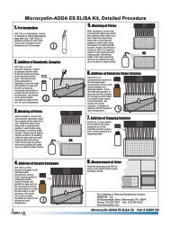
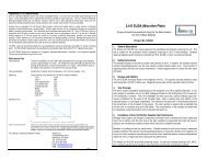
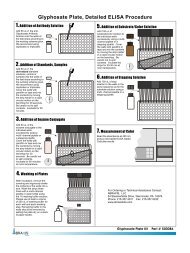
A. Reagents and Materials Provided1. <strong>Microtiter</strong> plate (12 X 8 strips) coated with a secondary antibody, in a resealable aluminum pouch2. Lyophilized <strong>BMAA</strong> Calibrators/Standards (6): 0, 5, 25, 100, 250, 500 ng/mL (ppb), must be reconstitutedbefore use, see Test Preparation (Section D)3. Antibody Solution (rabbit anti-<strong>BMAA</strong>), 6 mL4. <strong>BMAA</strong>-HRP Conjugate Solution, 6 mL5. Wash Solution (5X) Concentrate, 100 mL, must be diluted before use, see Test Preparation (Section D)6. Sample Diluent, 25 mL7. Substrate (Color) Solution (TMB), 16 mL8. Stop Solution, 12 mL (handle with care)B. Additional Materials (not delivered with the test kit)1. Micro-pipettes with disposable plastic tips (50-200 µL)2. Multi-channel pipette (50-250 µL) or stepper pipette with disposable plastic tips (50-250 µL)3. <strong>Microtiter</strong> plate reader (wave length 450 nm)4. Deionized or distilled water5. Paper towels or equivalent absorbent material6. Timer7. Tape or parafilmC. Sample CollectionCollect water samples in polypropylene (PP) or high density polyethylene (HDPE) containers. Do not use glasscontainers, as analyte will be lost due to adsorption.D. Test PreparationMicro-pipetting equipment and pipette tips for pipetting the standards and the samples are necessary. In order toequalize the incubation periods on the entire microtiter plate, a multi-channel pipette or a stepping pipette isrecommended for adding the enzyme conjugate, antibody, substrate, and stop solutions. Please only use thereagents and standards from one package lot in one test, as they have been adjusted in combination.1. Adjust the microtiter plate and the reagents to room temperature before use.2. Remove the number of microtiter plate strips required from the aluminum pouch. The remaining strips arestored in the aluminum pouch and zip-locked closed. Store the remaining kit in the refrigerator (4-8°C).3. The conjugate, antibody, substrate, and stop solutions are ready to use and do not require any furtherdilutions.4. The standards are provided in lyophilized form. To reconstitute, add 1 mL of deionized or distilled water toeach standard vial. Vortex thoroughly. Reconstituted standards may be used for up to one month (storefrozen).5. Dilute the Wash Solution (5X) Concentrate at a ratio of 1:5. If using the entire bottle (100 mL), add to 400 mLof deionized or distilled water and mix thoroughly.6. The stop solution must be handled with care as it contains diluted H2SO4.E. Working SchemeThe microtiter plate consists of 12 strips of 8 wells, which can be used individually for the test. The standards mustbe run with each test. Never use the values of standards which have been determined in a test performedpreviously.F. Assay Procedure1. Add 100 µL of the calibrator/standard solutions or samples into the wells of the test strips accordingto the working scheme given. Analysis in duplicate or triplicate is recommended.2. Add 50 µL of enzyme conjugate solution to the individual wells successively using a multi-channelpipette or a stepping pipette.3. Add 50 µL of antibody solution to the individual wells successively using a multi-channel pipette or astepping pipette. Cover the wells with parafilm or tape and mix the contents by moving the strip holderin a circular motion on the benchtop for 30 seconds. Be careful not to spill the contents.4. Incubate the strips for 90 minutes at room temperature.5. Remove the covering and decant the contents of the wells into a sink. Wash the strips four times usingthe diluted washing buffer solution. Please use at least a volume of 250 µL of washing buffer foreach well in each washing step. Remaining buffer in the wells should be removed by patting theinverted plate dry on a stack of paper towels.6. Add 150 µL of substrate (color) solution to the wells. Cover the wells with parafilm or tape and mixthe contents by moving the strip holder in a circular motion on the benchtop for 30 seconds. Be carefulnot to spill the contents. Incubate the strips for 30 minutes at room temperature. Protect the strips fromdirect sunlight.7. Add 100 µL of stop solution to the wells in the same sequence as for the substrate solution.8. Read the absorbance at 450 nm using a microplate ELISA photometer within 15 minutes after theaddition of stopping solution.G. EvaluationThe evaluation of the ELISA can be performed using commercial ELISA evaluation programs such as 4-Parameter (preferred) or Logit/Log. For a manual evaluation, calculate the mean absorbance value for eachof the standards. Calculate the %B/B0 for each standard by dividing the mean absorbance value for eachstandard by the Zero Standard (Standard 0) mean absorbance. Construct a standard curve by plotting the%B/B0 for each standard on the vertical linear (y) axis versus the corresponding <strong>BMAA</strong> concentration on thehorizontal logarithmic (x) axis on graph paper. %B/B0 for samples will then yield levels in ppb (or ng/mL) of<strong>BMAA</strong> by interpolation using the standard curve. Results can also be obtained by using a spreadsheet macroavailable from <strong>Abraxis</strong> upon request.The concentrations of the samples are determined using the standard curve run with each test. Sampleextracts showing a lower concentration of <strong>BMAA</strong> than standard 1 (5 ppb) should be reported as containing < 5ppb of <strong>BMAA</strong>. Samples showing a higher concentration than standard 5 (500 ppb) must be diluted furtherwith the provided sample diluent and re-analyzed.Semi-quantitative results can be derived by simple comparison of the sample absorbances to theabsorbances of the calibrators. Samples with lower absorbances than a calibrator will have concentrations of<strong>BMAA</strong> greater than the concentration of that calibrator. Samples which have higher absorbances than acalibrator will have concentrations of <strong>BMAA</strong> less than that calibrator.H. Standard CurveStd 0-Std 5: Standards(0; 5; 25; 100; 250; 500 ppb)Samp1, Samp2, etc.: SamplesFor demonstration purposes only. Not for use in sample interpretation.