Effects on oral soft tissue produced by a diode laser in vitro - Biolase
Effects on oral soft tissue produced by a diode laser in vitro - Biolase
Effects on oral soft tissue produced by a diode laser in vitro - Biolase

Create successful ePaper yourself
Turn your PDF publications into a flip-book with our unique Google optimized e-Paper software.
<str<strong>on</strong>g>Effects</str<strong>on</strong>g> <strong>on</strong> Oral Soft Tissue Produced <strong>by</strong> a<br />
Diode Laser In Vitro<br />
K. Goharkhay, MD, DMD, 1 A. Moritz, MD, DMD, 1 * P. Wilder-Smith, MD, DMD, 2<br />
U. Schoop, MD, DMD, 1 W. Kluger, MD, 1 S. Jakolitsch, MD, 1 and W. Sperr, MD, DMD 1<br />
1 Department of C<strong>on</strong>servative Dentistry, Dental School, University of Vienna,<br />
A-1090 Vienna, Austria<br />
2 Beckman Laser Institute and Medical Cl<strong>in</strong>ic, University of California, Irv<strong>in</strong>e,<br />
Irv<strong>in</strong>e, California 92697<br />
Background and Objectives: This <strong>in</strong>vestigati<strong>on</strong> determ<strong>in</strong>ed <strong>in</strong>cisi<strong>on</strong><br />
characteristics and <strong>soft</strong>-<strong>tissue</strong> damage result<strong>in</strong>g from standardized <strong>in</strong>cisi<strong>on</strong>s<br />
us<strong>in</strong>g a wide range of <strong>laser</strong> modes and parameters of a <strong>diode</strong><br />
<strong>laser</strong> at 810 nm.<br />
Study Design/Materials and Methods: Histologic exam<strong>in</strong>ati<strong>on</strong>s were<br />
performed to verify vertical and horiz<strong>on</strong>tal <strong>tissue</strong> damage as well as<br />
<strong>in</strong>cisi<strong>on</strong> depth and width.<br />
Results: Incisi<strong>on</strong> depth and width correlated str<strong>on</strong>gly with average<br />
powers, but not with <strong>laser</strong> parameters or the used tips. No <strong>laser</strong> damage<br />
was visible to the naked eye <strong>in</strong> the b<strong>on</strong>e underly<strong>in</strong>g the <strong>in</strong>cisi<strong>on</strong>s <strong>in</strong> the<br />
range between 0.5–4.5 W.<br />
C<strong>on</strong>clusi<strong>on</strong>: The remarkable cutt<strong>in</strong>g ability and the tolerable damage<br />
z<strong>on</strong>e clearly show that the <strong>diode</strong> <strong>laser</strong> is a very effective and, because of<br />
its excellent coagulati<strong>on</strong> ability, useful alternative <strong>in</strong> <strong>soft</strong>-<strong>tissue</strong> surgery<br />
of the <strong>oral</strong> cavity. Lasers Surg. Med. 25:401–406, 1999.<br />
© 1999 Wiley-Liss, Inc.<br />
Key words: <strong>in</strong>cisi<strong>on</strong> depth; mucosa; collateral <strong>tissue</strong> damage<br />
INTRODUCTION<br />
The scalpel and the c<strong>on</strong>venti<strong>on</strong>al electrosurgery<br />
unit are the <strong>in</strong>struments of choice for <strong>soft</strong><strong>tissue</strong><br />
surgery. In additi<strong>on</strong>, <strong>laser</strong>s are an alternative<br />
to c<strong>on</strong>venti<strong>on</strong>al surgical systems. Scalpels<br />
have been used for many years because of their<br />
ease of use, accuracy, and m<strong>in</strong>imal damage to the<br />
surround<strong>in</strong>g <strong>tissue</strong>. On the other hand, scalpels<br />
cannot provide the hemostasis that is helpful for<br />
use <strong>on</strong> highly vascular <strong>tissue</strong> [1].<br />
One characteristic difference between a <strong>laser</strong><br />
and a scalpel cut is the generati<strong>on</strong> of a coagulated<br />
<strong>tissue</strong> layer al<strong>on</strong>g the walls of the <strong>laser</strong> <strong>in</strong>cisi<strong>on</strong><br />
[2]. All <strong>laser</strong>-<strong>tissue</strong> <strong>in</strong>teracti<strong>on</strong>s produce some degree<br />
of <strong>tissue</strong> vaporizati<strong>on</strong> and a surround<strong>in</strong>g<br />
z<strong>on</strong>e of thermal necrosis [3]. This z<strong>on</strong>e of thermal<br />
damage should ideally be kept to a m<strong>in</strong>imum, as it<br />
may impede wound heal<strong>in</strong>g and graft take, and<br />
reduce tensile strength [2]. Cl<strong>in</strong>ical experience<br />
suggests some advantages for <strong>laser</strong> over scalpel<br />
surgical procedures of the <strong>oral</strong> <strong>tissue</strong>s. Advan-<br />
© 1999 Wiley-Liss, Inc.<br />
Lasers <strong>in</strong> Surgery and Medic<strong>in</strong>e 25:401–406 (1999)<br />
tages of this tool <strong>in</strong>clude greater precisi<strong>on</strong>, a relatively<br />
bloodless surgical and postsurgical course,<br />
sterilizati<strong>on</strong> of the surgical area, m<strong>in</strong>imal swell<strong>in</strong>g<br />
and scarr<strong>in</strong>g, coagulati<strong>on</strong>, vaporizati<strong>on</strong>, and<br />
cutt<strong>in</strong>g, m<strong>in</strong>imal or no sutur<strong>in</strong>g, and much less or<br />
no postsurgical pa<strong>in</strong> [4–6]. Research has c<strong>on</strong>sistently<br />
dem<strong>on</strong>strated that <strong>laser</strong> surgery can be<br />
performed safely <strong>by</strong> us<strong>in</strong>g parameters which protect<br />
underly<strong>in</strong>g b<strong>on</strong>e and tooth structures [7]. The<br />
factors that determ<strong>in</strong>e the <strong>in</strong>itial <strong>tissue</strong> effect <strong>in</strong>clude<br />
the <strong>laser</strong> wavelength, <strong>laser</strong> power, the available<br />
<strong>laser</strong> waveform (c<strong>on</strong>t<strong>in</strong>uous wave, chopped,<br />
and pulsed beams), <strong>tissue</strong> optical properties, and<br />
<strong>tissue</strong> thermal properties [8]. At this time, <strong>oral</strong><br />
<strong>laser</strong> applicati<strong>on</strong>s do not replace the majority of<br />
*Corresp<strong>on</strong>dence to: Prof. A. Moritz, Department of C<strong>on</strong>servative<br />
Dentistry, Dental School, University of Vienna, Waehr<strong>in</strong>gerstr.<br />
25a, A-1090 Vienna, Austria.<br />
Accepted 25 May 1999
402 Goharkhay et al.<br />
traditi<strong>on</strong>al scalpel procedures or the high-speed<br />
drill. Certa<strong>in</strong> <strong>laser</strong>s are ideal for specific <strong>oral</strong> procedures,<br />
but <strong>laser</strong>s cannot provide the same efficiency<br />
and efficacy as a scalpel for many different<br />
<strong>oral</strong> procedures [4]. Electrosurgery units produce<br />
adequate hemostasis but result <strong>in</strong> greater thermal<br />
<strong>in</strong>jury and also have the disadvantage of<br />
caus<strong>in</strong>g muscle fasciculati<strong>on</strong>s. Reports suggest<br />
that heal<strong>in</strong>g is delayed for electrosurgery wounds<br />
when compared with scalpel wounds [1]. The<br />
thermal and histologic events result<strong>in</strong>g from <strong>soft</strong><strong>tissue</strong><br />
<strong>in</strong>cisi<strong>on</strong> with different CO 2 <strong>laser</strong>s have already<br />
been determ<strong>in</strong>ed [2]. These exam<strong>in</strong>ati<strong>on</strong>s<br />
showed that <strong>in</strong>cisi<strong>on</strong> depths correlate positively<br />
with the average power [9–11]. Diode <strong>laser</strong>s have<br />
been used successfully for c<strong>on</strong>diti<strong>on</strong><strong>in</strong>g enamel<br />
and dent<strong>in</strong> surfaces. Furthermore, the <strong>diode</strong> <strong>laser</strong><br />
reveals a bactericidal effect and helps to reduce<br />
<strong>in</strong>flammati<strong>on</strong> <strong>in</strong> the root canal and <strong>in</strong> the period<strong>on</strong>tal<br />
pocket <strong>in</strong> additi<strong>on</strong> to scal<strong>in</strong>g [12–14].<br />
The aim of this study was to determ<strong>in</strong>e <strong>in</strong>cisi<strong>on</strong><br />
characteristics and <strong>soft</strong>-<strong>tissue</strong> damage result<strong>in</strong>g<br />
from standardized <strong>in</strong>cisi<strong>on</strong>s us<strong>in</strong>g a wide<br />
range of <strong>laser</strong> modes and parameters of a <strong>diode</strong><br />
<strong>laser</strong> at 810 nm.<br />
MATERIALS AND METHODS<br />
For this <strong>in</strong>vestigati<strong>on</strong>, 17 fresh pig mandibles<br />
were used no later than 6 hours after the<br />
animals’ death. Immediately after death, the<br />
mandibles were cooled until 1 hour before use and<br />
then returned to room temperature. Six standardized<br />
<strong>in</strong>cisi<strong>on</strong>s per <strong>laser</strong> parameter comb<strong>in</strong>ati<strong>on</strong>, 3<br />
cm <strong>in</strong> length, were made <strong>in</strong> the <strong>oral</strong> mucosa parallel<br />
to the border of the mandible. Three <strong>in</strong>cisi<strong>on</strong>s<br />
per parameter were positi<strong>on</strong>ed 5 mm below the<br />
g<strong>in</strong>gival marg<strong>in</strong>, and three <strong>in</strong> the thicker <strong>soft</strong> <strong>tissue</strong><br />
5 mm from the lower border of the mandible.<br />
The average thickness of these <strong>tissue</strong>s was 0.8–2<br />
mm. A total of 198 <strong>in</strong>cisi<strong>on</strong>s was made. The <strong>in</strong>cisi<strong>on</strong>s<br />
were performed <strong>in</strong> the anterior, middle, and<br />
posterior third of the mandible. The <strong>in</strong>cisi<strong>on</strong><br />
length was standardized, with a template positi<strong>on</strong>ed<br />
3 mm below the planned <strong>in</strong>cisi<strong>on</strong> site dur<strong>in</strong>g<br />
the performance of each <strong>in</strong>cisi<strong>on</strong>. The <strong>laser</strong><br />
handpiece was attached to a motorized device to<br />
standardize the <strong>in</strong>cisi<strong>on</strong> and to c<strong>on</strong>trol movements.<br />
Radiati<strong>on</strong> was applied <strong>by</strong> the handpiece at<br />
a speed of 10 mm/sec<strong>on</strong>d and was timed with a<br />
stopwatch. This experimental model has been<br />
used for many years <strong>in</strong> standardized studies<br />
[2,3,15].<br />
Treatment was carried out with 33 different<br />
sett<strong>in</strong>gs of the Dentek LD 15 <strong>diode</strong> <strong>laser</strong> (Dentek<br />
Austria GMBH, Gasselberg 53–54, A-8564 Gaisfeld,<br />
Austria). This <strong>laser</strong> has a wavelength of 810<br />
nm. The <strong>laser</strong> output power ranges from 0.5–15<br />
W. A pulse rate of 2–32 msec <strong>in</strong> pulsed mode and<br />
a frequency of 1.5–250 Hz can be used. The <strong>laser</strong><br />
can also be operated <strong>in</strong> c<strong>on</strong>t<strong>in</strong>uous wave mode.<br />
The target beam is generated <strong>by</strong> a helium-ne<strong>on</strong><br />
<strong>laser</strong> (533 nm, 1 mW).<br />
The applicati<strong>on</strong> occured with a 200- or 400-<br />
�m tip. Not <strong>on</strong>ly c<strong>on</strong>t<strong>in</strong>uous wave (cw), but also<br />
pulsed modes with a frequency of 25 Hz and 30msec<br />
pulse width, and 50 Hz and 10 msec, were<br />
used. The menti<strong>on</strong>ed output powers do not corresp<strong>on</strong>d<br />
to the output powers described <strong>on</strong> the <strong>laser</strong><br />
<strong>in</strong>strument, but were measured directly at the<br />
outflow of radiati<strong>on</strong> with a wattmeter. The 400-<br />
�m tip was used <strong>in</strong> a measured range of 0.5–4.5<br />
W, whereas a maximum of 2 W could be achieved<br />
for the th<strong>in</strong>ner 200-�m tip.<br />
Immediately after irradiati<strong>on</strong>, <strong>in</strong>cisi<strong>on</strong>s were<br />
dissected out with a marg<strong>in</strong> exceed<strong>in</strong>g 5 mm and<br />
divided with a scalpel. The b<strong>on</strong>e underly<strong>in</strong>g each<br />
<strong>in</strong>cisi<strong>on</strong> was marked, labeled, and photographed.<br />
The chief evaluati<strong>on</strong> factor for b<strong>on</strong>e was charr<strong>in</strong>g,<br />
which was selected as a gross <strong>in</strong>dicator of significant<br />
<strong>laser</strong>-<strong>in</strong>duced thermal damage.<br />
The <strong>soft</strong>-<strong>tissue</strong> samples were fixed directly <strong>in</strong><br />
10% neutral-buffered formal<strong>in</strong> for 15 days and<br />
were dehydrated for 8 days <strong>in</strong> a ris<strong>in</strong>g alcohol series.<br />
After treatment with terp<strong>in</strong>eol and toluol for<br />
10 days, the specimens were embedded <strong>in</strong> paraff<strong>in</strong><br />
for another 8 days. A total of 198 wax blocks was<br />
prepared, and 6-�m secti<strong>on</strong>s were cut rout<strong>in</strong>ely<br />
with the microtome and sta<strong>in</strong>ed with Serius red.<br />
Measurements were made from 15 slides per parameter<br />
and <strong>in</strong>cisi<strong>on</strong> site. A photographic record<br />
of the results was made. Incisi<strong>on</strong> depth and<br />
width, as well as depth and width of adjacent <strong>tissue</strong><br />
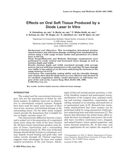
damage, were determ<strong>in</strong>ed. A typical slide<br />
with measurement locati<strong>on</strong>s is shown <strong>in</strong> Figure 1.<br />
RESULTS<br />
Mean <strong>in</strong>cisi<strong>on</strong> depths and widths, as well as<br />
mean collateral vertical and horiz<strong>on</strong>tal damage<br />
measurements and standard deviati<strong>on</strong>s, are presented<br />
<strong>in</strong> Tables 1 and 2. Mean <strong>in</strong>cisi<strong>on</strong> depths<br />
us<strong>in</strong>g cw and a 400-�m tip measured from 217.5–<br />
647.5 �m, and mean <strong>in</strong>cisi<strong>on</strong> widths from 78.8–<br />
357.5 �m. Us<strong>in</strong>g the 200-�m tip, mean <strong>in</strong>cisi<strong>on</strong><br />
depths of 405–605 �m and mean widths of 87.6–<br />
278.3 �m were measured. In the 50-Hz/10-msec<br />
pulsed mode, mean depths ranged from 295–<br />
737.5 �m. Mean widths of 162.5–400 �m were
measured for the 400-�m tip, while mean <strong>in</strong>cisi<strong>on</strong><br />
depths from 240–614.3 �m and mean widths from<br />
103.8–210.3 �m were determ<strong>in</strong>ed for the 200-�m<br />
tip. The 25-Hz/30 msec pulsed mode revealed<br />
mean depths from 298.5–527 �m and mean<br />
widths from 138.8–377.5 �m for the thicker tip,<br />
and mean <strong>in</strong>cisi<strong>on</strong> depths from 201.6–345 �m and<br />
widths from 115.2–244.3 �m for the th<strong>in</strong>ner tip.<br />
Mean vertical damage measured from 22.5–<br />
85.3 �m and mean horiz<strong>on</strong>tal damage from 28.3–<br />
98 �m, irrespective of the <strong>laser</strong> parameters and<br />
tips used.<br />
The depth and width of <strong>in</strong>cisi<strong>on</strong> correlated<br />
str<strong>on</strong>gly and positively with average powers, but<br />
not with <strong>laser</strong> parameters or the tips used.<br />
To the naked eye, no <strong>laser</strong> damage was visible<br />
<strong>in</strong> the b<strong>on</strong>e underly<strong>in</strong>g the <strong>in</strong>cisi<strong>on</strong>s, either <strong>in</strong><br />
the thicker <strong>soft</strong> <strong>tissue</strong> or <strong>in</strong> the th<strong>in</strong>ner <strong>soft</strong> <strong>tissue</strong>.<br />
DISCUSSION<br />
Histologic events result<strong>in</strong>g from <strong>soft</strong>-<strong>tissue</strong><br />
<strong>in</strong>cisi<strong>on</strong>s with different CO2 <strong>laser</strong>s have already<br />
Oral Soft Tissue and Diode Laser In Vitro 403<br />
Fig. 1. Incisi<strong>on</strong>al and collateral effects of <strong>diode</strong> <strong>laser</strong> <strong>in</strong> <strong>oral</strong> <strong>soft</strong> <strong>tissue</strong>.<br />
been determ<strong>in</strong>ed. Histologic effects are related to<br />
the parameters used and the beam characteristics<br />
rather than wavelength; greater damage to the<br />
collateral <strong>tissue</strong>s has been observed with the use<br />
of the c<strong>on</strong>stant wave mode. This effect enhances<br />
thermocoagulati<strong>on</strong> to achieve hemostasis and provide<br />
a bloodless surgical field. The desired results<br />
with the least risk of unwanted thermal damage<br />
can be achieved with very short pulses at the<br />
highest power density for the shortest time possible<br />
[2]. The extent of collateral thermal effects is<br />
smaller <strong>by</strong> a factor of about 2–3 for the superpulse<br />
mode <strong>in</strong> comparis<strong>on</strong> to the cw mode [16,17]. A<br />
wide range of cl<strong>in</strong>ical effects can be achieved c<strong>on</strong>sistently<br />
and predictably <strong>in</strong> <strong>soft</strong> <strong>tissue</strong>, depend<strong>in</strong>g<br />
<strong>on</strong> the parameter c<strong>on</strong>figurati<strong>on</strong> selected. The use<br />
of higher average powers correlates with <strong>in</strong>creas<strong>in</strong>g<br />
depths of <strong>in</strong>cisi<strong>on</strong>. Incisi<strong>on</strong> width and collateral<br />
damage are the results of complex <strong>in</strong>teracti<strong>on</strong>s<br />
between the different <strong>laser</strong> parameter variables,<br />
as menti<strong>on</strong>ed. Incisi<strong>on</strong> shape and width are<br />
str<strong>on</strong>gly mode-dependent. The cw mode produces
404 Goharkhay et al.<br />
TABLE 1. Incisi<strong>on</strong>al and Collateral <str<strong>on</strong>g>Effects</str<strong>on</strong>g> of 400-�m Tip*<br />
Average<br />
power<br />
(W)<br />
Pulse<br />
width<br />
(msec)<br />
Pulse<br />
repetiti<strong>on</strong><br />
(Hz)<br />
Mean ± SD<br />
<strong>in</strong>cisi<strong>on</strong> depth<br />
<strong>in</strong> �m (n)<br />
relatively wide, straight-sided <strong>in</strong>cisi<strong>on</strong>s. Therefore,<br />
this mode can cut or ablate large amounts of<br />
<strong>tissue</strong>. Comparable <strong>in</strong>cisi<strong>on</strong>s <strong>in</strong> depth can be<br />
achieved equally quickly and efficiently at lower<br />
average powers with the superpulse mode [3].<br />
Of the cl<strong>in</strong>ically comm<strong>on</strong> dental <strong>laser</strong>s, the<br />
CO 2 <strong>laser</strong> usually produces narrower z<strong>on</strong>es of<br />
damage <strong>in</strong> <strong>soft</strong> <strong>tissue</strong>s than does the Nd:YAG <strong>laser</strong><br />
because of the greater absorpti<strong>on</strong> of the CO 2<br />
wavelength <strong>by</strong> <strong>soft</strong> <strong>tissue</strong>s [18–20]. The average<br />
z<strong>on</strong>e of damage caused <strong>by</strong> CO 2 <strong>laser</strong>s after <strong>laser</strong><br />
<strong>in</strong>cisi<strong>on</strong> <strong>in</strong> <strong>soft</strong> <strong>tissue</strong>s is less than 0.6 mm [16,18,<br />
21–23]. This f<strong>in</strong>d<strong>in</strong>g is directly relevant to cl<strong>in</strong>ical<br />
Mean ± SD<br />
<strong>in</strong>cisi<strong>on</strong> width<br />
<strong>in</strong> �m (n)<br />
Mean ± SD<br />
vertical damage<br />
<strong>in</strong> �m (n)<br />
Mean ± SD<br />
horiz<strong>on</strong>tal<br />
damage <strong>in</strong> �m (n)<br />
0.5 cw 217.5 ± 12.6 (15) 78.8 ± 19.3 (15) 30 ± 4.0 (15) 29.8 ± 1.3 (15)<br />
1 cw 230 ± 18.7 (15) 119.3 ± 8.7 (15) 22.5 ± 6.5 (15) 37.5 ± 6.5 (15)<br />
1.5 cw 272.3 ± 12.3 (15) 175 ± 23.8 (15) 44.5 ± 5.3 (15) 56.3 ± 25 (15)<br />
2 cw 302.5 ± 22.2 (15) 205 ± 17.3 (15) 40 ± 8.2 (15) 77.5 ± 2.1 (15)<br />
2.5 cw 421.3 ± 19.3 (15) 277.5 ± 20.6 (15) 48.8 ± 4.8 (15) 76.8 ± 4.6 (15)<br />
3.5 cw 593 ± 9.5 (15) 300 ± 7.1 (15) 46.3 ± 5.1 (15) 73.3 ± 11.6 (15)<br />
4.5 cw 647.5 ± 9.6 (15) 357.5 ± 30.3 (15) 55 ± 4.1 (15) 98 ± 33.4 (15)<br />
0.5 30 25 298.5 ± 11.5 (15) 138.8 ± 8.5 (15) 35 ± 12.9 (15) 40 ± 4.1 (15)<br />
1 30 25 380 ± 40.8 (15) 162.5 ± 6.5 (15) 28.3 ± 2.4 (15) 51.8 ± 16 (15)<br />
1.5 30 25 416.3 ± 13.8 (15) 218.8 ± 1.7 (15) 23.3 ± 5.4 (15) 55.75 ± 4.3 (15)<br />
2 30 25 488.7 ± 12.6 (15) 275.7 ± 14.1 (15) 48.6 ± 7.7 (15) 49 ± 2.6 (15)<br />
2.5 30 25 476.3 ± 18 (15) 371.3 ± 8.5 (15) 44.9 ± 10.2 (15) 66.7 ± 5.8 (15)<br />
3.5 30 25 497 ± 12.6 (15) 367 ± 15 (15) 57.5 ± 6.5 (15) 72.5 ± 32 (15)<br />
4.5 30 25 527 ± 22.2 (15) 377.5 ± 45.7 (15) 56.3 ± 4.8 (15) 97.5 ± 12.6 (15)<br />
0.5 10 50 295 ± 13 (15) 213.75 ± 4.8 (15) 28.8 ± 3 (15) 37.5 ± 2.9 (15)<br />
1 10 50 477.5 ± 9.6 (15) 162.5 ± 8.7 (15) 23.8 ± 4.8 (15) 35 ± 5.8 (15)<br />
1.5 10 50 492.5 ± 6.5 (15) 248.8 ± 9.8 (15) 43.8 ± 4.8 (15) 63.3 ± 7 (15)<br />
2 10 50 590 ± 8.2 (15) 304.8 ± 12.7 (15) 34.5 ± 5.3 (15) 55 ± 5.8 (15)<br />
2.5 10 50 587 ± 20.6 (15) 372.7 ± 20.2 (15) 75 ± 20.4 (15) 78.8 ± 29.5 (15)<br />
3.5 10 50 707.5 ± 17.1 (15) 400 ± 8.2 (15) 70 ± 21.6 (15) 65 ± 12.9 (15)<br />
4.5 10 50 737.5 ± 9.6 (15) 377.5 ± 48 (15) 80 ± 35.8 (15) 87.2 ± 8.6 (15)<br />
*SD, standard deviati<strong>on</strong>.<br />
TABLE 2. Incisi<strong>on</strong>al and Collateral <str<strong>on</strong>g>Effects</str<strong>on</strong>g> of 200-�m Tip*<br />
Average<br />
power<br />
(W)<br />
Pulse<br />
width<br />
(msec)<br />
Pulse<br />
repetiti<strong>on</strong><br />
(Hz)<br />
Mean ± SD<br />
<strong>in</strong>cisi<strong>on</strong> depth<br />
<strong>in</strong> �m (n)<br />
Mean ± SD<br />
<strong>in</strong>cisi<strong>on</strong> width<br />
<strong>in</strong> �m (n)<br />
Mean ± SD<br />
vertical damage<br />
<strong>in</strong> �m (n)<br />
Mean ± SD<br />
horiz<strong>on</strong>tal<br />
damage <strong>in</strong> �m (n)<br />
0.5 cw 405 ± 20.8 (15) 98.3 ± 9.3 (15) 52 ± 4.3 (15) 53.9 ± 8.1 (15)<br />
1 cw 475.7 ± 8.9 (15) 87.6 ± 13.7 (15) 47.8 ± 4.7 (15) 55.5 ± 11.3 (15)<br />
1.5 cw 520.5 ± 29.7 (15) 138.3 ± 9.3 (15) 60.3 ± 5.6 (15) 59.8 ± 7.5 (15)<br />
2 cw 605 ± 16.38 (15) 278.3 ± 19.8 (15) 59.5 ± 8.6 (15) 62 ± 9.4 (15)<br />
0.5 30 25 201.6 ± 6.3 (15) 195.8 ± 5.7 (15) 29.3 ± 3.3 (15) 28.3 ± 3.3 (15)<br />
1 30 25 307.5 ± 9.6 (15) 115.2 ± 11.1 (15) 37.5 ± 6.5 (15) 31.5 ± 5 (15)<br />
1.5 30 25 262.5 ± 6.5 (15) 204.3 ± 9.9 (15) 60.3 ± 5.6 (15) 48.8 ± 3 (15)<br />
2 30 25 345 ± 12.9 (15) 244.3 ± 8.3 (15) 58.4 ± 11.6 (15) 42.7 ± 7.9 (15)<br />
0.5 10 50 240 ± 18.3 (15) 103.8 ± 11.1 (15) 38.8 ± 6.3 (15) 49.5 ± 4.2 (15)<br />
1 10 50 306.4 ± 7.7 (15) 111.8 ± 12.5 (15) 85.3 ± 8.5 (15) 60.6 ± 8.2 (15)<br />
1.5 10 50 566.3 ± 12.5 (15) 157.8 ± 13.3 (15) 81.4 ± 9 (15) 53.1 ± 8.5 (15)<br />
2 10 50 614.3 ± 11.8 (15) 210.3 ± 7.6 (15) 75.9 ± 6.9 (15) 56.2 ± 10.5 (15)<br />
*SD, standard deviati<strong>on</strong>.<br />
dentistry because of c<strong>on</strong>cerns regard<strong>in</strong>g possible<br />
damage to neighbor<strong>in</strong>g structures, such as teeth<br />
or b<strong>on</strong>e, dur<strong>in</strong>g <strong>soft</strong>-<strong>tissue</strong> <strong>laser</strong> surgery. Comparis<strong>on</strong>s<br />
of Nd:YAG and <strong>diode</strong> <strong>laser</strong> show that,<br />
when used <strong>in</strong> c<strong>on</strong>tact mode, these two <strong>laser</strong>s produce<br />
similar extents of <strong>tissue</strong> vaporizati<strong>on</strong> and<br />
z<strong>on</strong>es of thermal necrosis [24]. With 10–12 W, values<br />
of 0.625 and 0.79, respectively, and 0.48 and<br />
0.9 mm, respectively, are obta<strong>in</strong>ed with <strong>diode</strong> and<br />
Nd:YAG <strong>laser</strong>s [25,26]. Although the extent of <strong>tissue</strong><br />
vaporizati<strong>on</strong> at low powers is less for the <strong>diode</strong><br />
<strong>laser</strong>, these differences are not apparent at<br />
higher <strong>laser</strong> powers and energies that can be
cl<strong>in</strong>ically applied with commercially available<br />
units [24].<br />
However, the radiati<strong>on</strong> of a <strong>diode</strong> <strong>laser</strong><br />
shows a greater absorpti<strong>on</strong> and a smaller penetrati<strong>on</strong><br />
depth than that of a Nd:YAG <strong>laser</strong>, especially<br />
<strong>in</strong> blood-rich <strong>tissue</strong>. The wavelength of the<br />
<strong>diode</strong> <strong>laser</strong> is c<strong>on</strong>siderably more absorbed due to<br />
hemoglob<strong>in</strong> than that of the Nd:YAG <strong>laser</strong>. This<br />
causes not <strong>on</strong>ly a better <strong>in</strong>cisi<strong>on</strong> performance but<br />
also an excellent coagulati<strong>on</strong> of <strong>tissue</strong> [27]. The<br />
thickness of the charr<strong>in</strong>g layer and the coagulati<strong>on</strong><br />
layer, and <strong>in</strong>cisi<strong>on</strong> depth, are similar for the<br />
<strong>diode</strong> <strong>laser</strong> and the Nd:YAG <strong>laser</strong> with the same<br />
<strong>laser</strong> sett<strong>in</strong>gs [25].<br />
Advantages of the <strong>diode</strong> <strong>laser</strong> seen <strong>in</strong> our<br />
cl<strong>in</strong>ical rout<strong>in</strong>e are that it requires no anesthetics<br />
and that the wounds heal <strong>soft</strong>ly. Moreover, its<br />
simple use allows very good model<strong>in</strong>g of the g<strong>in</strong>giva.<br />
Favorable results <strong>in</strong> other dental areas encouraged<br />
us to determ<strong>in</strong>e the surgical effects <strong>on</strong><br />
<strong>soft</strong> <strong>tissue</strong> <strong>produced</strong> <strong>by</strong> a <strong>diode</strong> <strong>laser</strong>, us<strong>in</strong>g two<br />
different tips <strong>in</strong> c<strong>on</strong>t<strong>in</strong>uous wave mode and two<br />
pulsed modes. Accord<strong>in</strong>g to our results, the <strong>in</strong>cisi<strong>on</strong><br />
depth correlates str<strong>on</strong>gly and positively with<br />
the average power, whereas <strong>in</strong>cisi<strong>on</strong> shape and<br />
width depend neither <strong>on</strong> the mode used, nor <strong>on</strong><br />
the fiber. These results c<strong>on</strong>firm the f<strong>in</strong>d<strong>in</strong>gs reported<br />
<strong>by</strong> Judy et al. [25], who <strong>in</strong>vestigated the<br />
Nd:YAG <strong>laser</strong>. A possible explanati<strong>on</strong> may be the<br />
chopped operat<strong>in</strong>g mode of the <strong>diode</strong> <strong>laser</strong>.<br />
The horiz<strong>on</strong>tal and vertical damage z<strong>on</strong>e depends<br />
neither <strong>on</strong> the average power, nor <strong>on</strong> the<br />
mode used or fiber tip. When compared with the<br />
CO 2 <strong>laser</strong>, <strong>on</strong>e characteristic difference from the<br />
<strong>diode</strong> <strong>laser</strong> can be found, namely that no trend of<br />
greater damage to lateral <strong>tissue</strong>s with the c<strong>on</strong>stant<br />
wave mode at higher power levels can be<br />
observed. Also, no charr<strong>in</strong>g of b<strong>on</strong>e underly<strong>in</strong>g<br />
0.8-mm-thick <strong>soft</strong> <strong>tissue</strong> was observed with the<br />
c<strong>on</strong>t<strong>in</strong>uous wave mode, or with the pulsed mode<br />
at an average power of 4.5 W. Several authors<br />
have reported that the use of the CO 2 <strong>laser</strong> can<br />
result <strong>in</strong> possible damage to the underly<strong>in</strong>g b<strong>on</strong>e<br />
around teeth when cutt<strong>in</strong>g <strong>tissue</strong>s with either<br />
pulsed or c<strong>on</strong>t<strong>in</strong>uous wave CO 2 <strong>laser</strong>s [2,3,28].<br />
Clayman et al. [29] described m<strong>in</strong>imal damage to<br />
the b<strong>on</strong>e under g<strong>in</strong>giva treated with a CO 2 <strong>laser</strong>,<br />
but the g<strong>in</strong>giva healed well, although over a<br />
l<strong>on</strong>ger period of time. It is possible that the <strong>laser</strong><br />
wavelength is transmitted through the surface<br />
layer of the b<strong>on</strong>e <strong>in</strong>to the <strong>in</strong>ner cancellous <strong>tissue</strong>.<br />
Simple observati<strong>on</strong> of the surface does not preclude<br />
<strong>in</strong>ner damage. However, <strong>in</strong> this study, low-<br />
Oral Soft Tissue and Diode Laser In Vitro 405<br />
power sett<strong>in</strong>gs were used, so that the possibility of<br />
damage was extremely remote.<br />
Blood circulati<strong>on</strong> acts as a potential, not very<br />
significant coolant. One typical example of heat<br />
c<strong>on</strong>vecti<strong>on</strong> <strong>in</strong> <strong>tissue</strong> is heat transfer due to blood<br />
flow. Due to the low perfusi<strong>on</strong> of most <strong>tissue</strong>s,<br />
however, heat c<strong>on</strong>vecti<strong>on</strong> is negligible <strong>in</strong> a first<br />
approximati<strong>on</strong>. Only dur<strong>in</strong>g l<strong>on</strong>g exposures does<br />
it play a significant role [30]. Certa<strong>in</strong>ly an adverse<br />
cellular resp<strong>on</strong>se will over time change the histological<br />
picture of the <strong>tissue</strong>s affected. Yet the focus<br />
of this study was <strong>on</strong> the measurement of direct<br />
thermal damage, not <strong>on</strong> <strong>tissue</strong> resp<strong>on</strong>se to<br />
irradiati<strong>on</strong>.<br />
C<strong>on</strong>trary to other <strong>in</strong>vestigati<strong>on</strong>s [3,7], deeper<br />
<strong>in</strong>cisi<strong>on</strong>s could be achieved with the <strong>diode</strong> <strong>laser</strong><br />
than were achieved <strong>by</strong> other authors with the CO 2<br />
or Nd:YAG <strong>laser</strong> at the same power sett<strong>in</strong>g, even<br />
with fewer movements of the delivery system.<br />
Even the horiz<strong>on</strong>tal and vertical z<strong>on</strong>es of thermal<br />
damage are <strong>in</strong> a comparable range. These f<strong>in</strong>d<strong>in</strong>gs,<br />
i.e., the remarkable cutt<strong>in</strong>g ability and the<br />
tolerable damage z<strong>on</strong>e, clearly show that the <strong>diode</strong><br />
<strong>laser</strong> is a very effective and, because of its<br />
excellent coagulati<strong>on</strong> ability, useful alternative <strong>in</strong><br />
<strong>soft</strong>-<strong>tissue</strong> surgery of the <strong>oral</strong> cavity.<br />
REFERENCES<br />
1. Libo<strong>on</strong> J, Funkhouser W, Terris D. Comparis<strong>on</strong> of mucosal<br />
<strong>in</strong>cisi<strong>on</strong>s made <strong>by</strong> scalpel, CO 2 <strong>laser</strong>, electrocautery<br />
and c<strong>on</strong>stant-voltage electrocautery. Otolaryngol Head<br />
Neck Surg 1997;116:379–385.<br />
2. Wilder-Smith P, Arrastia AM, Liaw LH, Berns M. Incisi<strong>on</strong><br />
properties and thermal effects of thoursee CO 2 <strong>laser</strong>s<br />
<strong>in</strong> <strong>soft</strong> <strong>tissue</strong>. Oral Surg Oral Med Oral Pathol Oral<br />
Radiol Endod 1995;79:685–691.<br />
3. Wilder-Smith P, Dang J, Kurosaki T. Investigat<strong>in</strong>g the<br />
range of surgical effects <strong>on</strong> <strong>soft</strong> <strong>tissue</strong> <strong>produced</strong> <strong>by</strong> a carb<strong>on</strong><br />
dioxide <strong>laser</strong>. J Am Dent Assoc 1997;128:583–588.<br />
4. Pick RM, Colvard MD. Current status of <strong>laser</strong>s <strong>in</strong> <strong>soft</strong><br />
<strong>tissue</strong> dental surgery. J Period<strong>on</strong>tol 1993;64:589–602.<br />
5. Gold SI, Vilardi MA. Pulsed <strong>laser</strong> beam effects <strong>on</strong> g<strong>in</strong>giva.<br />
J Cl<strong>in</strong> Period<strong>on</strong>tol 1994;21:391–396.<br />
6. White JM, Goodis HE, Rose CL. Use of the pulsed<br />
Nd:YAG Laser for <strong>in</strong>tra<strong>oral</strong> <strong>soft</strong> <strong>tissue</strong> surgery. Lasers<br />
Surg Med 1991;11:455–561.<br />
7. Perry D, Goodis H, White J. In <strong>vitro</strong> study of the effects of<br />
Nd:YAG <strong>laser</strong> probe parameters <strong>on</strong> bov<strong>in</strong>e <strong>oral</strong> <strong>soft</strong> <strong>tissue</strong><br />
excisi<strong>on</strong>. Lasers Surg Med 1997;20:39–46.<br />
8. Douglas N, Dederich NH. Laser/<strong>tissue</strong> <strong>in</strong>teracti<strong>on</strong>. Alpha<br />
Omegan 1991;84:33–36.<br />
9. Mat<strong>in</strong> NH, Orikasa N, Kusakari H. <str<strong>on</strong>g>Effects</str<strong>on</strong>g> of CO 2 <strong>laser</strong><br />
irradiati<strong>on</strong> to period<strong>on</strong>tal <strong>tissue</strong>s. Int Soc Laser Dent<br />
1995; Abstracts 9.<br />
10. Walsh JT Jr, Deutsch TF. Pulsed CO 2 <strong>laser</strong> <strong>tissue</strong> ablati<strong>on</strong>.<br />
measurement of the ablati<strong>on</strong> rate. Lasers Surg Med<br />
1988;8:3:264–275.
406 Goharkhay et al.<br />
11. White JM, Goodis HE, Yessic MJ, Meyers TD. Histologic<br />
effects of a high repetiti<strong>on</strong> pulsed Nd:YAG <strong>laser</strong> <strong>on</strong> <strong>oral</strong><br />
<strong>soft</strong> <strong>tissue</strong>. In: Wigor HA, Featherst<strong>on</strong>e JDB, White JM,<br />
eds. Lasers <strong>in</strong> dentistry. Internati<strong>on</strong>al Society for Optical<br />
Eng<strong>in</strong>eer<strong>in</strong>g. Proc SPIE 2394; 1995. p 129–141.<br />
12. Moritz A, Schoop U, Goharkhay K, Wernisch J, Sperr W.<br />
Procedures for enamel and dent<strong>in</strong> c<strong>on</strong>diti<strong>on</strong><strong>in</strong>g: a comparis<strong>on</strong><br />
of c<strong>on</strong>venti<strong>on</strong>al and <strong>in</strong>novative methods. J Esthet<br />
Dent 1998;10:84–93.<br />
13. Moritz A, Schoop U, Goharkhay K, Doertbudak O, Sperr<br />
W. Rapid report: irradiati<strong>on</strong> of <strong>in</strong>fected root canals with a<br />
<strong>diode</strong> <strong>laser</strong> <strong>in</strong> vivo. Lasers Surg Med 1997;21:221–226.<br />
14. Moritz A, Schoop U, Goharkhay K, Wernisch J, Sperr W.<br />
Treatment of period<strong>on</strong>tal pockets with a <strong>diode</strong> <strong>laser</strong>. Lasers<br />
Surg Med 1998;22:302–311.<br />
15. Wilder-Smith P, Dang J, Kurosaki T, Neev J. The <strong>in</strong>fluence<br />
of <strong>laser</strong> parameter c<strong>on</strong>figurati<strong>on</strong>s at 9.3 micr<strong>on</strong>s <strong>on</strong><br />
<strong>in</strong>cisi<strong>on</strong>al and collateral effects <strong>in</strong> <strong>soft</strong> <strong>tissue</strong>. Oral Surg<br />
Oral Med Oral Path Oral Radiol Endod 1997;84:22–27.<br />
16. Fitzpatrick RE, Ruiz-Esparza J, Goldman MP. The depth<br />
of thermal necrosis us<strong>in</strong>g the CO 2 <strong>laser</strong>: a comparis<strong>on</strong> of<br />
the superpulsed mode and c<strong>on</strong>venti<strong>on</strong>al mode. J Dermatol<br />
Surg Oncol 1991;17:340–344.<br />
17. Bar-Am A, Less<strong>in</strong>g JB, Niv J, Brenner SH, Peyser MR.<br />
High and low-power CO 2 <strong>laser</strong>s. Comparis<strong>on</strong> of results<br />
for three cl<strong>in</strong>ical <strong>in</strong>dicati<strong>on</strong>s. J Reprod Med 1993;38:455–<br />
458.<br />
18. Nels<strong>on</strong> JS, Berns MW. Basic <strong>laser</strong> physics and <strong>tissue</strong> <strong>in</strong>teracti<strong>on</strong>s.<br />
C<strong>on</strong>temp Dermatol 1988;2:3–15.<br />
19. Luciano AA, Frishman GN, Maier DB. A comparative<br />
analysis of adhesi<strong>on</strong> reducti<strong>on</strong>, <strong>tissue</strong> effects, and <strong>in</strong>cis<strong>in</strong>g<br />
characteristics of electrosurgery, CO 2 <strong>laser</strong>, and<br />
Nd:YAG <strong>laser</strong> at operative laparoscopy: an animal study.<br />
Laparoendoscopic Surg 1992;2:287–292.<br />
20. Scherer H, Fuhrer A, Hopf J. Current status of <strong>laser</strong> sur-<br />
gery of benign diseases <strong>in</strong> the area of the <strong>soft</strong> palate.<br />
Laryngol Rh<strong>in</strong>ol Otol 1994;73:14–20.<br />
21. Zweig AD, Meierhofer B, Muller OM. Lateral thermal<br />
damage al<strong>on</strong>g pulsed <strong>laser</strong> <strong>in</strong>cisi<strong>on</strong>s. Laser Surg Med<br />
1990;10:262–274.<br />
22. McKenzie AL. A thoursee-z<strong>on</strong>e model of <strong>soft</strong>-<strong>tissue</strong> damage<br />
<strong>by</strong> a CO 2 <strong>laser</strong>. Phys Med Biol 1986;31:967–983.<br />
23. Polanyi TG. Laser physics: medical applicati<strong>on</strong>s. Otolaryngol<br />
Cl<strong>in</strong> North Am 1983;16:753–774.<br />
24. Wyman A, Duffy S, Sweetland HM, Sharp F, Rogers K.<br />
Prelim<strong>in</strong>ary evaluati<strong>on</strong> of a new high power <strong>diode</strong> <strong>laser</strong>.<br />
Lasers Surg Med 1992;12:506–509.<br />
25. Judy MM, Matthews L, Ar<strong>on</strong>off B, Hults D. Soft <strong>tissue</strong><br />
studies with 805 nm <strong>diode</strong> <strong>laser</strong> radiati<strong>on</strong>: thermal effects<br />
with c<strong>on</strong>tact tips and comparis<strong>on</strong> with effects of<br />
1064 nm Nd:YAG <strong>laser</strong> radiati<strong>on</strong>. Lasers Surg Med 1993;<br />
13:528–536.<br />
26. Schoursoder T, Brackett K, Joffe S. An experimental<br />
study of the effects of electrocautery and various <strong>laser</strong>s<br />
<strong>on</strong> gastro<strong>in</strong>test<strong>in</strong>al <strong>tissue</strong>. Surgery 1987;101:691–697.<br />
27. Rastegar S, Motamedi M, Jacques SL, Kim MB. Theoretical<br />
analysis of equivalency of high-power <strong>diode</strong> <strong>laser</strong><br />
(810 nm) and Nd:YAG <strong>laser</strong> (1064 nm) for coagulati<strong>on</strong> of<br />
<strong>tissue</strong>. Predicti<strong>on</strong>s for prostate coagulati<strong>on</strong>. [Proceed<strong>in</strong>gs<br />
of the Laser–Tissue Interacti<strong>on</strong> III. 21–24 Jan (1992).<br />
Los Angeles] Wash<strong>in</strong>gt<strong>on</strong>, Soc of Photo-Optical Instrumentati<strong>on</strong><br />
Eng<strong>in</strong>eers.<br />
28. Goldman L, Shumrick DA, Rockwell RJ, Meyer R. The<br />
<strong>laser</strong> <strong>in</strong> maxillofacial surgery. Arch Surg 1968;96:397—<br />
400.<br />
29. Clayman L, Fuller T, Beckman H. Heal<strong>in</strong>g of c<strong>on</strong>t<strong>in</strong>uouswave<br />
and rapid superpulsed, carb<strong>on</strong> dioxide, <strong>laser</strong> <strong>in</strong>duced<br />
b<strong>on</strong>e defects. J Oral Surg 197836:932–937.<br />
30. Niemz M. Laser <strong>tissue</strong> <strong>in</strong>teracti<strong>on</strong>s. Spr<strong>in</strong>ger, Berl<strong>in</strong>;<br />
1996. p 69.