obstacles to sensory fusion: rivalry & suppression
obstacles to sensory fusion: rivalry & suppression
obstacles to sensory fusion: rivalry & suppression

Create successful ePaper yourself
Turn your PDF publications into a flip-book with our unique Google optimized e-Paper software.
Vision Science III – Ocular Motility and Binocular Vision<br />
LECTURE 15 – OBSTACLES TO SENSORY FUSION: RIVALRY & SUPPRESSION<br />
4/16/02<br />
REVIEW TOPICS<br />
• What is hyperstereopsis<br />
• Fine and coarse stereopsis<br />
• Pulfrich phenomenon (effect)<br />
• Chromostereopsis<br />
• Longitudinal magnification with 60, 78 and 90 D fundus lenses<br />
• Free <strong>fusion</strong> stereogram<br />
• Random dot stereograms<br />
• Au<strong>to</strong>stereogram<br />
• Pass/fail criterion for normal stereopsis<br />
• Kinetic depth effect, biological motion, motion capture<br />
COURSE PROGRESS NOTES<br />
• Normal binocular vision (visual direction, mo<strong>to</strong>r & <strong>sensory</strong> <strong>fusion</strong>, ocular dominance, depth perception, stereopsis)<br />
• Obstacles <strong>to</strong> <strong>sensory</strong> <strong>fusion</strong> (<strong>rivalry</strong>, <strong>suppression</strong>, aniseikonia) {We are here.}<br />
• Neurophysiology & development of binocular vision<br />
• Binocular vision anomalies (amblyopia, strabismus, eccentric fixation, anomalous correspondence)<br />
RIVALRY<br />
Thus far we have been discussing normal binocular vision, in which the visual system fuses two images in<strong>to</strong> one<br />
percept. Fusion requires...<br />
• mo<strong>to</strong>r <strong>fusion</strong>, so the right and left eye images will fall on nearly corresponding retinal locations, and<br />
• <strong>sensory</strong> <strong>fusion</strong>, <strong>to</strong> combine the two retinal images in<strong>to</strong> one.<br />
Because of Panum’s area, it is not necessary for the right and left images <strong>to</strong> fall on exactly corresponding points; <strong>fusion</strong><br />
is still possible when the images are on slightly disparate retinal locations, as long as the disparity is not <strong>to</strong>o large.<br />
Retinal disparity naturally occurs for objects that are located off the horopter. The visual system correctly interprets the<br />
retinal disparity as a difference in depth, and the stereoscopic perception correctly corresponds with reality.<br />
In addition <strong>to</strong> having a slight disparity in retinal directions, the actual shape of the images in the two eyes may be<br />
slightly different. This occurs because of binocular parallax—the two eyes are viewing the same object from different<br />
positions.<br />
For example, if you hold your hand vertically, about 20 cm directly in front of your nose, the right and left eyes will be<br />
looking at opposite sides of your hand. Verify this by alternately occluding the two eyes. When binocularly viewing<br />
your hand, both sides are simultaneously visible in a single fused image.<br />
This demonstrates that in some situations the image presented <strong>to</strong> the right and left eyes may be different. This may<br />
occur naturally, as in this example, but some ocular anomalies, such as an acute oculomo<strong>to</strong>r paresis, may cause<br />
completely different images <strong>to</strong> fall on the two retinas. When attempting <strong>to</strong> fuse dissimilar images, the visual system is<br />
faced with conflicting data from the two eyes, a situation known as <strong>rivalry</strong>. The Dictionary of Visual Science defines<br />
<strong>rivalry</strong> as, “a competition or antagonism; a vying for supremacy.” The <strong>rivalry</strong> between the two eyes is sometimes called<br />
90
inocular <strong>rivalry</strong>, retinal <strong>rivalry</strong>, <strong>rivalry</strong> of the visual fields, or strife <strong>rivalry</strong>. Subcategories include color <strong>rivalry</strong>,<br />
con<strong>to</strong>ur <strong>rivalry</strong> or border <strong>rivalry</strong>.<br />
During <strong>rivalry</strong>, the person may see diplopia (one object, two images), but <strong>rivalry</strong> is more commonly associated with<br />
con<strong>fusion</strong> (two objects in the same visual direction). Quoting from Reading (Reading RW. Binocular Vision.<br />
Butterworth Publishers, Woburn, MA, 1983 p. 36),<br />
With a deviating eye two different objects are imaged on the two foveae, and the patient usually sees<br />
these in the same visual direction. This is known as con<strong>fusion</strong>. The figure also shows that the object<br />
fixated by the left eye, called the fixating eye, is imaged extrafoveally in the right eye, so the patient<br />
sees any given object as appearing <strong>to</strong> be in two locations. This constitutes diplopia.<br />
SUPPRESSION<br />
The visual system normally will not <strong>to</strong>lerate <strong>rivalry</strong> for long. It usually reconciles the dissimilar images by suppressing<br />
one. Reading defines <strong>suppression</strong> as, “the failure of one of the two monocular visual systems <strong>to</strong> perceive a normally<br />
visible object in all or part of the visual field.”<br />
The entire image from one retina may be suppressed (gross <strong>suppression</strong>), but in some cases, parts of the right eye’s<br />
visual field will be suppressed, at the same time that other parts of the left eye’s visual field are suppressed. This is<br />
illustrated by Fig. 5-4 in Steinman; you may experience <strong>rivalry</strong> and intermittent <strong>suppression</strong> yourself by viewing Fig.<br />
15.1 below. Figure 15.2 also shows that, in some cases only a small part of one image is <strong>suppression</strong>. Suppression of<br />
the fovea in one eye by the image in the other eye is called central <strong>suppression</strong>.<br />
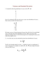
Figure 15.1. When free fusing these two images, look for a composite perception caused by the <strong>rivalry</strong> of<br />
dissimilar features.<br />
Figure 50.2. Free fuse the two left images and see if you notice that the center of the horizontal line is missing in<br />
the fused image.<br />
PRINCIPLES OF RIVALRY AND SUPPRESSION<br />
Suppression and <strong>rivalry</strong> are closely related. Rivalry suggests that, in the competition between the two images, one eye’s<br />
image may dominate part of the time (or in part of the visual field), while at other times the other dominates.<br />
Suppression is a long term dominance by one image over the other.<br />
91
Rivalry and <strong>suppression</strong> are not usually noticed in normal binocular viewing conditions, but these can become clinical<br />
problems in some circumstances. In an acute oculomo<strong>to</strong>r nerve paresis, the foveas may be pointing in different<br />
directions and receive completely different images. The patients will likely experience diplopia and con<strong>fusion</strong>. If the<br />
person cannot learn <strong>to</strong> reconcile the <strong>rivalry</strong> by suppressing either image, the only solution may be <strong>to</strong> occlude one eye.<br />
In a situation of binocular <strong>rivalry</strong>, which image, or which feature will be suppressed; that is, in the competition between<br />
two images, which one will win The manner in which the visual system deals with disparate retinal images depends,<br />
<strong>to</strong> some degree, on the nature of the images.<br />
Con<strong>to</strong>urs versus homogenous fields<br />
Con<strong>to</strong>urs in one image tend <strong>to</strong> dominate or suppress homogenous fields in the other image. This is illustrated by Fig.<br />
15.3. A black dot (con<strong>to</strong>ur) on a yellow field is presented <strong>to</strong> one eye, while the other eye sees a homogenous gray field.<br />
The binocular percept (right) is of the black dot, surrounded with a slightly brighter yellow corona on the<br />
yellow-gray field. The con<strong>to</strong>ur takes precedence over the empty field, and the corona shows that the zone of<br />
<strong>suppression</strong> extends beyond the con<strong>to</strong>ur itself.<br />
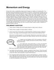
Figure 15.3. Con<strong>to</strong>urs tend <strong>to</strong> dominate homogenous field in binocular <strong>rivalry</strong>. When free fusing the two left frames, you should<br />
notice an image similar <strong>to</strong> that shown on the right.<br />
Similar, but unfusable images<br />
When two similar, though unfusable, con<strong>to</strong>urs are presented <strong>to</strong> either eye, the binocular percept includes a fused image<br />
with a combined local zone of <strong>suppression</strong> contributed by each image. If neither image dominates the other the other,<br />
the percept will combine parts from both. This is illustrated by Fig. 15.4.<br />
Figure 15.4 Rivalry between similar con<strong>to</strong>urs of equal dominance produces a composite with areas of local <strong>suppression</strong> from both<br />
images.<br />
Binocular luster and color <strong>fusion</strong><br />
92
When two identical images have opposite black versus white con<strong>to</strong>urs, the fused image contains a combination of both<br />
percepts, such that the object appears in stereoscopic depth and the dark background displays luster or a shiny<br />
appearance. This is illustrated by Fig. 15.5 and Steinman Fig. 5-9, 10. Steinman says that,<br />
It has been proposed that the shimmering appearance of luster mimics the shiny appearance<br />
of chrome because when viewing chrome, one eye tends <strong>to</strong> receive a bright reflection off the<br />
metal surface while the other does not, yielding the same type of luminance based <strong>rivalry</strong>.<br />
(p. 134)<br />
Figure 15.5. Images designed <strong>to</strong> illustrate luster, which results from the binocular <strong>rivalry</strong> between white and black con<strong>to</strong>urs.<br />
When different colors are presented <strong>to</strong> each eye, the result may be binocular color <strong>fusion</strong>—the color appears <strong>to</strong> be a<br />
mixture of the monocular hues. This delicate percept may be difficult <strong>to</strong> maintain, and the binocular image may<br />
alternate between the two original colors, or portions of the binocular image may contain a mixed color, while other<br />
portions may contain one color or the other. This may be tested using the yellow-green box (left) and the red box (right)<br />
in Fig. 15.6. The same effect can be seen by viewing a white field with anaglyph glasses.<br />
Figure 15.6. Free <strong>fusion</strong> color <strong>rivalry</strong> targets designed <strong>to</strong> demonstrate binocular color <strong>fusion</strong>.<br />
Suppression can be complicated, depending on the nature of the stimulus presented <strong>to</strong> each eye and the way the images<br />
are processed by the visual system. In general, the following trends are expected:<br />
• Bright features tend <strong>to</strong> suppress darker features<br />
• High contrast features tend <strong>to</strong> suppress low contrast features<br />
• Clear images tend <strong>to</strong> suppress blurry images<br />
• Foveal images tend <strong>to</strong> suppress peripheral images<br />
• Moving images tend <strong>to</strong> suppress stationary images<br />
• Images on the nasal retina of one eye tend <strong>to</strong> suppress images on the temporal retina of the other eye. (See<br />
Steinman Fig. 5-11.)<br />
93
CLINICAL MEASUREMENT OF SUPPRESSION<br />
Occasionally you may encounter <strong>suppression</strong> during a routine eye examination. During the von Graffe phoria test or<br />
when testing the BI and BO <strong>fusion</strong>al reserves, you expect the patient <strong>to</strong> see diplopia. If, during BI or BO testing, the<br />
patient never sees the break, but says that the chart appears <strong>to</strong> be moving <strong>to</strong> one side, you know that they are<br />
suppressing one eye.<br />
Some patients may be unable <strong>to</strong> do a von Graffe test, because they never see double, and the target may always appear<br />
<strong>to</strong> be moving <strong>to</strong> the side as prism is added. If they see single, but the target moves with increasing prism, you know that<br />
they are seeing with only one eye. You may then want <strong>to</strong> test for <strong>suppression</strong>. The Stereo Fly test includes a<br />
<strong>suppression</strong> check. Another simple test for <strong>suppression</strong> (as well as diplopia or <strong>fusion</strong>) is the Worth Four Dot test.<br />
It is possible that some patient may suppress one eye only during artificial viewing conditions (such as when looking<br />
through a phoropter in a dark room), but not during natural conditions. Although neither eye is suppressed most of the<br />
time, a person with a fragile binocular system may tend <strong>to</strong> suppress one eye some of the time. This may lead <strong>to</strong> more<br />
permanent <strong>suppression</strong>, therefore it is useful <strong>to</strong> have some clinical test <strong>to</strong> grade <strong>suppression</strong> proneness in the two eyes.<br />
One technique illustrated by Figure 3-8 in Reading (p. 35) uses Bagolini lenses and a set of neutral density filters. The<br />
Bagolini lenses have fine, barely visible striations, oriented at 135 degrees before the right eye and 45 degrees before<br />
the left eye. During normal binocular <strong>fusion</strong> the patient wearing the lenses will see a cross, while viewing a point<br />
source of light. If either eye is suppresses, only a single line will be seen. Also see Steinman Fig. 5-15.<br />
If asymmetric <strong>suppression</strong> proneness is suspected, it can be graded by the following procedure. Start with normal<br />
<strong>fusion</strong> and the binocular perception of a cross. Gradually add neutral density lenses before one eye until it is<br />
suppressed. For example, the right eye image may be suppressed when a 2.5 ND filter is used. Repeat the procedure<br />
for the other eye. For example, the left eye may be suppressed with the addition of a 1.0 ND filter. Such an asymmetric<br />
result indicates that the left eye is more prone <strong>to</strong> <strong>suppression</strong>.<br />
You could also test <strong>suppression</strong> proneness by increasing dioptric blur before either eye, though this usually yields more<br />
variable and less reliable results than if you use ND filters.<br />
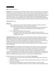
Figure 15.7. Bagolini lenses may be used <strong>to</strong> grade <strong>suppression</strong>. The binocularly fused view is shown on the right.<br />
SUPPRESSION FIELDS AND STEREOCAMPIMETRY<br />
When one eye is suppressed, usually only certain portions of its visual field are suppressed. For example, in order <strong>to</strong><br />
eliminate con<strong>fusion</strong> caused by different images on the two foveas, the area near the fovea of one eye may be<br />
94
suppressed. In addition, <strong>to</strong> eliminate diplopia, the retinal location receiving the diplopic image may also be suppressed.<br />
This is illustrated by Fig. 15.8, below, redrawn after Reading Fig. 3-9.<br />
Fixation<br />
OD <strong>suppression</strong> zone<br />
f<br />
f<br />
f<br />
Figure 15.9. When OS fixates on the dot, the star falls on the fovea of the esotropic OD. This leads <strong>to</strong> con<strong>fusion</strong><br />
and diplopia, which is solved by <strong>suppression</strong> a narrow zone on the OD retina.<br />
It is possible <strong>to</strong> measure the extent of local <strong>suppression</strong> in the visual field of one eye using a technique known as<br />
stereocampimetry. Figure 15.10 shows how it is possible <strong>to</strong> plot the zone of <strong>suppression</strong> in the deviating eye. In this<br />
example, the normal right eye observes a fixation target in a mirror separately from the deviating left eye. Test stimuli<br />
are presented <strong>to</strong> the left eye, which is pointed <strong>to</strong>ward a tangent screen, and the examiner tests for areas of <strong>suppression</strong>.<br />
These will appear <strong>to</strong> be sco<strong>to</strong>mas in that monocular visual field.<br />
Figure 50.8. Schematic diagram of one type of stereocampimeter. (Redrawn from Reading Fig. 3-10.)<br />
95

![Graduate Bulletin [PDF] - MFC home page - Appalachian State ...](https://img.yumpu.com/50706615/1/190x245/graduate-bulletin-pdf-mfc-home-page-appalachian-state-.jpg?quality=85)