Sonohysterography - AIUM
Sonohysterography - AIUM
Sonohysterography - AIUM

Create successful ePaper yourself
Turn your PDF publications into a flip-book with our unique Google optimized e-Paper software.
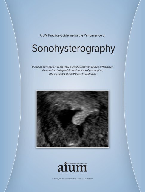
<strong>AIUM</strong> Practice Guideline for the Performance of<br />
<strong>Sonohysterography</strong><br />
Guideline developed in collaboration with the American College of Radiology,<br />
the American College of Obstetricians and Gynecologists,<br />
and the Society of Radiologists in Ultrasound<br />
© 2011 by the American Institute of Ultrasound in Medicine
The American Institute of Ultrasound in Medicine (<strong>AIUM</strong>) is a multidisciplinary<br />
association dedicated to advancing the safe and effective use<br />
of ultrasound in medicine through professional and public education,<br />
research, development of guidelines, and accreditation. To promote<br />
this mission, the <strong>AIUM</strong> is pleased to publish, in conjunction with<br />
the American College of Radiology (ACR), the American College<br />
of Obstetricians and Gynecologists (ACOG), and the Society of<br />
Radiologists in Ultrasound (SRU), this <strong>AIUM</strong> Practice Guideline for the<br />
Performance of <strong>Sonohysterography</strong>. We are indebted to the many<br />
volunteers who contributed their time, knowledge, and energy to bringing<br />
this document to completion.<br />
The <strong>AIUM</strong> represents the entire range of clinical and basic science<br />
interests in medical diagnostic ultrasound, and, with hundreds of<br />
volunteers, the <strong>AIUM</strong> has promoted the safe and effective use<br />
of ultrasound in clinical medicine for more than 50 years. This document<br />
and others like it will continue to advance this mission.<br />
Practice guidelines of the <strong>AIUM</strong> are intended to provide the medical<br />
ultrasound community with guidelines for the performance and recording<br />
of high-quality ultrasound examinations. The guidelines reflect<br />
what the <strong>AIUM</strong> considers the minimum criteria for a complete examination<br />
in each area but are not intended to establish a legal standard of<br />
care. <strong>AIUM</strong>-accredited practices are expected to generally follow the<br />
guidelines with recognition that deviations from these guidelines will<br />
be needed in some cases, depending on patient needs and available<br />
equipment. Practices are encouraged to go beyond the guidelines to<br />
provide additional service and information as needed.<br />
14750 Sweitzer Ln, Suite 100<br />
Laurel, MD 20707-5906 USA<br />
800-638-5352 • 301-498-4100<br />
www.aium.org<br />
Original copyright 2002; revised 2007, 2011—<strong>AIUM</strong> PRACTICE GUIDELINES—<strong>Sonohysterography</strong>
Effective April 18, 2011—<strong>AIUM</strong> PRACTICE GUIDELINES—<strong>Sonohysterography</strong><br />
I. Introduction<br />
The clinical aspects contained in specific sections of this guideline (Introduction, Indications<br />
and Contraindications, Specifications for Individual Examinations, and Equipment<br />
Specifications) were developed collaboratively by the American Institute of Ultrasound in<br />
Medicine (<strong>AIUM</strong>), the American College of Radiology (ACR), the American College of<br />
Obstetricians and Gynecologists (ACOG), and the Society of Radiologists in Ultrasound<br />
(SRU). Recommendations for physician qualifications, written request for the examination,<br />
procedure documentation, and quality control may vary among the 4 organizations and are<br />
addressed by each separately.<br />
This guideline has been developed to assist qualified physicians performing sonohysterography.<br />
Properly performed sonohysterography can provide information about the uterus,<br />
endometrium, and fallopian tubes. Additional studies may be necessary for complete<br />
diagnosis. Adherence to the following guideline will maximize the diagnostic benefit of sonohysterography.<br />
<strong>Sonohysterography</strong> is the evaluation of the endometrial cavity using the transcervical<br />
injection of sterile fluid. Various terms such as saline infusion sonohysterography and simply<br />
sonohysterography have been used to describe this technique. The primary goal of sonohysterography<br />
is to visualize the endometrial cavity in more detail than is possible with routine<br />
endovaginal sonography. 1 <strong>Sonohysterography</strong> can also be used to assess tubal patency. 3<br />
1<br />
II. Indications and Contraindications<br />
A. Indications 3–11<br />
Indications include but are not limited to evaluation of:<br />
1. Abnormal uterine bleeding;<br />
2. Uterine cavity, especially with regard to uterine myomas, polyps, and synechiae;<br />
3. Abnormalities detected on endovaginal sonography, including focal or diffuse<br />
endometrial or intracavitary abnormalities;<br />
4. Congenital abnormalities of the uterus;<br />
5. Infertility; and<br />
6. Recurrent pregnancy loss.
Effective April 18, 2011—<strong>AIUM</strong> PRACTICE GUIDELINES—<strong>Sonohysterography</strong><br />
B. Contraindications<br />
<strong>Sonohysterography</strong> should not be performed in a woman who is pregnant or who could be<br />
pregnant. This is usually avoided by scheduling the examination in the follicular phase of<br />
the menstrual cycle, after menstrual flow has essentially ceased but before the patient has<br />
ovulated. In a patient with regular cycles, sonohysterography should not in most cases be<br />
performed later than the 10th day of the menstrual cycle. <strong>Sonohysterography</strong> should not<br />
be performed in patients with a pelvic infection or unexplained pelvic tenderness, which<br />
could be due to pelvic inflammatory disease. Active vaginal bleeding is not a contraindication<br />
to the procedure but may make the interpretation more challenging.<br />
III. Qualifications and Responsibilities of the Physician<br />
See www.aium.org for <strong>AIUM</strong> Official Statements including Standards and Guidelines for the<br />
Accreditation of Ultrasound Practices and relevant Physician Training Guidelines.<br />
2<br />
IV. Written Request for the Examination<br />
The written or electronic request for an ultrasound examination should provide sufficient<br />
information to allow for the appropriate performance and interpretation of the examination.<br />
The request for the examination must be originated by a physician or other appropriately<br />
licensed health care provider or under the provider’s direction. The accompanying clinical<br />
information should be provided by a physician or other appropriate health care provider familiar<br />
with the patient’s clinical situation and should be consistent with relevant legal and local<br />
health care facility requirements.<br />
V. Specifications for Individual Examinations<br />
A. Patient Preparation<br />
Pelvic organ tenderness should be assessed during the preliminary endovaginal sonogram.<br />
If adnexal tenderness or pain suspicious for an active pelvic infection is found before fluid infusion,<br />
the examination should be deferred until after an appropriate course of treatment. In the<br />
presence of nontender hydrosalpinges, consideration may be given to administering antibiotics<br />
at the time of the examination; in this case, it is prudent to discuss the antibiotic regimen<br />
with the referring physician. A pregnancy test is advised when clinically indicated. Patients<br />
should be questioned about a latex allergy before use of a latex sheath. The optimal time to<br />
perform this test in a menstruating woman is after the bleeding ends but before ovulation.
Effective April 18, 2011—<strong>AIUM</strong> PRACTICE GUIDELINES—<strong>Sonohysterography</strong><br />
B. Procedure<br />
Preliminary endovaginal sonography with measurements of the endometrium and evaluation<br />
of the uterus, ovaries, and pelvic free fluid should be performed before sonohysterography.<br />
A speculum is used to allow visualization of the cervix. The presence of unusual pain, lesions,<br />
or purulent vaginal or cervical discharge may require rescheduling the procedure pending<br />
further evaluation. Before insertion, the catheter should be flushed with sterile fluid to avoid<br />
introducing air during the study. After cleansing the external os, the cervical canal and/or uterine<br />
cavity should be catheterized using aseptic technique, and appropriate sterile fluid should<br />
be instilled slowly by means of manual injection under real-time sonographic imaging. Imaging<br />
should include real-time scanning of the endometrial and cervical canal. 12<br />
C. Contrast Agent<br />
Appropriate sterile fluid such as normal saline or water should be used for sonohysterography.<br />
D. Images<br />
Precatheterization images should be obtained and recorded, in at least 2 planes, to show<br />
normal and abnormal findings. These images should include the thickest bilayer endometrial<br />
measurement on a sagittal image when possible.<br />
Once the uterine cavity is filled with fluid, a complete survey of the uterine cavity should be<br />
performed and representative images obtained to document normal and abnormal findings.<br />
If a balloon catheter is used for the examination, images should be obtained at the end of the<br />
procedure with the balloon deflated to fully evaluate the endometrial cavity, particularly the<br />
cervical canal and lower portion of the endometrial cavity.<br />
3<br />
Additional techniques such as color Doppler and 3-dimensional imaging may be helpful in<br />
evaluating both normal and abnormal findings. 13,14<br />
VI. Documentation<br />
Adequate documentation is essential for high-quality patient care. There should be a permanent<br />
record of the ultrasound examination and its interpretation. Images of all appropriate<br />
areas, both normal and abnormal, should be recorded. Variations from normal size should be<br />
accompanied by measurements. Images should be labeled with the patient identification, facility<br />
identification, examination date, and side (right or left) of the anatomic site imaged.<br />
An official interpretation (final report) of the ultrasound findings should be included in the<br />
patient’s medical record. Retention of the ultrasound examination should be consistent both<br />
with clinical needs and with relevant legal and local health care facility requirements.<br />
Reporting should be in accordance with the <strong>AIUM</strong> Practice Guideline for Documentation of an<br />
Ultrasound Examination.
Effective April 18, 2011—<strong>AIUM</strong> PRACTICE GUIDELINES—<strong>Sonohysterography</strong><br />
VII. Equipment Specifications<br />
<strong>Sonohysterography</strong> is usually conducted with a high-frequency endovaginal transducer. In cases<br />
of an enlarged uterus, additional transabdominal images during infusion may be required to fully<br />
evaluate the endometrium. The transducer should be adjusted to operate at the highest<br />
clinically appropriate frequency under the ALARA (as low as reasonably achievable) principle.<br />
VIII. Quality Control and Improvement, Safety, Infection Control,<br />
and Patient Education<br />
Policies and procedures related to quality control, patient education, infection control, and<br />
safety should be developed and implemented in accordance with the <strong>AIUM</strong> Standards and<br />
Guidelines for the Accreditation of Ultrasound Practices.<br />
Equipment performance monitoring should be in accordance with the <strong>AIUM</strong> Standards and<br />
Guidelines for the Accreditation of Ultrasound Practices.<br />
4<br />
IX. ALARA Principle<br />
The potential benefits and risks of each examination should be considered. The ALARA<br />
(as low as reasonably achievable) principle should be observed when adjusting controls that<br />
affect the acoustic output and by considering transducer dwell times. Further details on<br />
ALARA may be found in the <strong>AIUM</strong> publication Medical Ultrasound Safety, Second Edition.<br />
Acknowledgments<br />
This guideline was revised by the American Institute of Ultrasound in Medicine (<strong>AIUM</strong>)<br />
in collaboration with the American College of Obstetricians and Gynecologists (ACOG),<br />
the American College of Radiology (ACR) and the Society of Radiologists in Ultrasound<br />
(SRU) according to the process described in the <strong>AIUM</strong> Clinical Standards Committee<br />
Manual.<br />
Collaborative Committee<br />
Members represent their societies in the initial draft and final revision of this guideline.<br />
<strong>AIUM</strong><br />
Mert Bahtiyar, MD<br />
Kevin J. Doody, MD<br />
Daniel Skupski, MD<br />
Brad Van Voorhis, MD
Effective April 18, 2011—<strong>AIUM</strong> PRACTICE GUIDELINES—<strong>Sonohysterography</strong><br />
ACR<br />
Marcela Bohm-Velez, MD, Chair<br />
Debra L. Acord, MD<br />
Helena Gabriel, MD<br />
Ruth B. Goldstein, MD<br />
ACOG<br />
Daniel Breitkopf, MD<br />
Steven R. Goldstein, MD<br />
SRU<br />
Robert L. Bree, MD<br />
Theodore Dubinsky, MD<br />
Faye C. Laing, MD<br />
<strong>AIUM</strong> Clinical Standards Committee<br />
David Paushter, MD, Chair<br />
Leslie Scoutt, MD, Vice Chair<br />
Lisa Allen, BS, RDMS, RDCS, RVT<br />
Mert Bahtiyar, MD<br />
Harris L. Cohen, MD<br />
Lin Diacon, MD, RDMS, RPVI<br />
Judy Estroff, MD<br />
J. Christian Fox, MD, RDMS<br />
Charlotte Henningsen, MS, RT, RDMS, RVT<br />
Adam Hiett, MD, RDMS<br />
Lars Jensen, MD<br />
Christopher Moore, MD, RDMS, RDCS<br />
Steven Perlmutter, MD<br />
Olga Rasmussen, RDMS<br />
Carl Reading, MD<br />
Shia Salem, MD<br />
Daniel Skupski, MD<br />
Jay Smith, MD<br />
Lami Yeo, MD<br />
5
Effective April 18, 2011—<strong>AIUM</strong> PRACTICE GUIDELINES—<strong>Sonohysterography</strong><br />
6<br />
References<br />
1. Bree RL, Bowerman RA, Bohm-Velez M, et al. US evaluation of the uterus in patients with postmenopausal<br />
bleeding: a positive effect on diagnostic decision making. Radiology 2000; 216:260–264.<br />
2. Hajishafiha M, Zobairi T, Zanjani VR, Ghasemi-Rad M, Yekta Z, Mladkova N. Diagnostic value of<br />
sonohysterography in the determination of fallopian tube patency as an initial step of routine<br />
infertility assessment. J Ultrasound Med 2009; 28:1671–1677.<br />
3. Becker E Jr, Lev-Toaff AS, Kaufman EP, Halpern EJ, Edelweiss MI, Kurtz AB. The added value of<br />
transvaginal sonohysterography over transvaginal sonography alone in women with known or<br />
suspected leiomyoma. J Ultrasound Med 2002; 21:237–247.<br />
4. Breitkopf DM, Frederickson RA, Snyder RR. Detection of benign endometrial masses by endometrial<br />
stripe measurement in premenopausal women. Obstet Gynecol 2004; 104:120–125.<br />
5. Doubilet PM. Society of Radiologists in Ultrasound Consensus Conference statement on postmenopausal<br />
bleeding. J Ultrasound Med 2001; 20:1037–1042.<br />
6. Dubinsky TJ, Stroehlein K, Abu-Ghazzeh Y, Parvey HR, Maklad N. Prediction of benign and malignant<br />
endometrial disease: hysterosonographic-pathologic correlation. Radiology 1999; 210:393–397.<br />
7. Goldstein RB, Bree RL, Benson CB, et al. Evaluation of the woman with postmenopausal bleeding:<br />
Society of Radiologists in Ultrasound-Sponsored Consensus Conference statement. J Ultrasound<br />
Med 2001; 20:1025–1036.<br />
8. Goldstein SR. Use of ultrasonohysterography for triage of perimenopausal patients with unexplained<br />
uterine bleeding. Am J Obstet Gynecol 1994; 170:565–570.<br />
9. Laifer-Narin S, Ragavendra N, Parmenter EK, Grant EG. False-normal appearance of the endometrium<br />
on conventional transvaginal sonography: comparison with saline hysterosonography. AJR Am<br />
J Roentgenol 2002; 178:129–133.<br />
10. Laifer-Narin SL, Ragavendra N, Lu DS, Sayre J, Perrella RR, Grant EG. Transvaginal saline<br />
hysterosonography: characteristics distinguishing malignant and various benign conditions.<br />
AJR Am J Roentgenol 1999; 172:1513–1520.<br />
11. Mihm LM, Quick VA, Brumfield JA, Connors AF Jr, Finnerty JJ. The accuracy of endometrial biopsy<br />
and saline sonohysterography in the determination of the cause of abnormal uterine bleeding.<br />
Am J Obstet Gynecol 2002; 186:858–860.<br />
12. Lindheim SR, Sprague C, Winter TC III. Hysterosalpingography and sonohysterography: lessons in<br />
technique. AJR Am J Roentgenol 2006; 186:24–29.<br />
13. Benacerraf BR, Shipp TD, Bromley B. Improving the efficiency of gynecologic sonography with<br />
3-dimensional volumes: a pilot study. J Ultrasound Med 2006; 25:165–171.<br />
14. Ghate SV, Crockett MM, Boyd BK, Paulson EK. <strong>Sonohysterography</strong>: do 3D reconstructed images<br />
provide additional value AJR Am J Roentgenol 2008; 190:W227–W233.