Vol 3 - Cont J Pharm Sci - Wilolud Journals
Vol 3 - Cont J Pharm Sci - Wilolud Journals
Vol 3 - Cont J Pharm Sci - Wilolud Journals

You also want an ePaper? Increase the reach of your titles
YUMPU automatically turns print PDFs into web optimized ePapers that Google loves.
A. K. Jain et al: <strong>Cont</strong>inental J. <strong>Pharm</strong>aceutical <strong>Sci</strong>ences 3: 1 - 6, 2009<br />
allow the volatile solvent evaporate. The prepared microspheres were filtered, washed with distilled water and dried<br />
in vacuum.<br />
Characterization of microspheres<br />
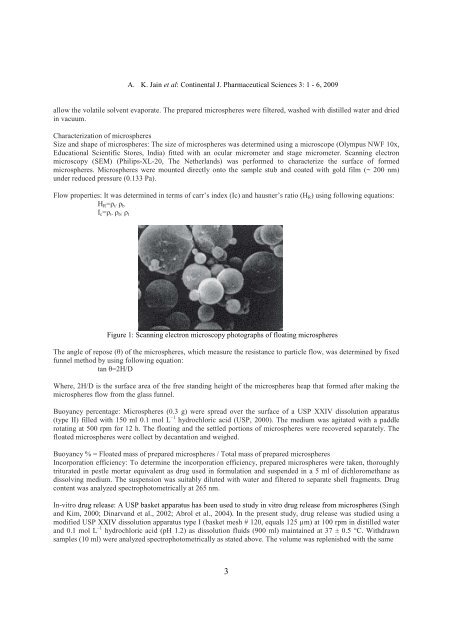
Size and shape of microspheres: The size of microspheres was determined using a microscope (Olympus NWF 10x,<br />
Educational <strong>Sci</strong>entific Stores, India) fitted with an ocular micrometer and stage micrometer. Scanning electron<br />
microscopy (SEM) (Philips-XL-20, The Netherlands) was performed to characterize the surface of formed<br />
microspheres. Microspheres were mounted directly onto the sample stub and coated with gold film (~ 200 nm)<br />
under reduced pressure (0.133 Pa).<br />
Flow properties: It was determined in terms of carr’s index (Ic) and hausner’s ratio (H R ) using following equations:<br />
H R =ρ t/ ρ b<br />
I c =ρ t- ρ b/ ρ t<br />
Figure 1: Scanning electron microscopy photographs of floating microspheres<br />
The angle of repose (θ) of the microspheres, which measure the resistance to particle flow, was determined by fixed<br />
funnel method by using following equation:<br />
tan θ=2H/D<br />
Where, 2H/D is the surface area of the free standing height of the microspheres heap that formed after making the<br />
microspheres flow from the glass funnel.<br />
Buoyancy percentage: Microspheres (0.3 g) were spread over the surface of a USP XXIV dissolution apparatus<br />
(type II) filled with 150 ml 0.1 mol L –1 hydrochloric acid (USP, 2000). The medium was agitated with a paddle<br />
rotating at 500 rpm for 12 h. The floating and the settled portions of microspheres were recovered separately. The<br />
floated microspheres were collect by decantation and weighed.<br />
Buoyancy % = Floated mass of prepared microspheres / Total mass of prepared microspheres<br />
Incorporation efficiency: To determine the incorporation efficiency, prepared microspheres were taken, thoroughly<br />
triturated in pestle mortar equivalent as drug used in formulation and suspended in a 5 ml of dichloromethane as<br />
dissolving medium. The suspension was suitably diluted with water and filtered to separate shell fragments. Drug<br />
content was analyzed spectrophotometrically at 265 nm.<br />
In-vitro drug release: A USP basket apparatus has been used to study in vitro drug release from microspheres (Singh<br />
and Kim, 2000; Dinarvand et al., 2002; Abrol et al., 2004). In the present study, drug release was studied using a<br />
modified USP XXIV dissolution apparatus type I (basket mesh # 120, equals 125 µm) at 100 rpm in distilled water<br />
and 0.1 mol L –1 hydrochloric acid (pH 1.2) as dissolution fluids (900 ml) maintained at 37 ± 0.5 °C. Withdrawn<br />
samples (10 ml) were analyzed spectrophotometrically as stated above. The volume was replenished with the same<br />
3