Vol 3 - Cont J Pharm Sci - Wilolud Journals
Vol 3 - Cont J Pharm Sci - Wilolud Journals
Vol 3 - Cont J Pharm Sci - Wilolud Journals

You also want an ePaper? Increase the reach of your titles
YUMPU automatically turns print PDFs into web optimized ePapers that Google loves.
A. K. Jain et al: <strong>Cont</strong>inental J. <strong>Pharm</strong>aceutical <strong>Sci</strong>ences 3: 1 - 6, 2009<br />
amount of fresh dissolution fluid each time to maintain the sink condition. All experiments were performed in<br />
triplicate. Linear regression was used to analyze the in vitro release mechanism.<br />
Statistical analysis: Experimental results were expressed as mean ± SD. Student’s t-test and one-way analysis of<br />
variance (ANOVA) were applied to check significant differences in drug release from different formulations.<br />
Differences were considered to be statistically significant at P< 0.05.<br />
RESULTS AND DISCUSSION<br />
Floating microspheres were prepared by the solvent evaporation method by using acrycoat S100 and chitosan (Table<br />
1). The SEM photographs showed that the fabricated microspheres were spherical with a smooth surface and<br />
exhibited a range of sizes within each batch (Fig. 1). The microspheres floated for prolonged time over the surface<br />
of the dissolution medium without any apparent gelation. Buoyancy percentage of the microspheres was shown in<br />
Table 2.<br />
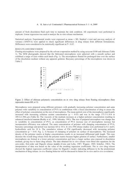
Figure 2: Effect of chitosan polymeric concentration on in vitro drug release from floating microspheres (bars<br />
represents mean±SD; n=3)<br />
Microspheres were prepared using different polymers with gradually increasing polymer concentration and same<br />
polymer with variability in concentration of PVA in combination with a fixed concentration of drug to assess the<br />
effect of polymer concentration on the size of microspheres. The mean particle size of the microspheres significantly<br />
increased with increasing cellulose acetate concentration (p < 0.05) and was in the range 375.1±1.818 to<br />
389.3±1.984 µm (Table II). The viscosity of the medium increases at a higher polymer concentration resulting in<br />
enhanced interfacial tension (Reddy et al., 1990; Ishizaka, 1981). The size of prepared microsphere was change due<br />
to variability in concentration of PVA, as concentration of PVA increase size of microspheres increase but<br />
incorporation efficiency was reduced. The same concentration of polymer with changing concentration of PVA,<br />
buoyancy percentage and period was increase till to 18 h. In vitro FM release studies were performed in 0.1 mol/L<br />
hydrochloric acid for 18 h. The cumulative release of FM significantly decreased with increasing polymer<br />
concentration (p < 0.05, Fig. 2, 3) because of clumping of polymer on surface of microspheres. The increased<br />
density of the polymer matrix at higher concentrations results in an increased diffusional path length. This may<br />
decrease the overall drug release from the polymer matrix because higher gelation of surface. Furthermore, smaller<br />
microspheres are formed at a lower polymer concentration and have a larger surface area exposed to dissolution<br />
medium, giving rise to faster drug release. The data obtained for in vitro release were fitted into equations for the<br />
zero-order, first-order and Higuchi release models (Costa and Lobo, 1987; Wagner, 1969; Schefter, 1963). The<br />
interpretation of data was based on the value of the resulting regression coefficients. The in vitro drug release<br />
showed the highest regression coefficient values for Higuchi’s model, indicating diffusion to be the predominant<br />
mechanism of drug release. in-vivo floating behavior will be discussed in future for in-vitro and in-vivo co-relation.<br />
4