January 2008 CDA Journal - Sandra Kalil Bussadori
January 2008 CDA Journal - Sandra Kalil Bussadori
January 2008 CDA Journal - Sandra Kalil Bussadori

Create successful ePaper yourself
Turn your PDF publications into a flip-book with our unique Google optimized e-Paper software.
pulp response<br />
cda journal, vol 36, nº 1<br />
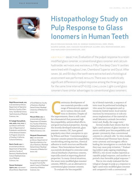
Histopathology Study on<br />
Pulp Response to Glass<br />
Ionomers in Human Teeth<br />
majid mousavinasab, dds; m. sadegh namazikhah, dmd, msed;<br />
nasrin sarabi, dds; hassan hosienpour jajarm, dds; maryam bidar, dds;<br />
and marjaneh ghavamnasiri, dds<br />
abstract objective: Evaluation of the pulpal response to a resinmodified<br />
glass ionomer, a conventional glass ionomer and calcium<br />
hydroxide. methods and materials: Fifty-five deep Class V cavities<br />
were lined with Vivaglass Liner, Chembond Superior and Dycal. After<br />
seven, 30, and 60 days the teeth were extracted and a histological<br />
assessment was performed. results: There was no statistically<br />
significant difference in pulpal response among the three groups<br />
for the same time interval (P>0.05). conclusion: Light-cured glass<br />
ionomers have similar advantages to conventional glass ionomers.<br />
authors<br />
Majid Mousavinasab, dds,<br />
is an assistant professor,<br />
Department of Operative<br />
Dentistry, Faculty of Dentistry,<br />
Yazd Dental School,<br />
Yazd University of Medical<br />
Sciences, Iran.<br />
M. Sadegh Namazikhah,<br />
dmd, msed, ficd, facd, is<br />
a professor and former<br />
chair of Department of<br />
Endodontics and director<br />
of the Advance Endodontic<br />
Program, University<br />
of Southern California,<br />
School of Dentistry.<br />
Nasrin Sarabi, dds, is<br />
an assistant professor,<br />
Department of Operative<br />
dentistry, Faculty of<br />
Dentistry, Mashhad Dental<br />
School, Mashhad University<br />
of Medical<br />
Sciences, Iran.<br />
Hassan Hosienpour<br />
Jajarm, dds, is an associate<br />
professor, Department<br />
of Oral Medicine, Faculty<br />
of Dentistry, Mashhad<br />
Dental School, Mashhad<br />
University of Medical Sciences,<br />
Iran.<br />
Maryam Bidar, dds, is<br />
an associate professor,<br />
Department of Endodontics,<br />
Faculty of Dentistry,<br />
Mashhad Dental School,<br />
Mashhad University of<br />
Medical Sciences, Iran.<br />
Marjaneh Ghavamnasiri,<br />
dds, is an associate<br />
professor, Department of<br />
Operative dentistry, Faculty<br />
of Dentistry, Mashhad<br />
Dental School, Mashhad<br />
University of Medical Sciences,<br />
Iran.<br />
acknowledgment<br />
This study was supported<br />
by a grant from the Research<br />
Council of Mashhad<br />
University of Medical<br />
Sciences, Iran.<br />
Continuous development of<br />
new materials provides a wide<br />
range of biomaterials appropriate<br />
to various clinical conditions<br />
in dentistry. Despite all<br />
the improvements, there is still a need<br />
for a biomaterial that possesses high<br />
biocompatibility, antimicrobial effects,<br />
and good mechanical properties. Among<br />
the recently developed materials, glass<br />
ionomer cements, GIC, have gained<br />
popularity since their conception in 1972<br />
by Wilson and Kent. 1 Conventional glass<br />
ionomer cements present biocompatibility,<br />
nonshrinking setting reaction, chemical<br />
adhesion to tooth structure and fluoride<br />
release. 2 New formulations have been<br />
successively developed to overcome some<br />
clinical drawbacks of previous cements,<br />
especially aimed at improving physical<br />
properties. 3 In many clinical situations,<br />
the newer resin-modified glass ionomer<br />
cements, RMGICs, are an alternative to<br />
conventional glass ionomer cements.<br />
To evaluate the biocompatibility<br />
of dental materials, a sequence of<br />
tests must be performed including in<br />
vitro assay for mutagenesis and cytotoxicity<br />
(initial tests), local toxicity<br />
reactions by intraosseous or subcutaneous<br />
implantation of the material in<br />
small laboratory animals (secondary<br />
tests) and, finally, the usage tests. 4<br />
Several studies have shown that the<br />
light-cured systems of glass ionomer cements<br />
exhibit poor biocompatibility and<br />
greater cytotoxicity than conventional<br />
cements in cultured cells. 5 In vitro studies<br />
of Vitrebond and Vitremer have shown<br />
some cytotoxic and mutagenic effects<br />
leading the investigators to conclude that<br />
they may cause pulp irritation. 5,6 Evaluating<br />
indirect pulp capping employing a<br />
RMGICs, two recent studies reported acceptable<br />
pulpal response, and another reported<br />
a less favorable pulpal response. 7-9<br />
This in vivo study evaluated the histological<br />
changes in pulp as a response to<br />
light-cured resin-modified glass ionomer<br />
and compared it with a conventional<br />
january <strong>2008</strong> 51
pulp response<br />
cda journal, vol 36, nº 1<br />
table 1<br />
Evaluation Criteria 11<br />
glass ionomer and a calcium hydroxide<br />
lining material in deep cavities.<br />
Methods and Materials<br />
The study population consisted of 19<br />
females and 12 males, ranging in age from<br />
13 to 32, with a mean age of 18 years. All<br />
of the patients required the extraction<br />
of permanent premolars for orthodontic<br />
reasons. The participants, and their<br />
parents or responsible persons, received<br />
an adequate explanation concerning the<br />
experimental rationale, clinical procedure,<br />
and possible risks. The parents and all the<br />
volunteers were asked to read and sign a<br />
consent form explaining the research protocol<br />
approved by the ethical guidelines.<br />
Patients were required to meet the<br />
following criteria.<br />
To be included in the study:<br />
n Permanent first premolars scheduled<br />
for orthodontic extraction<br />
n Scores of two or less using the<br />
periodontal screening record (evaluation<br />
consisted of examining the premolars<br />
with a periodontal probe)<br />
n Completed root formation<br />
To be excluded from the study:<br />
n Presence of caries<br />
n Presence of restorations<br />
n Presence of abrasion or erosion<br />
n Presence of pulpal symptoms<br />
or radiographic periapical lesions<br />
After local anesthesia the teeth were<br />
isolated with a rubber dam. A Class V<br />
cavity on the buccal surface of each tooth<br />
was prepared with a 440-diamond point<br />
(Shofu Inc, Kyoto 605-0983, Japan) in<br />
a high-speed handpiece under copious<br />
water spray coolant. New diamond points<br />
and burs were used after every four<br />
teeth. The axial wall was excavated using<br />
a carbide round bur at low speed until<br />
red feature of the pulp was observed.<br />
The 55 experimental teeth were<br />
divided into three groups. In the first<br />
Odontoblastic changes<br />
(Non) Remarkable change was not observed in the pulp<br />
(Slight) Disarrangement of odontoblasts was noted slightly below the cut dentinal tubules<br />
(Moderate) Disarrangement of odontoblasts was seen through most of the cut dentinal tubules<br />
(Severe) Disarrangement of odontoblasts was noted below the remaining dentin.<br />
Inflammatory cell infiltration<br />
(Non) None or a few inflammatory cells were observed through the pulp.<br />
(Slight) A few inflammatory cell infiltrations were noted below the cut dentinal tubules.<br />
(Moderate) Inflammatory cell remarkably observed below the remaining dentin.<br />
(Severe) Severe inflammatory cell infiltration was seen through the pulp.<br />
Reactionary dentin formation<br />
(Non) No abnormal or reparative dentin observed.<br />
(Slight) A small amount of reactionary dentin was noted.<br />
(Moderate) Reactionary dentin was observed below the almost-cut dentin.<br />
(Severe) Complete and large bulk of reactionary dentin was noted.<br />
group, Vivaglass Liner (Ivoclar Vivadent<br />
AG, Schaan, Lichtenstein) was applied<br />
to the axial wall of the cavity and then<br />
was light-cured for 20 seconds. In the<br />
second group, Chembond Superior<br />
(Dentsply, Detry, UK) was applied as<br />
a liner in the axial wall of the cavity;<br />
and in the third (control) group, Dycal<br />
(Dentsply, Milford, Del., USA) was<br />
applied. All of the materials were used<br />
according to manufacturer’s directions.<br />
After application, two layers of a copal<br />
varnish, Copalite (Cooley & Cooley LTD,<br />
Houston, Texas) were added. The cavities<br />
were restored with a high copper amalgam,<br />
Oralloy (Coltene Whaledent, USA).<br />
After seven, 30, and 60 days, the teeth<br />
were extracted under local anesthesia.<br />
The mesial and distal approximal<br />
surfaces of the teeth were reduced with<br />
a high-speed diamond bur under spray<br />
coolant until the pulp became almost<br />
visible through the remaining dentin to<br />
facilitate the penetration of the fixative<br />
solution. The surfaces were then fixed<br />
with a 10 percent neutral buffered formalin<br />
solution for one week. The teeth were<br />
demineralized with 10 percent ethylenediamine<br />
tetracetic acid (ETDA) with PH<br />
(7-7.4) as a demineralzing solution at 25<br />
degrees (Celsius) for 60 days, and each<br />
tooth was then embedded in paraffin.<br />
5µm-thick serial sections were prepared<br />
through the cavities and pulp, obtaining<br />
approximately 80 to 100 sections per cavity.<br />
They were placed on glass microslides<br />
and stained with either hematoxylin eosin<br />
for routine histological evaluation or<br />
Taylor’s modification of Gram’s staining<br />
technique for detecting microorganisms. 10<br />
The pulpal responses and the presence<br />
of bacteria in their cavities were<br />
evaluated using a light microscope (Zeiss,<br />
Germany). The RDT, remaining dentin<br />
thickness, was ranged as deep (0-0.4<br />
mm), moderate (0.4-0.7 mm), and shallow<br />
(more than 0.7 mm). Evaluation criteria<br />
for odontoblastic changes, inflammatory<br />
cell infiltration and reactionary<br />
dentin formation are shown in table 1. 11<br />
The results of odontoblastic changes, inflammatory<br />
cell infiltration, and reactionary<br />
dentin formation were statistically analyzed<br />
using the Kruskal Wallis and Mann-Whitney<br />
test at 95 percent level of confidence.<br />
Fisher’s Exact test (α = 0.05) was also used<br />
52 january <strong>2008</strong>
cda journal, vol 36, nº 1<br />
table 2<br />
Results of Histological Findings<br />
Time Intervals 7 days 30 days 60 days<br />
Experimental groups V C D V C D V C D<br />
Number of specimens 8 7 5 5 6 6 6 6 6<br />
Non 3 2 2 2 2 4 2 2 1<br />
Odontoblastic Slight 1 2 1 1 2 1 1 2 2<br />
changes Moderate 4 3 2 2 2 1 3 2 3<br />
Severe 0 0 0 0 0 0 0 0 0<br />
Non 0 2 2 3 2 5 2 2 3<br />
Inflammatory Slight 2 1 2 2 2 1 3 1 3<br />
cell infiltration Moderate 6 4 0 0 2 0 1 3 0<br />
Severe 0 0 1 0 0 0 0 0 0<br />
Non 8 7 5 2 5 3 2 2 1<br />
Reactionary Slight 0 0 0 3 1 3 4 4 5<br />
dentin Moderate 0 0 0 0 0 0 0 0 0<br />
formation Severe 0 0 0 0 0 0 0 0 0<br />
V: Vivaglass Liner; C: Chembond Superior; D: Dycal<br />
for understanding the correlation between<br />
pulpal responses with microorganisms and<br />
remaining dentin thickness in each group.<br />
Results<br />
Results of histological findings are<br />
shown in table 2.<br />
Bacterial penetration was observed<br />
in only six cases (five cases in cavity<br />
walls and only one case in pulp). There<br />
was no significant correlation between<br />
pulpal responses with dentinal thicknesses<br />
and microorganisms (P>0.05).<br />
In the Vivaglass Liner, there was a<br />
statistically significant difference in inflammatory<br />
cell response among three intervals<br />
(P
pulp response<br />
cda journal, vol 36, nº 1<br />
figure 1. Cavity preparation, remaining dentin<br />
thickness and pulp tissue. The odontoblast layer<br />
is disrupted and the cells were displaced into the<br />
dentinal tubules. Mild and scattered inflammatory<br />
cells are present. (Vivaglass Liner, seven<br />
days.) (H & E; 40X.)<br />
figure 3. A sample of Chembond Superior,<br />
seven days. Remnant of liner (L) and remaining<br />
dentin thickness (D). Odontoblast layer is<br />
disrupted. (H & E; 40X.)<br />
though pulpal responses in the same time<br />
intervals did not differ significantly among<br />
materials, inflammatory cell response in<br />
Vivaglass Liner after seven days was significantly<br />
more than in the 30- and 60-day<br />
groups. According to Geurtsen and others,<br />
HEMA and TEGDMA may be released<br />
from RMGI in the early 24 hours after<br />
polymerization. 5 Buillaguet and others<br />
also demonstrated the diffusion of HEMA<br />
through dentinal tubules, even against<br />
internal pressure. 24 The cytototicity of glass<br />
ionomer is reduced with time, as seen in<br />
the present study. 6 RMGIC has a burst release<br />
of fluoride and may also have a burst<br />
release of monomers that will be decreased<br />
with time. This finding agrees with the<br />
results observed by About and others. 25<br />
All of the testing materials in this<br />
study showed slight-to-moderate inflammatory<br />
reactions and no bacterial<br />
presence except in six cases. In this study,<br />
bacterial-staining data indicated that the<br />
lining and filling materials provided an<br />
figure 2. Moderate to severe aggregation of<br />
chronic inflammatory cells under the remaining<br />
dentin thickness. (Vivaglass Liner, seven days.) (H<br />
& E; 200X.)<br />
figure 4. Reactionary dentin formation<br />
(R) under the remaining dentin thickness (D).<br />
Remnant of liner (L) and pulp (P). (Vivaglass Liner,<br />
60 days.) (H & E; 40X.)<br />
almost complete seal against microleakage<br />
through all time intervals. There was only<br />
a reversible slight-to-moderate pulpal response,<br />
since the testing materials provided<br />
an excellent biological seal. This acceptable<br />
pulpal response was dependent upon<br />
the prevention of bacterial penetration<br />
or the lack of toxicity of glass ionomers.<br />
The results of this study showed there<br />
was no correlation between the presence<br />
or absence of microorganisms and remaining<br />
dentin thickness with the pulpal<br />
response. The authors’ finding corroborates<br />
the results of a study done by Sonoda<br />
and others. 11 This is probably due to<br />
the minimal changes in dentinal thickness<br />
prepared in this study and also due to a<br />
biologic seal which prevented the bacterial<br />
penetration through the pulpal tissue.<br />
If in this study the pulpal response<br />
to resin-modified glass ionomer after<br />
elimination of carious lesion had also<br />
been evaluated, the results of the study<br />
could better imitate clinical conditions. It<br />
is suggested that a study for evaluation of<br />
pulpal response to glass ionomer in deep<br />
carious lesions be done in the future.<br />
Conclusions<br />
The glass ionomer systems that were<br />
tested provided an almost complete seal<br />
against bacterial microleakage through<br />
all time intervals. No serious inflammatory<br />
reaction of the pulp was observed.<br />
The pulpal response to the Vivaglass<br />
Liner in seven days was significantly<br />
higher than the other intervals.<br />
In all groups, reactionary dentin<br />
formation after 60 days was more than<br />
other intervals. There was no significant<br />
difference in odontoblastic changes,<br />
reactionary dentin formation and inflammatory<br />
cell response among the groups for<br />
the same interval. There was no correlation<br />
between pulpal response with dentinal<br />
thickness and microorganisms.<br />
references<br />
1. Wilson AD, Kent BE, A new translucent cement in dentistry.<br />
The glass ionomer cement. Br Dent J 132(4):133-5, Feb. 15, 1972.<br />
2. Sasanaluckit P, Albustany KR, et al, Biocompatibility of glass<br />
ionomer cements. Biomaterials 14(2):906-16, October 1993.<br />
3. Mount GJ, Some physical and biological properties of glass<br />
ionomer cement. Int Dent J 45:135-40, 1995.<br />
4. Craig RG, Powers JM, Restorative Dental Materials, 11th ed.,<br />
London, Mosby Pub., 126-4, 2002.<br />
5. Geurtsen W, Spahl W, Leyhausen G, Residual monomer/additive<br />
release and variability in cytotoxicity of light-cured glass ionomer<br />
cements and compomers. J Dent Res 77(12):2012-9, 1998.<br />
6. Sidhu SK, Schmalz G, The biocompatibility of glass ionomer<br />
cement materials. A status report for the American <strong>Journal</strong> of<br />
Dentistry. Am J Dent 14(6):387-96, 2001.<br />
7. Costa CAS, Giro EMA, et al, Short-term evaluation of pulpodentin<br />
complex response to a resin-modified glass-ionomer<br />
cement and bonding agent applied in deep cavities. Dent<br />
Mater 19(8):739-46, December 2003.<br />
8. Murray PE, Hafez AA, et al, Bacterial microleakage and pulp<br />
inflammation associated with various restorative materials.<br />
Dent Mater 18(6):470-8, 2002.<br />
9. About I, Murray PE, et al, The effect of cavity restoration<br />
variables on odontoblast cell numbers and dental repair. J<br />
Dent 29(2):109-17, 2001.<br />
10. Rosai J, Ackerman’s surgical pathology, 8th ed., Quintessence<br />
Int publishing Co., 29, 1996.<br />
11. Sonoda H, Sasafuchi Y, et al, Pulpal response to a fluoride-releasing<br />
All-in-one resin bonding system. Oper Dent 27:271-7, 2002.<br />
12. Kinawi NA, The effect of different reinforced glass ionomer<br />
restorative cement on vital tooth structure. Egypt Dent J<br />
41(4):1479-84, 1995.<br />
54 january <strong>2008</strong>
cda journal, vol 36, nº 1<br />
13. Six N, Lasfargues JJ, Goldberg M, In vivo study of the pulp reaction<br />
to fuji IX, a glass ionomer cement. J Dent 28:413-22, 2000.<br />
14. Tarim B, Hafez AA, Cox CF, Pulpal response to a resinmodified<br />
glass-ionomer material on nonexposed and exposed<br />
monkey pulps. Quintessence Int 29:535-42, 1998.<br />
15. Subay RK, Cox CF, et al, Human pulp reaction to dentin<br />
bonded amalgam restoration. A histologic study. J Dent<br />
28(5):327-31, 2000.<br />
16. Fitzgerald M, Heys RJ, A clinical and histological evaluation<br />
of conservative pulpal therapy in human teeth. Oper Dent<br />
16(3):101-12, 1991.<br />
17. Gerzina TM, Hume WR, Diffusion of monomers from bonding<br />
resin-resin composite combinations through dentin in<br />
vitro. J Dent 24:125-8, 1996.<br />
18. Hanks CT, Craig RG, et al, Cytotoxic effects of resin<br />
components on cultured mammalian fibroblasts. J Dent Res<br />
18:1450-5, 1995.<br />
19. Heil J, Heiffeischeid G, et al, Genotoxicity of dental materials.<br />
Mutat Res 368:181-94, 1996.<br />
20. Leyhausen G, Abtahi M, et al, The biocompatibility of<br />
various resin-modified and one conventional glass-ionomer<br />
cement. Biomaterials 19:559-64, 1998.<br />
21. Schmalz G, Schweikl H , et al, Evaluation of a dentin barrier<br />
test by cytotoxicity testing of various dental cements. Endod<br />
22:112-5, 1996.<br />
22. Nascimento AB, Fontana UF, et al, Biocompatibility of a<br />
resin-modified glass ionomer cement applied as pulp capping<br />
in human teeth. Am J Dent 13(1):28-34, 2000.<br />
23. Hilton TJ, Cavity sealers, liners, and bases: current philosophies<br />
and indications for use. Oper Dent 21:134-46, 1996.<br />
24. Bouillaguet S, Wataha JC, et al, In vitro cytotoxicity and<br />
dentin permeability of HEMA. J Endod 22(5):244-8, May 1996.<br />
25. About I, Murray PE, et al, The effect of cavity restoration<br />
variables on odontoblast cell numbers and dental repair. J<br />
Dent 29(2):109-17, 2001.<br />
to request a printed copy of this article, please<br />
contact M.S. Namazikhah, DMD, MSEd, 6325 Topanga Canyon<br />
Blvd., Suite 515, Woodland Hills, Calif., 91367.<br />
january <strong>2008</strong> 55