tertiary structure of proteins involved in the bacillus subtilis cell ...
tertiary structure of proteins involved in the bacillus subtilis cell ...
tertiary structure of proteins involved in the bacillus subtilis cell ...

You also want an ePaper? Increase the reach of your titles
YUMPU automatically turns print PDFs into web optimized ePapers that Google loves.
14 TERTIARY STRUCTURE OF PROTEINS...<br />
A<br />
Spo0F<br />
Spo0B<br />
B<br />
Asp55~P<br />
N<br />
Thr84<br />
Phe103<br />
C<br />
N<br />
C<br />
áF<br />
áA<br />
áB<br />
áC<br />
áE<br />
áD<br />
C<br />
Spo0B<br />
Spo0F<br />
D<br />
E<br />
C<br />
F<br />
ó F<br />
S<strong>in</strong>R<br />
N<br />
SpoIIAB<br />
S<strong>in</strong>I<br />
Ser58~P<br />
SpoIIAB<br />
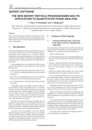
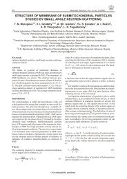
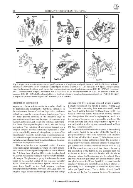
Fig. 2. Crys tal struc ture <strong>of</strong> some sporulation spe cific pro te<strong>in</strong>s. A. Com plex <strong>of</strong> two Spo0F mono mers with Spo0B dimer; am<strong>in</strong>oacid<br />
res i dues <strong>of</strong> Spo0F ac tive site are visu al ised on up per Spo0F mol e cule (PDB ID: 1F51). B. Ac tive site <strong>of</strong> N-Spo0A; phosphorylated<br />
Asp55 and am<strong>in</strong>oacid res i dues, which change <strong>the</strong>ir con for ma tion dur <strong>in</strong>g phosphorylation are shown (PDB ID: 1QMP). C. Com plex <strong>of</strong><br />
two C-ter mi nal do ma<strong>in</strong> <strong>of</strong> Spo0A <strong>in</strong> com plex with DNA; he li ces A-F are de picted (PDB ID: 1LQ1). D. Struc ture <strong>of</strong> S<strong>in</strong>R-S<strong>in</strong>I<br />
com plex (PDB ID: 1B0N). E. Phosphorylated form <strong>of</strong> SpoIIAA with site <strong>of</strong> phosphorylation po<strong>in</strong>t <strong>in</strong>g to sol vent (PDB ID: 1H4X). F.<br />
Com plex <strong>of</strong> SpoIIAB dimer with part <strong>of</strong> F mono mer (PDB ID: 1L0O).<br />
Ini ti a tion <strong>of</strong> sporulation<br />
Veg e ta tive <strong>cell</strong>s are able to mon i tor <strong>the</strong> num ber <strong>of</strong> <strong>cell</strong>s <strong>in</strong><br />
<strong>the</strong> pop u la tion and <strong>the</strong> amount <strong>of</strong> nu tri tional sub stances <strong>in</strong><br />
<strong>the</strong> en vi ron ment. In case <strong>of</strong> nu tri tion de pri va tion, <strong>the</strong> bac te -<br />
rial <strong>cell</strong> can en ter <strong>the</strong> pro cess <strong>of</strong> spore de vel op ment. There<br />
are many pro te<strong>in</strong>s <strong>in</strong> volved <strong>in</strong> <strong>the</strong> <strong>in</strong>i ti a tion stage <strong>of</strong><br />
sporulation that are im por tant for proper chro mo some seg -<br />
re ga tion, cytok<strong>in</strong>esis, <strong>cell</strong> length and <strong>cell</strong> shape de ter mi na -<br />
tion. Some <strong>of</strong> <strong>the</strong>se pro te<strong>in</strong>s play a cru cial role also dur <strong>in</strong>g<br />
sporulation. The <strong>in</strong>i ti a tion <strong>of</strong> sporulation de pends on a<br />
com plex se ries <strong>of</strong> ex ter nal and <strong>in</strong> ter nal sig nals and is str<strong>in</strong> -<br />
gently con trolled by a net work <strong>of</strong> reg u la tory pro te<strong>in</strong>s <strong>of</strong> <strong>the</strong><br />
phosphorelay. Re cently, <strong>the</strong> struc ture <strong>of</strong> some pro te<strong>in</strong>s <strong>in</strong> -<br />
volved <strong>in</strong> this adap tive re sponse were solved and ques tions<br />
partially answered regard<strong>in</strong>g <strong>in</strong>teractions between <strong>the</strong> compo<br />
nents <strong>of</strong> this reg u la tory net work.<br />
This phosphorelay is an ex panded ver sion <strong>of</strong> a twocom<br />
po nent sig nal transduction sys tem. The first com po -<br />
nent, a sen sor k<strong>in</strong>ase (up to five sporulation sen sor k<strong>in</strong> ases<br />
K<strong>in</strong> A-E have been iden ti fied [1]), is autophosphorylated<br />
by ATP on its histid<strong>in</strong>e res i due <strong>in</strong> re sponse to en vi ron men -<br />
tal changes. The phopshoryl group is sub se quently trans -<br />
ferred to <strong>the</strong> aspartyl res i due <strong>of</strong> <strong>the</strong> re sponse reg u la tor<br />
Spo0F. Then phosphotransferase Spo0B trans fers phos -<br />
phate from Spo0F to <strong>the</strong> fi nal re sponse reg u la tor,<br />
transcriptional ac ti va tor and repressor Spo0A [2]. Spo0F is<br />
a typ i cal s<strong>in</strong> gle do ma<strong>in</strong> re sponse reg u la tor that has an -<br />
struc ture with five -he li ces ar ranged around a cen tral<br />
-sheet con sist <strong>in</strong>g <strong>of</strong> five par al lel -strands [3] (Fig. 2A).<br />
The ac tive site com pris <strong>in</strong>g three aspartates Asp10, Asp11<br />
and Asp54, and <strong>the</strong> threon<strong>in</strong>e Thr82 and lys<strong>in</strong>e Lys104 res -<br />
i dues is sit u ated as a small pocket at <strong>the</strong> carboxy-ter mi nal<br />
end <strong>of</strong> <strong>the</strong> -sheet. The site <strong>of</strong> phosphorylation, Asp54 is at<br />
<strong>the</strong> bot tom <strong>of</strong> <strong>the</strong> pocket and is ac ces si ble to sol vent. The<br />
over all struc ture and ac tive site ge om e try <strong>of</strong> Spo0F is re -<br />
mark ably sim i lar to o<strong>the</strong>r reg u la tory do ma<strong>in</strong>s such as CheY<br />
[4], Fix J [5] or Spo0A [6].<br />
The phos phate ac cu mu lated on Spo0F is im me di ately<br />
de liv ered to Spo0A by <strong>the</strong> ac tion <strong>of</strong> Spo0B. Spo0B is a<br />
phosphotransferase with some func tional and struc tural<br />
sim i lar i ties with histid<strong>in</strong>e k<strong>in</strong> ases [7]. It is phosphorylated<br />
on its histid<strong>in</strong>e res i due and forms a dimer. The mono mer is<br />
made up <strong>of</strong> two do ma<strong>in</strong>s, an am<strong>in</strong>o-ter mi nal-helical hair -<br />
p<strong>in</strong> do ma<strong>in</strong> and a carboxy-ter mi nal do ma<strong>in</strong> with an /<br />
fold. The crys tal struc ture re veals that dimer is formed by<br />
<strong>the</strong> as so ci a tion <strong>of</strong> <strong>the</strong> hair p<strong>in</strong> he li cal do ma<strong>in</strong>s from two<br />
mol e cules to form a four-he lix bun dle with <strong>the</strong> site <strong>of</strong><br />
phosphorylation His30 pro trud <strong>in</strong>g <strong>in</strong>to <strong>the</strong> sol vent. There<br />
are two ac tive sites per dimer. The crys tal struc ture <strong>of</strong> <strong>the</strong><br />
com plex be tween Spo0F and Spo0B [8] re vealed how <strong>the</strong><br />
reg u la tory do ma<strong>in</strong> and phosphotransfer do ma<strong>in</strong> <strong>in</strong> ter act to -<br />
ge<strong>the</strong>r (Fig. 2A). The cocrystal con ta<strong>in</strong>ed two Spo0F mol e -<br />
cules per Spo0B dimer. The 1-he lix <strong>of</strong> each Spo0F<br />
mol e cule and <strong>the</strong> five - loops on <strong>the</strong> top <strong>of</strong> <strong>the</strong> mol e cule<br />
Krystalografická spoleènost