Transgenic tobacco plants expressing antimicrobial ... - Wdc-jp.biz
Transgenic tobacco plants expressing antimicrobial ... - Wdc-jp.biz
Transgenic tobacco plants expressing antimicrobial ... - Wdc-jp.biz

Create successful ePaper yourself
Turn your PDF publications into a flip-book with our unique Google optimized e-Paper software.
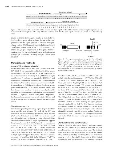
384 Enhanced resistance of transgenic <strong>tobacco</strong> express Lfc<br />
Figure1. The sequences of the amino acids and the nucleotides of signal peptide of PR1 and lactoferricin. For improved plant expression, codon<br />
choice was made according to the codon usage in <strong>tobacco</strong>. Shadowed letters show the signal peptide of <strong>tobacco</strong> PR1 protein, and * shows the stop<br />
codon.<br />
disease resistance to transgenic <strong>plants</strong>. In this study, we<br />
developed transgenic <strong>tobacco</strong> <strong>plants</strong> that carried the Lfc<br />
gene fused to the signal peptide of <strong>tobacco</strong> pathogenrelated<br />
protein (PR-1) under the control of the enhanced<br />
cauliflower mosaic virus (CaMV) 35S promoter. The<br />
effects of Lfc gene expression in transgenic <strong>tobacco</strong><br />
<strong>plants</strong> against the phytopathogenic bacteria Pseudomonas<br />
syringae pv. tabaci and the fungi Botrytis cinerea were<br />
analyzed.<br />
Materials and methods<br />
Assay of Lfc antibacterial activity<br />
Synthesized bovine Lfc (N-FKC RRW QWR MKK LGA PSI<br />
TCV RRA F-C) was purchased from BioGate Co. (Gifu, Japan).<br />
The in vitro antibacterial activity of Lfc was determined by<br />
the method described by Zhang et al. (1998) with a slight<br />
modification. P. syringae pv. maculicola (PMC8301) and<br />
Xanthomonas campestris pv. vesicatoria (#613) were a gift from<br />
Dr. Takikawa, University of Shizuoka. R. solanacearum (OE1-<br />
1) was collected in Aichi Prefecture, Japan. The bacteria were<br />
grown to OD600 of 0.3 in NB liquid medium (Difco), and<br />
1 ml aliquots were transferred to culture tubes. Synthetic Lfc<br />
was added to the culture tubes to obtain final concentrations of<br />
100 µmol l −1 , 10 µmol l −1 , 1 µmol l −1 or 0 µmol l −1 , respectively.<br />
The bacterial suspensions were serially diluted 10-fold and<br />
plated on NB agar. The colonies were counted after an overnight<br />
culture.<br />
Plasmid construction<br />
The chimeric peptide gene coding region (Figure 1) of the<br />
signal peptide from <strong>tobacco</strong> PR-1 and Lfc (Sig-Lfc) was<br />
synthesized by the recombinant polymerase chain reaction<br />
(PCR) method (Fujimoto et al. 1993). The first and second<br />
recombinant PCR were performed in a total volume of 100 µl.<br />
The first reaction contained 1× AmpliTaq Gold Buffer (Applied<br />
Biosystems), 0.25 mmol l −1 dNTPs, 1 µmol l −1 of recombinant<br />
primer 1 (5′-TTG TCT CTA CAC TTC TCT TAT TCC TAG<br />
TAA TAT CCC ACT CTT GCC GTG CC-3′) and recombinant<br />
primer 2 (5′-CGA GCT TCT TCA TAC GCC ATT GCC AAC<br />
GAC GGC ATT TAA AGG CAC GGC AAG A-3′), and 2.5 units<br />
of AmpliTaq Gold (Applied Biosystems). The second reaction<br />
contained 1× AmpliTaq Gold Buffer, 0.25 mmol l −1 dNTPs,<br />
1 µmol l −1 of recombinant primer 3 (5′-ATG GGA TTT GTT<br />
Figure2. The expression constructs for sig-Lfc. The pEC-sigLfc<br />
plasmid expression vector for right and left border regions of the Ti<br />
plasmid; Nos-pro and Nos-ter, promoter and terminator, respectively,<br />
of the nopaline synthase gene; NPTII, neomycin phosphotransferase<br />
II; 2×35S, duplicated enhancer CaMV 35S promoter; KGMMV, 5′<br />
no coding region of Kyuri green mottle mosaic virus; sig-Lfc, peptide<br />
coding sequence of sig-Lfc.<br />
CTC TTT TCA CAA TTG CCT TCA TTT CTT CTT GTC TCT<br />
ACA C-3′) and recombinant primer 4 (5′-TTA AAA GGC ACG<br />
ACG AAC GCA TGT TAT TGA AGG GGC TCC GAG CTT CTT<br />
CA-3′), 2.5 units of AmpliTaq Gold, and 1 µl of the first PCR<br />
solution. The first and second PCR mixture were denatured<br />
for 9 min at 94°C and then amplified via five cycles of 94°C<br />
for 1 min, 45°C for 1 min, and 72°C for 2 min followed by 20<br />
cycles of 94°C for 1 min, 60°C for 1 min, and 72°C for 2 min.<br />
This was followed by a final extension period of 10 min at 72°C.<br />
The PCR products were cloned into the pGEM T-Easy Vector<br />
System (Promega) and sequenced with the CEQ8000 System<br />
(Beckman Coulter). The vector including the Sig-Lfc gene was<br />
digested with BamHI and SacI. The DNA fragment containing<br />
the chimeric peptide was inserted into the pEC vector, which<br />
had a high-expression promoter, including the double enhancer<br />
of cauliflower mosaic virus 35S promoter and the Kyuri green<br />
mottle mosaic virus (KGMMV) 5′ no coding region (Fukuta<br />
and Kanbe 2002). The binary vector pEC-sigLfc is shown in<br />
Figure 2.<br />
Plant material and transformation<br />
Tobacco (Nicotiana tabacum cv. Samsun) seeds were treated<br />
with 1% NaOCl and 0.1% (v/v) Tween20 for 10 min and then<br />
rinsed three times with sterile deionized water. The sterilized<br />
seeds were germinated and grown on Murashige and Skoog’s<br />
(MS) medium (Murashige and Skoog 1962) and maintained<br />
under a 16-h/8-h (light/dark) photoperiod at 25°C. Excised<br />
young leaves were co-cultivated with the overnight culture of<br />
Copyright © 2012 The Japanese Society for Plant Cell and Molecular Biology